Submitted Successfully!
To reward your contribution, here is a gift for you: A free trial for our video production service.
Thank you for your contribution! You can also upload a video entry or images related to this topic.
Version Summary Created by Modification Content Size Created at Operation
1 -- 2804 2023-05-05 12:39:11 |
2 format correction Meta information modification 2804 2023-05-06 07:10:39 |

Video Upload Options

Do you have a full video?

Confirm

Are you sure to Delete?
Cite
If you have any further questions, please contact Encyclopedia Editorial Office.
Garrido, J.J. Axon Initial Segment and Neurodegenerative Diseases. Encyclopedia. Available online: https://encyclopedia.pub/entry/43858 (accessed on 01 July 2024).
Garrido JJ. Axon Initial Segment and Neurodegenerative Diseases. Encyclopedia. Available at: https://encyclopedia.pub/entry/43858. Accessed July 01, 2024.
Garrido, Juan José. "Axon Initial Segment and Neurodegenerative Diseases" Encyclopedia, https://encyclopedia.pub/entry/43858 (accessed July 01, 2024).
Garrido, J.J. (2023, May 05). Axon Initial Segment and Neurodegenerative Diseases. In Encyclopedia. https://encyclopedia.pub/entry/43858
Garrido, Juan José. "Axon Initial Segment and Neurodegenerative Diseases." Encyclopedia. Web. 05 May, 2023.
Axon Initial Segment and Neurodegenerative Diseases
Edit

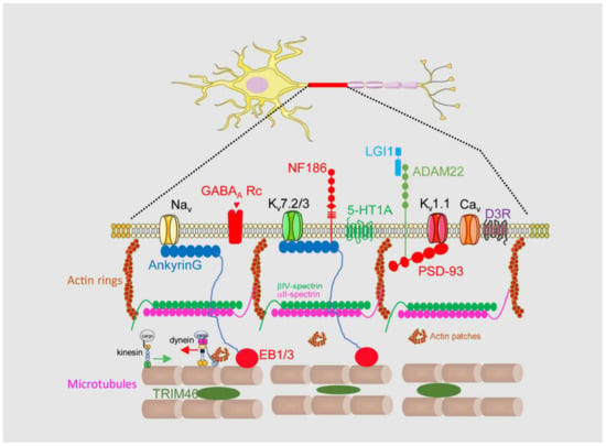
Brain channelopathies are a group of neurological disorders that result from genetic mutations affecting ion channels in the brain. Ion channels are specialized proteins that play a crucial role in the electrical activity of nerve cells by controlling the flow of ions such as sodium, potassium, and calcium. When these channels are not functioning properly, they can cause a wide range of neurological symptoms such as seizures, movement disorders, and cognitive impairment. In this context, the axon initial segment (AIS) is the site of action potential initiation in most neurons. This region is characterized by a high density of voltage-gated sodium channels (VGSCs), which are responsible for the rapid depolarization that occurs when the neuron is stimulated. The AIS is also enriched in other ion channels, such as potassium channels, that play a role in shaping the action potential waveform and determining the firing frequency of the neuron. In addition to ion channels, the AIS contains a complex cytoskeletal structure that helps to anchor the channels in place and regulate their function. 

axon initial segment brain disease voltage-gated ion channels

1. Introduction

1.1. Axon Initial Segment Structure and Physiology

Brain network functions depend on the coordinated action of multiple neurons and their regulation and support by glial cells. Each neuron in the network receives information through their dendritic tree and dendritic spines and processes and integrates this information in the soma. The changes in membrane potential arrive at an AIS, which then generate the corresponding action potentials to propagate the signal to the axon. Nodes of Ranvier are in charge of maintaining the signal until it arrives at the presynaptic terminal, which liberates neurotransmitters to produce a response in the next neuron. In this context, the AIS is the most reliable neuronal compartment, where action potentials are elicited [1][2]. An AIS acts as a gatekeeper controlling the output signal, and is characterized by a surprising structural plasticity, being able to modify its length, position, and composition to modulate the output signal when confronted with a deregulated input signal [3][4][5]. Furthermore, an AIS can also monitor the entry of proteins into the axon through the membrane diffusion barrier and control the cytoplasmic traffic, maintaining neuronal polarization [6]. In view of these important functions, it is easy to understand how the loss of AIS integrity or changes in their structure or structural plasticity leads to brain diseases or mental disorders. Moreover, the importance of AIS integrity for neuronal physiology is highlighted by the fact that AIS disruption precedes neuronal death [7].

1.2. Axon Initial Segment Composition and Structure

An AIS is a highly stable structure that is the first 20–60 μm of the axon (Figure 1). Its function as a membrane diffusion barrier is due to a high concentration of membrane proteins such as neurofascin, NrCAM, TAG1, ADAM22, and voltage-gated ion channels [6]. These membrane proteins are concentrated and tethered at the AIS through specific aminoacidic motifs that bind to AIS scaffold proteins such as ankyrinG, βIV-spectrin, or PSD-93 [8][9]. AIS proteins are organized into a complex network, which forms a diffusion barrier between the axonal and somatodendritic compartments of the neuron, thereby preventing the diffusion of proteins and lipids between these compartments [10]. Among them, ankyrinG and βIV-spectrin are essential in maintaining AIS structure and integrity, linking membrane proteins to the AIS cytoskeleton. AnkyrinG interacts with microtubules through EB proteins [11], while βIV-spectrin links ankyrinG to the actin cytoskeleton [12]. Both actin and microtubules at the AIS contain differential characteristics compared to the somatodendritic or axonal cytoskeleton. Microtubules at the AIS contain more detyrosinated and acetylated tubulin [13] and are more stable and act as a platform for axonal motor proteins [14] such as kinesin-1. Post-translational modifications (PTMs) of tubulin, such as acetylation, alter the ankyrinG distribution and impair kinesin-1 entry to the axon [15]. Tubulin PTMs confer a high degree of stability to the AIS, but additionally TRIM-46 (Tripartite motif containing 46) contributes to microtubule fasciculation [16]. Different actin structures have been identified at the AIS, forming actin rings and actin patches. Actin rings are supported by αII- and βIV-spectrin tetramers and are thought to participate in the initial axon structural plasticity [17], while actin patches may serve as a mechanism controlling the axonal entry of somatodendritic cargoes and their retrograde transport by dynein [18][19]. The AIS also contains an actin-related organelle, the cisternal organelle (CO), which is subject to structural changes associated with AIS plasticity during development and environmental changes [20][21]. Its function is unknown, but it was suggested to be putative Ca2+ store involved in calcium regulation at the AIS.
Figure 1. Axon initial segment structural and functional composition. The AIS is located in the first 20 to 60 μm of the axon and has a differentiated membrane composition with regard to somatodendritic and axonal domains. It is characterized by a high concentration of voltage-gated sodium (Nav) and potassium (Kv) channels anchored to ankyrinG or PSD-93 scaffold proteins. AnkyrinG links ion channels to microtubules through EB1/3 proteins and to actin cytoskeleton through its binding to βIV-spectrin. The AIS contains also voltage-gated calcium channels (Cav), serotonin, dopamine and GABAA receptors. TRIM46 binds microtubules and contribute to their fasciculation. Their special characteristics allows the control of axonal cargoes entry in the axon.
Among all AIS proteins, ankyrinG plays a crucial role at the structural and functional levels. AnkyrinG loss leads to AIS disassembly and consequently the loss of neuronal polarity and the appearance of dendritic spines at the former axon [22][23]. Functionally, the loss of ankyrinG impairs the concentration of sodium and some potassium voltage-gated ion channels, which contain an aminoacids sequence (AIS motif) for ankyrinG binding [24][25][26].
The complexity and specificity of the AIS structure, together with the still limited information available about its protein composition, renders it difficult to completely understand how action potentials are regulated and how voltage-gated ion channel density and location contribute to this regulation.

2. Axon Initial Segment, Mental Disorders, and Neurodegenerative Diseases

2.1. Mental Disorders

Bipolar disorder is a mental health condition characterized by episodes of manic and depressive symptoms. The exact causes of bipolar disorder are not fully understood, but it is believed to involve a complex interplay of genetic, environmental, and neurobiological factors. Research has suggested that abnormalities in the function and structure of neurons, including those in the AIS, may contribute to the development of bipolar disorder. Gene array studies in bipolar disease patients have shown decreased expression of potassium channel Kv1.1, while Kv7.2 and Kv7.3 channels had increased expression [27]. Different studies have highlighted the role of ANK3 gene single nucleotide polymorphims (SNPs) as a risk factor for bipolar disorder. These SNPs affect ankyrinG expression and concomitantly the proper anchoring of potassium channels [28]. Moreover, ANK3 mRNA can be regulated by the expression level of microRNAs in bipolar disorder patients, such as miR34a and miR10b-5p [29]. ankyrinG (480 kDa) is essential for AIS assembly and three missense mutations have been identified in humans. These mutations increase the AIS length, decrease the ankyrinG density, and prevent βIV spectrin recruitment. As a consequence, voltage-gated sodium channels are diffused along the longer AIS and action potential firing has a reduced temporal precision [30]. Furthermore, fore-brain specific knockout of ankyrinG in adult mice shows characteristics reminiscent of aspects of human mania [31].
Schizophrenia is a group of complex mental disorders characterized by psychotic, negative, and cognitive symptoms. Among other alterations, some studies have shown that individuals with schizophrenia have alterations in the expression and distribution of sodium channels in various regions of the brain, including the prefrontal cortex and hippocampus [32]. Loss of function (LoF) mutations and rare coding variants (RCVs) in sodium channels increase the risk of schizophrenia [32]. Mouse models related to schizophrenia have shown increased Nav1.2 and Nav1.6 channel expressions and changes in the electrophysiological properties of action potentials [33][34]. There is no clear evidence of the role of other potassium or calcium channels expressed in the AIS in schizophrenia. The link between ANK3 and schizophrenia remains unclear. Some GWAS studies have proposed an association between ANK3 SNPs and schizophrenia; however, a large GWAS did not show a significant association [35].

2.2. Neurodevelopmental Disorders

Neuronal polarization, functional polarity, and brain network physiology depend on proper early AIS development and maturation. Several neurodevelopmental disorders have been related to early alterations in AIS development. The mouse model of Angelman syndrome exhibited a significant increase in the AIS length in hippocampal neurons [36]. This syndrome is due to the loss of function of the UBE3A gene and is associated with intellectual disability, epilepsy, ataxia, and autism. An increased length is accompanied by a higher expression of voltage-gated sodium channels and ankyrinG. The same length increase has been detected in a fragile X mouse model (Fmr1−/y) that shows increased neuronal excitability [37]. More recently, the loss of a transcription factor, Pax6, was found to change the length and position of the AIS in prethalamic neurons [38]. Similarly, the loss of function of Rbfox proteins that regulate alternative splicing and posttranscriptional regulation leads to defects in AnkG localization and an immature electrophysiology [39], showing the importance of AnkG developmental splicing. In fact, neurons that express ankyrinG, including a 33 nucleotide cassette upstream of the first ZU5 domain, do generate significantly less action potentials, indicating a lower density of voltage-gated ion channels [30].
Autism spectrum disorders are also related to AIS alterations. Subjects with autism show a decreased GABA synthesis in the prefrontal cortex that can lead to downregulation of the GABAARa2 protein in the AIS of pyramidal neurons and affect the inhibitory control of action potentials, contributing to an excitation/inhibition imbalance [40]. Autism mouse models also show a shortening of the AIS in the primary somatosensory barrel cortex, similar to the AIS length change in the same region in ADHD (attention-deficit hyperactivity disorder) model rats [41]. The contribution of voltage-gated ion channels or other scaffold proteins to autism has been proposed, but results only allow to suggest that they contribute to an increased susceptibility to autism spectrum disorders.

2.3. Neuroinflammatory Diseases and Glial Cells

Neuroinflammatory diseases are a group of conditions that involve inflammation in the central nervous system (CNS). Inflammation is the normal response of the immune system to injury or infection, but in some cases, it can become chronic and contribute to the development of various neurological disorders. Neuroinflammatory diseases include, among others, multiple sclerosis (MS), amyotrophic lateral sclerosis (ALS), and also those derived from a brain trauma or brain stroke. Moreover, glial cell activation or alterations contribute significantly to this group of brain diseases.
Several studies have highlighted structural and functional changes in the AIS after brain damage due to blast wave, oxygen/glucose deprivation, or brain ischemia. Exposure to a blast wave generates a traumatic brain injury, leading to altered cognition, memory, and behavior. Translated to a rat model, the consequences are a shortening of the AIS and decreased neuronal excitability [42]. While the cellular and molecular changes are unknown, other models of brain injury have allowed us to understand several potential mechanisms. An oxygen and glucose deprivation model representative of brain ischemia allowed us to determine that sodium channels, structural proteins, and membrane proteins are proteolyzed by calpain [7]. As mentioned above, one of the mechanisms that activates calpain in the AIS is mediated by the purinergic P2X7 receptor. This receptor is not located in the AIS but its inhibition protects AIS voltage-gated ion channels and AIS structural proteins protecting the AIS integrity after middle cerebral artery occlusion (MCAO)-induced ischemia [3].
ALS and MS are characterized by alterations in nerve impulse conduction. In addition to myelin sheath alterations, models of both diseases show alterations in voltage-gated ion channel expression in the AIS and AIS plasticity. Studies have shown that the expression of Nav1.6 channels is increased in demyelinated axons in MS patients, activating the accumulation of intra-axonal calcium through the Na+/Ca2+ exchanger, which may contribute to the development of axonal dysfunction and neurodegeneration [43]. Motoneuron AISs in an ALS mice model G127X SOD1 were longer and thinner during the symptomatic stage and results suggest that the Nav1.6 channel expression increases [44]. However, G127X SOD1 mice show shorter AISs before the symptomatic stage, suggesting that AIS plasticity mechanisms may be a target for ALS. Other ALS models show reduced selective transport of cargoes before pathology appeared, suggesting AIS dysfunction [45].
A recent study on multiple sclerosis human tissue has shown that the gap between soma and AIS increases in pyramidal neurons and Purkinje cells in multiple sclerosis tissue and that the AIS is longer in cortical neurons of cortical inactive lesions [46]. Whether these AIS plasticity changes lead to voltage-gated ion channel expression or function alterations remains elusive, and further studies are necessary to decipher the potential role of the AIS in ALS and MS.
Glial cells play an important role in brain physiology and neuronal modulation. Astrocytes and microglia play a role in ischemia, ALS, or MS; however, their potential relation with the AIS needs further investigation. A small percentage of AISs in the cortex are associated and contact with microglial cells, and interestingly activated microglia lost its interaction with the AIS [47]. This interaction seems to have a physiological role and needs an intact AIS to be preserved. The role of this microglia–AIS contact in the regulation of voltage-gated ion channels is unknown; however, it is essential for chandelier cell GABAergic synaptogenesis in the AIS of neocortical pyramidal neurons [48].
Numerous studies point towards a crucial role of astrocytic ion channels linking the CNS microenvironment to astroglial responses, modulating health, inflammation, and ischemia. Even if astrocytes are non-excitable cells, these cells express a battery of potassium and calcium voltage-gated ion channels, as well as other ion channels that act as sensors and modulators [49]. Increased intracellular Ca2+ promotes ATP release from astrocytes, which modulates neuronal excitability and decreases the conduction speed in myelinated axons through their action on A2aR receptors and HCN2 channels in the AIS and nodes of Ranvier [50].
The role of microglia and astrocytes in brain diseases related to neuronal excitability and survival is increasingly studied; however, how these cells can participate in disease onset regulating their own ion channels and those in neurons remains unknown.

2.4. Autoimmune Diseases

A group of autoimmune diseases have been characterized as affecting structural and functional proteins in the AIS. Autoantibodies to βIV spectrin and TRIM46 have been detected in paraneoplastic CNS disorders [51][52][53]. AnkyrinG autoantibodies have been identified in some people affected by viral infections or suffering from cancer. AnkyrinG autoantibodies have been identified in AIS, nodes of Ranvier, and the subpial space in a patient living with human immunodeficiency virus (HIV) suffering steroid-responsive meningoencephalitis [54] and in a patient with a diagnosis of metastatic ovarian cancer and seizures [54]. Autoantibodies to Lgi1 are characteristic of limbic encephalitis [55]. Lgi1 interacts with the Kv1.1 channel and is required to modulate the Kv1.1 density in the AIS. Lgi1 knockout mice show increased neuronal excitability and suffered from seizures [56]. Together with Lgi1, Caspr2 also interacts with the Kv1.1 channel, and the appearance of Caspr2 autoantibodies is associated with autoimmune encephalitis [57]. Autoantibodies to voltage potassium channels are also related to Morvan’s syndrome and have been found in schizophrenia patients [58].

2.5. Alzheimer’s Disease

Recent studies have proposed an excitatory/inhibitory imbalance as a potential onset mechanism for Alzheimer’s disease. Actually, voltage-gated sodium channels and ankyrinG expression was significantly decreased in an Alzheimer’s disease mouse model APP/PS1 [59] from postnatal stages. Young APP/PS1 mice showed a reduced number of action potentials, while older APP/PS1 mice have an increased number of spikes and hyperexcitability [60]. This maybe the consequence of an increased number of cell surface Nav1.6 channels at around 7 months [61]. In fact, Nav1.6 knockdown is capable of ameliorating cognitive defects and reducing hyperexcitability in APP/PS1 mice [62]. Meanwhile, there are no data regarding Kv channel expression at the AIS in Alzheimer’s mice models, even though interaction of Kv7 channels with BACE1 was described [63]. Altered calcium homeostasis in neurons or glial cells is an early event observed in mouse models of Alzheimer disease [64]. Regarding the AIS, the potential role of the cisternal organelle in Ca2+ homeostasis deregulation has not been studied; however, the presence of ryanodine receptors coupled to Cav3 channels in the AIS [65] suggests the role of these channels in action potential deregulation.
Moreover, AIS shortening was identified in Alzheimer’s disease mouse models or patients [66][67][68]. The absence of AIS structural plasticity was demonstrated in a frontotemporal dementia model due to a tau protein mutation identified in humans and its relation to EB1/EB3 proteins and microtubules in the AIS [69].
In conclusion, the dysregulation of voltage-gated ion channels can have profound effects on neuronal function and contribute to the development of mental disorders and neurodegenerative diseases. Understanding the role of these channels in disease pathogenesis may lead to the development of new treatments and therapies for these devastating conditions. However, it is important to take into account that voltage-gated ion channels pathologies are not only due to alterations in channel structure or expression, but also due to changes in the neuronal domain where their function is exerted and changes in their scaffold and interacting proteins.

References

  1. Bender, K.J.; Trussell, L.O. The physiology of the axon initial segment. Annu. Rev. Neurosci. 2012, 35, 249–265.
  2. Kole, M.H.; Ilschner, S.U.; Kampa, B.M.; Williams, S.R.; Ruben, P.C.; Stuart, G.J. Action potential generation requires a high sodium channel density in the axon initial segment. Nat. Neurosci. 2008, 11, 178–186.
  3. Del Puerto, A.; Fronzaroli-Molinieres, L.; Perez-Alvarez, M.J.; Giraud, P.; Carlier, E.; Wandosell, F.; Debanne, D.; Garrido, J.J. ATP-P2X7 Receptor Modulates Axon Initial Segment Composition and Function in Physiological Conditions and Brain Injury. Cereb. Cortex 2015, 25, 2282–2294.
  4. Grubb, M.S.; Burrone, J. Activity-dependent relocation of the axon initial segment fine-tunes neuronal excitability. Nature 2010, 465, 1070–1074.
  5. Kuba, H. Plasticity at the axon initial segment. Commun. Integr. Biol. 2010, 3, 597–598.
  6. Rasband, M.N. The axon initial segment and the maintenance of neuronal polarity. Nat. Rev. Neurosci. 2010, 11, 552–562.
  7. Schafer, D.P.; Jha, S.; Liu, F.; Akella, T.; McCullough, L.D.; Rasband, M.N. Disruption of the axon initial segment cytoskeleton is a new mechanism for neuronal injury. J. Neurosci. 2009, 29, 13242–13254.
  8. Jenkins, S.M.; Bennett, V. Ankyrin-G coordinates assembly of the spectrin-based membrane skeleton, voltage-gated sodium channels, and L1 CAMs at Purkinje neuron initial segments. J. Cell Biol. 2001, 155, 739–746.
  9. Ogawa, Y.; Horresh, I.; Trimmer, J.S.; Bredt, D.S.; Peles, E.; Rasband, M.N. Postsynaptic density-93 clusters Kv1 channels at axon initial segments independently of Caspr2. J. Neurosci. 2008, 28, 5731–5739.
  10. Nakada, C.; Ritchie, K.; Oba, Y.; Nakamura, M.; Hotta, Y.; Iino, R.; Kasai, R.S.; Yamaguchi, K.; Fujiwara, T.; Kusumi, A. Accumulation of anchored proteins forms membrane diffusion barriers during neuronal polarization. Nat. Cell Biol. 2003, 5, 626–632.
  11. Leterrier, C.; Vacher, H.; Fache, M.P.; d’Ortoli, S.A.; Castets, F.; Autillo-Touati, A.; Dargent, B. End-binding proteins EB3 and EB1 link microtubules to ankyrin G in the axon initial segment. Proc. Natl. Acad. Sci. USA 2011, 108, 8826–8831.
  12. Leterrier, C. The Axon Initial Segment: An Updated Viewpoint. J. Neurosci. 2018, 38, 2135–2145.
  13. Sanchez-Ponce, D.; Munoz, A.; Garrido, J.J. Casein kinase 2 and microtubules control axon initial segment formation. Mol. Cell. Neurosci. 2011, 46, 222–234.
  14. Konishi, Y.; Setou, M. Tubulin tyrosination navigates the kinesin-1 motor domain to axons. Nat. Neurosci. 2009, 12, 559–567.
  15. Tapia, M.; Wandosell, F.; Garrido, J.J. Impaired function of HDAC6 slows down axonal growth and interferes with axon initial segment development. PLoS ONE 2010, 5, e12908.
  16. Harterink, M.; Vocking, K.; Pan, X.; Soriano Jerez, E.M.; Slenders, L.; Freal, A.; Tas, R.P.; van de Wetering, W.J.; Timmer, K.; Motshagen, J.; et al. TRIM46 Organizes Microtubule Fasciculation in the Axon Initial Segment. J. Neurosci. 2019, 39, 4864–4873.
  17. Papandreou, M.J.; Leterrier, C. The functional architecture of axonal actin. Mol. Cell. Neurosci. 2018, 91, 151–159.
  18. Eichel, K.; Shen, K. The function of the axon initial segment in neuronal polarity. Dev. Biol. 2022, 489, 47–54.
  19. Kuijpers, M.; van de Willige, D.; Freal, A.; Chazeau, A.; Franker, M.A.; Hofenk, J.; Rodrigues, R.J.; Kapitein, L.C.; Akhmanova, A.; Jaarsma, D.; et al. Dynein Regulator NDEL1 Controls Polarized Cargo Transport at the Axon Initial Segment. Neuron 2016, 89, 461–471.
  20. Benedeczky, I.; Molnar, E.; Somogyi, P. The cisternal organelle as a Ca(2+)-storing compartment associated with GABAergic synapses in the axon initial segment of hippocampal pyramidal neurones. Exp. Brain Res. 1994, 101, 216–230.
  21. Schluter, A.; Del Turco, D.; Deller, T.; Gutzmann, A.; Schultz, C.; Engelhardt, M. Structural Plasticity of Synaptopodin in the Axon Initial Segment during Visual Cortex Development. Cereb. Cortex 2017, 27, 4662–4675.
  22. Hedstrom, K.L.; Ogawa, Y.; Rasband, M.N. AnkyrinG is required for maintenance of the axon initial segment and neuronal polarity. J. Cell Biol. 2008, 183, 635–640.
  23. Sobotzik, J.M.; Sie, J.M.; Politi, C.; Del Turco, D.; Bennett, V.; Deller, T.; Schultz, C. AnkyrinG is required to maintain axo-dendritic polarity in vivo. Proc. Natl. Acad. Sci. USA 2009, 106, 17564–17569.
  24. Garrido, J.J.; Giraud, P.; Carlier, E.; Fernandes, F.; Moussif, A.; Fache, M.P.; Debanne, D.; Dargent, B. A targeting motif involved in sodium channel clustering at the axonal initial segment. Science 2003, 300, 2091–2094.
  25. Pan, Z.; Kao, T.; Horvath, Z.; Lemos, J.; Sul, J.Y.; Cranstoun, S.D.; Bennett, V.; Scherer, S.S.; Cooper, E.C. A common ankyrin-G-based mechanism retains KCNQ and NaV channels at electrically active domains of the axon. J. Neurosci. 2006, 26, 2599–2613.
  26. Zhou, D.; Lambert, S.; Malen, P.L.; Carpenter, S.; Boland, L.M.; Bennett, V. AnkyrinG is required for clustering of voltage-gated Na channels at axon initial segments and for normal action potential firing. J. Cell Biol. 1998, 143, 1295–1304.
  27. Smolin, B.; Karry, R.; Gal-Ben-Ari, S.; Ben-Shachar, D. Differential expression of genes encoding neuronal ion-channel subunits in major depression, bipolar disorder and schizophrenia: Implications for pathophysiology. Int. J. Neuropsychopharmacol. 2012, 15, 869–882.
  28. Imbrici, P.; Camerino, D.C.; Tricarico, D. Major channels involved in neuropsychiatric disorders and therapeutic perspectives. Front. Genet. 2013, 4, 76.
  29. Bavamian, S.; Mellios, N.; Lalonde, J.; Fass, D.M.; Wang, J.; Sheridan, S.D.; Madison, J.M.; Zhou, F.; Rueckert, E.H.; Barker, D.; et al. Dysregulation of miR-34a links neuronal development to genetic risk factors for bipolar disorder. Mol. Psychiatry 2015, 20, 573–584.
  30. Yang, R.; Walder-Christensen, K.K.; Lalani, S.; Yan, H.; Garcia-Prieto, I.D.; Alvarez, S.; Fernandez-Jaen, A.; Speltz, L.; Jiang, Y.H.; Bennett, V. Neurodevelopmental mutation of giant ankyrin-G disrupts a core mechanism for axon initial segment assembly. Proc. Natl. Acad. Sci. USA 2019, 116, 19717–19726.
  31. Zhu, S.; Cordner, Z.A.; Xiong, J.; Chiu, C.T.; Artola, A.; Zuo, Y.; Nelson, A.D.; Kim, T.Y.; Zaika, N.; Woolums, B.M.; et al. Genetic disruption of ankyrin-G in adult mouse forebrain causes cortical synapse alteration and behavior reminiscent of bipolar disorder. Proc. Natl. Acad. Sci. USA 2017, 114, 10479–10484.
  32. Rees, E.; Carrera, N.; Morgan, J.; Hambridge, K.; Escott-Price, V.; Pocklington, A.J.; Richards, A.L.; Pardinas, A.F.; Investigators, G.; McDonald, C.; et al. Targeted Sequencing of 10,198 Samples Confirms Abnormalities in Neuronal Activity and Implicates Voltage-Gated Sodium Channels in Schizophrenia Pathogenesis. Biol. Psychiatry 2019, 85, 554–562.
  33. Mi, Z.; Yang, J.; He, Q.; Zhang, X.; Xiao, Y.; Shu, Y. Alterations of Electrophysiological Properties and Ion Channel Expression in Prefrontal Cortex of a Mouse Model of Schizophrenia. Front. Cell. Neurosci. 2019, 13, 554.
  34. Page, S.C.; Sripathy, S.R.; Farinelli, F.; Ye, Z.; Wang, Y.; Hiler, D.J.; Pattie, E.A.; Nguyen, C.V.; Tippani, M.; Moses, R.L.; et al. Electrophysiological measures from human iPSC-derived neurons are associated with schizophrenia clinical status and predict individual cognitive performance. Proc. Natl. Acad. Sci. USA 2022, 119, e2109395119.
  35. Lam, M.; Chen, C.Y.; Li, Z.; Martin, A.R.; Bryois, J.; Ma, X.; Gaspar, H.; Ikeda, M.; Benyamin, B.; Brown, B.C.; et al. Comparative genetic architectures of schizophrenia in East Asian and European populations. Nat. Genet. 2019, 51, 1670–1678.
  36. Kaphzan, H.; Buffington, S.A.; Jung, J.I.; Rasband, M.N.; Klann, E. Alterations in intrinsic membrane properties and the axon initial segment in a mouse model of Angelman syndrome. J. Neurosci. 2011, 31, 17637–17648.
  37. Booker, S.A.; Simoes de Oliveira, L.; Anstey, N.J.; Kozic, Z.; Dando, O.R.; Jackson, A.D.; Baxter, P.S.; Isom, L.L.; Sherman, D.L.; Hardingham, G.E.; et al. Input-Output Relationship of CA1 Pyramidal Neurons Reveals Intact Homeostatic Mechanisms in a Mouse Model of Fragile X Syndrome. Cell Rep. 2020, 32, 107988.
  38. Tian, T.; Quintana-Urzainqui, I.; Kozic, Z.; Pratt, T.; Price, D.J. Pax6 loss alters the morphological and electrophysiological development of mouse prethalamic neurons. Development 2022, 149, dev200052.
  39. Jacko, M.; Weyn-Vanhentenryck, S.M.; Smerdon, J.W.; Yan, R.; Feng, H.; Williams, D.J.; Pai, J.; Xu, K.; Wichterle, H.; Zhang, C. Rbfox Splicing Factors Promote Neuronal Maturation and Axon Initial Segment Assembly. Neuron 2018, 97, 853–868.e6.
  40. Hong, T.; Falcone, C.; Dufour, B.; Amina, S.; Castro, R.P.; Regalado, J.; Pearson, W.; Noctor, S.C.; Martinez-Cerdeno, V. GABA(A)Ralpha2 is Decreased in the Axon Initial Segment of Pyramidal Cells in Specific Areas of the Prefrontal Cortex in Autism. Neuroscience 2020, 437, 76–86.
  41. Usui, N.; Tian, X.; Harigai, W.; Togawa, S.; Utsunomiya, R.; Doi, T.; Miyoshi, K.; Shinoda, K.; Tanaka, J.; Shimada, S.; et al. Length impairments of the axon initial segment in rodent models of attention-deficit hyperactivity disorder and autism spectrum disorder. Neurochem. Int. 2022, 153, 105273.
  42. Baalman, K.L.; Cotton, R.J.; Rasband, S.N.; Rasband, M.N. Blast wave exposure impairs memory and decreases axon initial segment length. J. Neurotrauma 2013, 30, 741–751.
  43. Craner, M.J.; Newcombe, J.; Black, J.A.; Hartle, C.; Cuzner, M.L.; Waxman, S.G. Molecular changes in neurons in multiple sclerosis: Altered axonal expression of Nav1.2 and Nav1.6 sodium channels and Na+/Ca2+ exchanger. Proc. Natl. Acad. Sci. USA 2004, 101, 8168–8173.
  44. Jorgensen, H.S.; Jensen, D.B.; Dimintiyanova, K.P.; Bonnevie, V.S.; Hedegaard, A.; Lehnhoff, J.; Moldovan, M.; Grondahl, L.; Meehan, C.F. Increased Axon Initial Segment Length Results in Increased Na(+) Currents in Spinal Motoneurones at Symptom Onset in the G127X SOD1 Mouse Model of Amyotrophic Lateral Sclerosis. Neuroscience 2021, 468, 247–264.
  45. Sasaki, S.; Warita, H.; Abe, K.; Iwata, M. Impairment of axonal transport in the axon hillock and the initial segment of anterior horn neurons in transgenic mice with a G93A mutant SOD1 gene. Acta Neuropathol. 2005, 110, 48–56.
  46. Senol, A.D.; Pinto, G.; Beau, M.; Guillemot, V.; Dupree, J.L.; Stadelmann, C.; Ranft, J.; Lubetzki, C.; Davenne, M. Alterations of the axon initial segment in multiple sclerosis grey matter. Brain Commun. 2022, 4, fcac284.
  47. Baalman, K.; Marin, M.A.; Ho, T.S.; Godoy, M.; Cherian, L.; Robertson, C.; Rasband, M.N. Axon initial segment-associated microglia. J. Neurosci. 2015, 35, 2283–2292.
  48. Gallo, N.B.; Berisha, A.; Van Aelst, L. Microglia regulate chandelier cell axo-axonic synaptogenesis. Proc. Natl. Acad. Sci. USA 2022, 119, e2114476119.
  49. Schmaul, S.; Hanuscheck, N.; Bittner, S. Astrocytic potassium and calcium channels as integrators of the inflammatory and ischemic CNS microenvironment. Biol. Chem. 2021, 402, 1519–1530.
  50. Lezmy, J.; Arancibia-Carcamo, I.L.; Quintela-Lopez, T.; Sherman, D.L.; Brophy, P.J.; Attwell, D. Astrocyte Ca(2+)-evoked ATP release regulates myelinated axon excitability and conduction speed. Science 2021, 374, eabh2858.
  51. Bartley, C.M.; Ngo, T.T.; Alvarenga, B.D.; Kung, A.F.; Teliska, L.H.; Sy, M.; DeRisi, J.L.; Rasband, M.N.; Pittock, S.J.; Dubey, D.; et al. betaIV-Spectrin Autoantibodies in 2 Individuals With Neuropathy of Possible Paraneoplastic Origin: A Case Series. Neurol. Neuroimmunol. Neuroinflamm. 2022, 9, e1188.
  52. Berghs, S.; Ferracci, F.; Maksimova, E.; Gleason, S.; Leszczynski, N.; Butler, M.; De Camilli, P.; Solimena, M. Autoimmunity to beta IV spectrin in paraneoplastic lower motor neuron syndrome. Proc. Natl. Acad. Sci. USA 2001, 98, 6945–6950.
  53. Valencia-Sanchez, C.; Knight, A.M.; Hammami, M.B.; Guo, Y.; Mills, J.R.; Kryzer, T.J.; Piquet, A.L.; Amin, A.; Heinzelmann, M.; Lucchinetti, C.F.; et al. Characterisation of TRIM46 autoantibody-associated paraneoplastic neurological syndrome. J. Neurol. Neurosurg. Psychiatry 2022, 93, 196–200.
  54. Bartley, C.M.; Ngo, T.T.; Cadwell, C.R.; Harroud, A.; Schubert, R.D.; Alvarenga, B.D.; Hawes, I.A.; Zorn, K.C.; Hunyh, T.; Teliska, L.H.; et al. Dual ankyrinG and subpial autoantibodies in a man with well-controlled HIV infection with steroid-responsive meningoencephalitis: A case report. Front. Neurol. 2022, 13, 1102484.
  55. Ohkawa, T.; Fukata, Y.; Yamasaki, M.; Miyazaki, T.; Yokoi, N.; Takashima, H.; Watanabe, M.; Watanabe, O.; Fukata, M. Autoantibodies to epilepsy-related LGI1 in limbic encephalitis neutralize LGI1-ADAM22 interaction and reduce synaptic AMPA receptors. J. Neurosci. 2013, 33, 18161–18174.
  56. Seagar, M.; Russier, M.; Caillard, O.; Maulet, Y.; Fronzaroli-Molinieres, L.; De San Feliciano, M.; Boumedine-Guignon, N.; Rodriguez, L.; Zbili, M.; Usseglio, F.; et al. LGI1 tunes intrinsic excitability by regulating the density of axonal Kv1 channels. Proc. Natl. Acad. Sci. USA 2017, 114, 7719–7724.
  57. Pinatel, D.; Hivert, B.; Boucraut, J.; Saint-Martin, M.; Rogemond, V.; Zoupi, L.; Karagogeos, D.; Honnorat, J.; Faivre-Sarrailh, C. Inhibitory axons are targeted in hippocampal cell culture by anti-Caspr2 autoantibodies associated with limbic encephalitis. Front. Cell. Neurosci. 2015, 9, 265.
  58. Zandi, M.S.; Irani, S.R.; Lang, B.; Waters, P.; Jones, P.B.; McKenna, P.; Coles, A.J.; Vincent, A.; Lennox, B.R. Disease-relevant autoantibodies in first episode schizophrenia. J. Neurol. 2011, 258, 686–688.
  59. Sun, X.; Wu, Y.; Gu, M.; Liu, Z.; Ma, Y.; Li, J.; Zhang, Y. Selective filtering defect at the axon initial segment in Alzheimer’s disease mouse models. Proc. Natl. Acad. Sci. USA 2014, 111, 14271–14276.
  60. Vitale, P.; Salgueiro-Pereira, A.R.; Lupascu, C.A.; Willem, M.; Migliore, R.; Migliore, M.; Marie, H. Analysis of Age-Dependent Alterations in Excitability Properties of CA1 Pyramidal Neurons in an APPPS1 Model of Alzheimer’s Disease. Front. Aging Neurosci. 2021, 13, 668948.
  61. Liu, C.; Tan, F.C.; Xiao, Z.C.; Dawe, G.S. Amyloid precursor protein enhances Nav1.6 sodium channel cell surface expression. J. Biol. Chem. 2015, 290, 12048–12057.
  62. Yuan, D.J.; Yang, G.; Wu, W.; Li, Q.F.; Xu, D.E.; Ntim, M.; Jiang, C.Y.; Liu, J.C.; Zhang, Y.; Wang, Y.Z.; et al. Reducing Nav1.6 expression attenuates the pathogenesis of Alzheimer’s disease by suppressing BACE1 transcription. Aging Cell 2022, 21, e13593.
  63. Hessler, S.; Zheng, F.; Hartmann, S.; Rittger, A.; Lehnert, S.; Volkel, M.; Nissen, M.; Edelmann, E.; Saftig, P.; Schwake, M.; et al. beta-Secretase BACE1 regulates hippocampal and reconstituted M-currents in a beta-subunit-like fashion. J. Neurosci. 2015, 35, 3298–3311.
  64. Di Benedetto, G.; Burgaletto, C.; Bellanca, C.M.; Munafo, A.; Bernardini, R.; Cantarella, G. Role of Microglia and Astrocytes in Alzheimer’s Disease: From Neuroinflammation to Ca(2+) Homeostasis Dysregulation. Cells 2022, 11, 2728.
  65. Lipkin, A.M.; Cunniff, M.M.; Spratt, P.W.E.; Lemke, S.M.; Bender, K.J. Functional Microstructure of Ca(V)-Mediated Calcium Signaling in the Axon Initial Segment. J. Neurosci. 2021, 41, 3764–3776.
  66. Anton-Fernandez, A.; Leon-Espinosa, G.; DeFelipe, J.; Munoz, A. Pyramidal cell axon initial segment in Alzheimer s disease. Sci. Rep. 2022, 12, 8722.
  67. Ma, F.; Akolkar, H.; Xu, J.; Liu, Y.; Popova, D.; Xie, J.; Youssef, M.M.; Benosman, R.; Hart, R.P.; Herrup, K. The Amyloid Precursor Protein Modulates the Position and Length of the Axon Initial Segment. J. Neurosci. 2023, 43, 1830–1844.
  68. Marin, M.A.; Ziburkus, J.; Jankowsky, J.; Rasband, M.N. Amyloid-beta plaques disrupt axon initial segments. Exp. Neurol. 2016, 281, 93–98.
  69. Sohn, P.D.; Huang, C.T.; Yan, R.; Fan, L.; Tracy, T.E.; Camargo, C.M.; Montgomery, K.M.; Arhar, T.; Mok, S.A.; Freilich, R.; et al. Pathogenic Tau Impairs Axon Initial Segment Plasticity and Excitability Homeostasis. Neuron 2019, 104, 458–470.e5.
More
Information
Subjects: Cell Biology
Contributor MDPI registered users' name will be linked to their SciProfiles pages. To register with us, please refer to https://encyclopedia.pub/register :
View Times: 354
Revisions: 2 times (View History)
Update Date: 06 May 2023
1000/1000
Video Production Service