Submitted Successfully!
To reward your contribution, here is a gift for you: A free trial for our video production service.
Thank you for your contribution! You can also upload a video entry or images related to this topic.
Version Summary Created by Modification Content Size Created at Operation
1 -- 1683 2023-05-05 05:04:36 |
2 language was corrected; description was corrected + 720 word(s) 2369 2023-05-05 15:33:24 | |
3 format corrected. -12 word(s) 2357 2023-05-05 15:40:31 |

Video Upload Options

Do you have a full video?

Confirm

Are you sure to Delete?
Cite
If you have any further questions, please contact Encyclopedia Editorial Office.
Oshchepkova, A.; Zenkova, M.; Vlassov, V. Extracellular Vesicles Application as Drug Delivery Carriers. Encyclopedia. Available online: https://encyclopedia.pub/entry/43818 (accessed on 27 July 2024).
Oshchepkova A, Zenkova M, Vlassov V. Extracellular Vesicles Application as Drug Delivery Carriers. Encyclopedia. Available at: https://encyclopedia.pub/entry/43818. Accessed July 27, 2024.
Oshchepkova, Anastasiya, Marina Zenkova, Valentin Vlassov. "Extracellular Vesicles Application as Drug Delivery Carriers" Encyclopedia, https://encyclopedia.pub/entry/43818 (accessed July 27, 2024).
Oshchepkova, A., Zenkova, M., & Vlassov, V. (2023, May 05). Extracellular Vesicles Application as Drug Delivery Carriers. In Encyclopedia. https://encyclopedia.pub/entry/43818
Oshchepkova, Anastasiya, et al. "Extracellular Vesicles Application as Drug Delivery Carriers." Encyclopedia. Web. 05 May, 2023.
Extracellular Vesicles Application as Drug Delivery Carriers
Edit

Extracellular vesicles (EVs) are membrane vesicles released into the extracellular milieu by cells of various origins. They contain different biological cargoes, protecting them from degradation by environmental factors. There is an opinion that EVs have a number of advantages over synthetic carriers, creating new opportunities for drug delivery. There are minimal requirements for carriers used for therapeutic nucleic acids (tNAs) delivery in the clinic. Briefly, they should provide for long circulation, low immunogenicity, and high intracellular accumulation of tNAs, simultaneously. In addition, the tNA delivery system must be biodegradable and act in a specific manner. A number of reviews have already discussed the use of EVs as a delivery system in various aspects: the EV surface molecules potentially involved in their interaction with recipient cells, the doses of EVs used for preclinical studies, strategies for engineering EVs with specific targeting ligands, approaches used for increasing the EV stability in the bloodstream. 

extracellular vesicles drug delivery nucleic acid therapeutics nanocarrier

1. Interactions of EVs with Immune Cells

Extracellular vesicles (EVs) are particles that are naturally secreted and taken up by cells. For this reason, these carriers are expected to be biocompatible. Indeed, EVs were shown to cause only negligible side effects on cell physiology or survival [1]. However, it is known that EVs make a significant contribution to the development of immune responses, especially if they originate from cells of the immune system. There are increasing pieces of evidence that EVs, released by nonimmune cells, can also participate in the development of immune responses [2]. Blood cells are often chosen for the production of EVs, which, as it has turned out, can stimulate inflammation [3][4]. EVs derived from other cellular sources may also cause a moderate proinflammatory response. Thus, SKOV3- or HEK293-derived EVs caused a slight activation of tumor necrosis factor-α (TNF-α) or interferon-α (INF-α) production by peripheral blood mononuclear cells (PBMCs) [5]. It seems that, regardless of the origin of EVs, they tend to have a visible effect on cells of the immune system, and such impacts have to be monitored in the case of long-term treatment.
The impact of nonimmune EVs on host immunity has been well illustrated by EVs isolated from cow milk and administrated to female Sprague–Dawley rats by oral gavage [6]. Today, a long-term treatment strategy with well-tolerated doses of therapeutics is preferred. Although this avoids acute reactions to therapy, the body develops chronic responses to exposure. After long-term medication (once daily for 15 days) with milk EVs (25 mg/kg b. wt), an increase in the level of granulocyte-macrophage colony-stimulating factor (GM-CSF) and a ~40% decrease in triglycerides were noted in the serum of healthy rats despite the absence of other serious changes [6].
Two models have been proposed to explain the action of milk EVs in the intestine. First, the uptake of EVs by enterocytes can cause the production of defective chylomicrons, which can be recognized and eliminated by intestinal macrophages. A more plausible explanation is that the macrophages engulf aggregates of chylomicrons and EVs. Recently, the associations of EVs and lipid particles, yielding aggregates, have been discussed [7][8][9]. In any case, the reduced level of triglycerides in the rat’s blood is easily explained by the macrophage-mediated elimination of chylomicrons from the intestine that prevents chylomicron movement into the lacteal (lymph capillary) and subsequent entry into the venous circulation [10].
Intrigue enough, these milk EVs (25 mg/kg b. wt.) had a long-term inhibitory effect (~4–5 weeks) on tumor growth in mice of human lung cancer (A549) xenografts exclusively after administration by oral gavage (three times a week), but not after intraperitoneal injection [6]. It looks like the action of EVs in the intestine was the origin of their therapeutic effect. The positive effect of EVs administered through the digestive tract was also confirmed by other observations [11].

2. EVs in the Bloodstream

Cancer is still one of the leading causes of death in the world, and EVs are considered promising carriers for therapeutics in oncology. Frequently, EVs are intravenously injected into the body based on the idea of their passive accumulation in a tumor site through the enhanced permeability and retention (EPR) effect [12][13]. However, the ability of EVs to overcome significant distances through the bloodstream seems questionable. Furthermore, the EPR effect seems to be overestimated in humans [12][13].
The first barrier on the way of EVs to the target sites (tumor) is blood phagocytes. A recent study showed that tumor EVs in the blood vessels of a zebrafish embryo were preferentially taken up by patrolling macrophages (functionally similar to human patrolling monocytes), endothelial cells, and putative hematopoietic stem cells [14]. An interaction of EVs with non-cellular blood components, such as low-density lipoproteins (LDLs) [7], also contributes to EV removal from the bloodstream. An electron microscopy assay of crude human plasma has shown that EVs frequently bind and fuse with lipoprotein-like structures [9]. In vitro data indicate that EVs secreted by highly malignant breast cancer cells metastasizing to the brain are able to form aggregates with LDLs, which can subsequently be taken up by monocytes [7].
Eventually, in addition to the removal of EVs directly from the blood, there is a risk of their accumulation in the reticuloendothelial-system-rich organs [15]. This could result in EVs failing to meet their targets. The use of high doses of EVs can lead to their intensive accumulation in the lungs that can cause asphyxia, as has been found in experiments with mice [16].

3. EVs as a Part of the Cellular Secretory Apparatus

The release of EVs is closely integrated with other secretory and degradation processes in a cell (Figure 1) [17][18][19][20][21][22]. This integration is suggested by the results of studies demonstrating that the secretion of EVs can be increased to compensate for lysosomal impairment [19][21] or autophagy inhibition [18]. Sometimes, the loading of drugs into EVs can be performed using the cellular mechanisms of vesicle production. This technique is called “indirect”, “endogenous”, or “preloading”. A tNA or another cargo is added to cells, after which the secreted EVs are isolated. However, the intersections between EVs and other secretory/degradation processes inside cells can reduce the effectiveness of this drug loading strategy. Particular, the exocytosis of cargo can be expected to occur simultaneously via several secretory pathways; e.g., recently, the presence of overexpressed alpha-synuclein was simultaneously detected in endosomes, lysosomes, and autophagosomes [20].
Figure 1. MVB interacts with other secretory and degradation processes in a cell. MVBs, autophagosomes, and amphisomes may undergo exocytosis or fusion with lysosomes, resulting in either degradation of their content or lysosomal exocytosis. It should be noted that some paths shown in the figure are hypothetical. The scheme was prepared on the basis of the reviews [17][19][23][24][25]. MVB—multivesicular body; PH—phagophore; CMA—chaperone-mediated autophagy; ER—endoplasmic reticulum; L—lysosome.
The connection of EVs with other secretory/degradation processes may additionally provide new opportunities to control the EV release and the search for proteins suitable for selective loading of tNAs into EVs. An important question is the link between EV secretion and autophagy; e.g., the autophagy-associated protein Atg5 (autophagy protein 5) was found to control the release of EVs by the regulation of multivesicular body (MVB) acidification [26]. Atg5 decreases the activity of V1V0-ATPase by detaching its Atp6v1e1 component [26]. The deactivation of V1V0-ATPase reduces MVB acidity and promotes EV release. The sorting of Atp6v1e1 into intraluminal vesicles (ILVs) of MVBs depends on LC3 (microtubule-associated protein 1A/1B light chain 3). Most likely, the LC3 recruitment into ILVs is provided by Atg5. Eventually, both LC3 and Atp6v1e1 are released by cells via EVs [26].
From a practical point of view, LC3 can be of interest for the selective loading of tNAs into EVs. Its lipidated form, LC3-II, can be involved in ribonucleoprotein (RNP) sorting into ILVs [27]. LC3-II can recruit proteins containing the LC3-interaction region (LIR) [22], although it is not clear whether it is absolutely required for the sorting of RNA-binding proteins (RBPs) into ILVs [27]. Notably, LIR was not found in the previously mentioned protein, Atp6v1e1 [26]. The use of LC3-mediated loading of RNPs into ILVs can be of interest to those aiming at the therapeutic application of small nucleolar RNAs (snoRNAs) [27].
In addition to Atg5, there is growing evidence that proteins traditionally associated with autophagy can control MVB biogenesis. For instance, a recent report demonstrated the role of isoform A of lysosome-associated membrane protein 2 (LAMP2A) and heat shock 70-kDa protein 8 (HSC70) in the loading of proteins, containing the KFERQ-like motif, into EVs [28]. Although it is well known that both LAMP2A and HSC70 are involved in chaperone-mediated autophagy (CMA) [22][25], it is quite possible that, in combination with other factors, they can change the fate of cargoes, directing them to EVs.

4. Influence of the Non-Cellular Environment on EVs

The interaction of exogenous EVs and cells is affected by various environmental factors. It has been found that the efficiency of EV internalization [29] and the biological effects mediated by EVs [30] can depend on the stiffness of the cell growth substrate; e.g., EVs secreted by highly malignant breast cancer cells can affect the activation, proliferation, motility, and contractility of fibroblasts only under conditions that mimic the stiffness of the tumor, but not healthy tissue [30].
A general recommendation for EVs designed for the delivery of therapeutics to tumor cells is to obtain EVs under conditions close to the tumor microenvironment. Thus, it was noted that EVs secreted by metastatic melanoma cells were more efficiently internalized by their own cells, when EVs were produced under the acidic pH of the growth medium (pH = 6.0) [31]. The rigidity of these “acidic” EVs was increased due to changes in the composition of the EV’s lipids [31]. Similarly, hypoxic tumor EVs were better internalized by cancer cells than normoxic EVs [32].
Sometimes, EVs show a greater affinity for their own cells [33][34]. In general, this is similar to how tumor cells prefer “acidic” or “hypoxic” EVs. In the case of tumor-derived EVs, this property of EVs has been proposed for use as a Trojan horse for drug delivery [34]. This trend may persist even after endogenous loading of cargo into EVs [35]. However, little is actually known about the effect of a cargo on the properties of EVs. Chemotherapeutic drugs can affect EVs after endogenous loading as they can influence the EV-secreting cells. It has been found that prolonged cell exposure to doxorubicin, sufficient for developing drug resistance, can change the protein profile of EVs secreted by these cells [36]. Usually, drug loading into EVs requires short-term treatment, which is expected to have no effect on the properties of EVs.
In nature, the changes in the architecture of EVs can occur as a result of a viral infection. Viruses can penetrate inside EVs, thereby avoiding the host’s immune defense [37] and providing viral ligands on the surface of EVs [38]. Viruses need to control the EV composition to provide their entry into the type of cells that can ensure their propagation. Whether endogenously loaded cargo can affect the EV composition, such as viruses, is not entirely clear. The EV-mediated intercellular communication is believed to exist for targeted transfer of biological cargo from one cell to another [39]. It is reasonable to assume that cells should control the EV specificity depending on the type of cargo and the type of target cells.

5. Manufacture of EVs

The low yield of EVs is the key problem limiting their clinical applications. Although some external stimuli (e.g., cell starvation, hypoxia, or an increase in Ca2+ concentration in the culture medium) are used to increase the secretion of EVs [40], the composition and properties of such EVs can differ significantly from those obtained under normal conditions. This has recently been confirmed for EVs secreted by starving cells, where starvation significantly affects the protein composition of EVs [41]. The altering of the EV content in response to environmental stresses has also been discussed in a recently published review [42]. In addition to activating the EV secretion in stress conditions, it is possible to overexpress some proteins in EV-secreting cells, which can enhance the EV release into the extracellular space [43]. Although it may have a lesser influence on the content and features of EVs, the use of exogenous genetic constructs still raises concerns due to the risk of their inclusion into EVs, followed by insertional mutagenesis.
Isolation of EVs is considered a laborious and expensive process that is also poorly standardized. Conventional isolation approaches include ultracentrifugation, ultrafiltration, size exclusion chromatography, immunoaffinity, and polymer precipitation [44]. Among these, ultracentrifugation is the most commonly used technique [45]. Emerging technologies include membrane-based separation approaches and microfluidics [44]. Finally, large-scale production of EVs is based on the use of bioreactors [44][46]. A convenient classification of methods for EV isolation is based on the degree of purity and yield [47]. Summing up, the higher the degree of EV purification, the less the amount in the output. However, the low yield of EVs is not only a problem for highly purified EV preparations; even the standard ultracentrifugation protocol is not always sufficient to meet the quantitative requirements of laboratory studies.
Due to the different approaches used to isolate and purify EVs, there are significant inconsistencies in the doses of EVs used in preclinical studies [48]. The amount of EVs is usually measured either by the total protein concentration or by the particle number in the EV preparations (techniques of EV characterizations were discussed in reviews [49][50][51][52][53]). However, the different methods for isolating EVs give different levels of contamination in the EV preparations with nonvesicular structures. This may become the reason for the distortion in the quantification of EVs, followed by the diversity in the used therapeutic doses [48].
For clinical research, EVs are usually obtained from the recipients themselves, since the use of autologous EVs reduces the risk of EVs being recognized by the immune system. The EV recognition as potentially dangerous objects may lead to their accelerated clearance following administration. Recently, it was observed that repeated intravenous administration of Expi293F-derived EVs to non-human primates (Macaca nemestrina) may elicit the EV-specific antibody responses that result in the accelerated clearance of EVs [54]. Since the lifetime of primary cell cultures is restricted, it may not be possible to obtain enough amounts of EVs for a therapeutic dose. Thus, the main limitation of the clinical use of EVs is the absence of an optimal biological source for their production.

References

  1. Vilanova-Perez, T.; Jones, C.; Balint, S.; Dragovic, R.; Dustin, M.L.; Yeste, M.; Coward, K. Exosomes derived from HEK293T cells interact in an efficient and noninvasive manner with mammalian sperm in vitro. Nanomedicine 2020, 15, 1965–1980.
  2. Hovhannisyan, L.; Czechowska, E.; Gutowska-Owsiak, D. The Role of Non-Immune Cell-Derived Extracellular Vesicles in Allergy. Front. Immunol. 2021, 12, 702381.
  3. Hezel, M.E.V.; Nieuwland, R.; Bruggen, R.V.; Juffermans, N.P. The Ability of Extracellular Vesicles to Induce a Pro-Inflammatory Host Response. Int. J. Mol. Sci. 2017, 18, 1285.
  4. Xu, L.; Liang, Y.; Xu, X.; Xia, J.; Wen, C.; Zhang, P.; Duan, L. Blood cell-derived extracellular vesicles: Diagnostic biomarkers and smart delivery systems. Bioengineered 2021, 12, 7929–7940.
  5. Kim, S.M.; Yang, Y.; Oh, S.J.; Hong, Y.; Seo, M.; Jang, M. Cancer-derived exosomes as a delivery platform of CRISPR/Cas9 confer cancer cell tropism-dependent targeting. J. Control. Release Off. J. Control. Release Soc. 2017, 266, 8–16.
  6. Munagala, R.; Aqil, F.; Jeyabalan, J.; Gupta, R.C. Bovine milk-derived exosomes for drug delivery. Cancer Lett. 2016, 371, 48–61.
  7. Busatto, S.; Yang, Y.; Walker, S.A.; Davidovich, I.; Lin, W.H.; Lewis-Tuffin, L.; Anastasiadis, P.Z.; Sarkaria, J.; Talmon, Y.; Wurtz, G.; et al. Brain metastases-derived extracellular vesicles induce binding and aggregation of low-density lipoprotein. J. Nanobiotechnol. 2020, 18, 162.
  8. Menard, J.A.; Cerezo-Magaña, M.; Belting, M. Functional role of extracellular vesicles and lipoproteins in the tumour microenvironment. Philosophical transactions of the Royal Society of London. Ser. B Biol. Sci. 2018, 373, 20160480.
  9. Busatto, S.; Yang, Y.; Iannotta, D.; Davidovich, I.; Talmon, Y.; Wolfram, J. Considerations for extracellular vesicle and lipoprotein interactions in cell culture assays. J. Extracell. Vesicles 2022, 11, e12202.
  10. Xiao, C.; Stahel, P.; Lewis, G.F. Regulation of Chylomicron Secretion: Focus on Post-Assembly Mechanisms. Cell. Mol. Gastroenterol. Hepatol. 2019, 7, 487–501.
  11. Cieślik, M.; Nazimek, K.; Bryniarski, K. Extracellular Vesicles-Oral Therapeutics of the Future. Int. J. Mol. Sci. 2022, 23, 7554.
  12. Shi, Y.; van der Meel, R.; Chen, X.; Lammers, T. The EPR effect and beyond: Strategies to improve tumor targeting and cancer nanomedicine treatment efficacy. Theranostics 2020, 10, 7921–7924.
  13. Wu, J. The Enhanced Permeability and Retention (EPR) Effect: The Significance of the Concept and Methods to Enhance Its Application. J. Pers. Med. 2021, 11, 771.
  14. Hyenne, V.; Ghoroghi, S.; Collot, M.; Bons, J.; Follain, G.; Harlepp, S.; Mary, B.; Bauer, J.; Mercier, L.; Busnelli, I.; et al. Studying the Fate of Tumor Extracellular Vesicles at High Spatiotemporal Resolution Using the Zebrafish Embryo. Dev. Cell 2019, 48, 554–572.e557.
  15. Piffoux, M.; Volatron, J.; Silva, A.K.A.; Gazeau, F. Thinking Quantitatively of RNA-Based Information Transfer via Extracellular Vesicles: Lessons to Learn for the Design of RNA-Loaded EVs. Pharmaceutics 2021, 13, 1931.
  16. Smyth, T.; Kullberg, M.; Malik, N.; Smith-Jones, P.; Graner, M.W.; Anchordoquy, T.J. Biodistribution and delivery efficiency of unmodified tumor-derived exosomes. J. Control. Release Off. J. Control. Release Soc. 2015, 199, 145–155.
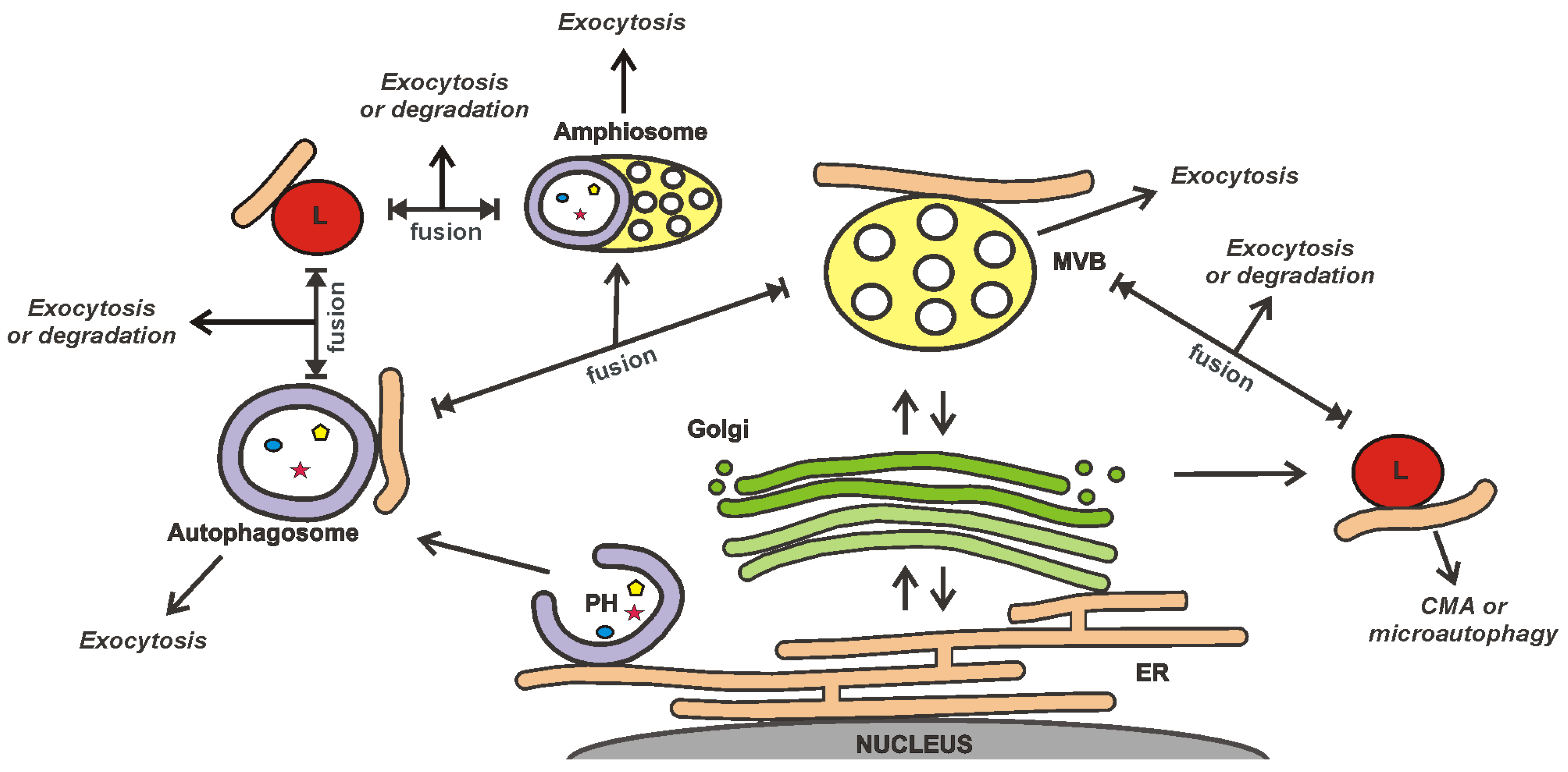
  17. Ponpuak, M.; Mandell, M.A.; Kimura, T.; Chauhan, S.; Cleyrat, C.; Deretic, V. Secretory autophagy. Curr. Opin. Cell Biol. 2015, 35, 106–116.
  18. Gudbergsson, J.M.; Johnsen, K.B. Exosomes and autophagy: Rekindling the vesicular waste hypothesis. J. Cell Commun. Signal. 2019, 13, 443–450.
  19. Buratta, S.; Tancini, B.; Sagini, K.; Delo, F.; Chiaradia, E.; Urbanelli, L.; Emiliani, C. Lysosomal Exocytosis, Exosome Release and Secretory Autophagy: The Autophagic- and Endo-Lysosomal Systems Go Extracellular. Int. J. Mol. Sci. 2020, 21, 2576.
  20. Zhao, X.; Guan, Y.; Liu, F.; Yan, S.; Wang, Y.; Hu, M.; Li, Y.; Li, R.; Zhang, C.X. SNARE Proteins Mediate α-Synuclein Secretion via Multiple Vesicular Pathways. Mol. Neurobiol. 2021, 59, 405–419.
  21. Sagini, K.; Buratta, S.; Delo, F.; Pellegrino, R.M.; Giovagnoli, S.; Urbanelli, L.; Emiliani, C. Drug-Induced Lysosomal Impairment Is Associated with the Release of Extracellular Vesicles Carrying Autophagy Markers. Int. J. Mol. Sci. 2021, 22, 2922.
  22. Leidal, A.M.; Debnath, J. Emerging roles for the autophagy machinery in extracellular vesicle biogenesis and secretion. FASEB Bioadvances 2021, 3, 377–386.
  23. Raiborg, C.; Wenzel, E.M.; Stenmark, H. ER-endosome contact sites: Molecular compositions and functions. EMBO J. 2015, 34, 1848–1858.
  24. Kohler, V.; Aufschnaiter, A.; Büttner, S. Closing the Gap: Membrane Contact Sites in the Regulation of Autophagy. Cells 2020, 9, 1184.
  25. Trivedi, P.C.; Bartlett, J.J.; Pulinilkunnil, T. Lysosomal Biology and Function: Modern View of Cellular Debris Bin. Cells 2020, 9, 1131.
  26. Guo, H.; Chitiprolu, M.; Roncevic, L.; Javalet, C.; Hemming, F.J.; Trung, M.T.; Meng, L.; Latreille, E.; Tanese de Souza, C.; McCulloch, D.; et al. Atg5 Disassociates the V(1)V(0)-ATPase to Promote Exosome Production and Tumor Metastasis Independent of Canonical Macroautophagy. Dev. Cell 2017, 43, 716–730.e717.
  27. Leidal, A.M.; Huang, H.H.; Marsh, T.; Solvik, T.; Zhang, D.; Ye, J.; Kai, F.; Goldsmith, J.; Liu, J.Y.; Huang, Y.H.; et al. The LC3-conjugation machinery specifies the loading of RNA-binding proteins into extracellular vesicles. Nat. Cell Biol. 2020, 22, 187–199.
  28. Ferreira, J.V.; da Rosa Soares, A.; Ramalho, J.; Máximo Carvalho, C.; Cardoso, M.H.; Pintado, P.; Carvalho, A.S.; Beck, H.C.; Matthiesen, R.; Zuzarte, M.; et al. LAMP2A regulates the loading of proteins into exosomes. Sci. Adv. 2022, 8, eabm1140.
  29. Stranford, D.M.; Hung, M.E.; Gargus, E.S.; Shah, R.N.; Leonard, J.N. A Systematic Evaluation of Factors Affecting Extracellular Vesicle Uptake by Breast Cancer Cells. Tissue Eng. Part A 2017, 23, 1274–1282.
  30. Schwager, S.C.; Bordeleau, F.; Zhang, J.; Antonyak, M.A.; Cerione, R.A.; Reinhart-King, C.A. Matrix stiffness regulates microvesicle-induced fibroblast activation. Am. J. Physiol. Cell Physiol. 2019, 317, C82–C92.
  31. Parolini, I.; Federici, C.; Raggi, C.; Lugini, L.; Palleschi, S.; De Milito, A.; Coscia, C.; Iessi, E.; Logozzi, M.; Molinari, A.; et al. Microenvironmental pH is a key factor for exosome traffic in tumor cells. J. Biol. Chem. 2009, 284, 34211–34222.
  32. Endzeliņš, E.; Ābols, A.; Bušs, A.; Zandberga, E.; Palviainen, M.; Siljander, P.; Linē, A. Extracellular Vesicles Derived from Hypoxic Colorectal Cancer Cells Confer Metastatic Phenotype to Non-metastatic Cancer Cells. Anticancer. Res. 2018, 38, 5139–5147.
  33. Jurgielewicz, B.J.; Yao, Y.; Stice, S.L. Kinetics and Specificity of HEK293T Extracellular Vesicle Uptake using Imaging Flow Cytometry. Nanoscale Res. Lett. 2020, 15, 170.
  34. Qiao, L.; Hu, S.; Huang, K.; Su, T.; Li, Z.; Vandergriff, A.; Cores, J.; Dinh, P.U.; Allen, T.; Shen, D.; et al. Tumor cell-derived exosomes home to their cells of origin and can be used as Trojan horses to deliver cancer drugs. Theranostics 2020, 10, 3474–3487.
  35. Sancho-Albero, M.; Navascués, N.; Mendoza, G.; Sebastián, V.; Arruebo, M.; Martín-Duque, P.; Santamaría, J. Exosome origin determines cell targeting and the transfer of therapeutic nanoparticles towards target cells. J. Nanobiotechnol. 2019, 17, 16.
  36. Wang, X.; Cheng, K.; Zhang, G.; Jia, Z.; Yu, Y.; Guo, J.; Hua, Y.; Guo, F.; Li, X.; Zou, W.; et al. Enrichment of CD44 in Exosomes From Breast Cancer Cells Treated With Doxorubicin Promotes Chemoresistance. Front. Oncol. 2020, 10, 960.
  37. Raab-Traub, N.; Dittmer, D.P. Viral effects on the content and function of extracellular vesicles. Nature reviews. Microbiology 2017, 15, 559–572.
  38. Vallhov, H.; Gutzeit, C.; Johansson, S.M.; Nagy, N.; Paul, M.; Li, Q.; Friend, S.; George, T.C.; Klein, E.; Scheynius, A.; et al. Exosomes containing glycoprotein 350 released by EBV-transformed B cells selectively target B cells through CD21 and block EBV infection in vitro. J. Immunol. 2011, 186, 73–82.
  39. Raposo, G.; Stahl, P.D. Extracellular vesicles: A new communication paradigm? Nature reviews. Mol. Cell Biol. 2019, 20, 509–510.
  40. Wang, S.; Li, C.; Yuan, Y.; Xiong, Y.; Xu, H.; Pan, W.; Pan, H.; Zhu, Z. Microvesicles as drug delivery systems: A new frontier for bionic therapeutics in cancer. J. Drug Deliv. Sci. Technol. 2023, 79, 104088.
  41. Böröczky, T.; Dobra, G.; Bukva, M.; Gyukity-Sebestyén, E.; Hunyadi-Gulyás, É.; Darula, Z.; Horváth, P.; Buzás, K.; Harmati, M. Impact of Experimental Conditions on Extracellular Vesicles’ Proteome: A Comparative Study. Life 2023, 13, 206.
  42. Dixson, A.C.; Dawson, T.R.; Di Vizio, D.; Weaver, A.M. Context-Specific regulation of extracellular vesicle biogenesis and cargo selection. Nat. Rev. Mol. Cell Biol. 2023, in press.
  43. Kojima, R.; Bojar, D.; Rizzi, G.; Hamri, G.C.; El-Baba, M.D.; Saxena, P.; Ausländer, S.; Tan, K.R.; Fussenegger, M. Designer exosomes produced by implanted cells intracerebrally deliver therapeutic cargo for Parkinson’s disease treatment. Nat. Commun. 2018, 9, 1305.
  44. Chen, J.; Li, P.; Zhang, T.; Xu, Z.; Huang, X.; Wang, R.; Du, L. Review on Strategies and Technologies for Exosome Isolation and Purification. Front. Bioeng. Biotechnol. 2021, 9, 811971.
  45. Akbar, A.; Malekian, F.; Baghban, N.; Kodam, S.P.; Ullah, M. Methodologies to Isolate and Purify Clinical Grade Extracellular Vesicles for Medical Applications. Cells 2022, 11, 186.
  46. Syromiatnikova, V.; Prokopeva, A.; Gomzikova, M. Methods of the Large-Scale Production of Extracellular Vesicles. Int. J. Mol. Sci. 2022, 23, 522.
  47. Clos-Sansalvador, M.; Monguió-Tortajada, M.; Roura, S.; Franquesa, M.; Borràs, F.E. Commonly used methods for extracellular vesicles’ enrichment: Implications in downstream analyses and use. Eur. J. Cell Biol. 2022, 101, 151227.
  48. Gupta, D.; Zickler, A.M.; El Andaloussi, S. Dosing extracellular vesicles. Adv. Drug Deliv. Rev. 2021, 178, 113961.
  49. Biagiotti, S.; Abbas, F.; Montanari, M.; Barattini, C.; Rossi, L.; Magnani, M.; Papa, S.; Canonico, B. Extracellular Vesicles as New Players in Drug Delivery: A Focus on Red Blood Cells-Derived EVs. Pharmaceutics 2023, 15, 365.
  50. Rajput, A.; Varshney, A.; Bajaj, R.; Pokharkar, V. Exosomes as New Generation Vehicles for Drug Delivery: Biomedical Applications and Future Perspectives. Molecules 2022, 27, 7289.
  51. Di Bella, M.A. Overview and Update on Extracellular Vesicles: Considerations on Exosomes and Their Application in Modern Medicine. Biology 2022, 11, 804.
  52. Davidson, S.M.; Boulanger, C.M.; Aikawa, E.; Badimon, L.; Barile, L.; Binder, C.J.; Brisson, A.; Buzas, E.; Emanueli, C.; Jansen, F.; et al. Methods for the identification and characterization of extracellular vesicles in cardiovascular studies: From exosomes to microvesicles. Cardiovasc. Res. 2023, 119, 45–63.
  53. Bağcı, C.; Sever-Bahcekapili, M.; Belder, N.; Bennett, A.P.S.; Erdener, Ş.E.; Dalkara, T. Overview of extracellular vesicle characterization techniques and introduction to combined reflectance and fluorescence confocal microscopy to distinguish extracellular vesicle subpopulations. Neurophotonics 2022, 9, 021903.
  54. Driedonks, T.; Jiang, L.; Carlson, B.; Han, Z.; Liu, G.; Queen, S.E.; Shirk, E.N.; Gololobova, O.; Liao, Z.; Nyberg, L.H.; et al. Pharmacokinetics and biodistribution of extracellular vesicles administered intravenously and intranasally to Macaca nemestrina. J. Extracell. Biol. 2022, 1, e59.
More
Information
Contributors MDPI registered users' name will be linked to their SciProfiles pages. To register with us, please refer to https://encyclopedia.pub/register : , ,
View Times: 342
Revisions: 3 times (View History)
Update Date: 05 May 2023
1000/1000
Video Production Service