Submitted Successfully!
To reward your contribution, here is a gift for you: A free trial for our video production service.
Thank you for your contribution! You can also upload a video entry or images related to this topic.
Version Summary Created by Modification Content Size Created at Operation
1 -- 2579 2023-05-04 13:08:35 |
2 format correct -5 word(s) 2574 2023-05-06 04:35:42 |

Video Upload Options

Do you have a full video?

Confirm

Are you sure to Delete?
Cite
If you have any further questions, please contact Encyclopedia Editorial Office.
Sivamaruthi, B.S.; Thangaleela, S.; Kesika, P.; Suganthy, N.; Chaiyasut, C. The Mesoporous Silica Nanoparticles. Encyclopedia. Available online: https://encyclopedia.pub/entry/43765 (accessed on 29 July 2024).
Sivamaruthi BS, Thangaleela S, Kesika P, Suganthy N, Chaiyasut C. The Mesoporous Silica Nanoparticles. Encyclopedia. Available at: https://encyclopedia.pub/entry/43765. Accessed July 29, 2024.
Sivamaruthi, Bhagavathi Sundaram, Subramanian Thangaleela, Periyanaina Kesika, Natarajan Suganthy, Chaiyavat Chaiyasut. "The Mesoporous Silica Nanoparticles" Encyclopedia, https://encyclopedia.pub/entry/43765 (accessed July 29, 2024).
Sivamaruthi, B.S., Thangaleela, S., Kesika, P., Suganthy, N., & Chaiyasut, C. (2023, May 04). The Mesoporous Silica Nanoparticles. In Encyclopedia. https://encyclopedia.pub/entry/43765
Sivamaruthi, Bhagavathi Sundaram, et al. "The Mesoporous Silica Nanoparticles." Encyclopedia. Web. 04 May, 2023.
The Mesoporous Silica Nanoparticles
Edit

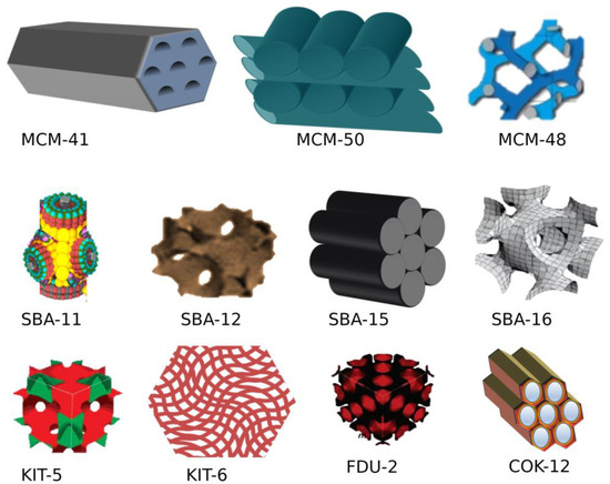
Mesoporous silica nanoparticles (MSNs) are a porous inorganic framework synthesized using the inorganic silica sources sodium silicates or silica tetraethyl orthosilicate as a precursor with quaternary ammonium salts as the surfactant. The size and porosity of MSNs are regulated by various factors such as surfactants, the source of silica, ion strength, aging duration, temperature, and pH. Highly ordered mesoporous silica came into the limelight after the fabrication of the silica-based material Mobile crystalline material 41 (MCM) by Kazuyuki Kuroda’s groups and scientists at Mobil Oil Corporation from aluminium silicate gel. MSNs have a unique porous solid framework with a large surface area facilitating functionalization with various functional groups for targeted drug delivery. MCM-41, MCM-48, and SBA-15 are the most common mesoporous silica materials with two-dimensional hexagonal and three-dimensional cubic structures possessing pore sizes of 2–10 nm.

nanoparticles mesoporous silica nanoparticles inflammatory diseases

1. Types of Mesoporous silica nanoparticles (MSNs)

Different forms of crystalline MSN are formed by altering the cationic surfactant to silica precursor ratio at basic conditions (pH 8–12). If this ratio is less than 1, then hexagonal two-dimensional mobile crystalline material 41 (2D MCM 41) is formed, and if the ratio is greater than 1, three-dimensional cubic MCM-48 is formed. The increase in the ratio leads to 3D lamellar MCM-50 [1]. Other mesostructured materials such as Santa Barbara Amorphous-11 (SBA-11), SBA-12, and SBA-15 were synthesized using poly (alkylene oxide) and alkyl poly (ethylene oxide) (PEO) oligomeric surfactants as templates [2]. FSM-16, folded sheets of mesoporous materials, were produced using quaternary ammonium surfactant and layered polysilicate kanemite as template and adsorbent, respectively, via catalysis. Several other MSNs of varying pore size, shape, and symmetry were fabricated and termed Technical Delft University (TUD-1), Hiroshima Mesoporous Material-33 (HMM-33), and Centrum voor Oppervlaktechemie en Katalyse/Centre for Research Chemistry and Catalysis (COK-12). MCM-41, MCM-50, SBA-15, and SBA-16 are the commonly used MSNs for drug delivery [3]. Figure 1 represent the types of MSN with their unique properties widely used for drug delivery.
Figure 1. Types of mesoporous silica based on shape and morphology. (Figure created using BioRender.com; accessed on 2 December 2022).

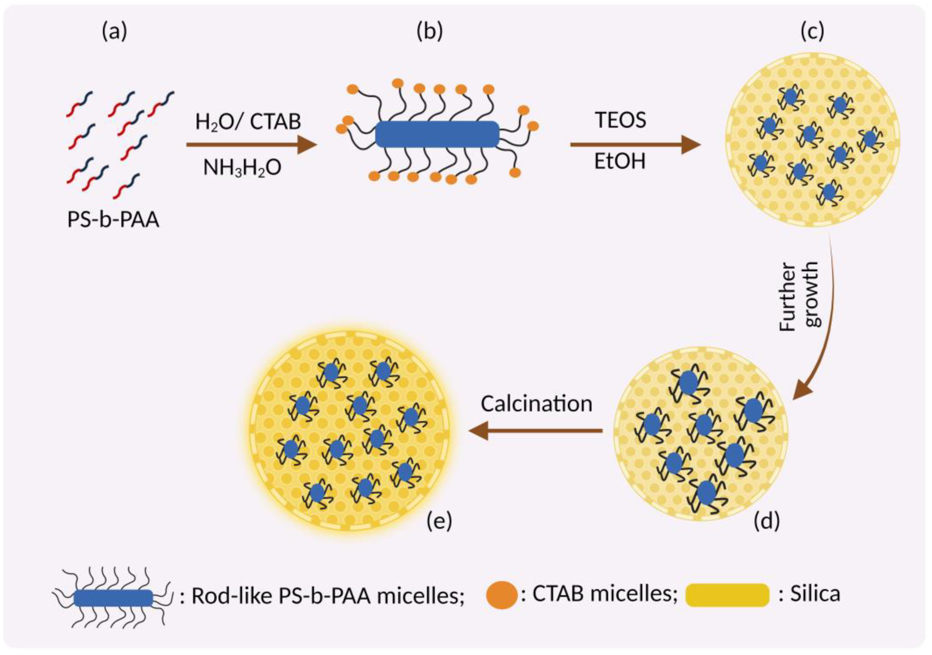
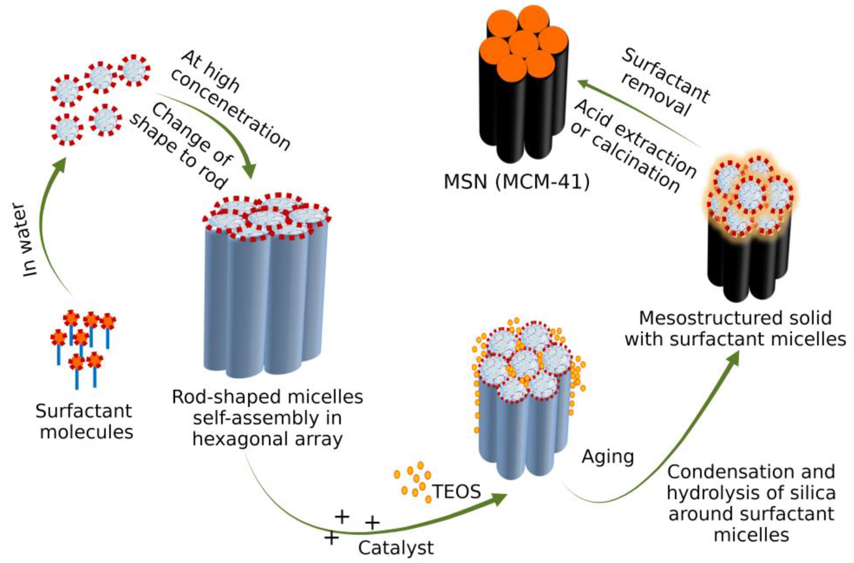
2. Fabrication of MSNs

Stober was a pioneer in synthesizing nanosized silica particles from micron-sized silica; hence it was termed Stober’s method. By manipulating the reaction parameters, MSNs of different sizes, morphology, and pore size were fabricated. Grun et al. modified Stober’s method for synthesizing spherical-shaped MCM-41. As pore size and volume play a key role in DDS, they can be controlled by altering the pH, temperature, surfactant concentration, and silica precursor. The components required to synthesize MSNs are an inorganic silica source/precursor, surfactant, template, and catalyst [4].
Predecessor: The most widely used silica precursors for MSN synthesis include glycerol-derived polyol-based silanes, orthosilicic acid, sodium metasilicate, tetraethyl orthosilicate (TEOS) or tetramethoxysilane (TMOS), Tetra methoxy vinyl silane (TMVS), and tetrakis (2-hydroxyethyl) orthosilicate (THEOS). TEOS and TMOS are poorly soluble and pH-dependent, replaced by glycerol-derived polyol-based silane precursors. MSNs of different shapes such as spherical, rod, and hexagonal tubular shapes can be obtained using 3-aminopropyltrimethoxysilane (APTMS), 3-[2-(2-aminoethyl amino) ethylamino] propyltrimethoxysilane (AEPTMS), ureidopropyltrimethoxysilane (UDPTMS), 3-isocyanatopropyltriethoxysilane (ICPTES), 3-cyanopropyltriethoxysilane (CPTES), and allyltrimethoxysilane (ALTMS) as organoalkoxysilanes precursor [5].
Surfactant: These are classified as below, based on the surface charges [6][7].
  • Cationic surfactants: Surfactants such as cetyl trimethyl ammonium chloride (CTAC), cetyl pyridinium chloride (CPyC), and cetyl tri methyl ammonium bromide (CTAB) with positively charged hydrophilic trimethyl ammonium/pyridinium groups as polar head and a sixteen-carbon hydrocarbon chain nonpolar tail act as a cationic surfactant.
  • Anionic surfactant: Low-cost, eco-friendly anionic surfactants such as sodium salts of alkyl carboxylic acids, phosphoric acid, and sulfonic acid with a negatively charged polar head and hydrocarbon nonpolar tail are widely used as an anionic surfactant.
  • Non-ionic surfactants: Non-ionic triblock copolymers such as alkyl poly (ethylene oxide) (PEO) oligomeric surfactants, poly (alkylene oxide) block copolymers, Triton X-100, polysorbate, and pluronic F 127 with a non-dissociable hydrophilic head which cannot ionize in aqueous solution act as a non-ionic surfactant.
  • Amphoteric/zwitterionic surfactants: Includes surfactants with a net neutral charge such as sodium dodecyl benzene sulfonate (SDBS), sodium dodecyl sulphate (SDS), phospholipids, betaines/sulfobetaine, amino acids, propyl ortho-silicate (TPOS), trimethoxy silane (TMS) and sodium metasilicate (Na2SiO3).
Catalysts: The catalyst used for the sol-gel method depends on the reaction. In an acid-catalyzed reaction, the hydrolysis is faster than the condensation reaction, which forms several small silica particles/networks of gels. In a base-catalyzed reaction, condensation is faster than hydrolysis resulting in larger silica particles/solid spheres forming. Diethanolamine (DEA), triethanolamine (TEA), hydrochloric acid, ammonia, and sodium hydroxide are commonly used as catalysts [8].

3. Mechanism of Formation of MSN

Hydrolysis of tetramethyl orthosilicate (TMOS), the precursor to silicate ions, tends to adsorb on the surfactant micelles. When the charge around the surfactant reduces, hydrolysis starts, followed by condensation of the silica precursor. Intermicellar repulsion prevents the formation of small silica aggregates, leading to the formation of discrete hexagonally ordered mesopores of silica [9] (Figure 2).
Figure 2. Formation of MSNs by Stober’s method. (a) Amphiphilic block copolymer (polystyrene-b-poly(acrylic acid), PS-b-PAA) and CTAB as co-templates in aqueous ammonia solution; (b) Formation of composite rod-like micelles due to electrostatic interaction between CTA+ and PAA; (c) Formation of core part by deposition of silicate of TEOS on rod-like micelles of CTAB-coated PS-b-PAA; (d) Dual mesoporous silica nanoparticles by self-assembly of TEOS with CTAB; (e) Calcination forming the final core-shell dual mesoporous silica spheres. (Figure created using BioRender.com; accessed on 2 December 2022).

3.1. Swelling Shrinking Mechanism

Tetraethyl orthosilicate (TEOS) is used as a precursor without solvent. TEOS in an aqueous solution shows phase separation but, under continuous stirring, forms an emulsion-like system. The addition of TEOS to the amphibolic surfactant CTAB solubilizes in the hydrophobic core forming large micelles. TEOS, on hydrolysis, becomes hydrophilic, is released as a monomer in the aqueous surrounding, and is adsorbed to CTAB via electrostatic attraction. Consumption of TEOS from the hydrophobic core reduces micelle size. Hydrolysis and condensation occur simultaneously, forming the silica shell around the micelles. The adjacent micelles aggregate to form a mesoporous structure [10].

3.2. Sol-Gel Method

The majority of MSN fabricated using the Stober technique undergo the sol-gel process. This process of MSN synthesis involves two stages, nucleation and growth. Nucleation involves hydrolysis of the precursor metal alkoxides TEOS in the presence of catalyst HCl/NH4OH solution to form silanol groups. Under an acidic environment, the alkoxy group (O-R) and a hydroxyl group (O-H) undergo protonation followed by subsequent deprotonation leaving an alcohol group R-OH. The growth stage involves the polymerization of silanol groups or between silanol and ethoxy groups where the -OH group of one partially hydrolyzed protonated precursor condenses with -OH groups of another partially hydrolyzed precursor molecule forming siloxane bridges (Si-O-Si), leading to the formation of oligomers and subsequent condensation, forming a mesoporous silica structure [11]. The hydrolysis and condensation of alkoxide monomers into a colloidal solution (sol) turns into an ordered polymer (gel) network. This process requires an acid or base as the catalyst.
The rate of hydrolysis depends on the ratio of Si/H2O. Under acidic conditions, fast hydrolysis occurs, while in basic conditions, condensation occurs. Multiple condensation results in a chain-like structure in the sol and network-like structure in the gel form. The sol-gel process can be modified to yield particles of the desired size and properties. The sol-gel method was used for the synthesis of MCM-41. CTAB acts as an amphiphilic surfactant and tends to assemble as a spherical micelle in water and precursor polysilicic acid. The silica precursor interacts with the surfactant by electrostatic and hydrogen bonding, forming amorphous silica, and then molding it to a mesoporous structure (Figure 3). The surfactant is removed by repeated washing and then calcinated.
Figure 4. Formation of MCM-41 by sol-gel method. (Figure created using BioRender.com; accessed on 2 December 2022).

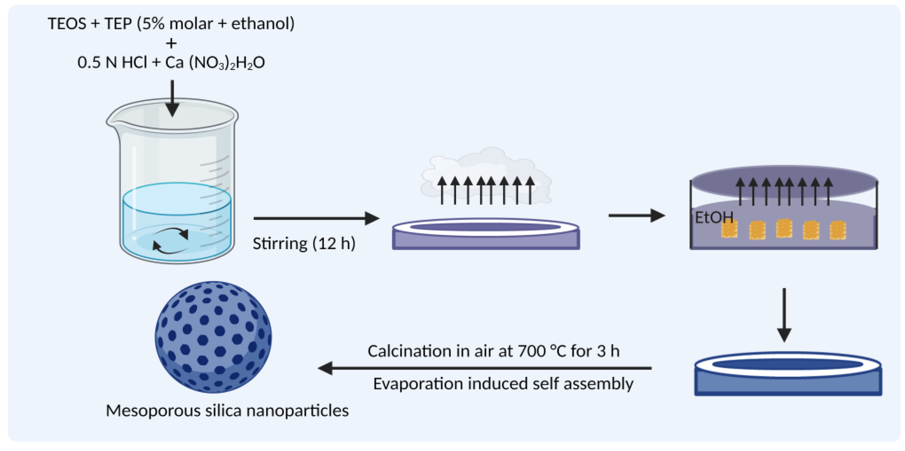
3.3. Evaporation-Induced Self-Assembly (EISA)

In the process of EISA, all the reactants change their concentration during evaporation, resulting in the silica precursor’s organization as the liquid-crystal-like template [12]. The precursor formulation TEOS is prepared in ethanol/water solvent containing the surfactant CTAB which is injected into an aerosol generator with sufficient air flow and pressure to produce a monodispersed solid droplet. During the alcohol evaporation, micelle formation is induced, which converts the silica-surfactant co-assembly into liquid crystal mesophase (Figure 4).
Figure 4. Formation of MSN by the evaporation-induced self-assembly method. (Figure created using BioRender.com; accessed on 2 December 2022).

3.4. Removal of Surfactant after Synthesis

Removal of surfactant from the final MSN product is essential for the following reasons.
  • For biomedical applications, the cytotoxic nature of surfactants affects living cells due to its interaction with the phospholipid layer of cells leading to lysis [13].
  • Surfactants reduce the pore size and volume, affecting the drug-loading efficiency and release kinetics [14].
  • The presence of surfactants affects surface functionalization.
Calcination: MSNs calcinated at 800 °C remove the surfactant forming hollow cylindrical MSNs. Calcination affects the surface functionalization as Si-OH bonds are converted to Si-O-Si bonds at higher temperatures, compressing the surface area and pore size. To overcome the limitation of calcination, a solvent extraction method is used to remove the surfactant [15].
Solvent extraction: The solvent used depends on the surfactant and experimental conditions.
Solvents such as water, ethanol, hydrochloric acids, ammonium nitrate, and other alcohols have been used. Although the solvent extraction method restores the pore size integrity, the complete removal of surfactant is difficult [16].
Chemical-assisted oxidation: Hydrogen peroxide is commonly used as a chemical oxidant to remove surfactants with an oxidation reaction, increasing the pore diameter with reduced pore volume and surface area. The number of silanol groups on the surface of MSNs is higher than in calcinated samples. Oxidants used are H2O2, ozone, KMnO4–H2O2, and NH4ClO4 [17].
Microwave digestion: Removal of surfactant is performed by exposing MSNs in HNO3-H2O2 solution or hexane and ethanol to microwaves for 2 min. This process increases the silanol groups with higher pore size, volume, and large surface area [18].

3.5. Factors That Influence MSN Particle Synthesis

The following factors influence MSNs particle synthesis [19].
(a)
Rate of silica interaction and condensation
(b)
Assembly kinetics, nucleation, and growth rates
(c)
Charge of silica: The rate of silane hydrolysis and condensation of the siloxane bond depends strongly on the charge states.
(d)
pH of reaction mixture: The silica charge depends on the reaction mixture’s pH. Hydrolysis of the Si–OR bond in silanes occurs faster in an acidic/basic environment compared to a neutral solution. At a pH below the isoelectric point (IEP of silica—2.0), the silica is positively charged, and its charge density increases with a decrease in pH. In reaction mixtures with a pH above the IEP, the silica becomes negatively charged and the charge density increases with the pH. At pH 2–4, the negatively charged silicates interact with positively charged surfactants via electrostatic and hydrogen bond interactions. At pH 4–7, the negative charge density of silicate increases; hence interaction with the surfactant occurs only through the electrostatic force [20].
Silica condensation reaction:
≡Si-OH + HO-Si≡ ⇌ ≡Si-O-Si≡ + HOH
≡Si-OH + RO-Si≡ ⇌ ≡Si-O-Si≡ + ROH
where R represents methyl, ethyl, propyl, and butyl groups, etc.
The condensation rate increases with an increase in the negative charge of silicate due to its nucleophilic attack.
(e)
Co-surfactants used: Alcohols such as ethanol and butanol influence the pore size, shape, and flexibility when their concentration increases. An increase in co-surfactant concentration disrupts the spherical shape of MSNs, forming amorphous particles with disordered pore sizes.
(f)
Solvent used: Alcohols such as ethanol, propanol, butanol, and pentanol enhance the pore formation in mesopores. The channel rotations of mesoporous materials are also modified by alcohol. Removal of surfactants after the synthesis of MSNs is promoted by alcohol with a high boiling point which prevents aggregation of MSNs. Long-chain alcohols help MSNs transit from one phase to another.
(g)
Silica sources: Sodium silicates, colloidal solutions, and organosilanes form mesoporous silicate structures more rapidly than other precursors [21].
(h)
Temperature: The critical temperature for determining the final properties of MSNs is between 10 and 130 °C, within which 25 °C is the optimum temperature [22].

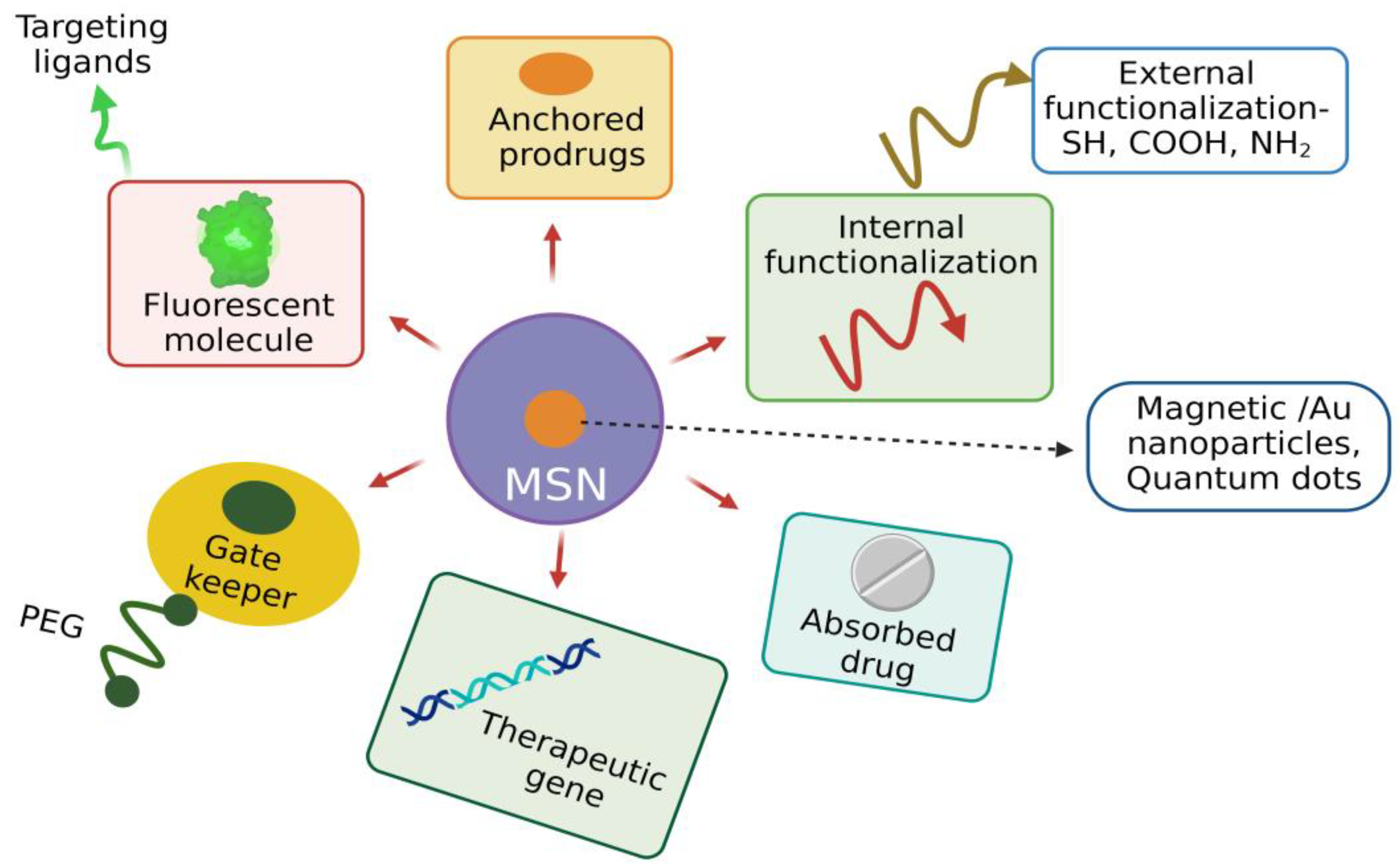
3.6. Functionalization of MSNs

Despite its unique physiochemical properties, the functionalization of MSNs is required to improve their physical and chemical properties to enhance their drug adsorption and sustained release at the target site [23]. The unique architecture of MSNs facilitates functionalization, i.e., introducing various moieties both in the internal and external surface of MSN, which plays a crucial role in targeted drug delivery. MSNs have two functional surfaces: a cylindrical pore surface and an exterior particle surface. The surface-containing silanol group can be selectively functionalized for controlled drug loading and targeted cell-specific drug release [24]. The organic silanes vinyltriethoxy silane (VTES), 3-aminopropyl triethoxysilane (APTES), methoxy-PEG-silane, and 3-mercaptopropyl trimethoxysilane make the fabricated MSNs versatile and promising candidates to perform a specific task.
Various surface-modifying functional groups include carboxyl groups, polymers such as PEG, phospholipids, polyethyleneimine (PEI), phospholipids, organic phosphates, and thiols. For the modification of the negatively charged surface, polymers such as 3-aminopropyltriethoxysilane, polylysines, and polyethyleneimine are used [25][26]. Diethoxydimethyl silane, trimethylchlorosilane, and polymethyl hydrosiloxane reduce the hydrophobicity of MSNs and improves the loading efficiency of hydrophobic drugs [27]. Generally, MSNs are functionalized in the internal/external surfaces, walls of pores, and their entry site. Entrapment of drugs with MSNs is based on various interactive forces such as covalent and hydrogen bonding, van der Waals interaction, and electrostatic forces depending on the functional group present at that site [28][29].
Functionalization influences the MSN’s behavior in biological fluids, such as solubility, dispersibility, drug loading, and drug-release kinetics. The surface modification of MSNs is carried out by co-condensation and post-synthetic modification. Co-condensation involves incorporating functional groups with silica precursors during the synthesis of MSNs. In contrast, the post-synthetic approach involves selectively attaching functional groups to the external and pore surfaces. Co-condensation has several advantages, including the wide range of organoalkoxysilanes that can be used, uniform homogenous distribution of functional groups, and high payload of the functional group without any adverse effect on the MSN structure [30].
Covalent attachment of ligands and imaging agents to MSNs will modify the characteristics of MSNs. For example, incorporating iron oxide into MSNs enhances the magnetic resonance imaging (MRI) properties and their therapeutic output [31]. For the delivery of hydrophobic drugs, the MSN surface is functionalized with hydrophobic groups, as these drugs cannot interact with the hydrophilic surface of MSNs. Delivery of nucleic acids is facilitated by functionalizing the surface of MSNs with the positively charged polymer PEI so that DNA/RNA can bind to the MSNs via the electrostatic force of attraction. This nonviral mode of gene delivery prevents enzymatic degradation of nucleic acids [32]. Functionalization of MSNs with a cationic polymer facilitates the delivery of antibiotics as MSNs interact with negatively charged bacterial cell walls and biofilms and quickly enter microbial cells [33]. MSNs can be conjugated with several ligands such as aptamers, growth factors, and vitamins, facilitating cellular entry by receptor-mediated endocytosis forming endosomes resulting in targeted drug delivery with reduced toxicity [34]. Incorporating photosensitizers into MSNs functionalized with specific ligands to cancer cells promotes photodynamic therapy for killing cancer cells more efficiently than radio- and chemotherapies [35]. Peripheral functionalization of MSNs facilitates the colloidal nature, chemical stability, specific targeting, and pore-gating capacity, improving the MSN’s biocompatibility and nontoxicity.
In addition, surface functionalization promotes the co-delivery of cargo with different properties for the synergistic effect to enhance the therapeutic response of the drug. For example, MSNs coated with PEI and loaded with osteostatin functionalized with SOST siRNA effectively treat osteoporosis [36]. Surface functionalization of MSNs with various moieties based on different therapeutic strategies enhances its application efficiency (Figure 5).
Figure 5. Functionalization of MSN as a promising theranostic agent enhancing its biocompatibility. (Figure created using BioRender.com; accessed on 2 December 2022).

References

  1. Zhang, J.; Luz, Z.; Goldfarb, D. EPR studies of the formation mechanism of the mesoporous materials MCM-41 and MCM-50. J. Phys. Chem. B. 1997, 101, 7087–7094.
  2. Zhao, D.; Huo, Q.; Feng, J.; Chmelka, B.F.; Stucky, G.D. Nonionic triblock and star diblock copolymer and oligomeric surfactant syntheses of highly ordered, hydrothermally stable, mesoporous silica structures. J. Am. Chem. Soc. 1998, 120, 6024–6036.
  3. Zhou, Y.; Quan, G.; Wu, Q.; Zhang, X.; Niu, B.; Wu, B.; Huang, Y.; Pan, X.; Wu, C. Mesoporous silica nanoparticles for drug and gene delivery. Acta Pharm. Sin. B. 2018, 8, 165–177.
  4. Grun, M.; Unger, K.K.; Matsumoto, A.; Tsutsumi, K. Novel Pathways for the Preparation of Mesoporous MCM-41 Materials: Control of Porosity and Morphology. Microporous Mesoporous Mater. 1999, 27, 207–216.
  5. Hu, Y.; Wang, J.; Zhi, Z.; Jiang, T.; Wang, S. Facile synthesis of 3D cubic mesoporous silica microspheres with a controllable pore size and their application for improved delivery of a water-insoluble drug. J. Colloid Interface Sci. 2011, 363, 410–417.
  6. Bonifaz, L.; Bonnyay, D.; Mahnke, K.; Rivera, M.; Nussenzweig, M.C.; Steinman, R.M. Efficient targeting of protein antigen to the dendritic cell receptor DEC-205 in the steady state leads to antigen presentation on major histocompatibility complex class I products and peripheral CD8+ T cell tolerance. J. Exp. Med. 2002, 196, 1627–1638.
  7. Mirzaei, M.; Zarch, M.B.; Darroudi, M.; Sayyadi, K.; Keshavarz, S.T.; Sayyadi, J.; Fallah, A.; Maleki, H. Silica mesoporous structures: Effective nanocarriers in drug delivery and nanocatalysts. Appl. Sci. 2020, 10, 7533.
  8. Selvarajan, V.; Obuobi, S.; Ee, P.L.R. Silica nanoparticles—a versatile tool for the treatment of bacterial infections. Front. Chem. 2020, 8, 602.
  9. Blin, J.L.; Impéror-Clerc, M. Mechanism of self-assembly in the synthesis of silica mesoporous materials: In situ studies by X-ray and neutron scattering. Chem. Soc. Rev. 2013, 42, 4071–4082.
  10. Yi, Z.; Dumée, L.F.; Garvey, C.J.; Feng, C.; She, F.; Rookes, J.E.; Mudie, S.; Cahill, D.M.; Kong, L. A new insight into growth mechanism and kinetics of mesoporous silica nanoparticles by in situ small angle X-ray scattering. Langmuir 2015, 31, 8478–8487.
  11. Danks, A.E.; Hall, S.R.; Schnepp, Z. The evolution of “sol–gel” chemistry as a technique for materials synthesis. Mater. Horiz. 2016, 3, 91–112.
  12. Fontecave, T.; Boissiere, C.; Baccile, N.; Plou, F.J.; Sanchez, C. Using evaporation-induced self-assembly for the direct drug templating of therapeutic vectors with high loading fractions, tunable drug release, and controlled degradation. Chem. Mater. 2013, 25, 4671–4678.
  13. Zhao, Q.; Wu, B.; Shang, Y.; Huang, X.; Dong, H.; Liu, H.; Chen, W.; Gui, R.; Li, J. Development of a nano-drug delivery system based on mesoporous silica and its anti-lymphoma activity. Appl. Nanosci. 2020, 10, 3431–3442.
  14. Hoffmann, F.; Fröba, M. Vitalising porous inorganic silica networks with organic functions—PMOs and related hybrid materials. Chem. Soc. Rev. 2011, 40, 608–620.
  15. Fuertes, A.B. Synthesis of ordered nanoporous carbons of tunable mesopore size by templating SBA-15 silica materials. Microporous Mesoporous Mater. 2004, 67, 273–281.
  16. Yang, L.M.; Wang, Y.J.; Luo, G.S.; Dai, Y.Y. Simultaneous removal of copolymer template from SBA-15 in the crystallization process. Microporous Mesoporous Mater. 2005, 81, 107–114.
  17. Büchel, G.; Denoyel, R.; Llewellyn, P.L.; Rouquerol, J. In situ surfactant removal from MCM-type mesostructures by ozone treatment. J. Mater. Chem. 2001, 11, 589–593.
  18. Manet, S.; Schmitt, J.; Impéror-Clerc, M.; Zholobenko, V.; Durand, D.; Oliveira, C.L.; Pedersen, J.S.; Gervais, C.; Baccile, N.; Babonneau, F.; et al. Kinetics of the formation of 2D-hexagonal silica nanostructured materials by nonionic block copolymer templating in solution. J. Phys. Chem. B 2011, 115, 11330–11344.
  19. Tella, J.O.; Adekoya, J.A.; Ajanaku, K.O. Mesoporous silica nanocarriers as drug delivery systems for anti-tubercular agents: A review. R. Soc. Open Sci. 2022, 9, 220013.
  20. Yu, J.; Shi, J.L.; Chen, H.R.; Yan, J.N.; Yan, D.S. Effect of inorganic salt addition during synthesis on pore structure and hydrothermal stability of mesoporous silica. Microporous Mesoporous Mater. 2001, 46, 153–162.
  21. Bharti, C.; Nagaich, U.; Pal, A.K.; Gulati, N. Mesoporous silica nanoparticles in target drug delivery system: A review. Int. J. Pharm. Investig. 2015, 5, 124–133.
  22. Kleitz, F.; Marlow, F.; Stucky, G.D.; Schüth, F. Mesoporous silica fibers: Synthesis, internal structure, and growth kinetics. Chem. Mater. 2001, 13, 3587–3595.
  23. Natarajan, S.K.; Selvaraj, S. Mesoporous silica nanoparticles: Importance of surface modifications and its role in drug delivery. RSC Adv. 2014, 4, 14328–14334.
  24. Hoffmann, F.; Cornelius, M.; Morell, J.; Fröba, M. Silica-based mesoporous organic– inorganic hybrid materials. Angew. Chem. Int. Ed. 2006, 45, 3216–3251.
  25. Tang, Q.; Xu, Y.; Wu, D.; Sun, Y. A study of carboxylic-modified mesoporous silica in controlled delivery for drug famotidine. J. Solid State Chem. 2006, 179, 1513–1520.
  26. Hartono, S.B.; Gu, W.; Kleitz, F.; Liu, J.; He, L.; Middelberg, A.P.; Yu, C.; Lu, G.Q.; Qiao, S.Z. Poly-L-lysine functionalized large pore cubic mesostructured silica nanoparticles as biocompatible carriers for gene delivery. ACS Nano 2012, 6, 2104–2117.
  27. Tang, Q.; Chen, Y.; Chen, J.; Li, J.; Xu, Y.; Wu, D.; Sun, Y. Drug delivery from hydrophobic-modified mesoporous silicas: Control via modification level and site-selective modification. J. Solid State Chem. 2010, 183, 76–83.
  28. Slowing, I.I.; Vivero-Escoto, J.L.; Trewyn, B.G.; Lin, V.S.Y. Mesoporous silica nanoparticles: Structural design and applications. J. Mater. Chem. 2010, 20, 7924–7937.
  29. Lv, X.; Zhang, L.; Xing, F.; Lin, H. Controlled synthesis of monodispersed mesoporous silica nanoparticles: Particle size tuning and formation mechanism investigation. Microporous Mesoporous Mater. 2016, 225, 238–244.
  30. Radu, D.R.; Lai, C.Y.; Wiench, J.W.; Pruski, M.; Lin, V.S.Y. Gatekeeping layer effect: A poly (lactic acid)-coated mesoporous silica nanosphere-based fluorescence probe for detection of amino-containing neurotransmitters. J. Am. Chem. Soc. 2004, 126, 1640–1641.
  31. Ye, F.; Laurent, S.; Fornara, A.; Astolfi, L.; Qin, J.; Roch, A.; Martini, A.; Toprak, M.S.; Muller, R.N.; Muhammed, M. Uniform mesoporous silica coated iron oxide nanoparticles as a highly efficient, nontoxic MRI T (2) contrast agent with tunable proton relaxivities. Contrast Media Mol. Imaging 2012, 7, 460–468.
  32. Meng, H.; Mai, W.X.; Zhang, H.; Xue, M.; Xia, T.; Lin, S.; Wang, X.; Zhao, Y.; Ji, Z.; Zink, J.I.; et al. Codelivery of an optimal drug/siRNA combination using mesoporous silica nanoparticles to overcome drug resistance in breast cancer in vitro and in vivo. ACS Nano 2013, 7, 994–1005.
  33. Álvarez, E.; González, B.; Lozano, D.; Doadrio, A.L.; Colilla, M.; Izquierdo-Barba, I. Nanoantibiotics Based in Mesoporous Silica Nanoparticles: New Formulations for Bacterial Infection Treatment. Pharmaceutics 2021, 13, 2033.
  34. Frickenstein, A.N.; Hagood, J.M.; Britten, C.N.; Abbott, B.S.; McNally, M.W.; Vopat, C.A.; Patterson, E.G.; MacCuaig, W.M.; Jain, A.; Walters, K.B.; et al. Mesoporous Silica Nanoparticles: Properties and Strategies for Enhancing Clinical Effect. Pharmaceutics 2021, 1, 570.
  35. Ahir, M.; Upadhyay, P.; Ghosh, A.; Sarker, S.; Bhattacharya, S.; Gupta, P.; Ghosh, S.; Chattopadhyay, S.; Adhikary, A. Delivery of dual miRNA through CD44-targeted mesoporous silica nanoparticles for enhanced and effective triple-negative breast cancer therapy. Biomater. Sci. 2020, 8, 2939–2954.
  36. Paris, J.L.; Vallet-Regí, M. Mesoporous Silica Nanoparticles for Co-Delivery of Drugs and Nucleic Acids in Oncology: A Review. Pharmaceutics 2020, 12, 526.
More
Information
Subjects: Others
Contributors MDPI registered users' name will be linked to their SciProfiles pages. To register with us, please refer to https://encyclopedia.pub/register : , , , ,
View Times: 1.1K
Revisions: 2 times (View History)
Update Date: 06 May 2023
1000/1000
Video Production Service