Submitted Successfully!
To reward your contribution, here is a gift for you: A free trial for our video production service.
Thank you for your contribution! You can also upload a video entry or images related to this topic.
Version Summary Created by Modification Content Size Created at Operation
1 -- 4181 2023-04-22 23:13:23 |
2 format correct -1 word(s) 4180 2023-04-23 03:04:57 | |
3 format correct Meta information modification 4180 2023-04-23 03:05:35 | |
4 format correct -3 word(s) 4177 2023-04-24 09:37:04 |

Video Upload Options

Do you have a full video?

Confirm

Are you sure to Delete?
Cite
If you have any further questions, please contact Encyclopedia Editorial Office.
Pilvenyte, G.; Ratautaite, V.; Boguzaite, R.; Ramanavicius, A.; Viter, R.; Ramanavicius, S. MIP Application for the Detection of Cancer Biomarkers. Encyclopedia. Available online: https://encyclopedia.pub/entry/43344 (accessed on 29 July 2024).
Pilvenyte G, Ratautaite V, Boguzaite R, Ramanavicius A, Viter R, Ramanavicius S. MIP Application for the Detection of Cancer Biomarkers. Encyclopedia. Available at: https://encyclopedia.pub/entry/43344. Accessed July 29, 2024.
Pilvenyte, Greta, Vilma Ratautaite, Raimonda Boguzaite, Arunas Ramanavicius, Roman Viter, Simonas Ramanavicius. "MIP Application for the Detection of Cancer Biomarkers" Encyclopedia, https://encyclopedia.pub/entry/43344 (accessed July 29, 2024).
Pilvenyte, G., Ratautaite, V., Boguzaite, R., Ramanavicius, A., Viter, R., & Ramanavicius, S. (2023, April 22). MIP Application for the Detection of Cancer Biomarkers. In Encyclopedia. https://encyclopedia.pub/entry/43344
Pilvenyte, Greta, et al. "MIP Application for the Detection of Cancer Biomarkers." Encyclopedia. Web. 22 April, 2023.
MIP Application for the Detection of Cancer Biomarkers
Edit

Biomarkers can provide critical information about cancer and many other diseases; therefore, developing analytical systems for recognising biomarkers is an essential direction in bioanalytical chemistry. Molecularly imprinted polymers (MIPs) have been applied in analytical systems to determine biomarkers. The most attractive way to replace natural biological recognition systems is based on applying molecularly imprinted polymers.

molecularly imprinted polymer (MIP) conducting polymer (CP) electrochemical sensor cancer biosensors prostate cancer biomarker breast cancer biomarker epithelial ovarian cancerbiomarker hepatocellular carcinoma biomarker biomarker detection diagnostics

1. Introduction

Biomarkers can provide critical information about cancer and many other diseases; therefore, developing analytical systems for recognising biomarkers is an essential direction in bioanalytical chemistry [1], therefore, biosensor development technologies are evolving rapidly. Conducting polymers are frequently used to advance the analytical performance of sensors [2][3][4][5][6][7][8][9][10][11][12][13][14][15], particularly for the selectivity towards targeted analytes [15][8]. MIPs are used as artificial receptors to detect target molecules in sensors that act as smart electrochemical output devices. Preparation of the MIP involves several steps (Figure 1). Molecularly imprinted polymers (MIPs) are created by polymerising functional monomers on different types of electrode surfaces such as pencil graphite electrode [2], graphite electrode [3], boron-doped nanocrystalline diamond [4][5], platinum electrode [6][7], or the gold-coated quartz crystal microbalance [8][9] and surface plasmon resonance sensors disks [10] to form complementary sites for target molecules. After analytes are removed, they leave fitting imprinted cavities to recognize target molecules such as low molar mass molecules (e.g. theophylline [4][5], caffeine [8], histamine [11], uric acid [9][12], tryptophan [3], etc.), or large molar mass substances such as proteins [6][7][13] or DNA [2], or even living entities such as bacteria [14]. The high specificity, selectivity, sensitivity for target molecules, and detection ability of MIP-based electrochemical biosensors open vast possibilities for disease diagnostics.

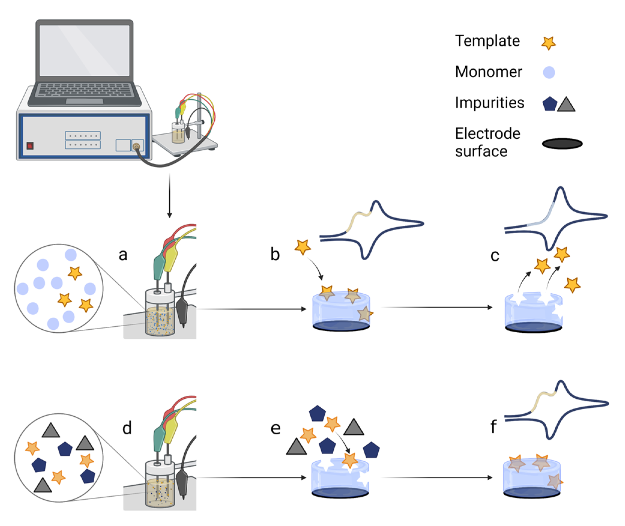
Figure 1. Scheme of the electrochemical polymerisation of the MIP directly on the electrode and the application of the MIP-based electrochemical biosensor: (a) polymerisation mixture preparation; (b) polymerisation of the polymer with template imprints layer on the electrode; (c) extraction of the template from the MIP; (d) application of the MIP-based sensor; (e) competitive interaction of target molecules on the MIP in presence of interferents; (f) detection of target molecules. All drawings were designed using Biorender.com, accessed on 26 December 2022.

2. Biomarker of Prostate Cancer—PSA

A prostate-specific antigen (PSA) is a protein produced by normal and malignant prostate gland cells. The PSA test measures the amount of PSA in the blood. Any PSA value greater than 10.0 ng/mL is considered a positive case, indicating a high risk of prostate cancer. Meanwhile, PSA values ranging from 4.0 to 10.0 ng/mL are a ‘grey area’ and indicate a higher-than-average risk of prostate cancer; thus, more tests may be needed. Lastly, PSA values below 4.0 ng/mL are considered negative, and such PSA values indicate a low risk of prostate cancer [16].
A novel dual-modality MIP-based biosensor for PSA and myoglobin (Myo) was demonstrated in a study by Karami et al. [17] (Figure 2D). The proposed electrochemical sensor was dedicated to simultaneously detecting two target analytes in serum and urine. For this purpose, a disposable gold screen-printed electrode (SPE) surface was combined with a molecular imprinting technique and an immunoassay approach. The MIP was obtained via free-radical polymerisation of methyl acrylate as a monomer and N, N’-methylenebisacrylamide as a crosslinker in the presence of the nanocomposite, which was made of Fe3O4 nanoparticles decorated with multi-wallet carbon nanotubes (MWCNT), graphene oxide, and specific antibodies for PSA. LOD of PSA and Myo was found to be 5.4 pg/mL and 0.83 ng/mL, respectively. The selectivity of the proposed sensor was evaluated regarding a relatively long list of interferents. In total, nine interferents were used: bovine serum albumin, cortisol, epidermal growth factor receptor, carcinoembryonic antigen, thrombin antigen, human thrombin, vascular endothelial growth factor, human albumin serum, and neuron-specific enolase. The selectivity test demonstrated that the proposed sensor is highly specific for the target analytes. The accuracy, reproducibility, stability, and regeneration test of the sensor and immunosensor are very promising for everyday analysis. In real samples, recovery of PSA and Myo was favourable.
Figure 2. MIP application for detection of cancer biomarkers. All drawings were designed using Biorender.com based on the reviewed articles. (A) The Ppy-based MIP with PSA imprints on a gold screen-printed electrode. (B) A gold electrode with glutaraldehyde–cysteamine matrix pretreatment coated with polytoluidine blue for analyte PSA entrapment. (C) Thiolated aptamer–PSA complex was immobilised on the gold electrode coated with polydopamine for analyte PSA entrapment. (D) A screen-printed electrode with self-assembled dithiodipropionic acid di(N-hydroxysuccinimide ester) with immobilised analytes, PSA, and Myo coated with polymethyl acrylate for imprinting. To detect PSA, MIP was exposed to Fe3O4 nanoparticles decorated with multi-walled carbon nanotubes, graphene oxide, and specific antibodies for PSA. (E) A gold screen-printed electrode with self-assembled cysteamine coated with Ppy for analyte CA-125 entrapment. (F) A gold screen-printed electrode coated with poly(2-aminophenol) for analyte CA15-3 entrapment. (G) The MIP based on the toluidine blue (TB) film was polymerised via glutaraldehyde on self-assembled 8-amino-1-octane thiol-modified AuSPE for analyte CA15-3 entrapment. (H) A fluorine-doped tin oxide-glass electrode coated with Ppy for analyte CA15-3 entrapment. (I) A glassy carbon electrode with polythionine and gold nanoparticles coated with polydopamine for analyte AFP entrapment. (J) Laser-scribed graphene electrode with gold nanoparticles coated with PEDOT for analyte HER-2 entrapment. (K) A gold screen-printed electrode coated with polyphenol for analyte HER-2 entrapment. All drawings were designed using Biorender.com, accessed on 26 December 2022.
For PSA detection, Abbasy et al. [18] developed a MIP biosensor for monitoring PSA using a poly(toluidine blue) on a modified AuE. The imprinted poly(toluidine blue) film was electropolymerised on the surface of an electrode in a pre-formed glutaraldehyde-cysteamine (GA-Cys A) matrix (Figure 2B). Due to the cross-linking procedure via the Au–S bonds, the resilience of the MIP against degradation was improved. According to the differential pulse voltammetry method, the biosensor showed a linear response from 0.001–0.06 μg/mL and a LOD of 0.001 μg/mL. This sensor was used to measure PSA in the plasma samples. Sadly, this sensor’s selectivity was tested regarding only two substances: human cancer antigen CEA and human serum albumin. Yazdani et al. [19] developed a more precise sensor by choosing the Py as a functional monomer and was electropolymerised on an AuSPE. Since organic and alkaline solvents could not be used due to the lack of stability of the SPEs, the PSA template was removed overnight with an oxalic acid solution. The LOD of 2.0 pg/mL and a linear range of 0.01–4 ng/mL were obtained (Figure 2A).
Nevertheless, this study did not demonstrate any selectivity tests with interfering substances. Results of the blood serum samples and simple method confirm that the nano biosensor could be successfully used in clinical diagnosis to detect a PSA. Jolly et al. [20] developed a more complex hybrid MIP/aptamer sensor for PSA (Figure 2C). First, a thiolated aptamer–PSA complex was immobilised on the AuE, then the dopamine was electropolymerised, and the PSA template was removed. Polydopamine (PDA) exhibits melanin-like conducting properties and can be used to prepare energy devices such as supercapacitors. PDA can provide numerous ways to provide bioelectronic sensors with excellent biocompatibility, self-repair ability, universal coating capability, and electrical conductivity [21]. The use of aptamers enhances selectivity and specificity. The LOD was 1pg/mL. Such aptamer-based MIPs for PSA were tested in a selectivity test regarding the capacitance changes when the human Kallikrein 2 protein and human serum albumin were used. The authors state that the human Kallikrein 2 protein (or Human glandular Kallikrein 2) is 80% homologous to PSA. The selectivity test demonstrated that the proposed sensor was selective and sensitive to PSA when the interfering substances were used in the samples. Tamboli et al. [22] improved this work with a MIP-based electrochemical sensor using a MOSFET method. The hybrid synthetic receptors were created by immobilising an aptamer–PSA complex on the gold electrode and putting it through multiple cycles of dopamine electropolymerisation. Highly specific cavities were formed, resulting in a very low LOD of 0.1 pg/mL and 1–10 pg/mL in diluted human plasma.
The researchs show that the PDA matrix on the AuE may be the most suitable model for PSA detection as they result in the lowest LOD value [20][22]. The MOSFET method provides precision, making LOD ten times lower than by using differential pulse voltammetry. The biosensor with Ppy is easy to fabricate, but washing takes time, and the resulting LOD is similar to that of the biosensor based on PDA. A dual-modality MIP-based biosensor shows promising results but is challenging to prepare, and antibody stability decreases with increasing temperature. The least sensitive sensor was obtained using a polytoluidine blue. A comparison of the selectivity tests used in the reviewed studies demonstrates that selectivity is the under-evaluated aspect of the electrochemical sensor development. The selectivity tests demonstrate the MIP’s significant precedence over the NIP, but a higher number of interfering substances could often be used in the interfering molecules test. All reviewed studies for PSA detection used AuE or AuSPE.

3. Biomarkers of Breast Cancer

3.1. CA15-3

Cancer antigen 15-3 (CA15-3) is a protein mainly produced by specific breast cancer cells. Some people with breast, lung, pancreatic, ovarian, and prostate cancer or non-cancerous diseases, such as endometriosis, pelvic inflammatory disease, liver disease, or during pregnancy, have higher levels of it in their blood. The increase in CA15-3 is most commonly due to breast cancer with metastases [23] and higher levels (≥30 U/mL values in the blood are considered high) when cancer has spread to the bones, liver, or both [24].
In the study of Ribeiro et al. [25], a disposable biosensor for breast cancer biomarker CA15-3 recognition via molecular imprinting using polytoluidine blue as a conducting film was described (Figure 2G). The polytoluidine blue film was electropolymerised via glutaraldehyde on a preformed SAM on an AuSPE. This structure gave greater stability to the adhesion of the film on the electrode surface. The biosensor results showed a linear response from 0.10 U/mL to 100 U/mL and a LOD below 0.10 U/mL. Given that CA15-3 is a high molecular weight glycoprotein (300–450 kDa), the selectivity assay of diluted artificial serum containing small molecules ranging from 58 to 180 Da and one larger molecule, such as bovine serum albumin (66 kDa), does not appear to be sufficient to compare precise selectivity. However, with further studies, this new biosensor appears to have the potential for point-of-care use, as it has a wide concentration range that allows direct analysis of samples above or well below the cut-off value of 30 U/mL. Santos et al. [26] also conducted a polymer to develop a sensor for CA15-3. Pyrrole was electropolymerised on a fluorine-doped tin oxide conductive glass. Ethanol was chosen for easy template removal. The biosensor resulted in a LOD of 1.07 U/mL, with a linear response of 1.44–13.2 U/mL. This sensor’s selectivity was evaluated using only two molecules: carcinoembryonic antigen and interleukin 6. Pacheco et al. [27] developed a MIP-based sensor to detect CA15-3 with a non-conducting poly(2-aminophenol) film on the surface of an AuE that resulted in a LOD of 1.5 U/mL (Figure 2F). Another breast cancer biomarker, the human epidermal growth factor receptor 2 extracellular domain, and cystatin C, a kidney function biomarker, were chosen to evaluate selectivity, and the results indicated that the human epidermal growth factor receptor 2 extracellular domain may influence the analysis. This MIP sensor provides easy-to-prepare, low-cost monitoring but with lower sensitivity than the sensors used in the previously mentioned studies.
AuSPE and FTO glass were included in the reviewed studies for CA15-3 detection. AuSPE with a polytoluidine blue matrix [25] resulted in the lowest LOD, following fluorine-doped tin oxide glass with a Ppy matrix [26], resulting in a similar LOD. The biosensor with a non-conducting polymer matrix poly(o-aminophenol) [27] showed the least sensitive detection compared to previous MIP sensors with polytoluidine blue and Ppy. In conclusion, based on the LOD, these electrochemical MIP biosensors demonstrate that molecular imprinting is suitable for detecting breast cancer CA15-3 biomarkers.

3.2. HER-2

Human epidermal growth factor receptor 2 (HER-2) is a significant biomarker in invasive breast cancer, according to the 2007 American Society of Clinical Oncology guidelines. Clinical HER-2 testing is performed using immunohistochemistry, ELISA analysis of serum or tumour cytosol, and the Western blot test for HER-2 protein overexpression. Immunohistochemistry is the most frequently used test to evaluate the HER-2 status in breast cancer and screening [28], though there are issues with the accuracy and consistency of immunohistochemistry results acquired from different laboratories. The ELISA approach is expensive and lacks clinical evidence for prognostic usefulness or comparability to other tests [29]. The cut-off value is 15.0 ng/mL for the serum-HER-2 concentration [30].
Lahcen et al. [31] developed a point-of-care nanostructured sensor for the cancer biomarker HER-2 (Figure 2J). The biosensing platform was fabricated of laser-scribed graphene (LSG) electrodes modified with nanostructured gold and MIP. LSG has high electrical and thermal conductivity, excellent mechanical stability, and a large specific surface area. The gold nanostructures enhance the surface area and promote protein adsorption, and MIP enhances selectivity and sensitivity. The functional monomer for conductive MIP was chosen from Py, aminothiophenol, and 3,4-ethylenedioxythiophene. First, the pyrrole was polymerised on LSG–gold nanostructures, and the results showed increased charge transfer resistance. The plausible reason for this was the too-thick Ppy layer. Next, polyaminothiophenol showed better interaction with the gold nanostructures layer due to thiol groups, but the peak current response was significantly hindered. Finally, the poly(3,4-ethylenedioxythiophene) polymer showed the best results among the three polymers on LSG–gold nanostructures. The achieved LOD of 0.43 ng/mL is clinically relevant for monitoring the breast cancer biomarker HER-2. The selectivity of the biosensor was tested in the presence of seven other molecules such as cardiac troponin-I, cardiac troponin-T, cardiac troponin-C, glucose, dopamine, myoglobin, and cholesterol. The results showed suitable selectivity for HER-2. Pacheco et al. [32] developed a sensor for breast cancer biomarker (HER-2-ECD) detection using a molecularly imprinted electrochemical sensor with non-conducting polymer polyphenol (Figure 2K). The LOD was 1.6 ng/mL, higher than the previous conducting MIP sensor. The sensor’s response to two interfering proteins was tested for the selectivity evaluation: CA 15-3 and cystatin C. The results showed that CA 15-3 may interfere with the analysis.
Both methods used gold, one combining AuSPE with a polyphenol matrix [32], the other using LSG with gold nanostructures and a poly(3,4-ethylenedioxythiophene) [31]. The results showed that the sensor with poly(3,4-ethylenedioxythiophene) had a lower LOD of HER-2 than the non-conducting polyphenol.

4. Biomarker of Epithelial Ovarian Cancer—CA-125

Cancer antigen 125 (CA-125) is a standard tumour biomarker used to diagnose epithelial ovarian cancer and monitor women who have been diagnosed with epithelial ovarian cancer during or after therapy. Because of its stability and widespread availability, CA-125 is still regarded as the best available biomarker for epithelial ovarian cancer. According to the clinical guidelines for ovarian cancer published by the National Institute for Health and Care Excellence, the most frequently quoted reference range for CA-125 in the diagnosis of epithelial ovarian cancer is 0–35 IU/mL [33]. The label-free amperometric immunosensor [34] is one of many approaches used with immunoassays. Measurements of CA-125 can be made using immunoradiometric assays, electrochemical immunoassays, ELISA, electrochemical immunosensors, electrochemiluminescence immunosensors on paper, quartz crystal microbalance, surface plasmon resonance, and plasmon resonance scattering. While sensitive, immunoradiometric tests, enzyme immunoassays, and ELISA are also complex, time-consuming, expensive, and imprecise, and toxically reagent-intensive. Immobilisation of immunoreagent onto the electrode surface is another issue with the electrochemical enzyme immunoassay [35].
Rebelo et al. [36] designed a MIP to determine CA-125 via the electropolymerisation of pyrrole on an AuSPE. For quantification, the square wave voltammetry and surface plasmon resonance were used. Results showed that biosensors based on electrochemical transduction presented a better performance with a lower LOD of 0.01 U/mL and a more extensive linear range between 0.01 and 500 U/mL. The sensor’s analytical performance was tested in an artificial serum medium to compare it to the results obtained in the buffer solution. Exposing the MIP sensor to artificial serum increased sensitivity, LOD (0.1 U/mL), and the linear concentration range was reduced, resulting in 0.1–500 U/mL. The quantification of CA-125 in serum is achievable even with a reduced working range because its threshold value is 35 U/mL. Furthermore, selectivity was demonstrated using interferences: CA 15-3 and artificial serum containing NaCl, NaHCO3, and bovine serum albumin. This biosensor provides accurate findings for the measurement of CA-125 based on the LOD (0.01 U/mL). For the determination of CA-125, several other detection approaches covering molecular imprinting were described, including a non-conducting polymer MIP sensor using polyphenol [37], resulting in a higher LOD (of 0.5 U/mL) than a reviewed article with a conducting polymer (LOD 0.01 U/mL) [36], magnetic MIP sensor (for CA-125 and CA15-3) [38], and the synthetic receptor built using the molecular imprinting approach [35].

5. Biomarker of Hepatocellular Carcinoma—AFP

Despite the several biomarkers proposed for hepatocellular carcinoma, AFP is still the most widely used criterion. In addition, patients with a bile duct, gastric, lung, certain ovarian and teratocarcinoma, or embryonal carcinoma of the testis carcinoma also have higher serum amounts [23].
AFP is a 70-kDa glycoprotein that transports bilirubin, fatty acids, retinoids, steroids, heavy metals, phytoestrogens, flavonoids, and perhaps medicines. Except for pregnant women, serum AFP levels rapidly decline after birth, leaving only negligible amounts (5 ng/mL) in healthy individuals [39]. According to the American Board of Internal Medicine, the reference range in serum is ˂10 ng/mL [40]. The 400 ng/mL AFP threshold in serum is more accurate in detecting hepatocellular carcinoma than the 200 ng/mL criterion [41]. Lai et al. [42] introduced a modified electrochemical MIP immunosensor for the detection of AFP (Figure 2I). MIP was prepared via electropolymerisation of the dopamine using AFP templates on a conducting polymer polythionine film with a fast charge transfer capacity to detect the AFP target molecules. Modifying GCE with gold nanoparticles and polythionine provided high conductivity, stability, and adequate biocompatibility, resulting in a low LOD of 0.8138 pg/mL. Good sensor recoveries were obtained using spiked human serum samples, although the selectivity of the biosensor was not investigated. With further improvements, the sensor may be suitable for monitoring AFP in patients. A dual MIP-based biosensor was developed for the AFP and carcinoembryonic antigen (CEA) [43]. For template imprinting, electropolymerisation of Py on the FTO electrode was performed in the presence of methyl orange, the AFP, and CEA antigens. Pyrrole monomers polymerise around the methyl orange template due to electrostatic interactions and hydrogen bonding. Methyl orange is vital in producing molecularly imprinted Ppy because it promotes the formation of Ppy rectangular nanotubes (Figure 3). Ppy conductivity increases with the increasing specific surface area, regardless of morphology [44], resulting in higher sensor sensitivity. CEA and AFP had linear ranges of 5–104 and 10–104 pg/mL and detection limits of 1.6 and 3.3 pg/mL, respectively. The biosensor provided accurate results for AFP and CEA determination in serum samples, as well as high sensitivity and stability over five weeks period. The selectivity of the developed sensor was evaluated against various interferences, PSA, HE4, CA19-9, CA125, and CA15-3, and showed excellent results [43].
Figure 3. The polymerisation of pyrrole (a) in the presence of methyl orange, (b) without dyes. (a) Electropolymerisation of pyrrole in the presence of methyl orange. Methyl orange creates a rectangular template for attaching pyrrole monomers. This results in the formation of polypyrrole rectangular nanotubes with more surface area. (b) Electropolymerisation of pyrrole without dyes, resulting in a polypyrrole with rounder edges and less surface area. All drawings were designed using Biorender.com, accessed on 26 December 2022.
A summary of the overviewed studies is given in Table 1 and includes information about polymers used in the design of MIP, type of electrode, method of analysis for the detection of biomarkers, and analytical parameters such as linear range and LOD.
Table 1. The summary of MIP application for detection of cancer biomarkers.

6. Detection of Small Molecule Cancer Biomarkers: 5-HIAA and Neopterin

Cancer can be suspected not only by detecting proteins but also by small-weight molecules. For example, 5-Hydroxyindole acetic acid (5-HIAA) is a serotonin metabolite [45]. Therefore, 5-HIAA is used to evaluate serotonin levels from urine samples. One of the reasons for elevated levels of 5-HIAA is a carcinoid tumour. Typical results are 2–9 mg of 5-HIAA over 24 h [40]. Generally, women have lower levels than men.
Moncer et al. [46] reported the development of an electrochemical biosensor on GCE for 5-HIAA detection based on molecularly imprinted Ppy. Choosing a suitable solvent for washing is one of the most crucial steps in preparing MIPs. It creates complementary imprinting cavities for selective rebinding by washing the templates and preserving the same three-dimensional polymer network structure. Using NaOH (0.1 to 0.2 M) as a washing solution results in a partial or even total detachment of the Ppy film from the GCE surface. This issue could be caused by gaseous species formation that destroys the adhesion of the film to the electrode surface [47]. Therefore, the relatively lower (0.05 M) concentration of NaOH was chosen to give the most efficient removal of 5-HIAA and ensure a good nanofilm adhesion to the electrode. The selectivity was tested with interfering compounds: tryptophan, tyrosine, serotonin, indole, indole-3-acetic acid, and l(+)-ascorbic acid. In addition, the interference of paracetamol, amoxicillin, ibuprofen, 17β-estradiol, and caffeine was also investigated due to their ability to distort normal 5-HIAA levels. Finally, a MIP sensor was applied to detect 5-HIAA in plasma, serum, and urine samples with a LOD of 15 pM/L. The results showed that this nano-sensor is a promising tool for the quick screening of 5-HIAA in urine without laborious pretreatment, where its normal levels in urine are found to be 4–9 mg/mL/24 h. This is owing to the characteristics of MIPs that can quickly screen the sample, selectively identify the template, and quantify it with high sensitivity.
Neopterin is another cancer biomarker with a small molecular weight. In addition, it is known as 1′,2′,3′-D-erythro-trihydroxypropylpterin. This is a type of unconjugated pteridine that is found in live cells as a metabolic product [48]. Increased neopterin concentrations are also significant in the progression of other diseases such as virus infections such as COVID-19, as well as in transplant rejection, autoimmune disorders, cardiovascular diseases, neurodegenerative diseases, and cancer. Because of its sensitivity to light, limited solubility, and low concentration in biological fluids, the analysis of neopterin is complicated [48][49]. In serum, the concentration values of neopterin in healthy individuals are up to 10 nM, while greater than 10 nM can be considered elevated [50][51]. A conducting polymer mix of bis–bithiophene derivatised with cytosine and bithiophene derivatised with boronic acid on a Pt electrode was used in a design of a molecularly imprinted sensor to detect neopterin [52]. The sensor’s sensitivity to neopterin was over three-and-a-half times that of pterin, nearly seventy times that of 6-biopterin, and nearly seventeen times that of creatinine. The sensor also did not respond to glucose or xanthine. The sensitivity of chemosensors to neopterin in real samples was similar to that of neopterin in aqueous solutions. The LOD of the sensor was 22 µM.

7. Conclusions

The overviewed studies have shown that MIPs can be successfully applied in developing biosensors for cancer biomarkers. MIP-based biosensors with conducting polymers gave better LODs for PSA, CA15-3, HER-2, CA-125, and neopterin biomarkers determination than non-conducting polymers. In conclusion, MIP-based electrochemical biosensors were used to detect various cancer biomarkers such as proteins (PSA, Myo, CA15-3, HER-2, CA-125) or small molecules (5-HIAA, neopterin). Therefore, it could be an alternative to expensive and time-consuming laboratory tests. In the reviewed articles, gold was mainly chosen for electrodes. Other types of electrodes, such as GCE and LSG electrodes, were combined with gold nanoparticles. SPE became a common choice recently due to its convenient clinical use. Since the determination of multiple biomarkers is usually required for cancer detection, it would be advantageous to develop biosensors capable of simultaneously detecting several biomarkers. Overall, these biosensors offer a simple, sensitive, and low-cost analysis required for the early diagnosis of cancer in comparison with routine laboratory tests. Many new technical developments of MIP-based biosensors are currently under intense study to help achieve this goal at the multidisciplinary interface of chemistry, biology, and material science. As discussed, MIP-based biosensors have many diagnostic possibilities for detecting various cancer biomarkers, such as proteins (PSA, Myo, CA15-3, HER-2, CA-125) or small-weight molecules (5-HIAA, neopterin) due to their robustness, sensitivity, and inexpensive analysis.

References

  1. Pilvenyte G.; Ratautaite V.; Boguzaite R.; Ramanavicius A.; Viter R.; Ramanavicius S. Molecularly Imprinted Polymers for the Determination of Cancer Biomarkers. Int. J. Mol. Sci. 2023, 24, 4105, https://doi.org/10.3390/ ijms24044105.
  2. V. Ratautaite, S.N. Topkaya, L. Mikoliunaite, M. Ozsoz, Y. Oztekin, A. Ramanaviciene, A. Ramanavicius Molecularly imprinted polypyrrole for DNA determination. Electroanalysis 2013, 25 (5), 1169–1177, https://doi.org/ 10.1002/elan.201300063.
  3. V. Ratautaite, E. Brazys, A. Ramanaviciene, A. Ramanavicius Electrochemical sensors based on l-tryptophan molecularly imprinted polypyrrole and polyaniline. J. Electroanal. Chem. 2022, 917, 116389, https://doi.org/10.1016/j.jelechem.2022.116389.
  4. I. Baleviciute, V. Ratautaite, A. Ramanaviciene, Z. Balevicius, J. Broeders, D. Croux, M. McDonald, F. Vahidpour, R. Thoelen, W.D. Ceuninck, K. Haenen, M. Nesladek, A. Reza, A. Ramanavicius Evaluation of theophylline imprinted polypyrrole film. Synth. Met. 2015, 209, 206–211, https://doi.org/10.1016/j.synthmet.2015.07.021.
  5. V. Ratautaite, S.D. Janssens, K. Haenen, M. Nesl´ adek, A. Ramanaviciene, I. Baleviciute, A. Ramanavicius Molecularly imprinted polypyrrole based impedimentric sensor for theophylline determination. Electrochim. Acta 2014, 130 (0), 361–367, https://doi.org/10.1016/j.electacta.2014.03.035.
  6. V. Ratautaite, R. Boguzaite, E. Brazys, D. Plausinaitis, S. Ramanavicius, U. Samukaite-Bubniene, M. Bechelany, A. Ramanavicius Evaluation of the interaction between SARS-CoV-2 spike glycoproteins and the molecularly imprinted polypyrrole. Talanta 2023, 253, 123981, https://doi.org/10.1016/j.talanta.2022.123981.
  7. V. Ratautaite, R. Boguzaite, E. Brazys, A. Ramanaviciene, E. Ciplys, M. Juozapaitis, R. Slibinskas, M. Bechelany, A. Ramanavicius Molecularly imprinted polypyrrole based sensor for the detection of SARS-CoV-2 spike glycoprotein. Electrochim. Acta 2022, 403, 139581, https://doi.org/10.1016/j.electacta.2021.139581.
  8. V. Ratautaite, D. Plausinaitis, I. Baleviciute, L. Mikoliunaite, A. Ramanaviciene, A. Ramanavicius Characterization of caffeine-imprinted polypyrrole by a quartz crystal microbalance and electrochemical impedance spectroscopy. Sens. Actuators B-Chem. 2015, 212, 63–71, https://doi.org/10.1016/j.snb.2015.01.109.
  9. D. Plausinaitis, L. Sinkevicius, U. Samukaite-Bubniene, V. Ratautaite, A. Ramanavicius Evaluation of electrochemical quartz crystal microbalance based sensor modified by uric acid-imprinted polypyrrole. Talanta 2020, 220, 21414, https://doi.org/10.1016/j.talanta.2020.121414.
  10. D. Balciunas, D. Plausinaitis, V. Ratautaite, A. Ramanaviciene, A. Ramanavicius Towards electrochemical surface plasmon resonance sensor based on the molecularly imprinted polypyrrole for glyphosate sensing. Talanta 2022, 241, 123252, https://doi.org/10.1016/j.talanta.2022.123252.
  11. V. Ratautaite, M. Nesladek, A. Ramanaviciene, I. Baleviciute, A. Ramanavicius Evaluation of histamine imprinted polypyrrole deposited on boron doped nanocrystalline diamond. Electroanalysis 2014, 26, 2458–2464, https://doi.org/10.1002/elan.201400294.
  12. V. Ratautaite, U. Samukaite-Bubniene, D. Plausinaitis, R. Boguzaite, D. Balciunas, A. Ramanaviciene, G. Neunert, A. Ramanavicius Molecular imprinting technology for determination of uric acid. Int. J. Mol. Sci. 2021, 22 (9), 5032, https://doi.org/10.3390/ijms22095032.
  13. A. Ramanaviciene, A. Ramanavicius Molecularly imprinted polypyrrole-based synthetic receptor for direct detection of bovine leukemia virus glycoproteins. Biosens. Bioelectron. 2004, 20 (6), 1076–1082, https://doi.org/10.1016/j.bios.2004.05.014.
  14. Liustrovaite V, Pogorielov M, Boguzaite R, Ratautaite V, Ramanaviciene A, Pilvenyte G, Holubnycha V, Korniienko V, Diedkova K, Viter R, Ramanavicius A. Towards Electrochemical Sensor Based on Molecularly Imprinted Polypyrrole for the Detection of Bacteria—Listeria monocytogenes. Polymers 2023, 15(7), 1597, https://doi.org/10.3390/polym15071597.
  15. Ramanavicius, S.; Jagminas, A.; Ramanavicius, A. Advances in Molecularly Imprinted Polymers Based Affinity Sensors. Polymers 2021, 13, 974, https://doi.org/10.3390/polym13060974.
  16. Sattarahmady, N.; Rahi, A.; Heli, H. A signal-on built in-marker electrochemical aptasensor for human prostate-specific antigen based on a hairbrush-like gold nanostructure. Sci. Rep. 2017, 7, 11238, https://doi.org/10.1038/s41598-017-11680-5.
  17. Karami, P.; Bagheri, H.; Johari-Ahar, M.; Khoshsafar, H.; Arduini, F.; Afkhami, A. Dual-modality impedimetric immunosensor for early detection of prostate-specific antigen and myoglobin markers based on antibody-molecularly imprinted polymer.. Talanta 2019, 202, 111–122, https://doi.org/10.1016/j.talanta.2019.04.061.
  18. Abbasy, L.; Mohammadzadeh, A.; Hasanzadeh, M.; Razmi, N. Development of a reliable bioanalytical method based on prostate specific antigen trapping on the cavity of molecular imprinted polymer towards sensing of PSA using binding affinity of PSA-MIP receptor: A novel biosensor.. Pharm. Biomed. Anal. 2020, 188, 113447, https://doi.org/10.1016/j.jpba.2020.113447.
  19. Yazdani, Z.; Yadegari, H.; Heli, H. A molecularly imprinted electrochemical nanobiosensor for prostate specific antigen determination.. Anal. Biochem. 2019, 566, 116–125, https://doi.org/10.1016/j.ab.2018.11.020.
  20. Jolly, P.; Tamboli, V.; Harniman, R.L.; Estrela, P.; Allender, C.J.; Bowen, J.L. Aptamer—MIP hybrid receptor for highly sensitive electrochemical detection of prostate specific antigen.. Biosens. Bioelectron. 2016, 75, 188–195. .
  21. Jin, Z.; Yang, L.; Shi, S.; Wang, T.; Duan, G.; Liu, X.; Li, Y. Flexible Polydopamine Bioelectronics. Adv. Funct. Mater 2021, 31, 2103391, https://doi.org/10.1002/adfm.202103391.
  22. Tamboli, V.K.; Bhalla, N.; Jolly, P.; Bowen, C.R.; Taylor, J.T.; Bowen, J.L.; Allender, C.J.; Estrela, P. Hybrid Synthetic Receptors on MOSFET Devices for Detection of Prostate Specific Antigen in Human Plasma. Anal. Chem. 2016, 88, 11486–11490, https://doi.org/10.1021/acs.analchem.6b02619.
  23. Diagnostic Tests. Tumor Markers . National Cancer Institute. SEER Training Modules. Retrieved 2023-4-16
  24. Cancer Antigen 15-3 (CA15-3) Test . Canadian Cancer Society. Retrieved 2023-4-16
  25. Ribeiro, J.A.; Pereira, C.M.; Silva, A.F.; Sales, M.G.F. Disposable electrochemical detection of breast cancer tumour marker CA 15-3 using poly(Toluidine Blue) as imprinted polymer receptor. Biosens. Bioelectron. 2018, 109, 246-254, https://doi.org/10.1016/j.bios.2018.03.011.
  26. Santos, A.R.T.; Moreira, F.T.C.; Helguero, L.A.; Sales, M.G.F. Antibody Biomimetic Material Made of Pyrrole for CA 15-3 and Its Application as Sensing Material in Ion-Selective Electrodes for Potentiometric Detection. Biosensors 2018, 8, 8, https://doi.org/10.3390/bios8010008.
  27. Pacheco, J.G.; Silva, M.S.V.; Freitas, M.; Nouws, H.P.A.; Delerue-Matos, C. Molecularly imprinted electrochemical sensor for the point-of-care detection of a breast cancer biomarker (CA 15-3). Sens. Actuat. B-Chem. 2018, 256, 905–912, https://doi.org/10.1016/j.snb.2017.10.027.
  28. Krishnamurti, U.; Silverman, J.F. HER2 in Breast Cancer: A Review and Update. Adv. Anat. Pathol. 2014, 21, 100–107, DOI: 10.1097/PAP.0000000000000015.
  29. Cole, K.D.; He, H.-J.; Wang, L. Breast cancer biomarker measurements and standards. Proteom.–Clin. Appl. 2013, 7, 17–29, https://doi.org/10.1002/prca.201200075.
  30. Shamshirian, A.; Aref, A.R.; Yip, G.W.; Ebrahimi Warkiani, M.; Heydari, K.; Razavi Bazaz, S.; Hamzehgardeshi, Z.; Shamshirian, D.; Moosazadeh, M.; Alizadeh-Navaei, R.; et al. Diagnostic value of serum HER2 levels in breast cancer: A systematic review and meta-analysis. BMC Cancer 2020, 20, 1049, https://doi.org/10.1186/s12885-020-07545-2.
  31. Lahcen, A.A.; Rauf, S.; Aljedaibi, A.; de Oliveira Filho, J.I.; Beduk, T.; Mani, V.; Alshareef, H.N.; Salama, K.N. Laser-scribed graphene sensor based on gold nanostructures and molecularly imprinted polymers: Application for Her-2 cancer biomarker detection. Sens. Actuat. B-Chem. 2021, 347, 130556, https://doi.org/10.1016/j.snb.2021.130556.
  32. Pacheco, J.G.; Rebelo, P.; Freitas, M.; Nouws, H.P.A.; Delerue-Matos, C. Breast cancer biomarker (HER2-ECD) detection using a molecularly imprinted electrochemical sensor. Sens. Actuat. B-Chem. 2018, 273, 1008–1014, https://doi.org/10.1016/j.snb.2018.06.113.
  33. Ovarian Cancer: Recognition and Initial Management . National Institute for Health and Care Excellence. Retrieved 2023-4-16
  34. Zhao, L.; Ma, Z. Facile synthesis of polyaniline-polythionine redox hydrogel: Conductive, antifouling and enzyme-linked material for ultrasensitive label-free amperometric immunosensor toward carcinoma antigen-125. Anal. Chim. Acta 2018, 997, 60–66, https://doi.org/10.1016/j.aca.2017.10.017.
  35. Büyüktiryaki, S.; Say, R.; Denizli, A.; Ersöz, A. Phosphoserine imprinted nanosensor for detection of Cancer Antigen 125. Talanta 2017, 167, 172–180, https://doi.org/10.1016/j.talanta.2017.01.093.
  36. Rebelo, T.S.C.R.; Costa, R.; Brandão, A.T.S.C.; Silva, A.F.; Sales, M.G.F.; Pereira, C.M. Molecularly imprinted polymer SPE sensor for analysis of CA-125 on serum. Anal. Chim. Acta 2019, 1082, 126–135, https://doi.org/10.1016/j.aca.2019.07.050.
  37. Viswanathan, S.; Rani, C.; Ribeiro, S.; Delerue-Matos, C. Molecular imprinted nanoelectrodes for ultra sensitive detection of ovarian cancer marker. Biosens. Bioelectron. 2012, 33, 179–183, https://doi.org/10.1016/j.bios.2011.12.049.
  38. Bahari, D.; Babamiri, B.; Salimi, A. Ultrasensitive molecularly imprinted fluorescence sensor for simultaneous determination of CA125 and CA15-3 in human serum and OVCAR-3 and MCF-7 cells lines using Cd and Ni nanoclusters as new emitters. Anal. Bioanal. Chem. 2021, 413, 4049–4061, https://doi.org/10.1007/s00216-021-03362-z.
  39. Trevisani, F.; Garuti, F.; Neri, A. Alpha-fetoprotein for Diagnosis, Prognosis, and Transplant Selection. Semin. Liver Dis. 2019, 39, 163–177, DOI: 10.1055/s-0039-1677768.
  40. ABIM (American Board Of Internal Medicine) Laboratory Test Reference Ranges—January 2022 . American Board of Internal Medicine. Retrieved 2023-4-16
  41. Zhang, J.; Chen, G.; Zhang, P.; Zhang, J.; Li, X.; Gan, D.n.; Cao, X.; Han, M.; Du, H.; Ye, Y.A.; et al. The threshold of alpha-fetoprotein (AFP) for the diagnosis of hepatocellular carcinoma: A systematic review and meta-analysis. PLoS ONE 2020, 15, e0228857, https://doi.org/10.1371/journal.pone.0228857.
  42. Lai, Y.; Zhang, C.; Deng, Y.; Yang, G.; Li, S.; Tang, C.; He, N. A novel α-fetoprotein-MIP immunosensor based on AuNPs/PTh modified glass carbon electrode. Chin. Chem. Lett. 2019, 30, 160–162, https://doi.org/10.1016/j.cclet.2018.07.011.
  43. Taheri, N.; Khoshsafar, H.; Ghanei, M.; Ghazvini, A.; Bagheri, H. Dual-template rectangular nanotube molecularly imprinted polypyrrole for label-free impedimetric sensing of AFP and CEA as lung cancer biomarkers. Talanta 2022, 239, 123146, https://doi.org/10.1016/j.talanta.2021.123146.
  44. Li, Y.; Bober, P.; Trchová, M.; Stejskal, J. Polypyrrole prepared in the presence of methyl orange and ethyl orange: Nanotubes versus globules in conductivity enhancement. J. Mater. Chem. C 2017, 5, 4236–4245, DOI https://doi.org/10.1039/C7TC00206H.
  45. Ewang-Emukowhate, M.; Nair, D.; Caplin, M. The role of 5-hydroxyindoleacetic acid in neuroendocrine tumors: The journey so far. Int. J. Endocr. Oncol. 2019, 6, IJE17, https://doi.org/10.2217/ije-2019-0001.
  46. Moncer, F.; Adhoum, N.; Catak, D.; Monser, L. Electrochemical sensor based on MIP for highly sensitive detection of 5-hydroxyindole-3-acetic acid carcinoid cancer biomarker in human biological fluids. Anal. Chim. Acta 2021, 1181, 338925, https://doi.org/10.1016/j.aca.2021.338925.
  47. Turco, A.; Corvaglia, S.; Mazzotta, E. Electrochemical sensor for sulfadimethoxine based on molecularly imprinted polypyrrole: Study of imprinting parameters. Biosens. Bioelectron. 2015, 63, 240–247, https://doi.org/10.1016/j.bios.2014.07.045.
  48. Del Sole, R.; Scardino, A.; Lazzoi, M.R.; Mergola, L.; Scorrano, S.; Vasapollo, G. A molecularly imprinted polymer for the determination of neopterin. Microchim. Acta 2013, 180, 1401–1409, https://doi.org/10.1007/s00604-013-0982-y.
  49. Robertson, J.; Gostner, J.M.; Nilsson, S.; Andersson, L.-M.; Fuchs, D.; Gisslen, M. Serum neopterin levels in relation to mild and severe COVID-19. BMC Infect. Dis. 2020, 20, 942, https://doi.org/10.1186/s12879-020-05671-7.
  50. Berdowska, A.; Zwirska-Korczala, K. Neopterin measurement in clinical diagnosis. J. Clin. Pharm. Ther. 2001, 26, 319–329, https://doi.org/10.1046/j.1365-2710.2001.00358.x.
  51. Eisenhut, M. Neopterin in Diagnosis and Monitoring of Infectious Diseases. J. Biomark. 2013, 2013, 196432, https://doi.org/10.1155/2013/196432.
  52. Sharma, P.S.; Wojnarowicz, A.; Sosnowska, M.; Benincori, T.; Noworyta, K.; D’Souza, F.; Kutner, W. Potentiometric chemosensor for neopterin, a cancer biomarker, using an electrochemically synthesized molecularly imprinted polymer as the recognition unit. Biosens. Bioelectron. 2016, 77, 565–572, https://doi.org/10.1016/j.bios.2015.10.013.
More
Information
Subjects: Polymer Science
Contributors MDPI registered users' name will be linked to their SciProfiles pages. To register with us, please refer to https://encyclopedia.pub/register : , , , , ,
View Times: 449
Revisions: 4 times (View History)
Update Date: 24 Apr 2023
1000/1000
Video Production Service