Submitted Successfully!
To reward your contribution, here is a gift for you: A free trial for our video production service.
Thank you for your contribution! You can also upload a video entry or images related to this topic.
Version Summary Created by Modification Content Size Created at Operation
1 -- 2175 2023-04-02 04:28:43 |
2 layout -607 word(s) 1568 2023-04-03 02:48:04 | |
3 layout + 2 word(s) 1570 2023-04-03 09:03:21 |

Video Upload Options

Do you have a full video?

Confirm

Are you sure to Delete?
Cite
If you have any further questions, please contact Encyclopedia Editorial Office.
Komatsuya, K.; Kikuchi, N.; Hirabayashi, T.; Kasahara, K. Gangliosides and Ganglioside GD3-Binding Proteins. Encyclopedia. Available online: https://encyclopedia.pub/entry/42718 (accessed on 03 May 2024).
Komatsuya K, Kikuchi N, Hirabayashi T, Kasahara K. Gangliosides and Ganglioside GD3-Binding Proteins. Encyclopedia. Available at: https://encyclopedia.pub/entry/42718. Accessed May 03, 2024.
Komatsuya, Keisuke, Norihito Kikuchi, Tetsuya Hirabayashi, Kohji Kasahara. "Gangliosides and Ganglioside GD3-Binding Proteins" Encyclopedia, https://encyclopedia.pub/entry/42718 (accessed May 03, 2024).
Komatsuya, K., Kikuchi, N., Hirabayashi, T., & Kasahara, K. (2023, April 02). Gangliosides and Ganglioside GD3-Binding Proteins. In Encyclopedia. https://encyclopedia.pub/entry/42718
Komatsuya, Keisuke, et al. "Gangliosides and Ganglioside GD3-Binding Proteins." Encyclopedia. Web. 02 April, 2023.
Gangliosides and Ganglioside GD3-Binding Proteins
Edit

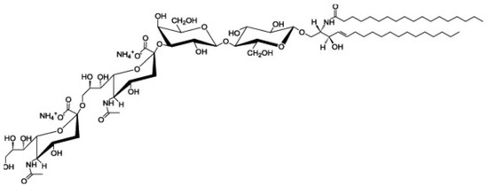
Ganglioside GD3 is a major ganglioside in neuronal progenitor cells. Highly sialylated gangliosides, GM1, GD1a, GD1b, GT1b are the main gangliosides in adult neurons. GD3 is implicated in cell attachment and cell-to-cell interaction during embryogenesis. Anti-ganglioside GD3 monoclonal antibody (clone:R24) coimmunoprecipitates heterotrimeric G protein Goα, GPI-anchored neuronal cell adhesion molecule TAG-1, Src-family kinase Lyn and Csk -binding protein Cbp from rat cerebellar granule cells. Ganglioside GD3 is involved in the migration of granule cells during the early stage of cerebellar development via these GD3-binding proteins.

gangliosides GPI-anchored proteins Src-family kinases lipid rafts heterotrimeric G proteins GD3 cerebellar granule cells migration phosphacan chondroitin sulfate proteoglycans

1. Gangliosides

Glycosphingolipids are found in the outer leaflet of the plasma membrane of all vertebrate cells and are thought to play functional roles in the regulation of cellular proliferation and differentiation [1]. Gangliosides, sialic acid-containing glycosphingolipids, are particularly abundant in the nervous system [2]. The species and amounts of gangliosides undergo profound changes during development, suggesting that they may play fundamental roles in this process. The plasma membrane lipids are not homogeneously distributed and the membranes contain microdomains or compartments. Low-density detergent-resistant membrane (DRM) fractions are isolated from cells by sucrose density gradient centrifugation [3]. The membrane fractions are rich in glycosphingolipids, sphingomyelin, cholesterol, glycosylphosphatidylinositol (GPI)-anchored proteins, and a variety of signaling molecules, such as Src family kinases and α-subunit of the heterotrimeric G proteins. These observations indicate the possible presence of glycosphingolipid-rich microdomains, referred to as lipid rafts, in cells and their involvement in signal transduction [4].
Ganglioside GD3 is a major ganglioside in neuronal progenitor cells (Figure 1). Highly sialylated gangliosides, GM1, GD1a, GD1b, GT1b are the main gangliosides in adult neurons. GM2/GD2 synthase knockout (KO) mice, expressing only ganglioside GM3 and GD3, showed mild neurological dysfunction at birth and progressive neurodegenerative changes. In contrast, double KO (DKO) mice, of GM2/GD2 synthase and GD3 synthase, expressing only ganglioside GM3, exhibit severe neurodegeneration with earlier onset and wider pathology distribution. Progressive tremor and staggering gait are observed in the DKO mice, suggesting that neurodegeneration occurred in the cerebellum [5]. GM2/GD2 synthase gene deficiency causes hereditary spastic paraplegia. GM3 synthase deficiency causes severe neurological disorders such as infantile epilepsy, mental retardation, and visual disorders [6]. Interestingly, GPI-anchored proteins and raft markers, caveolin-1 and flotillin-1, dispersed from DRM rafts in the cerebella of DKO mice [5]. These results suggest that gangliosides are involved in the formation of lipid rafts and the maintenance of neurons.
Figure 1. Structure of ganglioside GD3 (cited from https://avantilipids.com/product/860060 (accessed on 9 March 2023)).

2. Ganglioside GD3-Binding Proteins in Cerebellar Granule Cells

Ganglioside GD3 is important as a precursor of the b and c series ganglioside. Researchers isolated GD3 synthase (α2,8-sialyltransferase) cDNA and found that the GD3 synthase expression was regulated in stage- and spatio-restricted manners in the rat central nervous system [7]. To clarify the function of ganglioside GD3, researchers identified ganglioside GD3-binding proteins in the cerebellum by coimmunoprecipitation experiments using an anti-ganglioside GD3 antibody. Researchers demonstrated that anti-ganglioside GD3 monoclonal antibody (clone:R24) coimmunoprecipitates proteins of 40, 53, 56, 80  and 135 kDa from rat cerebellar granule cells.[1][2][3][4][5][6][7][8][9][10][11][12][13][14][15][16][17][18][19]

The cerebellar cortex is organized in four layers, including the external granular layer (EGL), molecular layer (ML), Purkinje cell layer (PCL), and internal granular layer (IGL) during development. Granule cells pass through all the cortical layers of the cerebellum [8]. First, granule progenitor cells migrate tangentially within the EGL, where they differentiate into immature granule cells. Stromal-cell derived factor-1 (SDF-1α), a chemokine expressed by meninges (also known as CXCL12), is an attractive guidance cue for tangential migration and a meningeal attractant of granule cells [9]. Immature cerebellar granule cells expressing the CXCR4 receptor migrate under the pial surface and meninges in response to the SDF-1α attractive guidance cue. SDF-1α prevents radial migration by chemoattracting granule cells toward the pia. These immature granule cells pause within the premigratory zone of the EGL before migrating through the ML and PCL to the IGL, and then they change their direction by migrating radially along the processes of Bergmann glial cells through the ML. However, the mechanism by which immature granule cells pause at the EGL/ML interface remains obscure.

2.1. Heterotrimeric G Protein Goα

The 40 kDa protein was identified as the α-subunit of the heterotrimeric G protein Go (Goα) [10]. Goα undergoes translocation to the DRM rafts in the early stage of cerebellar development in an activation-dependent manner. SDF-1α induces the chemoattraction of cerebellar granule cells. SDF-1α is the biological ligand for CXCR4, a G protein-coupled receptor. Treatment with SDF-1α stimulated GTPγS binding to Goα and caused Goα translocation to the DRM fractions and RhoA translocation to the membrane fraction. Chemokine SDF-1α is an attractive guidance cue for tangential migration and a meningeal attractant of granule cells [11]. Immature cerebellar granule cells expressing CXCR4 receptor migrate under the pial surface and meninges in response to the SDF-1α attractive action. Mice lacking either CXCR4 or SDF-1α display abnormal migration of granule cells in the cerebellum [12].
The linkage of Goα to the saturated acyl chains by palmitoylation and myristoylation is considered to facilitate Goα translocation to lipid rafts. The linkage of Gγ to prenyl residues, which contain unsaturated bonds, is considered to facilitate exclusion from the lipid rafts. In cerebellar granule cells, the Goαβγ heterotrimer was also excluded from the lipid rafts. This is probably due to the predominant effect of the Gγ prenyl group over the fatty acids of Goα on the partitioning of the heterotrimer in the rat cerebellum. Therefore, the signal-dependent translocation of Goα to the lipid rafts may be a consequence of the dissociation of the heterotrimer into two components, an α subunit and βγ complex.

2.2. Src-Family Kinase Lyn

The 53/56 kDa protein was identified as Src-family kinase Lyn by sequential immunoprecipitation with anti-Lyn antibody [13]. R24 treatment of primary cerebellar cultures induced Lyn activation and rapid tyrosine phosphorylation of an 80 kDa protein. These results suggest the functional association of ganglioside GD3 with Lyn. It is assumed that GD3 crosslinking by R24 treatment leads to coalescence of lipid rafts. This may induce clustering of Lyn and transphosphorylation of tyrosine residue in the kinase domain (activation site Tyr 397: homologous to c-Src Tyr419). Lyn contains a myristoylation site at glycine-2 and a palmitoylation site at cysteine-3.

2.3. GPI-Anchored Neuronal Cell Adhesion Molecule TAG-1

GPI-anchored proteins have been implicated in transmembrane signaling, nevertheless they lack intracellular domains. GPI-anchored proteins are mainly associated with lipid rafts. It is thought that association with src-family kinases in lipid rafts is important for GPI-anchored proteins in signal transduction. Researchers attempted to identify the cell-surface molecules involved in Lyn signaling because Lyn is a nonreceptor-type tyrosine kinase, and it had been found that the 135 kDa protein was identified as TAG-1, a GPI-anchored neuronal cell adhesion molecule and that the antibody-mediated cross-linking of TAG-1 induced Lyn activation and rapid tyrosine phosphorylation of 80 kDa protein in DRM raft fractions of cerebellar granule cells [14][15].
Phosphacan, a chondroitin sulfate proteoglycan, is a repulsive cue of cerebellar granule cells [15]. The GPI-anchored neural adhesion molecule TAG-1 is a binding partner of phosphacan [16], suggesting that the repulsive effect of phosphacan is possibly because of its interaction with TAG-1. The repulsive effect was greatly reduced on primary cerebellar granule cells of TAG-1-deficient mice [17]. Phosphacan was present in the ML and IGL, but not in the EGL of the rat cerebellum on postnatal days 1, 4, 7, 11, 15, and 20 and in adulthood. In contrast, transient TAG-1 expression was observed exclusively within the inner part of the EGL on postnatal days 1, 4, 7, and 11. Therefore, TAG-1-expressing cerebellar granule cells may be in contact with phosphacan at the EGL/ML interface. The overlap of the TAG-1 and phosphacan staining, which means that phosphacan and TAG-1 are interacting, is detected in the postnatal day 11 rat. These findings suggest that phosphacan may be a barrier-forming molecule that is responsible for the selective repulsion of TAG-1-expressing cerebellar granule cells via GD3 rafts to attenuate radial migration signaling. Furthermore, the down-regulation of TAG-1 after postnatal day 11 may enable the switching from the tangential migration to radial migration granule cells to the IGL.

2.4. Csk-Binding Protein Cbp

The 80 kDa protein was identified as the Csk (C-terminal Src kinase)-binding protein (Cbp) [18]. Cbp contains palmitoylation sites at cysteine-37, 40. R24 treatment induces tyrosine phosphorylation of Cbp in DRM raft fractions of cerebellar granule cells. These observations suggest that Cbp is a substrate of Lyn. Western blotting with anti-phosphotyrosine antibody demonstrated that the total tyrosine phosphorylation level was higher in the postnatal day 4 developing cerebellum than that in the adult one, and the tyrosine-phosphorylated proteins were highly accumulated in the DRM raft fraction of the postnatal day 4 cerebellum. Lyn protein was present in the DRM raft fraction of both postnatal day 4 and adult cerebella. However, the active form of Lyn was highly accumulated in the DRM raft fraction prepared from the postnatal day 4 cerebellum compared with the DRM raft fraction of the adult one. Tyrosine-phosphorylated 80 kDa protein was immunoprecipitated by the anti-Cbp antibody from the DRM raft fraction prepared from the postnatal day 4 cerebellum. Furthermore, Cbp phosphorylated at tyrosine-314 were highly accumulated in the DRM raft fraction prepared from the postnatal day 4 cerebellum. These findings suggest that TAG-1 transduces intracellular signal at GD3 rafts via Lyn/Cbp during migration of cerebellar granule cells. Taken together, researchers concluded that GD3 rafts are involved in the migration of granule cells during the early postnatal development. To support this idea, a previous study demonstrated that the lack of GD3 synthase reduces in vitro migration of cerebellar granule cells [19].

References

  1. Yamakawa, T.; and Nagai, Y. Glycolipids at the cell surface and their biological functions. Trends Biochem. Sci. 1978, 3, 128–131.
  2. Hakomori, S. Glycosphingolipids in cellular interaction, differentiation, and oncogenesis. Annu Rev Biochem 1981, 50, 733-764, doi:10.1146/annurev.bi.50.070181.003505.
  3. Brown, D.A.; Rose, J.K. Sorting of GPI-anchored proteins to glycolipid-enriched membrane subdomains during transport to the apical cell surface. Cell 1992, 68, 533-544, doi:10.1016/0092-8674(92)90189-j.
  4. Simons, K.; Gerl, M.J. Revitalizing membrane rafts: new tools and insights. Nat Rev Mol Cell Biol 2010, 11, 688-699, doi:10.1038/nrm2977.
  5. Ohmi, Y.; Tajima, O.; Ohkawa, Y.; Mori, A.; Sugiura, Y.; Furukawa, K. Gangliosides play pivotal roles in the regulation of complement systems and in the maintenance of integrity in nerve tissues. Proc Natl Acad Sci U S A 2009, 106, 22405-22410, doi:10.1073/pnas.0912336106.
  6. Furukawa, K.; Ohmi, Y.; Yesmin, F.; Tajima, O.; Kondo, Y.; Zhang, P.; Hashimoto, N.; Ohkawa, Y.; Bhuiyan, R.H. Novel Molecular Mechanisms of Gangliosides in the Nervous System Elucidated by Genetic Engineering. Int J Mol Sci 2020, 21, doi:10.3390/ijms21061906.
  7. Nara, K.; Watanabe, Y.; Maruyama, K.; Kasahara, K.; Nagai, Y.; Sanai, Y. Expression cloning of a CMP-NeuAc:NeuAc alpha 2-3Gal beta 1-4Glc beta 1-1'Cer alpha 2,8-sialyltransferase (GD3 synthase) from human melanoma cells. Proc Natl Acad Sci U S A 1994, 91, 7952-7956, doi:10.1073/pnas.91.17.7952.
  8. Galas, L.; Bénard, M.; Lebon, A.; Komuro, Y.; Schapman, D.; Vaudry, H.; Vaudry, D.; Komuro, H. Postnatal Migration of Cerebellar Interneurons. Brain Sci 2017, 7, doi:10.3390/brainsci7060062.
  9. Zhu, Y.; Yu, T.; Zhang, X.C.; Nagasawa, T.; Wu, J.Y.; Rao, Y. Role of the chemokine SDF-1 as the meningeal attractant for embryonic cerebellar neurons. Nat Neurosci 2002, 5, 719-720, doi:10.1038/nn881.
  10. Yuyama, K.; Sekino-Suzuki, N.; Sanai, Y.; Kasahara, K. Translocation of activated heterotrimeric G protein Galpha(o) to ganglioside-enriched detergent-resistant membrane rafts in developing cerebellum. J Biol Chem 2007, 282, 26392-26400, doi:10.1074/jbc.M705046200.
  11. Chédotal, A. Should I stay or should I go? Becoming a granule cell. Trends Neurosci 2010, 33, 163-172, doi:10.1016/j.tins.2010.01.004.
  12. Ma, Q.; Jones, D.; Borghesani, P.R.; Segal, R.A.; Nagasawa, T.; Kishimoto, T.; Bronson, R.T.; Springer, T.A. Impaired B-lymphopoiesis, myelopoiesis, and derailed cerebellar neuron migration in CXCR4- and SDF-1-deficient mice. Proc Natl Acad Sci U S A 1998, 95, 9448-9453, doi:10.1073/pnas.95.16.9448.
  13. Kasahara, K.; Watanabe, Y.; Yamamoto, T.; Sanai, Y. Association of Src family tyrosine kinase Lyn with ganglioside GD3 in rat brain. Possible regulation of Lyn by glycosphingolipid in caveolae-like domains. J Biol Chem 1997, 272, 29947-29953, doi:10.1074/jbc.272.47.29947.
  14. Kasahara, K.; Watanabe, K.; Takeuchi, K.; Kaneko, H.; Oohira, A.; Yamamoto, T.; Sanai, Y. Involvement of gangliosides in glycosylphosphatidylinositol-anchored neuronal cell adhesion molecule TAG-1 signaling in lipid rafts. J Biol Chem 2000, 275, 34701-34709, doi:10.1074/jbc.M003163200.
  15. Kasahara, K.; Watanabe, K.; Kozutsumi, Y.; Oohira, A.; Yamamoto, T.; Sanai, Y. Association of GPI-anchored protein TAG-1 with src-family kinase Lyn in lipid rafts of cerebellar granule cells. Neurochem Res 2002, 27, 823-829, doi:10.1023/a:1020265225916.
  16. Milev, P.; Maurel, P.; Häring, M.; Margolis, R.K.; Margolis, R.U. TAG-1/axonin-1 is a high-affinity ligand of neurocan, phosphacan/protein-tyrosine phosphatase-zeta/beta, and N-CAM. J Biol Chem 1996, 271, 15716-15723, doi:10.1074/jbc.271.26.15716.
  17. Komatsuya, K.; Iguchi, T.; Fukuyama, M.; Kawashima, I.; Ogura, K.; Kikuchi, N.; Shimoda, Y.; Takeda, Y.; Shimonaka, M.; Yamamoto, N.; et al. Phosphacan acts as a repulsive cue in murine and rat cerebellar granule cells in a TAG-1/GD3 rafts-dependent manner. J Neurochem 2022, 163, 375-390, doi:10.1111/jnc.15709.
  18. Sekino-Suzuki, N.; Yuyama, K.; Miki, T.; Kaneda, M.; Suzuki, H.; Yamamoto, N.; Yamamoto, T.; Oneyama, C.; Okada, M.; Kasahara, K. Involvement of gangliosides in the process of Cbp/PAG phosphorylation by Lyn in developing cerebellar growth cones. J Neurochem 2013, 124, 514-522, doi:10.1111/jnc.12040.
  19. Santiago, M.F.; Scemes, E. Neuroblast migration and P2Y(1) receptor mediated calcium signalling depend on 9-O-acetyl GD3 ganglioside. ASN Neuro 2012, 4, 357-369, doi:10.1042/AN20120035.
More
Information
Subjects: Cell Biology
Contributors MDPI registered users' name will be linked to their SciProfiles pages. To register with us, please refer to https://encyclopedia.pub/register : , , ,
View Times: 988
Revisions: 3 times (View History)
Update Date: 03 Apr 2023
1000/1000