Submitted Successfully!
To reward your contribution, here is a gift for you: A free trial for our video production service.
Thank you for your contribution! You can also upload a video entry or images related to this topic.
Version Summary Created by Modification Content Size Created at Operation
1 -- 2170 2023-03-18 20:37:29 |
2 format correct + 1 word(s) 2171 2023-03-20 02:16:28 | |
3 format correct -3 word(s) 2168 2023-03-20 09:18:44 | |
4 format correct Meta information modification 2168 2023-03-20 09:19:25 | |
5 Fixed typos. -31 word(s) 2137 2023-03-20 16:45:40 |

Video Upload Options

Do you have a full video?

Confirm

Are you sure to Delete?
Cite
If you have any further questions, please contact Encyclopedia Editorial Office.
Bansil, R.; Constantino, M.A.; Su-Arcaro, C.; Liao, W.; Shen, Z.; Fox, J.G. Motility of Helicobacter spp.. Encyclopedia. Available online: https://encyclopedia.pub/entry/42321 (accessed on 08 May 2024).
Bansil R, Constantino MA, Su-Arcaro C, Liao W, Shen Z, Fox JG. Motility of Helicobacter spp.. Encyclopedia. Available at: https://encyclopedia.pub/entry/42321. Accessed May 08, 2024.
Bansil, Rama, Maira A. Constantino, Clover Su-Arcaro, Wentian Liao, Zeli Shen, James G. Fox. "Motility of Helicobacter spp." Encyclopedia, https://encyclopedia.pub/entry/42321 (accessed May 08, 2024).
Bansil, R., Constantino, M.A., Su-Arcaro, C., Liao, W., Shen, Z., & Fox, J.G. (2023, March 18). Motility of Helicobacter spp.. In Encyclopedia. https://encyclopedia.pub/entry/42321
Bansil, Rama, et al. "Motility of Helicobacter spp.." Encyclopedia. Web. 18 March, 2023.
Motility of Helicobacter spp.
Edit

Helicobacter spp., including the well-known human gastric pathogen H. pylori, can cause gastric diseases in humans and other mammals. They are Gram-negative bacteria that colonize the gastric epithelium and use their multiple flagella to move across the protective gastric mucus layer. Different Helicobacter spp. differ in the number and arrangement of flagella, as well as in the size and shape of their cell body. The most well-studied of these is H. pylori, which is unipolar and lophotrichous (multiple flagella at one pole). Some of these bacteria are bipolar, e.g., the lophotrichous H. suis, and the monotrichous H. cetorum. The motility of H. pylori, H. suis, and H. cetorum was described in the review in Microorganisms 2023 linked above. 

Helicobacter motility unipolar bacteria bipolar bacteria gastric mucin Helicobacter spp.

1. Dependence of H. pylori Swimming on Cell Shape and Number of Flagella

The motility and chemotaxis of H. pylori have been discussed in earlier reviews focused on explaining the flagella and chemotaxis molecular machinery [1]. By tracking bacteria microscopically several researchers examined the motility of H. pylori. These studies show that the swimming characteristics of H. pylori depend on the shape of the cell, the size of the bacterium, and the number of flagella. Early studies examined the swimming of H. pylori in viscous polymer solutions to mimic the viscous mucus environment [2] and compared the helical H. pylori with straight–rod E. coli to address the effect of the helical shape [3]. Karim et al. [3] showed that both H. pylori and C. jejuni swam faster than E. coli in aqueous cultures, presumably due to their helical body shape [3].  Martinez et al. [4]  examined the effect of the helical shape of H. pylori in detail, using time-resolved phase contrast microscopy to compare the swimming of wild-type, helical H. pylori strains with isogenic, straight-rod mutants (Δcsd4 and Δcsd6) which lacked the helical-shape-determining peptidoglycan peptidases, Csd4 or Csd6 [5]. They noted that the helical shape of the cell body confers about a 7–20% advantage in swimming speed, depending on the strain. They also found that the swimming speed of H. pylori varies considerably among different strains due to differences in cell size and the number of flagella. The distribution of speeds in any given sample is very broad due to variations in cell size, shape, and the number of flagella, as well as temporal changes in speed during swimming. To examine the effect of varying the number of flagella, they compared the wild-type B128 strain of H. pylori (median number of flagella = 3) with its isogenic flagellar mutants, fliOΔC (median 1 flagellum) and sRNA_T (median 4 flagella). They observed that the speed was increased in the sRNA_T mutant, which has an extra flagellum compared to the wild-type, and decreased in the case of fliOΔC, which has on average only one flagellum [4].

Bacteria tracking studies show that H. pylori exhibit a run-reverse-reorient swimming mechanism [6][7][8][9][10] in aqueous broth as well as in viscous solutions. In the case of H. pylori, an unbundled state of tumbling or re-orientation can occur [6][9][10] between the forward and reverse runs. The analysis of trajectories using the methods developed by Theves et al. [11] to obtain the distributions of run speeds, change in orientation angle, and reversal frequency shows that the reversal frequency decreases for H. pylori swimming in viscous methylcellulose and porcine gastric mucin (PGM) solutions compared to Brucella broth (BB10) [6].

By imaging H. pylori at high magnification (100×) and at 100–200 frames per second, Constantino et al. [9] were able to track the motion of individual bacteria as they swam. The trajectory of a single bacterium at high magnification showed a characteristic oscillation superimposed on the track of its motion. This feature arises from the rotation of the cell body. To maintain a net zero torque at the flagellar pole, the cell body rotates in the opposite sense relative to the flagellar bundle. The rotation of the cell body can be seen as a change in the relative orientation of its microscopic image as it swims.  The movies in [9] show the flagellar bundle rotating counterclockwise during the forward run (the bacterium acting as a pusher) and rotating in the opposite sense (clockwise) during the reverse motion, with the bacterium acting as a puller. By imaging the circular motion of bacteria swimming close to surfaces, Antani et al. [7] showed that H. pylori swim at a faster speed when they move as a pusher with flagella rotating counterclockwise, as compared to the reverse run as a puller with flagella rotating in the clockwise rotation. By examining mutants lacking the phosphorylated response regulator CheY-P, they concluded that CheY-P binding to the flagellar motor promotes the clockwise rotation in H. pylori. Howitt et al. [8] showed that mutants of H. pylori lacking ChePep, a protein that is required for polar localization of some of the chemotaxis components, show more frequent reversals and sustained swimming in the reverse direction, implying that ChePep plays an important role in controlling the directional persistence of motility.
Dependence of H. pylori swimming speed on the rheology and pH of the medium. As H. pylori have to swim through mucus in order to colonize the gastric epithelium it is important to examine the effect of the viscosity and pH of the medium on the swimming speed. Studies of H. pylori swimming in solutions of the synthetic polymer methylcellulose show that the swimming speed increases with increasing viscosity in the low viscosity regime, followed by a decrease at higher viscosities [6][12]. Celli et al. [13] examined the swimming of H. pylori in PGM which contains Muc5AC, the major high molecular weight glycoprotein component responsible for the rheological properties of gastric mucus [14][15]. PGM is known to form a gel at pH < 4, while it remains a viscous solution above that pH [15]. Celli et al. [13] observed that, in the absence of urea, H. pylori bacteria did not swim in PGM mucin gels at pH 4 and lower, although their flagellar bundle rotated. Upon adding urea, the bacteria were observed to swim, and the pH of the medium went up as indicated by a fluorescent dye in the external medium. Urea hydrolysis produces NH3, which increases the pH of the mucin solution, causing it to de-gel and enabling the bacteria to swim [13]. Thus, H. pylori utilize the urease-mediated hydrolysis of urea not only to survive in the highly acidic gastric environment [16] but also to swim in the mucus layer. Su et al. [10] compared the pH-dependent swimming characteristics of H. pylori in PGM solutions and BB10 culture broth. They found that in both PGM and BB10, the swimming speed decreased as the pH fell below 4, but in BB10 the bacteria continue to swim at pH < 4, whereas in PGM the bacteria are stuck in various ways to the gel network, their flagella rotate, but they do not swim. They also noted that decreasing pH leads to a decreased fraction of motile bacteria, with a decreased fraction of fast swimmers in the distribution of speeds and net displacement of trajectories. The combination of the effect of pH on the flagellar motors and the sol-gel transition of mucin leads to optimal swimming at a slightly acidic pH of around 5 in porcine gastric mucin.

2. Motility of the Bipolarly Flagellated H. suis

Studies of several bacteria with bipolar flagella have shown that they swim with one or both flagella extended (E) away from the body acting as a pusher or a puller, or wrapped (W), in which case the flagellar bundle reverses its orientation and rotates while wrapped around the body [17][18][19]. Wrapped flagella have also been observed in unipolar bacteria [20]. For the long and tightly coiled (6–8 turns) bipolar bacterium H. suis, Constantino et al. [21] were able to visualize the thick bundles of 8–12 flagella and track their motion by using time-resolved phase contrast microscopy at 100× magnification and frame rates of 100–200 fps. Their study showed that, regardless of media, the flagellar bundles of H. suis also assume one of two configurations, E or W. These two configurations correspond to different modes of swimming determined by whether the flagellar bundles at the opposite poles are extended or wrapped around the cell body. This is illustrated schematically in Figure 1.
Figure 1. H. suis bacterium corresponding to either an extended (E) or a wrapped (W) configuration of the flagellar bundle. The EE mode corresponds to both bundles extended, EW to one extended and the other wrapped, and WW to both wrapped. Adapted from Constantino [21] under a Creative Commons Attribution 4.0 International License.
Wrapping the flagellar bundle around the body provides additional propulsion. H. suis predominantly swim with the lagging flagella extended behind the body and the leading flagella wrapped around the body (EW mode). During a smaller fraction of the runs, H. suis swim with both bundles extended away from the body (EE mode) or wrapped around the body (WW mode). However, in the EE or WW modes, the speed was greatly reduced, and the trajectories showed many more reorientations, suggesting that in the EE case, the two flagellar bundles were both acting as pushers and thus negating each other’s action. The fast-swimming EE mode, with one flagellum acting as a pusher and the other as a puller, was not observed. The WW mode could correspond to both flagella acting as pullers in opposite directions. The trajectories in the EW mode are almost linear, whereas, in the WW and EE modes, the bacteria travel lesser distances and display trajectories that show characteristics in between a ballistic and a random-walk motion. Calculations using the Method of Regularized Stokeslets for different modes of the flagellar bundles, including a rolling motion for the wrapped flagellar bundle and the usual pusher-puller mechanisms for the extended bundle, were in qualitatively good agreement with the experimental findings [21].

3. Motility of H. cetorum, a Monotrichous Bipolar Fusiform Bacterium

H. cetorum is a fusiform, slightly helical bacterium with bipolar, monotrichous flagella [22]. Constantino [23] reported a live-cell tracking motility study of H. cetorum in BB10. The trajectories of H. cetorum  resemble those of H. pylori and H. suis, with the characteristic cell body rotation superimposed on linear tracks. Analysis of the trajectories of H. cetorum indicates a tendency to swim in the same direction for long periods without much reorientation over the tracks. The single flagellum of H. cetorum is very thin and could not be visualized in time-resolved phase contrast microscopy, unlike the thick bundles of the lophotrichous H. pylori and H. suis bacteria.

4. Comparison of the Motility of H. pylori, H. suis, and H. cetorum
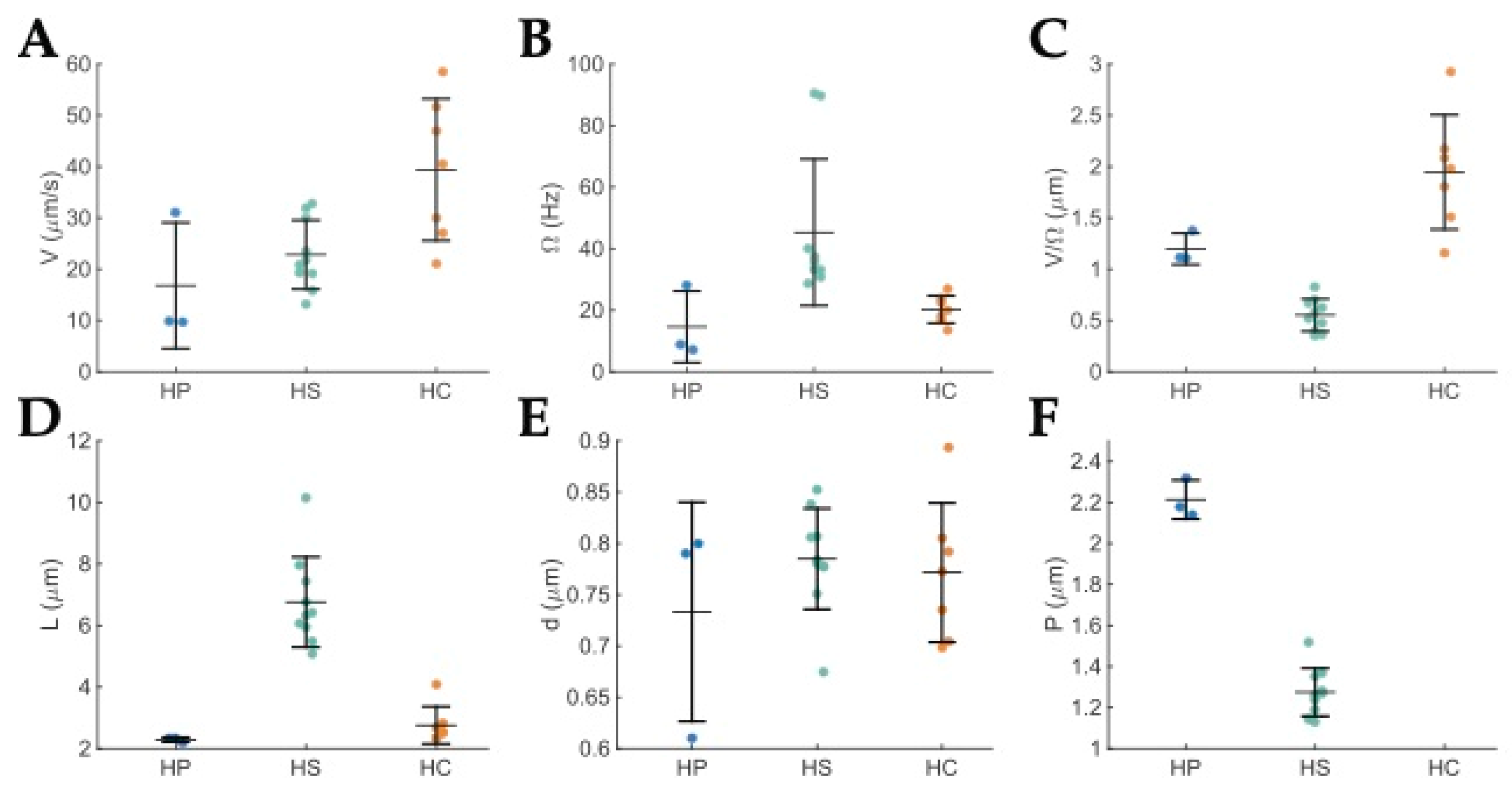
A comparison of all the measured swimming and shape parameters for H. cetorum, H. suis, and H. pylori is shown in Figure 2. Table 1 gives the average speed V, cell body rotation rate Ω, and cell shape parameters: length L, diameter d, and pitch P for these three Helicobacter spp. For this comparison, the researchers utilized the H. suis data swimming in the EW configuration because that corresponds to an optimum run [21].
Figure 2. Dot plots of the measured motility and shape parameters for H. pylori in blue (HP), H. suis in green (HS), and H. cetorum in orange (HC) swimming in BB10 broth. The horizontal lines indicate the mean and the vertical lines indicate the standard deviation. (A) Average speed, V for each run. (B) Body rotation rate, Ω for each run. (C) Distance traveled per revolution, V/Ω, for each run. (D) Body length, L of each bacterium. (E) Body diameter, d of each bacterium. (F) Body pitch, P of each bacterium; H. cetorum is fusiform, it was only slightly helical, and pitch could not be measured from the images obtained in this study. It can be approximated by the body length. The H. pylori data shown here are the same as in [9]. The H. suis data correspond to the EW configuration. Adapted from Constantino [23].
Table 1. Average speed V, cell body rotation rate Ω, and cell shape parameters: length L, diameter d, pitch P of H. pylori, H. suis, and H. cetorum in BB10 broth. The H. suis data are for the EW case, as runs can only be observed in that mode.
A comparison of the swimming of H. suis in PGM and culture broth BB10 at different pH values [23] shows that it too exhibits pH-dependent swimming similar to H. pylori; H. suis exhibits fewer reversals in broth than H. pylori. In gastric mucin, H. suis bacteria swim the fastest around pH 6, while H. pylori show peak swimming speed around pH 5. Both species have a reduced speed and rate of body rotation at pH 4 in gastric mucin and are stuck in the gel phase of mucin. The frequency of reversals decreases in mucin for both H. suis and H. pylori. Both of these Helicobacter spp. swim with increased directional persistence in gastric mucin, resulting in straighter swimming trajectories which may be advantageous in moving across the highly viscous mucus lining of the stomach.

References

  1. Lertsethtakarn, P.; Ottemann, K.M.; Hedrixson, D.R. Motility and chemotaxis in Campylobacter and Helicobacter. Annu. Rev. Microbiol. 2011, 65, 389–410.
  2. Yoshiyama, H.; Nakamura, H.; Kimoto, M.; Okita, K.; Nakazawa, T. Chemotaxis and motility of Helicobacter pylori in a viscous environment. J. Gastroenterol. 1999, 34, 18–23.
  3. Karim, Q.N.; Logan, R.P.; Puels, J.; Karnholz, A.; Worku, M.L. Measurement of motility of Helicobacter pylori, Campylobacter jejuni, and Escherichia coli by real time computer tracking using the Hobson BacTracker. J. Clin. Pathol. 1998, 51, 623–628.
  4. Martínez, L.E.; Hardcastle, J.M.; Wang, J.; Pincus, Z.; Tsang, J.; Hoover, T.R.; Bansil, R.; Salama, N.R. Helicobacter pylori strains vary cell shape and flagellum number to maintain robust motility in viscous environments. Mol. Microbiol. 2016, 99, 88–110.
  5. Sycuro, L.K.; Wyckoff, T.J.; Biboy, J.; Born, P.; Pincus, Z.; Vollmer, W.; Salama, N.R. Multiple Peptidoglycan Modification Networks Modulate Helicobacter pylori’s Cell Shape, Motility, and Colonization Potential. PLoS Pathog. 2012, 8, e1002603.
  6. Martínez, L.E.; Hardcastle, J.M.; Wang, J.; Pincus, Z.; Tsang, J.; Hoover, T.R.; Bansil, R.; Salama, N.R. Helicobacter pylori strains vary cell shape and flagellum number to maintain robust motility in viscous environments. Mol. Microbiol. 2016, 99, 88–110.
  7. Antani, J.D.; Sumali, A.X.; Lele, T.P.; Lele, P.P. Asymmetric random walks reveal that the chemotaxis network modulates flagellar rotational bias in Helicobacter pylori. eLife 2021, 10, e63936.
  8. Howitt, M.R.; Lee, J.Y.; Lertsethtakarn, P.; Vogelmann, R.; Joubert, L.M.; Ottemann, K.M.; Amieva, M.R. Chepep controls helicobacter pylori infection of the gastric glands and chemotaxis in the epsilonproteobacteria. MBio 2011, 2, e00098-11.
  9. Constantino, M.A.; Jabbarzadeh, M.; Fu, H.C.; Bansil, R. Helical and rod-shaped bacteria swim in helical trajectories with little additional propulsion from helical shape. Sci. Adv. 2016, 2, e1601661.
  10. Su, C.; Bieniek, K.; Liao, W.; Constantino, M.A.; Decker, S.M.; Turner, B.S.; Bansil, R. Comparison of motility of H. pylori in broth and mucin reveals the interplay of effect of acid on the bacterium and the rheology of the medium it swims in. bioRxiv 2020.
  11. Theves, M.; Taktikos, J.; Zaburdaev, V.; Stark, H.; Beta, C. A bacterial swimmer with two alternating speeds of propagation. Biophys. J. 2013, 105, 1915–1924.
  12. Worku, M.L.; Sidebotham, R.L.; Baron, H.J.; Misiewicz, J.J.; Logan, R.P.; Keshavarz, T.; Karim, Q.N. Motility of Helicobacter pylori in a viscous environment. Eur. J. Gastroenterol. Hepatol. 1999, 11, 1143–1150.
  13. Celli, J.P.; Turner, B.S.; Afdhal, N.H.; Keates, S.; Ghiran, I.; Kelly, C.P.; Ewoldt, R.H.; McKinley, G.H.; So, P.T.C.; Erramilli, S.; et al. Helicobacter pylori moves through mucus by reducing mucin viscoelasticity. Proc. Natl. Acad. Sci. USA 2009, 106, 14321–14326.
  14. Bansil, R.; Celli, J.P.; Hardcastle, J.M.; Turner, B.S. The influence of mucus microstructure and rheology in Helicobacter pylori infection. Front. Immunol. 2013, 4, 310.
  15. Celli, J.P.; Turner, B.S.; Afdhal, N.H.; Ewoldt, R.H.; Mckinley, G.H.; Bansil, R.; Erramilli, S. Rheology of gastric mucin exhibits a pH-dependent sol-gel transition. Biomacromolecules 2007, 8, 1580–1586.
  16. Clyne, M.; Labigne, A.; Drumm, B. Helicobacter pylori requires an acidic environment to survive in the presence of urea. Infect. Immun. 1995, 63, 1669–1673.
  17. Grognot, M.; Taute, K. More than propellers: How flagella shape bacterial motility behaviors. Curr. Opin. Microbiol. 2021, 61, 73–81.
  18. Thormann, K.M.; Beta, C.; Kühn, J.M. Wrapped up: The Motility of Polarly Flagellated Bacteria. Annu. Rev. Microbiol. 2022, 76, 349–367.
  19. Murat, D.; Hérisse, M.; Espinosa, L.; Bossa, A.; Alberto, F.; Wu, L.F. Opposite coordinated rotation of amphitrichous flagella governs oriented swimming and reversals in a magnetotactic spirillum. J. Bacteriol. 2015, 197, 3275–3282.
  20. Hintsche, M.; Waljor, V.; Großmann, R.; Kühn, J.M.; Thormann, K.M.; Peruani, F.; Beta, C. A polar bundle of flagella can drive bacterial swimming by pushing, pulling, or coiling around the cell body. Sci. Rep. 2017, 7, 16771.
  21. Constantino, M.A.; Jabbarzadeh, M.; Fu, H.C.; Shen, Z.; Fox, J.G.; Haesebrouck, F.; Lindén, S.; Bansil, R. Bipolar lophotrichous Helicobacter suis combine extended and wrapped flagella bundles to exhibit multiple modes of motility. Sci. Rep. 2018, 8, 14415.
  22. Harper, C.G.; Feng, Y.; Xu, S.; Taylor, N.S.; Kinsel, M.; Dewhirst, F.E.; Paster, B.J.; Greenwell, M.; Levine, G.; Rogers, A.; et al. Helicobacter cetorum sp. nov., a Urease Positive Helicobacter Species Isolated from Dolphins and Whales. J. Clin. Microbiol. 2002, 40, 4536–4543.
  23. Constantino, M.A. Investigating Effects of Morphology and Flagella Dynamics on Swimming Kinematics of Different Helicobacter Species Using Single-Cell Imaging. Ph.D. Thesis, Boston University, Boston, MA, USA, 2017.
More
Information
Subjects: Microbiology
Contributors MDPI registered users' name will be linked to their SciProfiles pages. To register with us, please refer to https://encyclopedia.pub/register : , , , , ,
View Times: 431
Entry Collection: Gastrointestinal Disease
Revisions: 5 times (View History)
Update Date: 20 Mar 2023
1000/1000