Submitted Successfully!
To reward your contribution, here is a gift for you: A free trial for our video production service.
Thank you for your contribution! You can also upload a video entry or images related to this topic.
Version Summary Created by Modification Content Size Created at Operation
1 -- 3511 2023-03-15 11:40:40 |
2 Reference format revised. + 7 word(s) 3518 2023-03-16 07:32:44 | |
3 remove blank line Meta information modification 3518 2023-03-16 07:33:06 |

Video Upload Options

Do you have a full video?

Confirm

Are you sure to Delete?
Cite
If you have any further questions, please contact Encyclopedia Editorial Office.
Singh, S.; Dutta, J.; Ray, A.; Karmakar, A.; Mabalirajan, U. Airway Epithelium in Asthma Pathobiology. Encyclopedia. Available online: https://encyclopedia.pub/entry/42230 (accessed on 06 July 2024).
Singh S, Dutta J, Ray A, Karmakar A, Mabalirajan U. Airway Epithelium in Asthma Pathobiology. Encyclopedia. Available at: https://encyclopedia.pub/entry/42230. Accessed July 06, 2024.
Singh, Sabita, Joytri Dutta, Archita Ray, Atmaja Karmakar, Ulaganathan Mabalirajan. "Airway Epithelium in Asthma Pathobiology" Encyclopedia, https://encyclopedia.pub/entry/42230 (accessed July 06, 2024).
Singh, S., Dutta, J., Ray, A., Karmakar, A., & Mabalirajan, U. (2023, March 15). Airway Epithelium in Asthma Pathobiology. In Encyclopedia. https://encyclopedia.pub/entry/42230
Singh, Sabita, et al. "Airway Epithelium in Asthma Pathobiology." Encyclopedia. Web. 15 March, 2023.
Airway Epithelium in Asthma Pathobiology
Edit

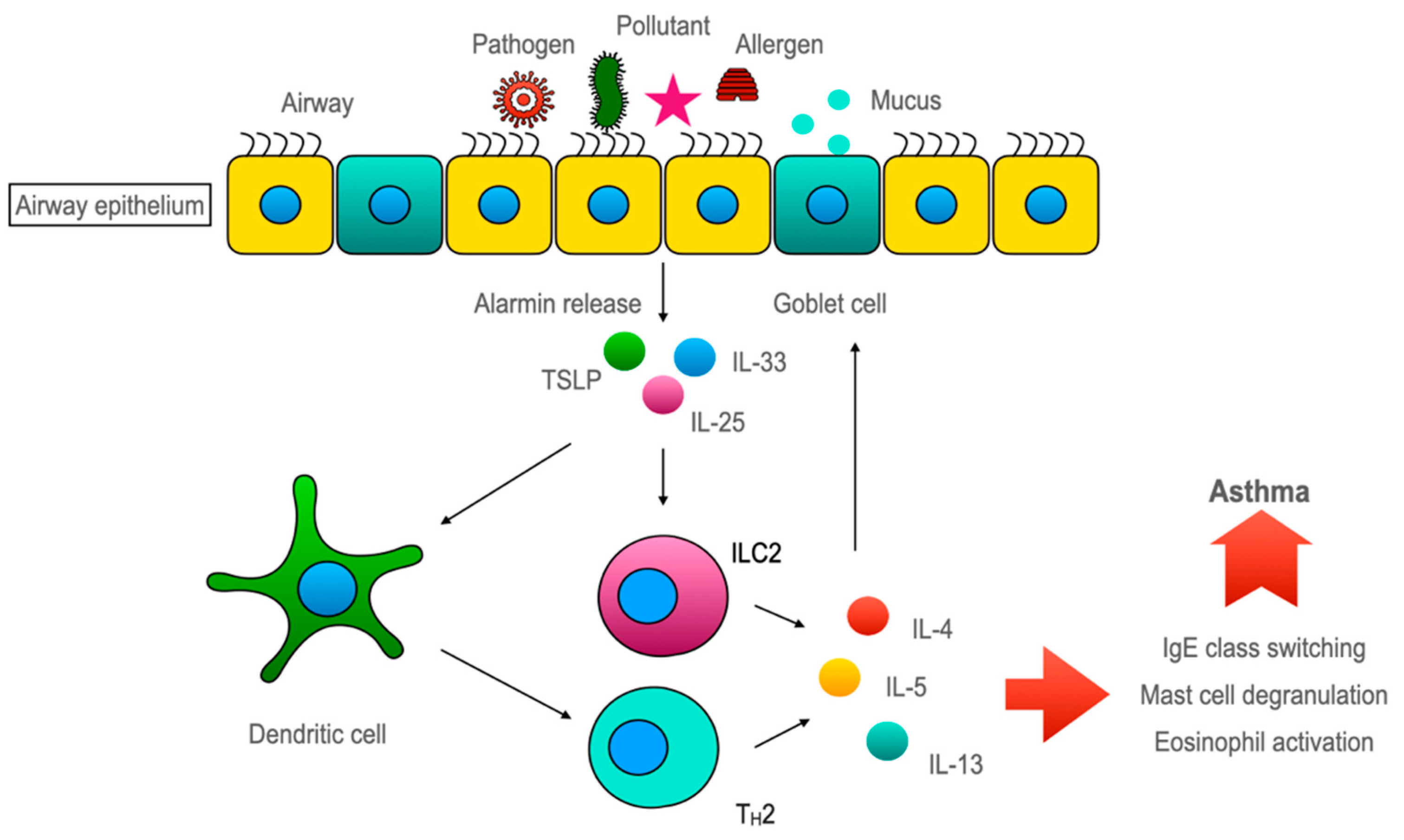
The features of allergic asthma are believed to be mediated mostly through the Th2 immune response. In this Th2-dominant concept, the airway epithelium is presented as the helpless victim of Th2 cytokines. Asthma researchers started believing in that the airway epithelium played a crucial role, as alarmins, which are the inducers of type 2 innate lymphoid cell (ILC2), are almost exclusively secreted by the airway epithelium. This underscores the eminence of airway epithelium in asthma pathogenesis. However, the airway epithelium has a bipartite functionality in sustaining healthy lung homeostasis and asthmatic lungs. On the one hand, the airway epithelium maintains lung homeostasis against environmental irritants/pollutants with the aid of its various armamentaria, including its chemosensory apparatus and detoxification system. Alternatively, it induces an ILC2-mediated type 2 immune response through alarmins to amplify the inflammatory response. However, the available evidence indicates that restoring epithelial health may attenuate asthmatic features.

allergic asthma airway epithelium alarmins

1. Barrier Function of Airway Epithelium

1.1. Importance of Epithelial Barrier in Maintaining Homeostasis

As a first line of nonspecific defense, the anatomical barriers such as, skin and mucosal membrane protects our body system from environmental insults. Essentially, these barriers in our body have two major functions: (a) organ-specific functions and (b) the maintenance of organ homeostasis against external aggressors. For example, the stratum corneum, which is the outermost epidermal layer, is essential in limiting water loss by transcutaneous evaporation to maintain the water content of our body [1]. In addition, the skin barrier is also crucial in providing defense against the invasion of various external molecules and microbes. Similarly, the intestinal epithelium is essential in the absorption of nutrients, water, and electrolytes, along with the homeostatic role of restraining the entry of allergens, microbes, and other foreign molecules into the intestinal wall. Likewise, alveolar epithelium is fundamentally involved in gaseous exchanges, surfactant production, and the regulation of ions and water transport to maintain the fluid balance on the alveolar surface, in addition to the protective function against environmental irritants/pollutants [2]. The airway epithelial cells, which line the entire region from the trachea to the terminal bronchi, are lined up by the ciliated cells and the Clara cells at the region of conducting airways (trachea, bronchi, and bronchioles). However, airway epithelium does not have any special organ-specific function like gas exchange or nutrient absorption, even though it regulates water/ions transport. However, airway epithelial cells serve as sentries in restricting the entry of inspired airway luminal contents beneath the epithelial layer and in removing/neutralizing the various toxic/irritant substances from the inhaled air to prevent the access of these irritants to alveoli where vital gas exchange happens. In order to perform this key function, the airway epithelial layer has numerous types of machinery, like physical barriers, chemical barriers, special cellular machinery, etc.
Meanwhile, the regulation of the airway epithelial barrier function is emerging as a crucial checkpoint in asthma pathobiology. Numerous studies have evidenced that the respiratory epithelium is compromised in asthmatic conditions. Additionally, it has been noted that asthma patients’ bronchial epithelial cells have abnormal antimicrobial response patterns. According to biopsy studies carried out in children, structural alterations in the respiratory epithelium may take place prior to the beginning of airway inflammation. There is a theory that claims that structural and functional dysfunction in the respiratory epithelium leads to an aberrant immune system and structural cell signaling, which, in turn, causes remodeling, inflammation, and allergic airway hypersensitivity [3].

1.2. Anatomical Barrier Role of Airway Epithelium

Epithelial junctional complexes act as crucial signaling hubs and threat detectors, interacting with the microenvironment and monitoring self-defense. This function of the epithelial barrier of the bronchial epithelial cells and its structural integrity is mainly conferred by the adhesive forces of the intercellular junctions. The major intercellular junction proteins documented in preserving the barrier include tight junctions, the adherens junctions, and the (hemi) desmosomes [4]. The tight and adherens junctions together form the apical junctional complex (AJC) and are present at the apicolateral border of the airway epithelial cells (Figure 1).
Figure 1. Scheme diagram to illustrate the structural changes in the airway epithelial barrier in asthmatics compared to healthy airway epithelium. When the asthmatic airway epithelium is exposed to allergens and air pollutants, it causes disruption to the tight epithelial junction and adherens junction. This leads to heightened mucosal permeability, effectuating more inhaled particles and allergens present in the subepithelial region and promoting innate and adaptive immune responses. This is accompanied by pulmonary neuroendocrine cells (PNECs) hyperplasia, a loss of ciliated cell numbers, goblet cell metaplasia, mucus hypersecretion, the thickening of the basal membrane, subepithelial fibrosis, increased airway smooth muscle mass, and the excess deposition of extracellular matrix.
The claudin multigene family, which encodes for the tetraspan transmembrane proteins, are the core tight junction proteins [5]. They interact with the claudins on the adjacent cells and forge a barrier that regulates the paracellular diffusion of ions and solutes. The alveolar epithelium also expresses over a dozen claudins, out of which claudin-3 (cldn-3), claudin-4 (cldn-4), and claudin-18(cldn-18) are predominantly expressed [6]. The other claudins expressed by the alveolar epithelia include claudin-5 (cldn-5) and claudin-7 (cldn-7), which also aids in maintaining the alveolar epithelial barrier. The loss of these proteins leads to increased permeability and causes a loss of barrier function [7].
The adherens junctions that are present beneath the tight junction comprise the cadherin (E-cadherin) and the catenin (α-catenin, β-catenin, and p120) families that mediate cell-to-cell adhesion [8][9]. The deletion of E-cadherin in lung epithelia not only causes epithelial denudation with a loss of ciliated cells but also suppresses the differentiation of club cells and, thus, limits epithelial healing after damage due to the stem cell properties of club cells [10]. The sustained loss of E-cadherin also evokes the differentiation of epithelia into a mesenchymal phenotype, a process otherwise called epithelial-mesenchymal transition (EMT). This EMT is known to cause subepithelial fibrosis, which is the main feature in airway remodeling in asthmatics and increases the severity and complexity of the disease.
The tight and the adherens junction proteins are linked to cytosolic proteins on one end and to the actin cytoskeleton on the other end to form “cytosolic plaques” [11]. A dominant plaque protein is found in the family of zonula occludens (ZO), which cement the intracellular domains of the tight and adherens junction with various cytoskeletal components and actin-binding proteins, like α-catenin, α-actinin, and vinculin. It is now becoming evident that a lack of these certain junctional complexes is sufficient to trigger the alarm of the immune defenses. 

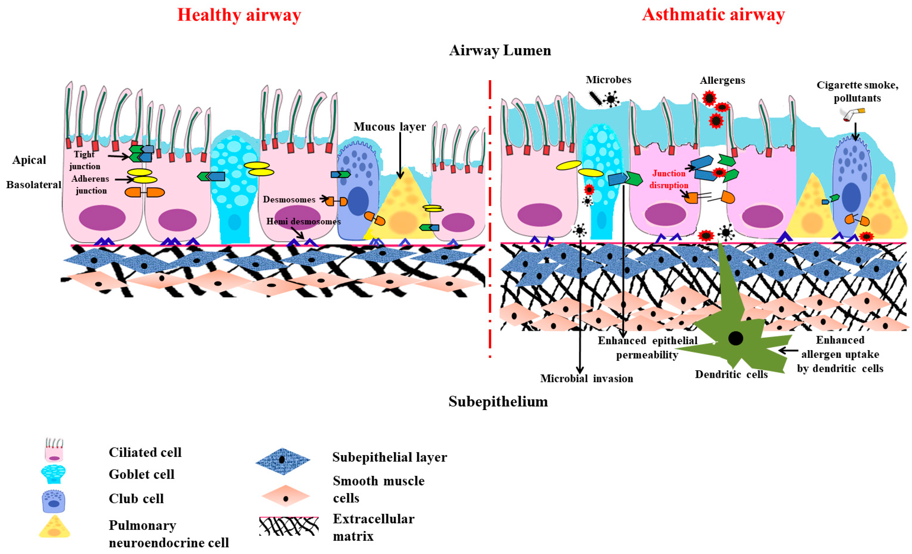
1.3. Chemical Barrier Role of Airway Epithelium

Given the incessant exposure of the lungs to noxious substances, the surface of the airway is layered up with a highly evolved fluid lining that is exchanged dynamically to aid in the mucociliary clearance of antigens. The produced mucus layer and the periciliary layer (PCL) are the two separate layers that make up this fluid lining, also known as airway surface liquid (ASL). The secreted mucins are glycoconjugates with threonine-rich domains, and these act as biophysical “rafts” or barriers to convey the pathogens out of the conducting airways [12]. Mucins, like MUC13, MUC16, and MUC4, which are tethered to the epithelial cells, bestow a direct host defense barrier at the epithelial surface that can be removed by pathogen- or host-associated proteases, releasing microbes to the mucociliary ‘escalator’ for elimination. On the contrary, MUC5AC, MUC5BA, and MUC2, which are secreted by the airways, create a mucous gel that hinders bacterial aggregation, binds microbial pathogens, and impairs their ability to adhere [12]. Even though the increased secretion and release of airway mucus is a noisome accompaniment to environmental cues and infections, mucins help maintain airway homeostasis and eliminate pathogens and cellular debris during recovery from injury or infection. Airway disorders viz., chronic obstructive pulmonary disease (COPD), bronchiectasis, cystic fibrosis, and asthma, all exhibit excessive goblet cell differentiation and mucus hyperproduction [13].
The epithelial cells and the submucosal glands, which form an integral part of the innate immune system, secrete many host-defense proteins, including lysozymes, lactoferrin, lipocalin-2, defensins, cathelicidin, surfactant proteins, acute-phase proteins, psoriasin (S100A7) proteins, and palate, lung, and nasal epithelium clone (PLUNC) proteins [14]. The lung epithelial cells act as immune sentinels, and this sets up an innate immune response owing to an ample repertoire of cytosolic, membrane-bound, and endosomal pattern-recognition receptors (PRRs) to identify various pathogens [15].

1.4. Physiological Barrier in Airway

The airway defense responses that guard the lungs and the rest of the body against inhaled irritants like cigarette smoke and aerosols include bronchoconstriction, which is a crucial and effective reflex mechanism [16]. However, the chemical irritants stimulate the sensory nerves present in the respiratory tract, and the generated action potential is conducted by the vagus nerves (to the brain stem). This instantly causes bronchoconstriction via the cholinergic efferent pathway accompanied by the hypersecretion of mucus, coughing, and dyspneic sensations [17]. Thereof, the conjecture that bronchoconstriction is a physiological protective mechanism, becomes a clinical symptom as this also confines the entry of the air and, thus, difficulty in breathing occurs subsequent to the bronchoconstriction.

1.5. Special Cellular Machinery in Airway Epithelial Barrier

Surprisingly, airway epithelial layers also have special machinery, like pulmonary neuroendocrine cells (PNECs), that produce an array of neuropeptides, neurotransmitters, and amines to sense the environmental air and catabolize the irritants/toxicants of the air to neutralize them [18][19]. Therefore, these cells are considered intrapulmonary sensors that also sense hypoxia by chemoreception [20]. Indeed, like the traditional chemoreceptors present in the nose, the PNEC clusters that are part of the epithelial layer have been shown to have olfactory receptors so that they can sense the toxicants in the inspired air and react to prevent the further entry of such toxicants through induction of bronchoconstriction [21].

1.6. Barrier Function of Airway Epithelium against Air Pollutants and Pathogens

Air pollutants are typically categorized as ultrafine, fine, and coarse, depending on their size, source, and nature (gases or particles). The main sources of indoor air pollution include stoves, biological substances (including mold), microplastics, and household dust, whereas automobile, industrial (urban), and agricultural (rural) activity are the major reason for outdoor pollution [22]. Airborne particulate matter (PM) is a heterogeneous mixture of solids and aerosols, which can include heavy metals, airborne dust, and nanoparticles discharged from chemical factories, wildfire smoke, vehicle exhaust, and volcanic eruptions. Urban homes have PM2.5- and PM10-rich indoor pollutants along with higher NO2 levels [23]. Since fine PM penetrates the narrow airways more deeply than coarse PM, it is particularly dangerous to breathe it in. Inhalation of fine particulates has been linked to the development of asthma and COPD. A correlation has been put forward between increased levels of outdoor pollution, particularly NO2, PM2.5, and black carbon, and the onset and development of childhood asthma along with lowered lung function [24].
In addition to pollutants and allergens, pathogens also have a major role in disrupting the epithelial barrier. Adivitiya et al. have shown the impact Sars Cov2 has on mucociliary clearance through protein network analysis. The spike (S) protein of the virus utilizes ACE2 (angiotensin-converting enzyme 2) and TMPRSS2 protease, which are present in the ciliated and secretory cells, to enter the host. The receptor-binding domain of S also interacts with CD209, a lectin protein found in the epithelial cell, to facilitate virus entry. 

2. The Victim Role of Airway Epithelium in Asthma Pathogenesis

2.1. Role of Th2 Cytokines in Airway Epithelial Barrier Dysfunction

It is generally believed that T helper 2 lymphocytes play a crucial role in asthma development after the initial sensitization phase (with allergen exposure). Upon repeated secondary exposures to the same allergen, Th2 cells accumulate in the lungs, and the features of allergic asthma develop. This is popularly referred to as type2/Th2 asthma, whereby such Th2 cells release various cytokines, like IL-4, IL-5, and IL-13, in response to allergens [25].
(a) IL-4 and IL-13: Both IL4 and IL-13 are crucial Th2 cytokines responsible for immunoglobulin class switching to enhance IgE production [26], leading to the degranulation of mast cells and basophils. The released proinflammatory meditators not only caused bronchoconstriction, but also cause airway epithelial injury. Treatment with both IL-4 and IL-13 cytokines showed reduced expression in the apical junctional complex proteins that encompass both the tight junction and adherent junction [27]. Treatment with IL-4 in HBEC (human bronchial epithelial cells) increases permeability in epithelial cells. There is reduced transepithelial electrical resistance that, in turn, leads to increased allergen sensitization and allergen uptake. IL-4 and IL-13 also cause a reduction in the ciliary beating of the ciliated epithelial cells and, in turn, in mucociliary clearance [28]. IL-13 also plays a major role in the secretion of periostin (POSTN), which is an essential biomarker in asthma. Periostin is an extracellular protein present in the matrix. It is a downstream product of the IL-13 pathway, signifying type 2 immunity. POSTN release is then coupled with epithelial mesenchyme transition, which is also shown in vitro in Beas2B cell lines. Thus, POSTN plays a major role in airway remodeling [29]. Overall, these cytokines are responsible for injuries to the airway epithelium. In addition to causing airway epithelial injury through oxidative stress, both IL-4 and IL-13 have been shown to cause perturbation in airway epithelial integrity with barrier dysfunction [27][30].
(b) IL-5: IL-5 plays a crucial role in differentiation, activation, maturation, and the recruitment of eosinophil that releases major basic cationic proteins, like major basic protein-1 (MBP-1), eosinophil cationic protein (ECP), eosinophil derived neurotoxin (EDN), and eosinophil peroxidase (EPO-1), that induce oxidative stress, causing an injury to the airway epithelium [31].

2.2. Mitochondrial Dysfunction in Asthmatic Airway Epithelium

The earlier clinical study found the presence of an increased number of mitochondria with altered structures in asthmatic children [32]. Later, a lab demonstrated the involvement of mitochondrial dysfunction in asthma pathogenesis [33]. The lab also demonstrated a reduction in the cytochrome c oxidase (COX), which is the primary enzyme of the electron transport chain (ETC) residing in mitochondria’s inner mitochondrial membrane (IMM), which transfers electrons from the cytochrome c to oxygen in the lungs of mice with allergic airway inflammation. 

While it is known that both IL-4 and IL-13 promote IgE class switching through STAT-6, they also induce an enzyme: 12/15-lipoxygenase (12/15-LOX, also called 15-lipooxygenase in humans) [34]. Even though the role of 5-lipoxygenase (5-LOX) is well established in asthma pathogenesis, the detailed role of 12/15-lipoxygenase was demonstrated. 12/15-LOX is one of the enzymes responsible for cellular suicide via the programmed disappearance of mitochondria and other cellular organelles from the reticulocytes and for immature fibroblasts converting into red blood cells and mature fibroblasts, respectively [35][36][37]. This is essential for the uninterrupted functions of red blood cells and mature fibroblasts, as the presence of organelles in these cells disturbs their functions, such as effective gas exchange and clear vision, respectively. Though it was known earlier that 12/15-LOX was increased in asthmatic lungs [38][39], its role in mitochondrial dysfunction was not known. The mere overexpression of 12/15-LOX in naïve mice causes mitochondrial dysfunction, along with the development of asthma-like features, indicating the pathogenetic role of 12/15-LOX [40].

3. Governing/Immune Role of Airway Epithelium in Allergic Airway Inflammation

3.1. Less Dominant Role of Inflammation in Causing Epithelial Barrier Dysfunction

The Th2 immune response has been demonstrated as the causative factor for inciting the loss of epithelial layer integrity. However, numerous pieces of literature also suggest the possibility of inflammation-independent epithelial cell dysfunction. Numerous asthma susceptibility genes, for instance, IL33, IL1RL1, MUC5AC, TSLP, CDHR3, and KIF3A, are expressed in the airway epithelium. This highlights the significance of the airway epithelium in the development of asthma [41]. Anomalies in the epithelial barrier due to disruptions in the tight and adherens junctions have been proclaimed to be involved in allergen sensitization and asthma advancement [42]. All these indicate that asthmatic people might have a compromised and dysfunctional epithelial barrier. Genome-wide association studies in asthmatics have shown the association of various genes with asthma susceptibility and how these genes are also expressed in the airway epithelium [43].
In addition to the inherent barrier defects seen in asthma patients, allergens directly affect the lung epithelial barrier, the first layer of defense in them. Allergens, such as house dust mites (HDMs), pollens, cockroach extracts, and fungi, produces or contain proteases and hence disturb the epithelial barrier, causing increased sensitization [44]. Inhaled allergens or proteases can cause epithelial cells to recognize and react by activating a variety of PRRs, including TLR and PAR. NF-κB activation is stirred up by these activated receptor signals. This, in turn, causes transcriptional activation of myriads’ pro-inflammatory genes, including cytokines and chemokines. As the role of NOD-like receptors in allergic inflammation is complex and context-dependent [45], the role of innate immune receptors in allergic inflammation is complex.

3.2. Less Dominant Role of Inflammation in Causing Mitochondrial Dysfunction in Airway Epithelia

Similar to epithelial barrier dysfunction, mitochondrial dysfunction in asthmatic airway epithelia can additionally be unaided by inflammation. Investigations and research have already determined the possibility of inflammation and oxidative stress-induced mitochondrial dysfunction in asthmatic airway epithelia. When there was a forced reduction in the expression of certain ETC enzymes in airway epithelium, allergic airway inflammation features got developed [46]. This indicates the possibility of inflammation-independent mitochondrial dysfunction in asthma pathogenesis.

3.3. Airway Epithelium Induces ILC2-Mediated Type 2 Immune Response through Alarmins

So far, the conventional rationale about the airway epithelium is of a ‘victim’ that often comes under recurrent stress from the inflammatory system in asthma. Albeit, over the last decade, mounting evidence has shifted the paradigm for the role of airway epithelium towards a more upstream modulator in airway inflammation. Several PRRs, including TLR2, TLR4, NOD1 [47][48], and PAR1-4, are expressed by airway epithelial cells [49][50]. Upon stimulation, they activate various signaling pathways that lead to the enlistment of immune cells and also the Th2 immune response [51]. One major group of cytokines which is recognized as important in this epithelium-driven immune response is called ‘alarmins’ [52]. ‘Alarmins’, specifically thymic stromal lymphopoietin (TSLP), interleukin-33, and interleukin-25, are released by airway epithelium upon cellular stress or damage caused by an allergen, pathogen, or pollutant exposure ultimately skews the immune response toward type 2 [53][54]. The concept of alarmins was introduced by Joost J Oppenheim. He used this umbrella term to describe a group of host proteins that are released upon cell damage or pathogen challenge, which recruit and activate both innate and adaptive immunity and galvanize the whole immune response through ‘early signals’ [52]. Although airway epithelium is considered the major source of alarmins but innate, adaptive, and other structural cells can also secrete alarmins [55] (Figure 2).
Figure 2. Airway epithelia regulating epi-immune response upon allergen/pathogen exposure. Pathogens or allergens disrupt the airway epithelia. Disrupted epithelia releases alarmins (IL-25, IL-33, and TSLP). Alarmins activate dendritic cells for Th2 polarization of the immune response. On the other hand, alarmins can directly activate ILC2 cells to secret IL-4, IL-5, and IL-13 cytokines. The dysregulation of this pathway leads to asthma pathogenicity.

4. Conclusions

When the treatment or control of asthma was insufficient, there was a need to modify the existing concept of asthma pathogenesis. Thus, the concept of asthma pathogenesis changed periodically from a neuropsychological disease to airway inflammation. Parallel to this conceptual change, the treatment strategy also changed from “calming mind” to anti-inflammatory drugs. For a long time, it has been believed that the Th2 immune response is the main driver of the entire pathophysiological features of asthma. When asthma was considered a Th2-dominant inflammatory disease, it was believed that almost everything was solved in asthma pathogenesis. However, this Th2-dominant hypothesis could not explain the poor correlation between airway inflammation and airway remodeling, severe asthma endotypes [Th2-high (eosinophilic) and Th2-low (non-eosinophilic)], therapy resistance in a certain percentage of asthmatics, etc. This indicated a need to change the concept used in asthma pathogenesis. In this context, it was always believed that the airway epithelium was a helpless victim of the immune cells. Although this was demonstrated to be partially true, it seems this is not the entire story. In this context, the literature in the last decade has emphasized the importance of airway epithelium in asthma pathogenesis. The airway epithelium has dual roles in healthy lung homeostasis and asthmatic lungs. The airway epithelium maintains lung homeostasis against environmental irritants, with its armamentaria, including anatomical barriers, chemical barriers, chemosensory apparatus, like pulmonary neuroendocrine cells, a detoxification system for inhaled xenobiotics, etc. However, if the external allergens load is severe and beyond the capacity of the airway epithelium, with or without the inherent epithelial barrier dysfunction in asthmatics, the airway epithelium starts secreting special biomolecules called alarmins that induce the ILC2-mediated type 2 immune response. This role is pathogenetic, as these alarmins tend to amplify the inflammatory response in contrast to the earlier homeostatic role. However, the exact role of these alarmins in asthma pathogenesis is yet to be investigated. The inflammatory mediators released from Th2 cells induce oxidative stress and also epithelial cell death. If the external irritants are severe, as is the case with cigarette smoke, the airway epithelium also releases IL-17A and IL-8, which are crucial in neutrophilic asthma. Current asthma pathogenesis knowledge is mostly based on the concept of “inflammation-induced epithelial injury”. So, it is obvious that therapeutic strategies have not focused on improving epithelial functioning in asthmatics, holding the belief that airway inflammation is the upstream event when an epithelial injury is caused.

References

  1. Sparr, E.; Millecamps, D.; Isoir, M.; Burnier, V.; Larsson, Å.; Cabane, B. Controlling the hydration of the skin though the application of occluding barrier creams. J. R. Soc. Interface 2012, 10, 20120788.
  2. Guillot, L.; Nathan, N.; Tabary, O.; Thouvenin, G.; Le Rouzic, P.; Corvol, H.; Amselem, S.; Clement, A. Alveolar epithelial cells: Master regulators of lung homeostasis. Int. J. Biochem. Cell Biol. 2013, 45, 2568–2573.
  3. Georas, S.N.; Rezaee, F. Epithelial barrier function: At the front line of asthma immunology and allergic airway inflammation. J. Allergy Clin. Immunol. 2014, 134, 509–520.
  4. Hartsock, A.; Nelson, W.J. Adherens and tight junctions: Structure, function and connections to the actin cytoskeleton. Biochim. Biophys. Acta 2008, 1778, 660–669.
  5. Overgaard, C.E.; Mitchell, L.A.; Koval, M. Roles for claudins in alveolar epithelial barrier function. Ann. N. Y. Acad. Sci. 2012, 1257, 167–174.
  6. Soini, Y. Claudins in lung diseases. Respir. Res. 2011, 12, 70.
  7. Wittekindt, O.H. Tight junctions in pulmonary epithelia during lung inflammation. Pflug. Arch 2017, 469, 135–147.
  8. Niessen, C.M.; Gottardi, C.J. Molecular components of the adherens junction. Biochim. Biophys. Acta 2008, 1778, 562–571.
  9. Fu, R.; Jiang, X.; Li, G.; Zhu, Y.; Zhang, H. Junctional complexes in epithelial cells: Sentinels for extracellular insults and intracellular homeostasis. FEBS J. 2022, 289, 7314–7333.
  10. Mitamura, Y.; Ogulur, I.; Pat, Y.; Rinaldi, A.O.; Ardicli, O.; Cevhertas, L.; Brüggen, M.C.; Traidl-Hoffmann, C.; Akdis, M.; Akdis, C.A. Dysregulation of the epithelial barrier by environmental and other exogenous factors. Contact Dermat. 2021, 85, 615–626.
  11. Luissint, A.C.; Parkos, C.A.; Nusrat, A. Inflammation and the Intestinal Barrier: Leukocyte-Epithelial Cell Interactions, Cell Junction Remodeling, and Mucosal Repair. Gastroenterology 2016, 151, 616–632.
  12. Whitsett, J.A.; Alenghat, T. Respiratory epithelial cells orchestrate pulmonary innate immunity. Nat. Immunol. 2015, 16, 27–35.
  13. Kaushik, M.S.; Chakraborty, S.; Veleri, S.; Kateriya, S. Mucociliary Respiratory Epithelium Integrity in Molecular Defense and Susceptibility to Pulmonary Viral Infections. Biology 2021, 10, 95.
  14. Zhang, N.; Van Crombruggen, K.; Gevaert, E.; Bachert, C. Barrier function of the nasal mucosa in health and type-2 biased airway diseases. Allergy 2016, 71, 295–307.
  15. Leiva-Juárez, M.M.; Kolls, J.K.; Evans, S.E. Lung epithelial cells: Therapeutically inducible effectors of antimicrobial defense. Mucosal Immunol. 2018, 11, 21–34.
  16. Gu, X.; Karp, P.H.; Brody, S.L.; Pierce, R.A.; Welsh, M.J.; Holtzman, M.J.; Ben-Shahar, Y. Chemosensory functions for pulmonary neuroendocrine cells. Am. J. Respir. Cell Mol. Biol. 2014, 50, 637–646.
  17. Gu, Q.; Lee, L.Y. Neurophysiology: Neural Control of Airway Smooth Muscle. In Encyclopedia of Respiratory Medicine, Four-Volume Set 2006; Laurent, G.J., Shapiro, S.D., Eds.; Elsevier: Amsterdam, The Netherlands, 2006; pp. 138–145.
  18. Cutz, E.; Yeger, H.; Pan, J. Pulmonary neuroendocrine cell system in pediatric lung disease-recent advances. Pediatr. Dev. Pathol. Off. J. Soc. Pediatr. Pathol. Paediatr. Pathol. Soc. 2007, 10, 419–435.
  19. Song, H.; Yao, E.; Lin, C.; Gacayan, R.; Chen, M.H.; Chuang, P.T. Functional characterization of pulmonary neuroendocrine cells in lung development, injury, and tumorigenesis. Proc. Natl. Acad. Sci. USA 2012, 109, 17531–17536.
  20. Noguchi, M.; Furukawa, K.T.; Morimoto, M. Pulmonary neuroendocrine cells: Physiology, tissue homeostasis and disease. Dis. Model Mech. 2020, 13, dmm046920.
  21. Gu, Q.; Lee, l.-y. Neural Control of Airway Smooth Muscle. World J. Anesthesiol. 2020.
  22. Guarnieri, M.; Balmes, J.R. Outdoor air pollution and asthma. Lancet 2014, 383, 1581–1592.
  23. Toskala, E.; Kennedy, D.W. Asthma risk factors. Int. Forum. Allergy Rhinol. 2015, 5 (Suppl. 1), S11–S16.
  24. Tiotiu, A.I.; Novakova, P.; Nedeva, D.; Chong-Neto, H.J.; Novakova, S.; Steiropoulos, P.; Kowal, K. Impact of Air Pollution on Asthma Outcomes. Int. J. Environ. Res. Public Health 2020, 17, 6212.
  25. León, B.; Ballesteros-Tato, A. Modulating Th2 Cell Immunity for the Treatment of Asthma. Front. Immunol. 2021, 12, 637948.
  26. Barnes, P.J. Th2 cytokines and asthma: An introduction. Respir. Res. 2001, 2, 64.
  27. Saatian, B.; Rezaee, F.; Desando, S.; Emo, J.; Chapman, T.; Knowlden, S.; Georas, S.N. Interleukin-4 and interleukin-13 cause barrier dysfunction in human airway epithelial cells. Tissue Barriers 2013, 1, e24333.
  28. Hellings, P.W.; Steelant, B. Epithelial barriers in allergy and asthma. J. Allergy Clin. Immunol. 2020, 145, 1499–1509.
  29. Burgess, J.K.; Jonker, M.R.; Berg, M.; Ten Hacken, N.T.; Meyer, K.B.; van den Berge, M.; Nawijn, M.C.; Heijink, I.H. Periostin: Contributor to abnormal airway epithelial function in asthma? Eur. Respir. J. 2021, 57, 2001286.
  30. Schmidt, H.; Braubach, P.; Schilpp, C.; Lochbaum, R.; Neuland, K.; Thompson, K.; Jonigk, D.; Frick, M.; Dietl, P.; Wittekindt, O.H. IL-13 Impairs Tight Junctions in Airway Epithelia. Int. J. Mol. Sci. 2019, 20, 3222.
  31. Amin, K.; Janson, C.; Bystrom, J. Role of eosinophil granulocytes in allergic airway inflammation endotypes. Scand. J. Immunol. 2016, 84, 75–85.
  32. Konrádová, V.; Čopová, C.; Suková, B.; Houštěk, J. Ultrastructure of the bronchial epithelium in three children with asthma. Pediatr. Pulmonol. 1985, 1, 182–187.
  33. Mabalirajan, U.; Dinda, A.K.; Kumar, S.; Roshan, R.; Gupta, P.; Sharma, S.K.; Ghosh, B. Mitochondrial structural changes and dysfunction are associated with experimental allergic asthma. J. Immunol. 2008, 181, 3540–3548.
  34. Heydeck, D.; Thomas, L.; Schnurr, K.; Trebus, F.; Thierfelder, W.E.; Ihle, J.N.; Kühn, H. Interleukin-4 and -13 induce upregulation of the murine macrophage 12/15-lipoxygenase activity: Evidence for the involvement of transcription factor STAT6. Blood 1998, 92, 2503–2510.
  35. Rapoport, S.M.; Schewe, T.; Wiesner, R.; Halangk, W.; Ludwig, P.; JanickeHöhne, M.; Tannert, C.; Hiebsch, C.; Klatt, D. The lipoxygenase of reticulocytes. Purification, characterization and biological dynamics of the lipoxygenase; its identity with the 96 respiratory inhibitors of the reticulocyte. Eur. J. Biochem. 1979, 96, 545–561.
  36. Watson, A.; Doherty, F.J. Calcium promotes membrane association of reticulocyte 15-lipoxygenase. Biochem. J. 1994, 298, 377–383.
  37. van Leyen, K.; Duvoisin, R.M.; Engelhardt, H.; Wiedmann, M. A function for lipoxygenase in programmed organelle degradation. Nature 1998, 395, 392–395.
  38. Chu, H.W.; Balzar, S.; Westcott, J.Y.; Trudeau, J.B.; Sun, Y.; Conrad, D.J.; Wenzel, S.E. Expression and activation of 15-lipoxygenase pathway in severe asthma: Relationship to eosinophilic phenotype and collagen deposition. Clin. Exp. Allergy 2002, 32, 1558–1565.
  39. Chanez, P.; Bonnans, C.; Chavis, C.; Vachier, I. 15-lipoxygenase: A Janus enzyme? Am. J. Respir. Cell Mol. Biol. 2002, 27, 655–658.
  40. Mabalirajan, U.; Rehman, R.; Ahmad, T.; Kumar, S.; Leishangthem, G.D.; Singh, S.; Dinda, A.K.; Biswal, S.; Agrawal, A.; Ghosh, B. 12/15-lipoxygenase expressed in non-epithelial cells causes airway epithelial injury in asthma. Sci. Rep. 2013, 3, 1540.
  41. Heijink, I.H.; Kuchibhotla, V.N.S.; Roffel, M.P.; Maes, T.; Knight, D.A.; Sayers, I.; Nawijn, M.C. Epithelial cell dysfunction, a major driver of asthma development. Allergy 2020, 75, 1902–1917.
  42. Aghapour, M.; Ubags, N.D.; Bruder, D.; Hiemstra, P.S.; Sidhaye, V.; Rezaee, F.; Heijink, I.H. Role of air pollutants in airway epithelial barrier dysfunction in asthma and COPD. Eur. Respir. Rev. Off. J. Eur. Respir. Soc. 2022, 31, 210112.
  43. Loxham, M.; Davies, D.E. Phenotypic and genetic aspects of epithelial barrier function in asthmatic patients. J. Allergy Clin. Immunol. 2017, 139, 1736–1751.
  44. Runswick, S.; Mitchell, T.; Davies, P.; Robinson, C.; Garrod, D.R. Pollen proteolytic enzymes degrade tight junctions. Respirology 2007, 12, 834–842.
  45. Guryanova, S.V.; Gigani, O.B.; Gudima, G.O.; Kataeva, A.M.; Kolesnikova, N.V. Dual Effect of Low-Molecular-Weight Bioregulators of Bacterial Origin in Experimental Model of Asthma. Life 2022, 12, 192.
  46. Aguilera-Aguirre, L.; Bacsi, A.; Saavedra-Molina, A.; Kurosky, A.; Sur, S.; Boldogh, I. Mitochondrial dysfunction increases allergic airway inflammation. J. Immunol. 2009, 183, 5379–5387.
  47. Barton, J.L.; Berg, T.; Didon, L.; Nord, M. The pattern recognition receptor Nod1 activates CCAAT/enhancer binding protein beta signalling in lung epithelial cells. Eur. Respir. J. 2007, 30, 214–222.
  48. Farkas, L.; Stoelcker, B.; Jentsch, N.; Heitzer, S.; Pfeifer, M.; Schulz, C. Muramyldipeptide modulates CXCL-8 release of BEAS-2B cells via NOD2. Scand. J. Immunol. 2008, 68, 315–322.
  49. Kauffman, H.F. Innate immune responses to environmental allergens. Clin. Rev. Allergy Immunol. 2006, 30, 129–140.
  50. Kato, A.; Favoreto, S.; Avila, P.C.; Schleimer, R.P. TLR3- and Th2 cytokine-dependent production of thymic stromal lymphopoietin in human airway epithelial cells. J. Immunol. 2007, 179, 1080–1087.
  51. Hammad, H.; Lambrecht, B.N. Dendritic cells and epithelial cells: Linking innate and adaptive immunity in asthma. Nat. Reviews Immunol. 2008, 8, 193–204.
  52. Oppenheim, J.J.; Yang, D. Alarmins: Chemotactic activators of immune responses. Curr. Opin. Immunol. 2005, 17, 359–365.
  53. Licona-Limón, P.; Kim, L.K.; Palm, N.W.; Flavell, R.A. TH2, allergy and group 2 innate lymphoid cells. Nat. Immunol. 2013, 14, 536–542.
  54. Smithgall, M.D.; Comeau, M.R.; Yoon, B.R.; Kaufman, D.; Armitage, R.; Smith, D.E. IL-33 amplifies both Th1- and Th2-type responses through its activity on human basophils, allergen-reactive Th2 cells, iNKT and NK cells. Int. Immunol. 2008, 20, 1019–1030.
  55. Gauvreau, G.M.; Bergeron, C.; Boulet, L.P.; Cockcroft, D.W.; Côté, A.; Davis, B.E.; Leigh, R.; Myers, I.; O’Byrne, P.M.; Sehmi, R. Sounding the alarmins-The role of alarmin cytokines in asthma. Allergy 2022, 78, 402–417.
More
Information
Subjects: Biology
Contributors MDPI registered users' name will be linked to their SciProfiles pages. To register with us, please refer to https://encyclopedia.pub/register : , , , ,
View Times: 281
Revisions: 3 times (View History)
Update Date: 16 Mar 2023
1000/1000
Video Production Service