Submitted Successfully!
To reward your contribution, here is a gift for you: A free trial for our video production service.
Thank you for your contribution! You can also upload a video entry or images related to this topic.
Version Summary Created by Modification Content Size Created at Operation
1 -- 2756 2023-03-02 10:15:23 |
2 format correct Meta information modification 2756 2023-03-03 01:55:46 | |
3 format correct + 6 word(s) 2762 2023-03-03 02:03:33 | |
4 format correct + 2 word(s) 2764 2023-03-03 02:04:54 | |
5 format correct -1 word(s) 2763 2023-03-03 02:06:27 |

Video Upload Options

Do you have a full video?

Confirm

Are you sure to Delete?
Cite
If you have any further questions, please contact Encyclopedia Editorial Office.
Teixeira, I.D.; Carvalho, E.; Leal, E.C. Use of Green Antimicrobials in Diabetic Foot Ulcers. Encyclopedia. Available online: https://encyclopedia.pub/entry/41809 (accessed on 14 May 2024).
Teixeira ID, Carvalho E, Leal EC. Use of Green Antimicrobials in Diabetic Foot Ulcers. Encyclopedia. Available at: https://encyclopedia.pub/entry/41809. Accessed May 14, 2024.
Teixeira, Ines D., Eugenia Carvalho, Ermelindo C. Leal. "Use of Green Antimicrobials in Diabetic Foot Ulcers" Encyclopedia, https://encyclopedia.pub/entry/41809 (accessed May 14, 2024).
Teixeira, I.D., Carvalho, E., & Leal, E.C. (2023, March 02). Use of Green Antimicrobials in Diabetic Foot Ulcers. In Encyclopedia. https://encyclopedia.pub/entry/41809
Teixeira, Ines D., et al. "Use of Green Antimicrobials in Diabetic Foot Ulcers." Encyclopedia. Web. 02 March, 2023.
Use of Green Antimicrobials in Diabetic Foot Ulcers
Edit

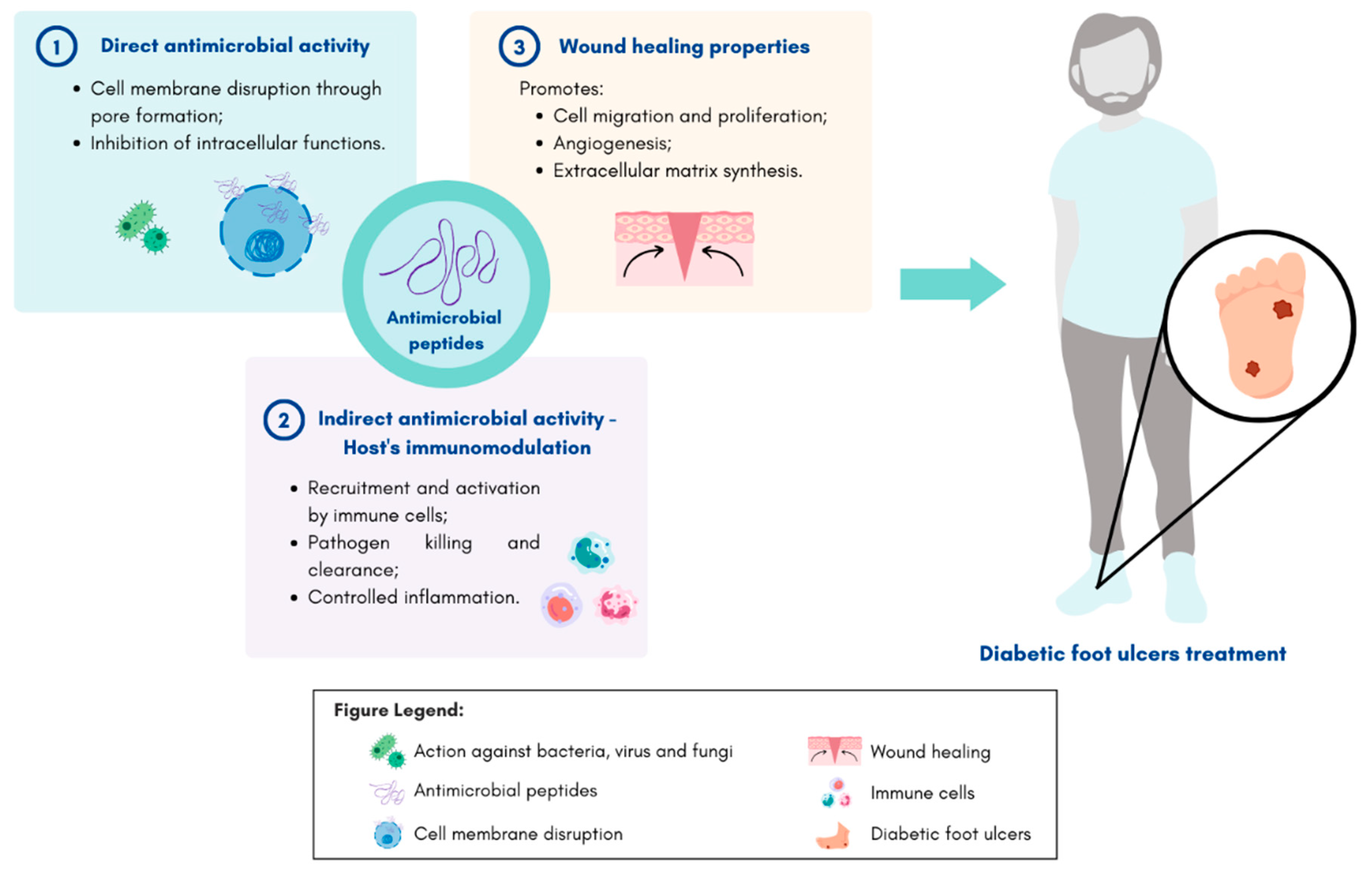
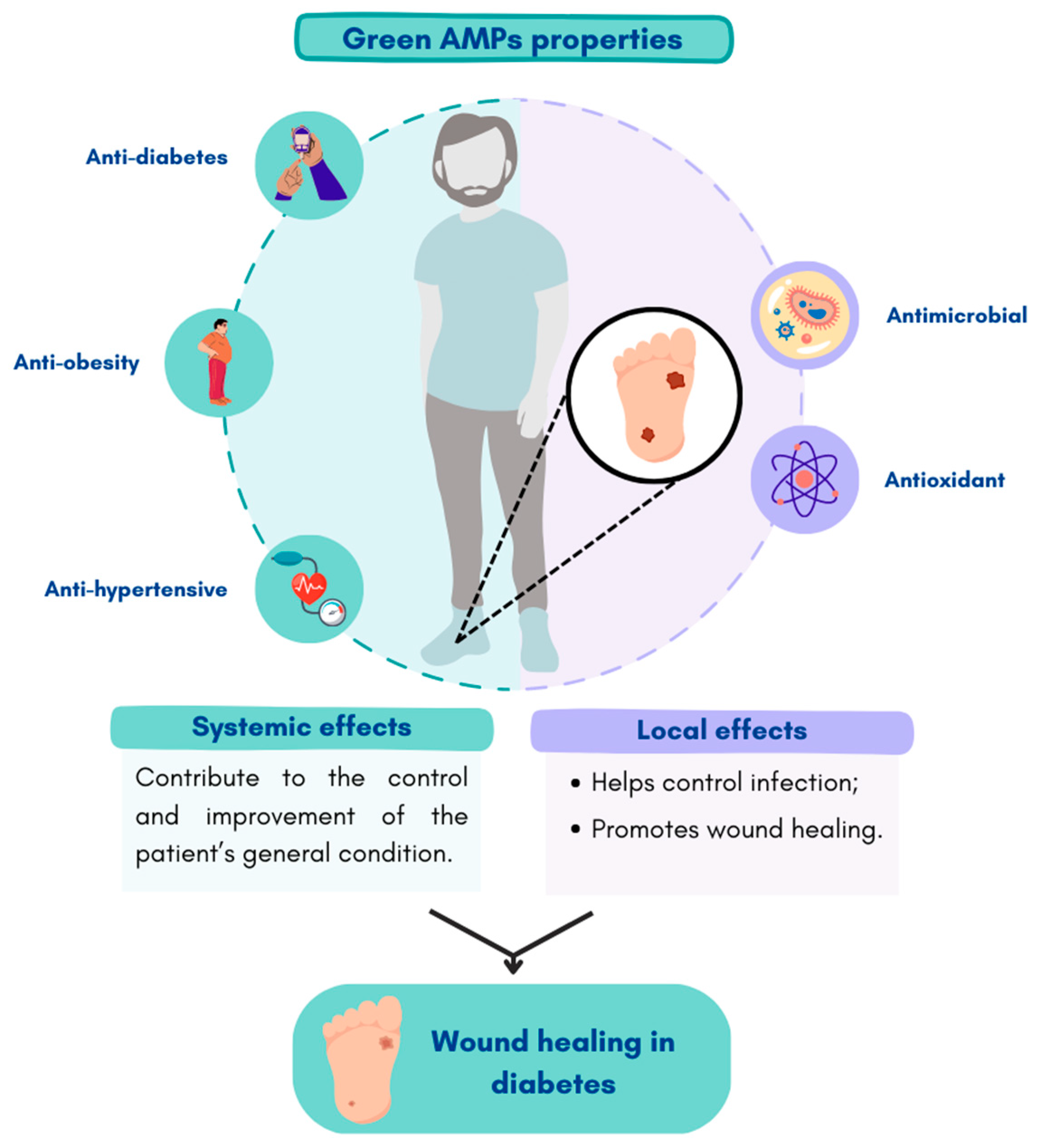
Diabetic foot ulcers (DFU) are one of the most serious and devastating complications of diabetes and account for a significant decrease in quality of life and costly healthcare expenses worldwide. This condition affects around 15% of diabetic patients and is one of the leading causes of lower limb amputations. DFUs generally present poor clinical outcomes, mainly due to the impaired healing process and the elevated risk of microbial infections which leads to tissue damage. Antimicrobial resistance poses a rising threat to global health, thus hampering DFU treatment and care. Faced with this reality, it is pivotal to find greener and less environmentally impactful alternatives for fighting these resistant microbes. Antimicrobial peptides are small molecules that play a crucial role in the innate immune system of the host and can be found in nature. Some of these molecules have shown broad-spectrum antimicrobial properties and wound-healing activity, making them good potential therapeutic compounds to treat DFUs. 

diabetic foot ulcer peptides antimicrobial peptides

1. Diabetic Foot Ulcers

Wound healing is a dynamic and complex biological process aimed to restore skin function after trauma [1]. This process is achieved through four successive phases with limited overlap: hemostasis, inflammation, proliferation, and remodeling [1][2].
The hemostasis phase initiates immediately after skin injury and is characterized by the constriction of the damaged blood vessels and the activation of platelets. Platelet aggregations will promote the formation of a fibrin clot that seals the damaged endothelium, stopping the bleeding [1][2]. Once the bleeding is controlled, the inflammatory phase begins. Damaged cells release chemokines and cytokines, which recruit inflammatory cells to the site of injury. Neutrophils are the first cells mobilized to the wound site and are responsible for microbial clearance [1][2][3]. These cells are then followed by pro-inflammatory macrophages (M1), which induce the clearing of apoptotic cells, including neutrophils, and secrete growth factors and cytokines that promote the inflammatory response by attracting adaptive immune system cells to the site of injury [1][2][3]. As the inflammatory stage is resolved, these cells undergo a phenotypic alteration to a reparative state (M2) that promotes tissue regeneration [3]. The proliferative phase begins once the inflammation decreases. This phase is characterized by the formation of granulation tissue, contraction of the wound edges, re-epithelization, and neovascularization [1][2][4]. The final step of wound healing is the remodeling phase, which includes collagen fiber reorganization, remodeling, and maturation of the scar tissue, as well as an increase in its tensile strength [1][2][4].
For a wound to heal successfully, all four phases need to be very well coordinated in the right order and within the appropriate time period [1][2]. Nevertheless, under diabetic conditions, this process is impaired resulting in the occurrence of chronic non-healing wounds in a state of persistent pro-inflammation [1][2][3]. This is characterized by an accumulation of immune cells, an increase in the M1/M2 macrophage ratio, increased generation of reactive oxygen species (ROS), and pro-inflammatory cytokines [1]. Moreover, about 60% of diabetic foot ulcers (DFUs) develop bacterial infections which contribute to the failure to heal chronic wounds [1][2].
Patients with DFIs are regularly hospitalized and are frequently exposed to numerous courses of antibiotics [5][6][7]. Today, the emergence and spread of bacteria that are resistant to conventional antibiotics, including those used within the hospital environment, impose a rising threat to global health [6][7]. This problem greatly hampers DFU treatment. For this reason, the development of alternative compounds with the capacity to downregulate the inflammatory response and control pathogen infection is urgently required.

2. Microbiota in Diabetic Foot Ulcers

The skin is the largest organ in the human body, and it has an essential role as a multifunctional barrier, protecting our body against pathogens or toxic substances [8]. The human skin contains a large and diverse composition of living microorganisms known as the skin microbiota. Most of these microorganisms are harmless and even advantageous to their host, protecting against invasion by more pathogenic organisms and contributing to skin homeostasis. However, the disruption in this balanced microbiota system can enhance the susceptibility to skin disorders, including infections [8][9].
The physical and chemical characteristics of the skin surface may vary according to environmental and specific host factors. These features can influence colonization by the skin microbiota and determine unique sets of microorganisms [8]. Under diabetic conditions, skin integrity is compromised by several factors, contributing to impaired tissue regeneration and alterations in the local skin microbiome [10][11]. Persistent hyperglycemia creates an excessive nutrient source for microbes and reduces innate immunity by causing poor chemotaxis, phagocytosis, and cleansing of pathogens by neutrophils [11]. Moreover, peripheral vascular disease hampers the action of the host’s immune response, in part due to reduced blood flow [11]. Peripheral neuropathy aggravates minor traumas and increases forefoot pressure, facilitating the entry of microbes [10][11]. All these factors contribute to microbial colonization, biofilm formation, and clinical infections that impair wound healing and contribute to serious complications including osteomyelitis and lower limb amputation [10][11]. Approximately 60% of all DFU cases are estimated to develop DFI [2].
The DFU microbiota has been extensively studied [5][11][12][13][14]. A longitudinal study of patients with DFU (n = 100) used shotgun metagenomic sequencing to profile chronic wound microbiota and investigated its role in clinical outcomes and the response to therapy. The majority of bacteria present in diabetic foot ulcers were Gram-positive strains, such as Staphylococcus aureus, methicillin-resistant S.aureus (MRSA), and Corynebacterium striatum, as well as Gram-negative bacteria, including Pseudomonas aeruginosa and Alcaligenes faecalis [11]. These results were in accordance with other microbial studies that also include Escherichia coli and Proteus spp. as the most predominant isolated strains from DFU [7][11][12]. Moreover, some anaerobes have also been identified in the deep tissue within diabetic wounds, including Peptostreptococcus spp., Bacteroides spp., Prevotella spp., and Clostridium spp. [2][5][11].
The majority of DFI are polymicrobial in nature, and mixed microorganisms, including fungi, are frequently prevalent. The prevalence of pathogenic fungal species and subsequent mycotic infections are responsible for an increased risk of diabetic foot syndrome development and poor clinical outcomes [2][5][11][12][13]. The most commonly isolated fungi are Candida spp., Trichophyton spp., Aspergillus spp., Trichosporon spp., and Cladosporium herbarum [2][11][12][13].
DFUs are considered polymicrobial ecosystems composed of highly dynamic and diverse microbial communities [5][14]. These microorganisms can exist independently or can organize into functionally equivalent pathogroups (FEP), where commensal and pathogenic bacteria co-aggregate symbiotically in a self-produced protective polysaccharide matrix with transformed phenotype known as biofilms [2][14]. Since biofilms hamper local access to antimicrobial agents and the host’s immune system, the wound healing becomes stalled and infection is very difficult to resolve, further promoting chronic infected wounds [2][5][14]. A prospective study revealed that approximately 97.6% of microbial isolates from chronic DFUs were multi-drug resistant (MDR) with 46.3% of MDR isolates having the ability to form biofilms. Staphylococcus aureus is the biofilm forming most predominant strain [15].

3. Antimicrobial Peptides

To combat multi-drug-resistant bacterial infections, there is a clear need for the development of novel alternative compounds and therapeutic strategies. In recent years, increasing attention has been paid to antimicrobial peptides (AMPs) as a novel class of antimicrobials with great clinical potential [6].
Antimicrobial peptides, also known as host defense peptides, are effector molecules of the innate immune system which can be found in all living organisms [6][7][16]. These small molecules play important roles in fighting infection through broad-spectrum antimicrobial activity, host’s immunomodulatory functions, as well as other functions relevant to wound healing (Figure 1) [9].
Figure 1. Schematic representation of antimicrobial peptides mechanisms of action and their potential application on diabetic foot ulcer treatment. Figure created in Canva.com (accessed on 24 January 2023).
There is a large diversity of known AMPs: more than 5000 antimicrobial peptides have been characterized and synthesized, and this number is expected to increase in the coming years [17]. Based on their structure, AMPs can be classified into four different groups: α-helical, extended, β-sheet, and cyclic [6][16][18]. Their secondary structures provide each peptide with a functional specificity [16].
Upon injury, the innate immune system recognizes pathogen-associated molecular patterns (PAMPs), including lipopolysaccharides (LPS) [2]. This, in turn, leads to the production of AMPs by skin resident cells, such as keratinocytes, and by infiltrating leukocytes, circulating neutrophils, and tissue macrophages [18][19]. As a result of the overexpression of these small molecules, the body is able to respond to injury and infection quickly and effectively [2][6][18].
The most frequent mechanism of action of AMPs consists of targeting bacterial cell membranes directly [2][6]. AMPs often include positively charged residues and multiple hydrophobic residues, which allow them to settle electrostatic interactions between their cationic membrane and the anionic bacterial membranes [6]. As a result, the pathogenic bacterial membrane is disrupted, often through the formation of pores. This subsequently leads to the insertion of AMPs into the membrane, causing bacterial cell death [2][6]. Additionally, it has also been described that some AMPs have the ability to cross bacterial cell membranes without affecting their integrity. These peptides inhibit essential bacterial intracellular functions such as nucleic acid and protein synthesis, as well as enzyme activity, thus leading to cell death [20].
In addition to the direct eradication of microbes, AMPs can have an indirect antimicrobial effect by modulating and enhancing the hosts’ adaptative immune responses [2][9][19]. These peptides can act as chemoattractants, recruiting and activating immune cells. This leads to the increasing expression of proinflammatory cytokines, thereby suppressing potentially harmful inflammation [2][6][16][18]. AMPs are also able to promote wound healing by the induction of cell migration, proliferation and differentiation, re-epithelization, support of angiogenesis, and enhancement of extracellular matrix synthesis [2][9].
In human skin, cathelicidins and defensins are the most prevalent classes of endogenous AMPs, particularly cathelicidin LL-37 and human β-defensins (hBDs 1–3) [18][19].
It is important to note, however, that under diabetic conditions, the expression and/or activity level of endogenous AMPs may be greatly affected. This in turn may contribute to the inadequate infection control and impaired wound healing often observed in the presence of diabetes [2]. Gonzalez-Curiel et al. have demonstrated increased susceptibility to infectious diseases in patients with type 2 diabetes due to lower levels of CAMP (LL-37) and DEFB4 (hBD-2) genes in peripheral blood cells [21]. Moreover, Al-Shibly et al. showed that although DFUs may express some endogenous AMPs, their expression levels are inefficient to suppress secondary infections and promote wound healing [22].
Therefore, in order to maximize the activity of these peptides at the wound site and enhance wound healing, it is necessary to maintain or increase their expression and activity levels as well as overcome some of their limitations. The use of chemical modifications could be considered a potential strategy for increasing the stability of these AMPs within the DFU microenvironment, decreasing their toxicity, and improving their antimicrobial and wound-healing functions [2].

4. Therapeutic Use of Green Antimicrobial Peptides in Diabetic Foot Ulcer

AMPs are a promising alternative for infected wounds since they are active against a wide range of Gram-positive and Gram-negative bacteria. In addition, AMPs can exhibit immunomodulatory and angiogenic properties, stimulate cell proliferation and migration, and accelerate wound healing [2]. Although the AMPs referred above were obtained using green processes and can be relevant as a therapeutic option for DFUs (Table 1), some of their inherent steps still need to be improved to develop a completely green protocol. Only a few AMPs are being extracted using more environmentally friendly approaches: Cn-AMPs, SP-1, KLENCNYAVELGK, Lyngbyazothrins mixture C/D, and Laxaphycin A, B, and B3. This may be explained by the recent increase in the awareness for the need of more sustainable and safer processes. AMPs extracted by greener methodologies include the Cn-AMPs: three antimicrobial peptides extracted from coconut water [23]. The extraction of these AMPs has been achieved through simple methodologies that did not use toxic solvents. Nevertheless, reverse phase chromatography (HPLC) was used in their purification stage. In order to reduce the generation of organic waste and accomplish a complete green extraction protocol, this technique could be replaced by new promising and eco-friendly approaches such as mixed-mode chromatography (MMC), multicolumn counter-current solvent gradient purification (MCSGP), or even supercritical fluid chromatography (SFC) [24]. These peptides showed antimicrobial activity against multiple Gram-positive and Gram-negative bacteria and fungi [23]. Interestingly, further studies investigated the promiscuity of these peptides and demonstrated that in addition to their antimicrobial activities, they also present activity against cancerous cells and immunostimulatory effects [25][26][27]. The immunostimulatory effect is a very important property, particularly in diabetes due to the low-grade inflammation, and it is difficult to have a controlled inflammatory phase of wound healing. This is of utmost importance as it will dictate the progression of wound closure [28][29].
Another example is SP-1, an AMP extracted by enzymatic hydrolysis from the cyanobacteria Spirulina platensis. The peptide KLENCNYAVELGK was successfully extracted from pepsin hydrolysates derived from Limnospira sp. This peptide showed antibacterial activity against Escherichia coli and Staphylococcus aureus [30]. It also presents antioxidant, anti-hypertensive, anti-diabetes, and anti-obesity activities [31][32]. Most of these peptides have antioxidant properties which are important to promote cell function in wounds under diabetic conditions, particularly, cell migration and proliferation, as well as angiogenic properties [33][34]. Moreover, for a proper wound healing, the overall condition of the patient is very important. The anti-hypertensive and anti-diabetic properties of the peptides will contribute to the control and improvement of the patient’s general condition, and consequently better wound healing. In this sense, peptides that improve the general condition of diabetes could make a better contribution to the treatment of DFUs (Figure 2). In addition, some peptides such as PvD1, cycloviolacin O2, pitipeptolides C-F, and laxaphycins A, B, and B3 have shown to have high cytotoxicity against tumor cells [35][36][37]. This is an important property since diabetic patients present an elevated risk of developing different types of cancers [38][39].
Figure 2. Effects of more environmentally friendly AMPs as potential therapeutic agents in chronic DFUs. Figure created in Canva.com (accessed on 21 February 2023).
Furthermore, the peptides in the Lyngbyazothrins mixture C/D and Laxaphycin A, B, and B3 obtained by greener methodologies also showed activity against common pathogens found in chronic DFUs [40][41]. The cyclic undecapeptides Lyngbyazothrins C/D showed antimicrobial activity against Escherichia coli, Pseudomonas aeruginosa, Serratia marcesens, and Bacillus subtilis [40]. Cyanobacterial peptides laxaphycins A, B, and B3 demonstrated antibacterial activity against Listeria monocytogenes, Bacillus cereus, and Staphylococcus aureus [41].
In addition to the aforementioned peptides, there are others obtained by less eco-friendly methodologies that present interesting properties concerning DFUs treatment. A study reported a peptide known as AQ-1766 that was extracted from the marine microalgae Tetraselmis suecica, with high activity against E. coli, P. aeruginosa, MRSA, L. monocytogenes, and M. luteus [42]. The fact that this particular AMP possesses high activity against MRSA, an antibiotic-resistant and prevalent pathogen in infected chronic DFUs, makes it an interesting and therapeutic approach for DFUs. Moreover, further research demonstrated that the peptide Snakin-Z also exhibited high antioxidant activity in addition to its antimicrobial properties [43].
Plants and microalgae have been considered sustainable and attractive sources of novel AMPs. Nevertheless, many of the extraction and production processes currently in use still employ hazardous substances [41][44]. The industrial scale-up production of AMPs is still a significant challenge due to highly expensive chemical methodologies [27].
Similarly, the current synthesis methodologies in use are still not completely green, in part due to their use of or the creation of toxic by-products. However, efforts are being made in order to develop more sustainable methodologies. This can be achieved by the replacement of hazardous substances and techniques with more sustainable options. Thus, it is imperative that the current synthesis of synthetic AMPs already identified as potential therapeutic agents for DFUs can be produced using greener techniques.
The potential therapeutic role of most of these peptides for DFU has not yet been investigated. However, due to their multiple properties, it would be important to further study the effects of novel greener AMPs as promising therapeutic agents for non-healing chronic infected wounds.
Table 1. Antimicrobial peptides extracted from sustainable sources as therapeutic options for DFU.

References

  1. Petkovic, M.; Sørensen, A.E.; Leal, E.C.; Carvalho, E.; Dalgaard, L.T. Mechanistic actions of microRNAs in diabetic wound healing. Cells 2020, 9, 2228.
  2. Da Silva, J.; Leal, E.C.; Carvalho, E. Bioactive antimicrobial peptides as therapeutic agents for infected diabetic foot ulcers. Biomolecules 2021, 11, 1894.
  3. Guo, S.A.; DiPietro, L.A. Factors affecting wound healing. J. Dent. Res. 2010, 89, 219–229.
  4. Singer, A.J.; Clark, R.A.F. Cutaneous Wound Healing. New Engl. J. Med. 1999, 341, 738–746.
  5. Ramirez-Acuña, J.M.; Cardenas-Cadena, S.A.; Marquez-Salas, P.A.; Garza-Veloz, I.; Perez-Favila, A.; Cid-Baez, M.A.; Flores-Morales, V.; Martinez-Fierro, M.L. Diabetic foot ulcers: Current advances in antimicrobial therapies and emerging treatments. Antibiotics 2019, 8, 193.
  6. Browne, K.; Chakraborty, S.; Chen, R.; Willcox, M.D.; Black, D.S.; Walsh, W.R.; Kumar, N. A new era of antibiotics: The clinical potential of antimicrobial peptides. Int. J. Mol. Sci. 2020, 21, 7047.
  7. Uivaraseanu, B.; Bungau, S.; Tit, D.M.; Fratila, O.; Rus, M.; Maghiar, T.A.; Pantis, C.; Vesa, C.M.; Zaha, D.C. Clinical, pathological and microbiological evaluation of diabetic foot syndrome. Medicina 2020, 56, 380.
  8. Grice, E.A.; Segre, J.A. The skin microbiome. Nat. Rev. Microbiol. 2011, 9, 244–253.
  9. Rademacher, F.; Gläser, R.; Harder, J. Antimicrobial peptides and proteins: Interaction with the skin microbiota. Exp. Dermatol. 2021, 30, 1496–1508.
  10. Kalan, L.R.; Meisel, J.S.; Loesche, M.A.; Horwinski, J.; Soaita, I.; Chen, X.; Uberoi, A.; Gardner, S.E.; Grice, E.A. Strain-and species-level variation in the microbiome of diabetic wounds is associated with clinical outcomes and therapeutic efficacy. Cell Host Microbe 2019, 25, 641–655.
  11. Kalan, L.R.; Brennan, M.B. The role of the microbiome in nonhealing diabetic wounds. Ann. N. Y. Acad. Sci. 2019, 1435, 79–92.
  12. Kareliya, H.; Bichile, L.; Bal, A.; Varaiya, A.; Bhalekar, P. Fungal Infection in Diabetic Foot a Clinicomicrobiological Study. Acta Sci. Microbiol. 2019, 2, 49–55.
  13. Kalan, L.; Loesche, M.; Hodkinson, B.P.; Heilmann, K.; Ruthel, G.; Gardner, S.E.; Grice, E.A. Redefining the Chronic-Wound Microbiome: Fungal Communities Are Prevalent, Dynamic, and Associated with Delayed Healing. MBio 2016, 7, e01058-16.
  14. Pouget, C.; Dunyach-Remy, C.; Pantel, A.; Schuldiner, S.; Sotto, A.; Lavigne, J.P. Biofilms in diabetic foot ulcers: Significance and clinical relevance. Microorganisms 2020, 8, 1580.
  15. Banu, A.; Noorul Hassan, M.M.; Rajkumar, J.; Srinivasa, S. Spectrum of bacteria associated with diabetic foot ulcer and biofilm formation: A prospective study. Australas. Med. J. 2015, 8, 280–285.
  16. Fry, D.E. Antimicrobial peptides. Surg. Infect. 2018, 19, 804–811.
  17. Depta, J.; Małkowska, P.; Wysokińska, M.; Todorska, K.; Sierawska, O.; Hrynkiewicz, R.; Bebnowska, D.; Niedźwiedzka-Rystwej, P. Therapeutic Role of Antimicrobial Peptides in Diabetes Mellitus. Biologics 2022, 2, 92–106.
  18. Gaiser, R.A. Antimicrobial Peptides and the Interplay between Microbes and Host: Towards Preventing Porcine Infections with Streptococcus Suis. Ph.D. Thesis, Wageningen University and Research, Wageningen, The Netherlands, 2016.
  19. Gallo, R.L.; Nizet, V. Endogenous production of antimicrobial peptides in innate immunity and human disease. Curr. Allergy Asthma Rep. 2003, 3, 402–409.
  20. Zhang, L.J.; Gallo, R.L. Antimicrobial peptides. Curr. Biol. 2016, 26, R14–R19.
  21. Gonzalez-Curiel, I.; Trujillo, V.; Montoya-Rosales, A.; Rincon, K.; Rivas-Calderon, B.; De Haro-Acosta, J.; Marin-Luevano, P.; Lozano-Lopez, D.; Enciso-Moreno, J.A.; Rivas-Santiago, B. 1,25-dihydroxyvitamin D3 induces LL-37 and HBD-2 production in keratinocytes from diabetic foot ulcers promoting wound healing: An in vitro model. PLoS ONE 2014, 9, e111355.
  22. Al-Shibly, I.K.; Alhamdany, M.H.; Al-Kaif, R.A.I.; Al-Kaif, L.A. Immunological Base Behind the Increased Susceptibility of Diabetic Patients for Infections. Indian J. Public Health 2019, 10, 3047–3051.
  23. Mandal, S.M.; Dey, S.; Mandal, M.; Sarkar, S.; Maria-Neto, S.; Franco, O.L. Identification and structural insights of three novel antimicrobial peptides isolated from green coconut water. Peptides 2009, 30, 633–637.
  24. Lima, P.G.; Oliveira, J.T.; Amaral, J.L.; Freitas, C.D.; Souza, P.F. Synthetic antimicrobial peptides: Characteristics, design, and potential as alternative molecules to overcome microbial resistance. Life Sci. 2021, 278, 119647.
  25. Silva, O.N.; Porto, W.F.; Migliolo, L.; Mandal, S.M.; Gomes, D.G.; Holanda, H.H.; Silva, R.S.P.; Dias, S.C.; Costa, M.P.; Costa, C.R.; et al. Cn-AMP1: A new promiscuous peptide with potential for microbial infections treatment. Pept. Sci. 2012, 98, 322–331.
  26. Santana, M.J.; de Oliveira, A.L.; Queiroz Júnior, L.H.; Mandal, S.M.; Matos, C.O.; Dias, R.D.O.; Franco, O.L.; Lião, L.M. Structural insights into Cn-AMP1, a short disulfide-free multifunctional peptide from green coconut water. FEBS Lett. 2015, 589, 639–644.
  27. Sathya, R.; MubarakAli, D.; MohamedSaalis, J.; Kim, J.W. A systemic review on microalgal peptides: Bioprocess and sustainable applications. Sustainability 2021, 13, 3262.
  28. Leal, E.C.; Emanuelli, T.; Santos, D.; Moura, J.; Fonseca, A.C.R.; Burgeiro, A.; Carvalho, E. Dysregulation of endoplasmic reticulum stress response in skin wounds in a streptozotocin-induced diabetes mouse model. J. Mol. Endocrinol. 2023.
  29. Leal, E.C.; Carvalho, E.; Tellechea, A.; Kafanas, A.; Tecilazich, F.; Kearney, C.; Kuchibhotla, S.; Auster, M.E.; Kokkotou, E.; Mooney, D.J.; et al. Substance P promotes wound healing in diabetes by modulating inflammation and macrophage phenotype. Am. J. Pathol. 2015, 185, 1638–1648.
  30. Sathya, R.; MubarakAli, D.; Mehboob Nousheen, M.G.; Vasimalai, N.; Thajuddin, N.; Kim, J.-W. An Investigation of Pepsin Hydrolysate of Short Antibacterial Peptides Derived from Limnospira Sp. Appl. Biochem. Biotechnol. 2022, 194, 5580–5593.
  31. Sun, Y.; Chang, R.; Li, Q.; Li, B. Isolation and characterization of an antibacterial peptide from protein hydrolysates of Spirulina platensis. Eur. Food Res. Technol. 2016, 242, 685–692.
  32. Yücetepe, A.; Özçelik, B. Bioactive peptides isolated from microalgae Spirulina platensis and their biofunctional activities. Akad. Gıda 2016, 14, 412–417.
  33. Figueiredo, A.; Leal, E.C.; Carvalho, E. Protein tyrosine phosphatase 1B inhibition as a potential therapeutic target for chronic wounds in diabetes. Pharmacol. Res. 2020, 159, 104977.
  34. Leal, E.C.; Carvalho, E. Heme Oxygenase-1 as Therapeutic Target for Diabetic Foot Ulcers. Int. J. Mol. Sci. 2022, 23, 12043.
  35. Figueira, T.N.; Oliveira, F.D.; Almeida, I.; Mello, É.O.; Gomes, V.M.; Castanho, M.A.; Gaspar, D. Challenging metastatic breast cancer with the natural defensin PvD1. Nanoscale 2017, 9, 16887–16899.
  36. Gerlach, S.L.; Rathinakumar, R.; Chakravarty, G.; Göransson, U.; Wimley, W.C.; Darwin, S.P.; Mondal, D. Anticancer and chemosensitizing abilities of cycloviolacin O2 from Viola odorata and psyle cyclotides from Psychotria leptothyrsa. Pept. Sci. 2010, 94, 617–625.
  37. Qamar, H.; Hussain, K.; Soni, A.; Khan, A.; Hussain, T.; Chénais, B. Cyanobacteria as Natural Therapeutics and Pharmaceutical Potential: Role in Antitumor Activity and as Nanovectors. Molecules 2021, 26, 247.
  38. Kim, D.S.; Scherer, D.S.P.E. Obesity, Diabetes, and Increased Cancer Progression. Diabetes Metab. J. 2021, 45, 799–812.
  39. Wang, M.; Yang, Y.; Liao, Z. Diabetes and cancer: Epidemiological and biological links. World J. Diabetes 2020, 11, 227–238.
  40. Zainuddin, E.N.; Jansen, R.; Nimtz, M.; Wray, V.; Preisitsch, M.; Lalk, M.; Mundt, S. LyngbyazothrinsA−D, Antimicrobial CycliUndecapeptides from the Cultured Cyanobacterium Lyngbya sp. J. Nat. Prod. 2009, 72, 2080.
  41. Dussault, D.; Vu, K.D.; Vansach, T.; Horgen, F.D.; Lacroix, M. Antimicrobial effects of marine algal extracts and cyanobacterial pure compounds against five foodborne pathogens. Food Chem. 2016, 199, 114–118.
  42. Guzmán, F.; Wong, G.; Román, T.; Cárdenas, C.; Alvárez, C.; Schmitt, P.; Albericio, F.; Rojas, V. Identification of antimicrobial peptides from the microalgae Tetraselmis suecica (Kylin) Butcher and bactericidal activity improvement. Mar. Drugs 2019, 17, 453.
  43. Zare-Zardini, H.; Tolueinia, B.; Hashemi, A.; Ebrahimi, L.; Fesahat, F. Antioxidant and cholinesterase inhibitory activity of a new peptide from Ziziphus jujuba fruits. Am. J. Alzheimer’s Dis. Other Dement.® 2013, 28, 702–709.
  44. Ishida, K.; Matsuda, H.; Murakami, M.; Yamaguchi, K. Kawaguchipeptin B, an antibacterial cyclic undecapeptide from the cyanobacterium Microcystis aeruginosa. J. Nat. Prod. 1997, 60, 724–726.
  45. Games, P.D.; Dos Santos, I.S.; Mello, É.O.; Diz, M.S.; Carvalho, A.O.; de Souza-Filho, G.A.; Cunha, M.; Vasconcelos, I.M.; Ferreira, B.D.S.; Gomes, V.M. Isolation, characterization and cloning of a cDNA encoding a new antifungal defensin from Phaseolus vulgaris L. seeds. Peptides 2008, 29, 2090–2100.
  46. Daneshmand, F.; Zare-Zardini, H.; Ebrahimi, L. Investigation of the antimicrobial activities of Snakin-Z, a new cationic peptide derived from Zizyphus jujuba fruits. Nat. Prod. Res. 2013, 27, 2292–2296.
  47. Pränting, M.; Lööv, C.; Burman, R.; Göransson, U.L.F.; Andersson, D.I. The cyclotide cycloviolacin O2 from Viola odorata has potent bactericidal activity against Gram-negative bacteria. J. Antimicrob. Chemother. 2010, 65, 1964–1971.
  48. Song, W.; Kong, X.; Hua, Y.; Chen, Y.; Zhang, C.; Chen, Y. Identification of antibacterial peptides generated from enzymatic hydrolysis of cottonseed proteins. LWT 2020, 125, 109199.
  49. Chahardoli, M.; Fazeli, A.; Ghabooli, M. Recombinant production of bovine Lactoferrin-derived antimicrobial peptide in tobacco hairy roots expression system. Plant Physiol. Biochem. 2018, 123, 414–421.
  50. Łojewska, E.; Sakowicz, T.; Kowalczyk, A.; Konieczka, M.; Grzegorczyk, J.; Sitarek, P.; Skała, E.; Czarny, P.; Sliwinski, T.; Kowalczyk, T. Production of recombinant colicin M in Nicotiana tabacum plants and its antimicrobial activity. Plant Biotechnol. Rep. 2020, 14, 33–43.
  51. Patiño-Rodríguez, O.; Ortega-Berlanga, B.; Llamas-González, Y.Y.; Flores-Valdez, M.A.; Herrera-Díaz, A.; Montes-de-Oca-Luna, R.; Korban, S.S.; Alpuche-Solís, A.G. Transient expression and characterization of the antimicrobial peptide protegrin-1 in Nicotiana tabacum for control of bacterial and fungal mammalian pathogens. Plant Cell Tissue Organ Cult. PCTOC 2013, 115, 99–106.
  52. Montaser, R.; Paul, V.J.; Luesch, H. Pitipeptolides C–F, antimycobacterial cyclodepsipeptides from the marine cyanobacterium Lyngbya majuscula from Guam. Phytochemistry 2011, 72, 2068–2074.
More
Information
Contributors MDPI registered users' name will be linked to their SciProfiles pages. To register with us, please refer to https://encyclopedia.pub/register : , ,
View Times: 479
Entry Collection: Peptides for Health Benefits
Revisions: 5 times (View History)
Update Date: 03 Mar 2023
1000/1000