Submitted Successfully!
To reward your contribution, here is a gift for you: A free trial for our video production service.
Thank you for your contribution! You can also upload a video entry or images related to this topic.
Version Summary Created by Modification Content Size Created at Operation
1 -- 4201 2023-02-18 00:15:16 |
2 format correction Meta information modification 4170 2023-02-21 01:46:20 | |
3 format correction Meta information modification 4170 2023-02-21 01:47:42 | |
4 format correction Meta information modification 4170 2023-02-21 01:49:56 |

Video Upload Options

Do you have a full video?

Confirm

Are you sure to Delete?
Cite
If you have any further questions, please contact Encyclopedia Editorial Office.
Godbout, K.; Tremblay, J.P. Application of Prime Editing to Liver Hereditary Diseases. Encyclopedia. Available online: https://encyclopedia.pub/entry/41378 (accessed on 27 July 2024).
Godbout K, Tremblay JP. Application of Prime Editing to Liver Hereditary Diseases. Encyclopedia. Available at: https://encyclopedia.pub/entry/41378. Accessed July 27, 2024.
Godbout, Kelly, Jacques P. Tremblay. "Application of Prime Editing to Liver Hereditary Diseases" Encyclopedia, https://encyclopedia.pub/entry/41378 (accessed July 27, 2024).
Godbout, K., & Tremblay, J.P. (2023, February 18). Application of Prime Editing to Liver Hereditary Diseases. In Encyclopedia. https://encyclopedia.pub/entry/41378
Godbout, Kelly and Jacques P. Tremblay. "Application of Prime Editing to Liver Hereditary Diseases." Encyclopedia. Web. 18 February, 2023.
Application of Prime Editing to Liver Hereditary Diseases
Edit

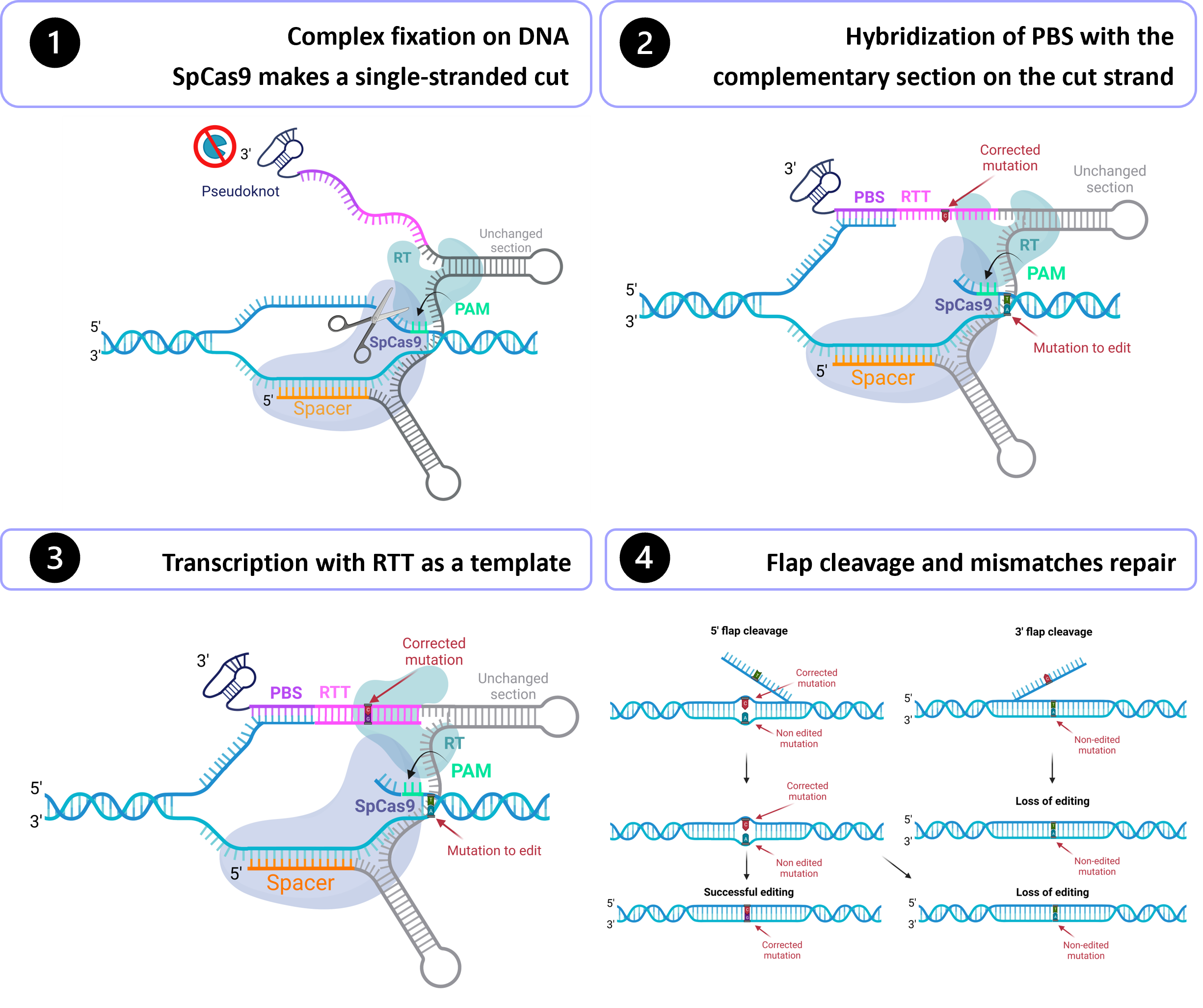
Gene therapy holds tremendous potential in the treatment of inherited diseases. Unlike traditional medicines, which only treat the symptoms, gene therapy has the potential to cure the disease by addressing the root of the problem: genetic mutations. The discovery of CRISPR/Cas9 in 2012 paved the way for the development of those therapies. Improvement of this system led to the recent development of an outstanding technology called prime editing. This system can introduce targeted insertions, deletions, and all 12 possible base-to-base conversions in the human genome. Since the first publication on prime editing in 2019, groups all around the world have worked on this promising technology to develop a treatment for genetic diseases. Liver diseases are currently the most studied field for human gene therapy by prime editing. To date, prime editing has been attempted in preclinical studies for tyrosinemia type 1, alpha-1-antitrypsin deficiency, phenylketonuria, DGAT1-deficiency, bile salt export pump deficiency, liver cancer, and for a liver disease caused by a mutation in the DNMT1 gene.

prime editing gene therapy inherited diseases genetic diseases CRISPR/Cas9 liver diseases gene editing

1. Introduction

Gene therapy offers enormous potential in the treatment of genetic diseases. Its potency lies in addressing the genetic root of the problem, unlike traditional medicines, which only treat the symptoms. By correcting the mutations, gene therapy has the potential to cure hereditary diseases. Sherkow et al. [1] defined gene therapy as “the intentional, expected permanent, and specific alteration of the DNA sequence of the cellular genome, for a clinical purpose”. The first approved gene therapy occurred in 1990, when a foreign gene was inserted into a kid’s immune cells [2]. Gene therapies first took the form of DNA insertion into the host genome [3]. In the 2000s, tools allowing the introduction of modifications at specific target sites in the genome were developed, including zinc-finger nucleases (ZFNs) [4] and transcription activator-like effector nucleases (TALENs) [5].
A milestone in the development of gene therapies was the discovery of CRISPR/Cas9 in 2012. This system involves a Cas9 nuclease that induces a double-strand DNA break at a precise place in the genome. The Cas9 is directed at the right sequence in the genome by a guide RNA. This guide is a single RNA strand complementary to an 18–24 nucleotides (nt) sequence in the genome [6][7]. When the complex is fixed on the complementary DNA sequence and the Cas9 recognizes a protospacer adjacent motif (PAM), the complex is activated. This PAM sequence varies depending on the microorganism of origin of the Cas9. For example, the most widely used Cas9 is from Streptococcus pyogenes (SpCas9) and recognizes the PAM 5′-NGG-3′ [8]. Once this small sequence is recognized, Cas9 will cut 3 nt upstream of the PAM [9]. The cell will then repair its DNA by non-homologous end joining (NHEJ), microhomology-mediated end joining (MMEJ), or homology-directed repair (HDR). NHEJ is an imprecise mechanism where broken ends of DNA are joined together, which often leads to insertions or deletions of nucleotides [10]. MMEJ is also an imprecise mechanism that can lead to undesired insertions, deletions, or even translocations [11]. This mechanism works by aligning short homologous sequences that are between the broken ends [12][13]. HDR repairs the damage using a homologous donor DNA, leading to a precise repair of the cut [10].
The evolution and refinement of the CRISPR/Cas9 technology have driven the development of base editing. This system exists in three versions: cytosine base editors (CBEs) [14], adenine base editors (ABEs) [15], and C to G base editors (CGBEs) [16]. CBE can install C > T and G > A mutations, while ABE can induce A > G and T > C mutations [17], and CGBE can generate C-to-G transversions [16][18]. Those editors change all the intended base pairs in a precise window (for example, CBE switches all C•G base pairs located in the window to T•A base pairs). Compared to CRISPR/Cas9, the advantages of this technology are that base editing does not require a double-strand break (the system uses a modified D10A Cas9) and does not need an exogenous DNA template, which leads to the reduction of unwanted indels. Base editing also leads to much more precise correction of the mutation by its ability to target a particular codon. However, those systems [19] cannot be applied when the change of a base pair in the window (other than the desired one) would lead to a non-silent mutation.
In October 2019, David R. Liu’s group released an outstanding discovery called prime editing [20]. This system can mediate targeted insertions, deletions, and all 12 possible base-to-base conversions. This mechanism makes DNA modifications with unprecedented precision and has substantial advantages over the traditional CRISPR/Cas9 and base editing systems (Table 1). Derived from the CRISPR/Cas9 system, this new technology is composed of a Cas9 nuclease fused with a reverse transcriptase (RT) at its 3′ extremity and a prime editing guide RNA (pegRNA) (Figure 1). The combination of Cas9 and RT forms the prime editor (PE). The pegRNA is composed of a spacer sequence, a primer binding site (PBS), a reverse transcriptase template (RTT), and a common region that binds to the Cas9 and the RT [19]. First, the complex binds to the DNA, guided by the spacer sequence in the pegRNA. The Cas9 recognizes a PAM and cuts 3 nt upstream. However, instead of creating a double-stranded cut, the modified H840A Cas9 from the prime editing induces a single-stranded nick [20]. Then, the PBS hybridizes to its complementary sequence located on the cut strand. Then, the RT will use the RTT as a template to transcribe the cut strand. At this point, one of the strands has a duplicated section, so the cell will have to remove one of the two sections to put back the DNA double-stranded again. The mismatch will be resolved by a 5′ flap or a 3′ flap. If a 3′ flap happens, the correction will be kept, but the editing will be lost if a 5′ flap occurs [20].
To introduce any modifications, the desired correction needs to be introduced in the RTT sequence, since it serves as a template for the transcription. There are currently several versions of the prime editor, the most popular being PE2 and PE3. PE3 is similar to PE2 but has an additional guide RNA that will induce a nick on the strand not initially cut by Cas9. It will promote the replacement of the unedited strand by forcing the cell to use the edited strand as a template. This increases the chances of retaining the edit at the mismatch repair step. When a position is given, this one is defined according to the reference system based on the initial Cas9 cleavage site. Thus, nucleotides downstream of this site will have a positive position (e.g., +5 means five nucleotides after the cut site towards 3′), and nucleotides upstream of the cut site will have a negative position (e.g., −5 means five nucleotides before the cut site towards 5′).
Table 1. Comparison of advantages and disadvantages of CRISPR/Cas9, base editing, and prime editing systems.
  CRISPR/Cas9 Base Editing Prime Editing
Off-target
effects
Significant off-target effects Little or no off-target effects Little or no off-target effects
  • Possibility of non-specific indels at DSB site [21];
  • DNA donor template can lead to plasmid integration in the genome;
  • Possible genome-wide off-targets.
  • No DSB;
  • Bystander base edits within a narrow window of 4–10 nt [22];
  • Genome-wide off-targets studies need to be made.
  • No DSB;
  • No bystander edits;
  • Genome-wide off-targets studies need to be made.
Flexibility
  • Can introduce insertions, deletions, and all types of substitutions.
  • Can introduce C > T, G > A, A > G, T > C and C > G substitutions only.
  • The consideration of bystander edits makes base editing more stringent on the possible sites [22].
  • Can introduce insertions, deletions, and all types of substitutions.
  • Less stringent PAM requirements [23].
Programmability 1 Only if a DNA donor template is given Yes Yes
Efficient in vivo
delivery
Currently possible Currently possible
(but more difficult than CRISPR/Cas9 because of its larger size)
Need to be improved
(too big for conventional vehicles)
1 Possibility to determine the issue of editing.

2. Application of Prime Editing to Liver Hereditary Diseases

There are several published studies on the correction by prime editing of mutations causing liver diseases or on the generation of animal models with these mutations (Table 1). Liver diseases are indeed currently the most studied field for human gene therapy by prime editing.
Table 1. Prime editing studies on correcting or introducing mutations causing liver diseases.
Disease Gene Mutation Goal 1 Prime
Editor
% of
Editing
Cells or Animal Models Length (nt) Edit
Position from the Nick
Delivery Method Prime Editor Form Comments Reference
Spacer PBS RTT
Liver cancer CTNNB1 6 nt deletion I PE3 30 Liver organoid 20 12 17 +1 Electroporation Plasmid   Schene 2020 [24]
Bile salt export pump deficiency ABCB11 D482G A > G 20 20   +7 The PAM is also mutated (+5 G > A silent mutation)
DGAT1-deficiency DGAT1 S210del Del
CCT
C 21 Patient-derived intestinal cells 20    
Bile salt export pump deficiency ABCB11 R1153H G > A 0 Patient-derived liver organoids        
Alpha-1-antitrypsin deficiency SERPINA1 E342K G > A I PE2 1.9 HEK293T cells 20 13 27   Lipo 2000 Plasmid   Liu 2021 [25]
PE3 9.9
PE2* 6.4
PE3* 15.8
C PE2 2.1 PiZ mice Hydrodynamic TVI
PE2* 6.7
PE3 3.1 AAV8
PE3 0.83 hPSCs 20 9 13 +3 Electroporation Plasmid   Habib 2022 [26]
PE2-NGA 2.0–3.0 HEK293T cells 20 13 20   Lipo 2000 Plasmid   Lung 2021 [27]
PE3-NGA 3.0–5.0
PE2-NGA 1.99 Human primary fibroblasts
Liver disease dnmt1 G > C I Intein-split PE2∆RnH 15 C57BL/6J pups 21   AAV8 Plasmid   Böck 2022 [28]
PE2∆RnH 35.9 C57BL/6J adult mice   AdV
58.2 C57BL/6J pups
Phenylketonuria Pahenu2 F263S T > C C Intein-split PE2∆RnH <1% Pahenu2 mice 20 13 19   AAV8 Plasmid  
PE2∆RnH 2.0 Adult Pahenu2 mice AdV
6.9 Neonates Pahenu2 mice
PE3∆RnH 11.1
PE3 19.6 HEK293T cells 16 Lipo 2000
PE3 19.7 19
Tyrosinemia type 1 fah   C PEDAR 0.76 FahΔExon5 mice         Hydrodynamic injection Plasmid   Jiang 2022 [29]
G > A PE3 2.3 HT1-mCdHs 20 11 15   Electroporation Plasmid sgRNA of PE3 nick in position -4 Kim 2021 [30]
34.3 HT1 mice Transplantation  
c.706G > A PE3 61 Fahmut/mut mice 20     +10 Hydrodynamic TVI Plasmid   Jang 2022 [31]
PE2 33
FAH 18.7 HEK293T cells Lentiviral vector    
1 The goal is either the introduction (I) of the mutation or the correction (C) of the mutation. Abbreviations: AAV8 = Adeno-Associated Virus Serotype 8; AdV = human adenoviral vector 5; hPSCs = human pluripotent stem cells; Lipo 2000 = Lipofectamine 2000; TVI = tail vein injection.
Schene et al. [24] were the first to publish an original article in 2020 about prime editing for liver diseases. They generated disease model organoids by prime editing and aimed to correct patient-derived disease models by prime editing. First, the authors introduced a 6 nt deletion in the CTNNB1 gene that may lead to liver cancer. Using the PE3 system and a pegRNA composed of a 20 nucleotides spacer, a 12 nucleotides PBS, and a 17 nucleotides RTT, they introduced in liver organoids the deletion at the +1 position in 30% of the cells. By using PE3 and a silent mutation in the PAM (to avoid a second prime editing event on an already edited allele), they introduced the A > G substitution (at +7 position in the pegRNA) in 20% of the liver organoid clones in the ABCB11 gene that causes bile salt export pump deficiency. Next, the authors tried to correct mutations in patient-derived intestinal cells and liver organoids. Using PE3, they successfully corrected 21% of the S210del in the DGAT1 gene. However, they did not obtain any editing for the correction of the R1153H mutation in the ABCB11 gene and the E342K mutation in the SERPINA1 gene in patient-derived liver organoids. They used the PE3 system, but the authors mentioned that they did not conduct any pegRNA design optimization, which is crucial for good prime editing efficiency.
Liu et al. [25] also published on prime editing for liver disease. They worked on the correction of the E342K mutation in the SERPINA1 gene which causes Alpha-1 antitrypsin (A1AT) deficiency. The E342K mutation is the most common one causing this disease. It is the same mutation that Schene et al. [24] failed to correct, highlighting that pegRNA design optimization is crucial. Liu et al. [25] first used PE2 to introduce the mutation into HEK293T cells. The classic PE2 requires an NGG PAM. Unfortunately, there were none near the site to be edited. Thus, they selected one that was more distant. Their pegRNA had a 20 nt spacer, a 13 nt PBS, and a 27 nt RTT. This pegRNA and PE2 achieved only 1.9% editing in HEK293T cells. However, with the same pegRNA and PE3, they achieved 9.9% editing. With this pegRNA and PE* [25] (PE that has a nuclear localization signal optimization), they obtained 6.4% of editing with PE2* and 15.8% with PE3*. They then attempted to correct in vivo a mutation in PiZ mice [25] (mouse model for the mutation E342K in the SERPINA1 gene). To do so, they delivered prime editing’s plasmid DNA by hydrodynamic tail vein injection. They obtained 2.1% editing with PE2 and 6.7% with PE2*. AAVs capacity being at most 5 kb [32], prime editing cannot be delivered using a single AAV. To address that issue, the authors tested to deliver the split-intein prime editors via AAVs by tail vein injection. By injecting a low dose of dual AAV8-PE3 (2 × 10¹¹ viral genome), they detected 0.6% of editing after two weeks, 2.3% after six weeks, and 3.1% after ten weeks. However, the authors hypothesized that because the PAM is far from the edited site, the efficiency of prime editing is not at its best and that the utilization of a nearer PAM will lead to greater efficiency.
In the same month that Liu et al. [25] released their study, Habib et al. [26] published an article on prime editing in hiPSCs (human induced pluripotent stem cells) by generating a doxycycline-inducible prime editing platform. They also attempted to correct by prime editing the E342K mutation (1024 G > A) in the SERPINA1 gene in hiPSCs derived from a patient with alpha 1-antitrypsin (A1AT) deficiency. First, with their doxycycline-inducible prime editing platform using PE3, the authors demonstrated that creating hiPSC cell lines containing a desired mutation for the HEK3 and RNF2 locus was possible. They obtained similar conclusions by comparing their pegRNA constructs for these loci with the results in hiPSCs of David Liu’s group (the creators of prime editing) in HEK293T cells. For example, both groups concluded that, compared to the HEK3 locus, efficient prime editing at the RNF2 site required a longer PBS sequence. That conclusion suggests that the optimum PBS length depends on the sequence and, thus, might have the same optimal length even when used in different cell types. Moreover, transition substitutions (position +1 to +33), transversion substitutions (position +1 to +33), small indels (1–3 bp at +10 position), large deletions (up to 80 bp) and large insertions (up to 42 bp) are possible at the HEK3 target site in hPSCs. However, Habib et al. [26] failed to introduce small indels at the +17 and +21 positions for a mutation in the RNF2 gene.
Another exciting aspect of Habib et al.’s [26] study is the section explaining why the PE3 system generates indel mutations. To understand what the main factor was, the authors tested five different scenarios: a single DSB induced by wild-type (wt) Cas9 and a sgRNA, a single nick induced by nCas9-RT and a sgRNA, a double DSB induced by wt Cas9 and two sgRNAs, a double nick induced by nCas9-RT and two sgRNAs, and finally the PE3 system (double nick induced by nCas9-RT, one pegRNA, and one sgRNA). From this experiment, the authors concluded that the unintended indels are not directly caused by double nicking but by the combinatory activity of the RT and the pegRNA.
In November 2021, Genesis Lung released his thesis [27] on the correction by prime editing of the E342K mutation in the SERPINA1 gene that causes the A1AT deficiency. Lung worked on the same mutation as Schene et al. [24], Liu et al. [25], and Habib et al. [26], whose work has been described above. The last three mentioned authors failed to demonstrate a detectable correction of the mutation [25]. They used a PE that required an NGG PAM, which caused the targeted nucleotide to be far from the cut site, thus hindering good efficiency. In their original paper, Anzalone et al. [20] proposed an RTT length between 10 and 16 nt, which is much shorter than what Liu et al. [25] used (27 nt) [26]. Lung [27] attempted to correct the E342K mutation in the SERPINA1 gene using a prime editor using another PAM. He first designed a variant of the prime editor that could use different PAMs (NGG, NGA, NGC) and conjugated them with pegRNAs that had different PBS (13 or 17 nt) and RTT lengths (between 10 and 20 nt). Liu reached up to 3% editing for the variants using an NGA PAM in HEK293T cells containing a lentivirus cassette with the E342K mutation. The best pegRNA had a spacer of 20 nt, a PBS of 13 nt, and an RTT of 20 nt. With PE3, he reached 3 to 5% editing. Those experiments were conducted by transfecting plasmids DNA with Lipofectamine 2000. He also tried to transfect the prime editing components in their RNA form, which can improve efficiency [33]. However, in this case, the efficiency was not significantly different between the use of DNA or RNA. He next tried to correct the E342K mutation in human primary fibroblasts. By using NGA-PE2, he obtained 1.99% of editing. In his thesis, Lung also described that overexpression of the three prime repair exonuclease 2 (TREX2) gene might decrease prime editing efficiency. It has been shown that this nuclear protein has 3′–5′ exonuclease activities [34]. Non-engineered pegs being really sensible to degradation by exonuclease, the presence of TREX2 may decrease prime editing efficiency by degrading pegs from their 3′ extremity, thus reducing the available number of pegs. Hence, the knockout of this gene may help prime editing. It is to note that since Lung’s master’s thesis is not a peer-reviewed article. Therefore, those results should be taken with caution.
Böck et al. [28]. worked on the editing of two other loci, dnmt1 and Pahenu2, both of which code for proteins expressed in the liver. They first tested two variations of the prime editor, the intein-split PE2∆RnH [28] and its unsplit version. AAV8 delivered the split version, and the unsplit version, being larger, was delivered by human adenoviral vector 5 (AdV). The latter virus can contain a larger cargo but is much more immunogenic. With the split version, they achieved 15% editing in vivo of the dnmt1 locus. Since they assumed that the unsplit version would give a better result, the authors also tested it in vivo, and they obtained much better results when the prime editor was not split. In neonatal mice, they obtained 58.2% of editing; in adult mice, they obtained 35.9%. Next, they tested the correction of a mutation at the Pahenu2 locus (F263S, c.835T > C), this mutation leading to phenylketonuria, a liver disease. They first tested their pegRNAs in an HEK293T cell line that had stably integrated exon 7 of the Pahenu2 allele. Their two best constructs had a spacer of 20 nt, a PBS of 13 nt, and an RTT of 16 or 19 nt. These two pegRNAs resulted in an editing of nearly 20%. The one with a 19 nt RTT had fewer off-targets; this pegRNA was thus chosen for the in vivo experiments. For the in vivo experiments, a mouse model of phenylketonuria (Pahenu2 mouse model [28]) was used. First, with the split version of the prime editor delivered by AAV8, they obtained meager results (less than 1% with PE2 and less than 2% with PE3). They then used the unsplit version of the prime editor delivered by an AdV. With the PE2 version in adult mice, they obtained only 2% editing. However, the results were better when they treated neonate mice. With PE2, they obtained 6.9% editing, and with PE3, they obtained 11.1% editing. This percentage was sufficient to lead to a therapeutic reduction of blood phenylalanine, without even inducing detectable off-target mutations and without leading to prolonged liver inflammation. Their results are encouraging. This project’s major problem in pursuing clinical translation is that they deliver a massive dose of virus (7 × 10¹⁴ vector genome/kg). In addition to being very expensive, using that amount of virus results in the induction of the immune system. They however noted that the percentage of edited hepatocytes was maintained even 12 weeks after the injection.
Jiang et al. [29] optimized prime editing to delete and replace long genomic sequences. The authors combined the PE with two pegRNAs, and they called it the PE-Cas9-based deletion and repair (PEDAR) method. They tested their method to remove a 1.38 kb pathogenic insertion in the FAH gene in a mouse model of tyrosinemia and replace it with a 19 bp sequence. They successfully delete the fragment and repair the deletion junction to restore FAH expression in the liver. They detected FAH-expressing hepatocytes on PEDAR-treated liver sections and obtained 0.76% of correction. Even if this is low, edited hepatocytes gained a growth advantage and eventually repopulated the liver. Indeed, forty days after the treatment, widespread FAH patches were observed in PEDAR-treated mouse liver sections and edited hepatocytes showed normal morphology.
Kim et al. [30] also worked on hereditary tyrosinemia type 1. They studied the correction by prime editing of a G > A point mutation (position +10 in their pegRNA) in the FAH gene in chemically derived hepatic progenitors (CdHs) from a mouse model of hereditary tyrosinemia (HT1 mice) [30]. After the treatment by prime editing, they grafted those cells into the liver of HT1 mice to study the repopulation of the liver by the corrected cells. First, the authors generated CdHs from HT1 mice. Next, they electroporated the cells with PE3, a sgRNA nicking at position -4, and a pegRNA that contained a spacer of 20 nt, a PBS of 11 nt, and an RTT of 15 nt. They obtained 2.3% editing without any off-target effects. Next, these authors tested the possibility of an ex vivo therapeutic transplantation of a corrected HT1-mCdHs-PE3b (chemically derived hepatic progenitors that are from HT1 mice and that have been treated with PE3b) cell population into the livers of HT1 mice. Because the bulk population of cells had a sufficient editing efficiency, they did not need to isolate cell clones. They thus directly grafted the bulk population of treated cells in the liver of HT1 mice. HT1-mCdHs-PE3b transplanted mice survived for more than 160 days, compared to control mice injected with PBS that died before 90 days. Liver damage of transplanted mice had also significantly decreased. After 140 days, the authors showed that the FAH-positive cell population in the HT1-mCdHs-PE3b transplanted liver repopulated the liver. Because of the repopulation of those cells, the percentage of edits in the liver increased from 2.3 to 34.3%.
Jang et al. [31] also studied the correction by prime editing of a mutation in the FAH gene. They worked on the G > A point mutation at the last nucleotide of exon 8. This mutation causes exon 8 skipping and results in loss of function of FAH, which causes hereditary tyrosinemia type 1. They first tested some pegRNAs in mutant Fah target sequence–containing HEK293T cells. The best one had a spacer of 20 nt, and the edit was at position +10 from the cut site. By using PE2, they obtained 18.7% of editing in vitro. They next tested their pegRNA in vivo in a mouse model of hereditary tyrosinemia type 1 (Fahmut/mut). The authors tested both PE2 and PE3. The first good news was that treated mice survived till the end of the experimental period (40 days for experiments with PE3 and 60 days for experiments with PE2), unlike the sick control mice which showed substantial weight loss and died before day 30 of the experiment. With these results, they demonstrated that the treatment prevented the mice from losing weight and prolonged their survival. Quantitative analyses were conducted following the mouse sacrifice. In the PE3-treated Fahmut/mut mice, they observed that 12% of the Fah mRNA contained the exon 8, compared to the control Fahmut/mut mice that did not have any. The frequency of FAH+ cells in the liver was also quantified. At day 40, PE3-treated mice had on average 61% FAH+ cells. At day 60, PE2-treated mice had on average 33% FAH+ cells. They also determined the percentage of editing by deep sequencing. They observed 11.5% editing in PE3-treated mice and 6.9% in PE2 treated-mice. This percentage was lower than the frequency of FAH+ cells because most hepatocytes are polyploid [35] and because hepatocyte DNA was mixed with nonparenchymal cell DNA. Therefore, the frequency of FAH+ cells is more indicative of clinical improvement. The authors also investigated the presence of indels at or near the targeted nucleotide and at potential off-target sites of the pegRNA. No off-target mutations were detected.
In short, for liver diseases, prime editing has been used to correct mutations causing DGAT1-deficiency, bile salt export pump deficiency, alpha-1-antitrypsin deficiency, phenylketonuria, and tyrosinemia type 1. Prime editing has also been used to generate cell or animal models for liver cancer, bile salt export pump deficiency, alpha-1-antitrypsin deficiency, and a liver disease caused by a mutation in the DNMT1 gene.

References

  1. Sherkow, J.S.; Zettler, P.J.; Greely, H.T. Is it “gene therapy”? J. Law Biosci. 2018, 5, 786–793.
  2. September 14, 1990: The Beginning. Hum. Gene Ther. 1990, 1, 371–372.
  3. Tamura, R.; Toda, M. Historic Overview of Genetic Engineering Technologies for Human Gene Therapy. Neurol. Med.-Chir. 2020, 60, 483–491.
  4. Urnov, F.D.; Rebar, E.J.; Holmes, M.C.; Zhang, H.S.; Gregory, P.D. Genome editing with engineered zinc finger nucleases. Nat. Rev. Genet. 2010, 11, 636–646.
  5. Sun, N.; Zhao, H. Transcription activator-like effector nucleases (TALENs): A highly efficient and versatile tool for genome editing. Biotechnol. Bioeng. 2013, 110, 1811–1821.
  6. Sander, J.D.; Joung, J.K. CRISPR-Cas systems for editing, regulating and targeting genomes. Nat. Biotechnol. 2014, 32, 347–355.
  7. Zhang, F.; Wen, Y.; Guo, X. CRISPR/Cas9 for genome editing: Progress, implications and challenges. Hum. Mol. Genet. 2014, 23, R40–R46.
  8. Jinek, M.; Chylinski, K.; Fonfara, I.; Hauer, M.; Doudna, J.A.; Charpentier, E. A Programmable dual-RNA-guided DNA endonuclease in adaptive bacterial immunity. Science 2012, 337, 816–821.
  9. Wu, X.; Kriz, A.J.; Sharp, P.A. Target specificity of the CRISPR-Cas9 system. Quant. Biol. 2014, 2, 59–70.
  10. Miyaoka, Y.; Berman, J.R.; Cooper, S.B.; Mayerl, S.J.; Chan, A.H.; Zhang, B.; Karlin-Neumann, G.A.; Conklin, B.R. Systematic quantification of HDR and NHEJ reveals effects of locus, nuclease, and cell type on genome-editing. Sci. Rep. 2016, 6, 23549.
  11. Sfeir, A.; Symington, L.S. Microhomology-Mediated End Joining: A Back-up Survival Mechanism or Dedicated Pathway? Trends Biochem. Sci. 2015, 40, 701–714.
  12. Sakuma, T.; Nakade, S.; Sakane, Y.; Suzuki, K.-I.T.; Yamamoto, T. MMEJ-assisted gene knock-in using TALENs and CRISPR-Cas9 with the PITCh systems. Nat. Protoc. 2015, 11, 118–133.
  13. Zhang, C.; Meng, X.; Wei, X.; Lu, L. Highly efficient CRISPR mutagenesis by microhomology-mediated end joining in Aspergillus fumigatus. Fungal Genet. Biol. 2016, 86, 47–57.
  14. Komor, A.C.; Kim, Y.B.; Packer, M.S.; Zuris, J.A.; Liu, D.R. Programmable editing of a target base in genomic DNA without double-stranded DNA cleavage. Nature 2016, 533, 420–424.
  15. Gaudelli, N.M.; Komor, A.C.; Rees, H.A.; Packer, M.S.; Badran, A.H.; Bryson, D.I.; Liu, D.R. Programmable base editing of A•T to G•C in genomic DNA without DNA cleavage. Nature 2017, 551, 464–471.
  16. Kurt, I.C.; Zhou, R.; Iyer, S.; Garcia, S.P.; Miller, B.R.; Langner, L.M.; Grünewald, J.; Joung, J.K. CRISPR C-to-G base editors for inducing targeted DNA transversions in human cells. Nat. Biotechnol. 2020, 39, 41–46.
  17. Porto, E.M.; Komor, A.C.; Slaymaker, I.M.; Yeo, G.W. Base editing: Advances and therapeutic opportunities. Nat. Rev. Drug Discov. 2020, 19, 839–859.
  18. Cao, T.; Liu, S.; Qiu, Y.; Gao, M.; Wu, J.; Wu, G.; Liang, P.; Huang, J. Generation of C-to-G transversion in mouse embryos via CG editors. Transgenic Res. 2022, 31, 445–455.
  19. Anzalone, A.V.; Koblan, L.; Liu, D.R. Genome editing with CRISPR–Cas nucleases, base editors, transposases and prime editors. Nat. Biotechnol. 2020, 38, 824–844.
  20. Anzalone, A.V.; Randolph, P.B.; Davis, J.R.; Sousa, A.A.; Koblan, L.W.; Levy, J.M.; Chen, P.J.; Wilson, C.; Newby, G.A.; Raguram, A.; et al. Search-and-replace genome editing without double-strand breaks or donor DNA. Nature 2019, 576, 149–157.
  21. Merkle, F.T.; Neuhausser, W.M.; Santos, D.; Valen, E.; Gagnon, J.A.; Maas, K.; Sandoe, J.; Schier, A.F.; Eggan, K. Efficient CRISPR-Cas9-Mediated Generation of Knockin Human Pluripotent Stem Cells Lacking Undesired Mutations at the Targeted Locus. Cell Rep. 2015, 11, 875–883.
  22. Wang, Q.; Yang, J.; Zhong, Z.; Vanegas, J.A.; Gao, X.; Kolomeisky, A.B. A general theoretical framework to design base editors with reduced bystander effects. Nat. Commun. 2021, 12, 6529.
  23. Kantor, A.; McClements, M.E.; MacLaren, R.E. CRISPR-Cas9 DNA Base-Editing and Prime-Editing. Int. J. Mol. Sci. 2020, 21, 6240.
  24. Schene, I.F.; Joore, I.P.; Oka, R.; Mokry, M.; van Vugt, A.H.M.; van Boxtel, R.; van der Doef, H.P.J.; van der Laan, L.J.W.; Verstegen, M.M.A.; van Hasselt, P.M.; et al. Prime editing for functional repair in patient-derived disease models. Nat. Commun. 2020, 11, 5352.
  25. Liu, P.; Liang, S.-Q.; Zheng, C.; Mintzer, E.; Zhao, Y.G.; Ponnienselvan, K.; Mir, A.; Sontheimer, E.J.; Gao, G.; Flotte, T.R.; et al. Improved prime editors enable pathogenic allele correction and cancer modelling in adult mice. Nat. Commun. 2021, 12, 2121.
  26. Habib, O.; Habib, G.; Hwang, G.-H.; Bae, S. Comprehensive analysis of prime editing outcomes in human embryonic stem cells. Nucleic Acids Res. 2022, 50, 1187–1197.
  27. Lung, G. Precise Correction of A1AT E342K by Modified NGA PAM Prime Editing and Determination of Prime Editing Inhibition by TREX2. Master’s Thesis, Harvard University Division of Continuing Education, Cambridge, MA, USA, 2021.
  28. Böck, D.; Rothgangl, T.; Villiger, L.; Schmidheini, L.; Matsushita, M.; Mathis, N.; Ioannidi, E.; Rimann, N.; Man Grisch-Chan, H.; Kreutzer, S.; et al. In Vivo Prime Editing of a Metabolic Liver Disease in Mice. Sci. Transl. Med. 2022, 14, eabl9238.
  29. Jiang, T.; Zhang, X.-O.; Weng, Z.; Xue, W. Deletion and replacement of long genomic sequences using prime editing. Nat. Biotechnol. 2021, 40, 227–234.
  30. Kim, Y.; Hong, S.-A.; Yu, J.; Eom, J.; Jang, K.; Yoon, S.; Hong, D.H.; Seo, D.; Lee, S.-N.; Woo, J.-S.; et al. Adenine base editing and prime editing of chemically derived hepatic progenitors rescue genetic liver disease. Cell Stem Cell 2021, 28, 1614–1624.e5.
  31. Jang, H.; Jo, D.H.; Cho, C.S.; Shin, J.H.; Seo, J.H.; Yu, G.; Gopalappa, R.; Kim, D.; Cho, S.-R.; Kim, J.H.; et al. Application of prime editing to the correction of mutations and phenotypes in adult mice with liver and eye diseases. Nat. Biomed. Eng. 2021, 6, 181–194.
  32. Godbout, K.; Tremblay, J.P. Delivery of RNAs to Specific Organs by Lipid Nanoparticles for Gene Therapy. Pharmaceutics 2022, 14, 2129.
  33. Li, H.; Busquets, O.; Verma, Y.; Syed, K.M.; Kutnowski, N.; Pangilinan, G.R.; Gilbert, A.L.; Bateup, H.S.; Rio, D.C.; Hockemeyer, D.; et al. Highly efficient generation of isogenic pluripotent stem cell models using prime editing. Elife 2022, 11, e79208.
  34. Bothmer, A.; Phadke, T.; Barrera, L.A.; Margulies, C.M.; Lee, C.S.; Buquicchio, F.; Moss, S.; Abdulkerim, H.S.; Selleck, W.; Jayaram, H.; et al. Characterization of the interplay between DNA repair and CRISPR/Cas9-induced DNA lesions at an endogenous locus. Nat. Commun. 2017, 8, 13905.
  35. Wilkinson, P.D.; Delgado, E.R.; Alencastro, F.; Leek, M.P.; Roy, N.; Weirich, M.P.; Stahl, E.C.; Otero, P.A.; Chen, M.I.; Brown, W.K.; et al. The Polyploid State Restricts Hepatocyte Proliferation and Liver Regeneration in Mice. Hepatology 2019, 69, 1242–1258.
More
Information
Contributors MDPI registered users' name will be linked to their SciProfiles pages. To register with us, please refer to https://encyclopedia.pub/register : ,
View Times: 661
Revisions: 4 times (View History)
Update Date: 21 Feb 2023
1000/1000
Video Production Service