Submitted Successfully!
To reward your contribution, here is a gift for you: A free trial for our video production service.
Thank you for your contribution! You can also upload a video entry or images related to this topic.
Version Summary Created by Modification Content Size Created at Operation
1 -- 2915 2023-02-15 02:44:01 |
2 format change Meta information modification 2915 2023-02-15 03:07:19 |

Video Upload Options

Do you have a full video?

Confirm

Are you sure to Delete?
Cite
If you have any further questions, please contact Encyclopedia Editorial Office.
Huang, T.; Zhao, C.; Xue, M.; Cao, Y.; Chen, L.; Chen, J.; Sun, Y.; Zeng, J. Agrimonolide from Agrimonia pilosa Ledeb.. Encyclopedia. Available online: https://encyclopedia.pub/entry/41235 (accessed on 29 June 2024).
Huang T, Zhao C, Xue M, Cao Y, Chen L, Chen J, et al. Agrimonolide from Agrimonia pilosa Ledeb.. Encyclopedia. Available at: https://encyclopedia.pub/entry/41235. Accessed June 29, 2024.
Huang, Ting, Chun-Cao Zhao, Man Xue, Yun-Feng Cao, Liang-Kang Chen, Jian-Xing Chen, Yi-Jie Sun, Jia Zeng. "Agrimonolide from Agrimonia pilosa Ledeb." Encyclopedia, https://encyclopedia.pub/entry/41235 (accessed June 29, 2024).
Huang, T., Zhao, C., Xue, M., Cao, Y., Chen, L., Chen, J., Sun, Y., & Zeng, J. (2023, February 15). Agrimonolide from Agrimonia pilosa Ledeb.. In Encyclopedia. https://encyclopedia.pub/entry/41235
Huang, Ting, et al. "Agrimonolide from Agrimonia pilosa Ledeb.." Encyclopedia. Web. 15 February, 2023.
Agrimonolide from Agrimonia pilosa Ledeb.
Edit

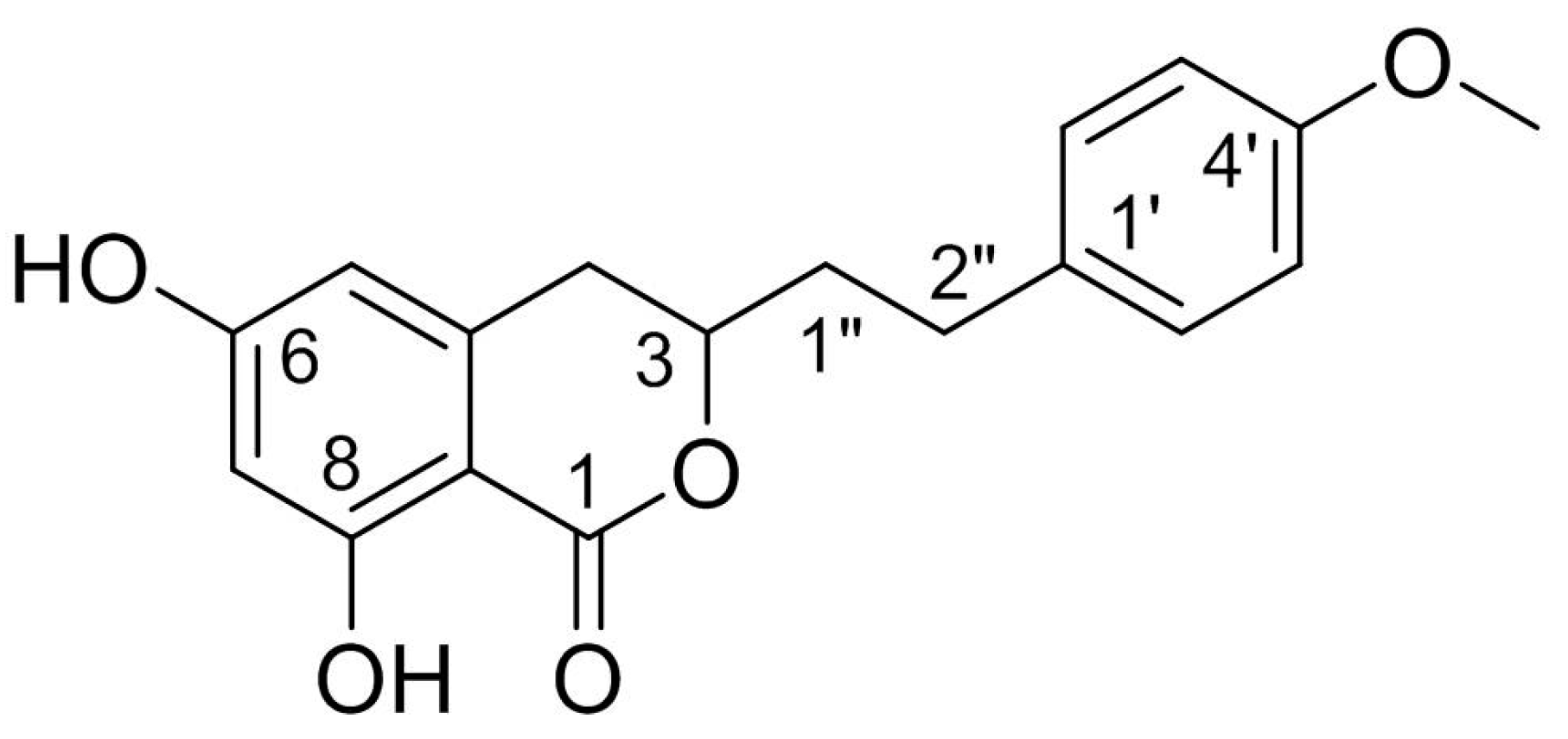
Agrimonolide (AM), which is a derivative of isocoumarins, is found mainly in the herb Agrimonia pilosa Ledeb. This compound is highly lipophilic and readily crosses the blood–brain barrier. Interest has grown in the use of AM as a multitarget natural treatment for various diseases, such as cancer, inflammation, hepatic injury, myocardial damage, and diabetes mellitus. The potential mechanisms of these pharmacological effects have been clarified at cellular and molecular levels. AM shows no cytotoxicity over a range of concentrations in different types of cells, providing evidence for its good safety profile in vitro. These findings indicate that AM is a promising medicinal agent.

agrimonolide Agrimonia pilosa Ledeb. pharmacological effect safety

1. Introduction

Agrimonolide (AM), also known as agrimolide, is a bioactive compound that naturally occurs in the plants Agrimonia pilosa Ledeb. [1] and Spiraea formosana Hayata [2]. AM was first isolated from the fresh root of A. pilosa in 1958 by a Japanese scholar [1]. In 2004, AM was subsequently found in the fresh stems of S. formosana [2].
Structurally, AM is a derivative of isocoumarins and its molecular formula is C18H18O5. The chemical name of AM is 3,4-dihydro-6,8-dihydroxy-3-[2-(4-methoxyphenyl) ethyl]-1H-2-benzopyran-1-one. This racemate compound possesses one methoxyl and two phenolic hydroxyls, one of which is chelated to a lactonic C = O. The chemical structure of AM is shown in Figure 1.
Figure 1. Chemical structure of AM.
Investigation of the bioactivities of AM has demonstrated that this compound has a wide range of potential pharmacological activities [3][4][5][6][7][8][9][10][11][12]. According to the current literature, AM acts as an α1A adrenergic receptor antagonist [3]. AM also exerts anti-inflammatory [4], antitumor [5][6], antioxidant [7], hepatoprotective [8], and myocardial protective effects [9][10]. Furthermore, AM effectively increases insulin-mediated glycogen levels in hepatocytes, suggesting that it could be a promising natural product for the prevention and treatment of diabetes mellitus [11][12].
In recent years, there have been several reviews on the botany, phytochemistry, ethnopharmacology, pharmacology, and traditional uses of A. pilosa [13][14][15][16], from which AM is isolated. However, there are few systematic summaries of the knowledge about this compound. In particular, important information regarding AM, such as aspects of its in vivo pharmacokinetics, biodistribution, bioavailability, and the application of drug delivery systems, is lacking. These obstacles limit the use and development of AM.

2. Sources

2.1. Derivation from Plants

As mentioned above, AM has been found in the plants of A. pilosa and S. formosana, which belong to the Rosaceae family. A. pilosa is a perennial herb with an erect stem that is 30–120 cm in height, and it grows along roadsides or in grassy areas at diverse altitudes. This plant is distributed in China, central Europe, the former Soviet Union, Mongolia, North Korea, Japan, and northern Vietnam [17]. A. pilosa is used in traditional Chinese medicine for mainly treating hemoptysis, metrorrhagia, hematemesis, and bloody dysentery [18]. S. formosana is a shrub endemic to Taiwan that grows in alpine woodlands at an altitude of 2100–2950 m [19]. The tender leaves, fruits, and roots of this plant have traditionally been used as diuretics, antidotes, and analgesics to treat inflammation, cough, headache, and toothache [20][21].
Conventional approaches are usually used to extract and separate AM from the above-mentioned plants. Table 1 summarizes the extraction and separation methods of AM [1][2][4][8][11][22][23]. The extraction and isolation process of AM from S. formosana can be summarized as follows [2]. Briefly, the fresh stems of S. formosana are extracted with hot ethanol, and the water suspension of the ethanol extract is subjected to a liquid-liquid partition to obtain chloroform, n-butanol, and water subfractions. AM is then separated from the chloroform subfraction using a combination of silica gel column chromatography. Finally, 5.6 mg of AM is obtained from 8.6 kg of S. formosana, equivalent to the content of 0.65 mg/kg. This result indicates that the content of AM in S. formosana is low.
Table 1. Extraction and separation methods of AM.

Parts

Methods of Extraction and Isolation

Yield

Content

Ref.

Fresh stems

8.6 kg of S. formosana is extracted with hot ethanol, and the water suspension of the ethanol extract is subjected to a liquid-liquid partition to obtain chloroform, n-butanol, and water subfractions, respectively. The chloroform subfraction is then fractionated by silica gel column chromatography.

5.6 mg

0.65 mg/kg

[2]

Fresh roots

10 kg of A. pilosa is extracted with methanol, and the extract is shaken with diethyl ether. The soluble part is boiled several times with petroleum ether, and the residue is heated and extracted repeatedly with benzene. Finally, the precipitated crystals are recrystallized from benzene and then from methanol.

3000–4000 mg

300–400 mg/kg

[1]

Dried plant

50 kg of A. pilosa is extracted with 60% ethanol, and the 30% ethanol elution part of macroporous resin is separated by silica gel column chromatography, recrystallization, ODS column chromatography, Sephadex LH-20 gel column chromatography and preparative high-performance liquid chromatography.

202 mg

4.04 mg/kg

[22]

Dried aerial parts

13 kg of A. pilosa is extracted with methanol and the extract is suspended in water. The suspension is partitioned between hexane, ethyl acetate, and n-butanol. The ethyl acetate fraction is then fractionated by repeated silica gel column chromatography.

43.7 mg

3.36 mg/kg

[11]

NA

Ethyl acetate fraction of methanol extract of A. pilosa is chromatographed repeatedly with silica gel columns and purified by preparative thin layer chromatography.

6.5 mg

NA

[4]

Dried roots

290 g of A. pilosa is extracted with hot water and the filtrated aqueous solution is partitioned with ethyl acetate and n-butanol, successively. The ethyl acetate soluble fraction is chromatographed by silica gel column repeatedly.

44 mg

151.7 mg/kg

[8]

Dried plant

500 g of A. pilosa is extracted with 70% ethanol. The extract is then eluted with different concentrations of ethanol on the macroporous resin. The 50% ethanol eluted fractions is collected and used for subsequent high-speed counter-current chromatography separation.

385.2 mg

770.4 mg/kg

[23]

NA: not available.
A large number of reports have described the process of extracting and separating AM from A. pilosa. Low toxicity solvents, such as absolute methanol and 60–70% ethanol solutions, are usually used to extract AM. Conventional separation techniques, such as liquid–liquid extraction, silica gel column chromatography, recrystallization, macroporous resin, and preparative high-performance liquid chromatography, have been used to separate AM. AM yields vary from 3–4 mg/kg to 300–400 mg/kg [1][8][11][22]. These classic separation methods are complicated, inefficient, and time consuming. However, in recent years, high-speed counter-current chromatography (HSCCC) coupled with ultraviolet detection or evaporative light-scattering detection has been considered an efficient protocol for separating AM from A. pilosa [23]. The AM yield obtained using this method is higher than that using conventional methods, producing an AM content of 770 mg/kg. Therefore, HSCCC is a powerful technique for separating AM from A. pilosa.
In summary, there are substantial differences in AM yields extracted from A. pilosa owing to differences in the origin of plants, the extraction and separation methods used, extraction parts, and other potential factors. Notably, A. pilosa has a much higher content of AM than S. formosana, which explains why A. pilosa is the major source of AM.

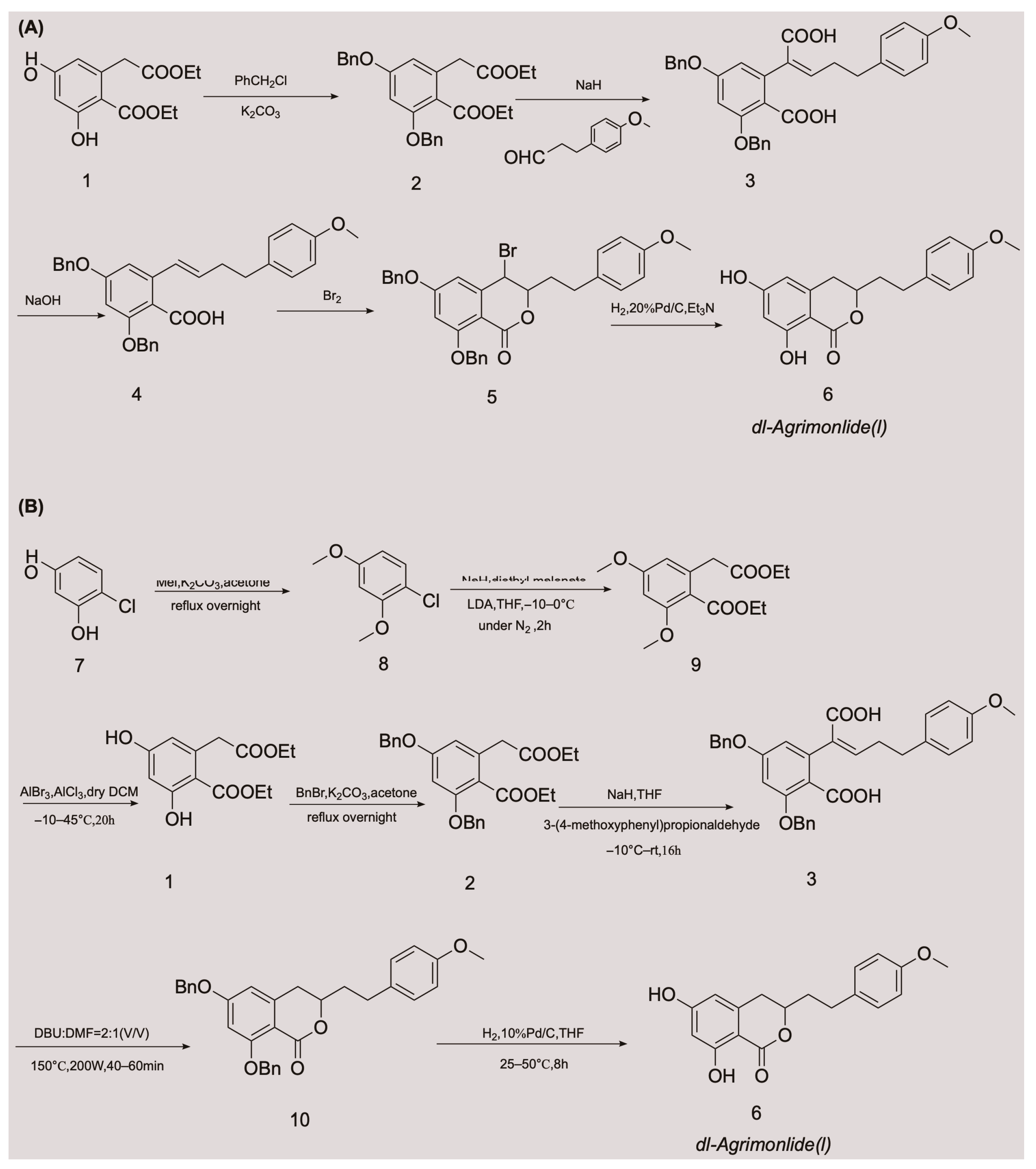
2.2. Obtaining AM by Chemical Synthesis

The low accumulation of AM in plants, its cumbersome extraction and separation processes, and the overexploitation of natural resources are generally considered the main driving factors for its high production cost. These factors are also the main causes of supply shortages of AM. These obstacles have made chemical synthesis an appealing alternative method for obtaining AM. Although AM has a variety of pharmacological activities, there have been relatively few advances in its chemical synthesis.
A few attempts have been made to synthesize AM, with variable success. In 1976, Yamato et al. synthesized the racemate of AM in five steps for the first time, and confirmed its structure using nuclear magnetic resonance imaging [24]. They started with compound 1 and obtained compound 2 by protecting two phenolic hydroxyl groups with a benzyl group. An overall yield of 2.6% AM was obtained through a series of reactions, including Stobbe condensation, ester hydrolysis, benzyl decarboxylation, bromine addition, and reduction [24]. The chemical synthesis route is shown in Figure 2A. Unfortunately, there were no subsequent reports of the chemical synthesis process of AM for many years. In 2018, a Chinese invention patent was published that contained a novel chemical synthesis method for AM [25]. The authors improved on the synthesis route of Yamato et al., and constructed the 3,4-dihydroisocoumarin core structure using microwave-assisted intramolecular esterification for the first time. The synthetic process began with 4-chlororesorcinol as the material, and the goal product was obtained in seven steps. The chemical synthesis route is shown in Figure 2B. This synthetic route increased the overall yield of AM to 20.7% [25][26], which is nearly eight times higher than that of Yamato et al. This exciting result suggests that microwave-assisted synthesis is a promising approach for the chemical construction of AM.
Figure 2. Chemical synthesis routes of AM [24][25][26].

3. Properties

3.1. Physicochemical Properties

Physicochemical properties, such as solubility in water and organic solvents, the acid dissociation constant, the oil/water partition coefficient, and chemical stability, are key factors that affect the pharmacokinetics, biopharmaceutics, and quality of drugs. The physicochemical properties of AM are summarized in detail in Table 2 [27][28][29].
Table 2. Physicochemical properties of AM.
AM is a white powder with a molecular weight of 314.3 g/moL [28]. AM is soluble in chloroform, dichloromethane, ethyl acetate, dimethyl sulfoxide, acetone, and other solutions [30], but is only minimally soluble in petroleum ether. This solubility suggests that AM is lipophilic. The partition coefficient and distribution coefficient are important parameters that describe the lipophilicity of a compound, which can be used to help predict the in vivo permeability. The reported partition coefficient and distribution coefficient values of AM are 3.649 and 2.949, respectively [27], indicating that AM has low solubility and moderate permeability. Such compounds usually have good intestinal tract permeability because there is a good balance between dissolution and passive diffusion penetration. According to the biopharmaceutics classification system, drug substances are categorized into four classes on the basis of their solubility parameter and permeability to biomembranes for evaluating the desired results of a formulation on oral bioavailability [31]. A low water solubility and poor oral bioavailability limit the biological effects of many natural products in vivo. The existing data suggest that AM belongs to biopharmaceutics classification system Class II and is likely to exhibit dissolution rate-limited absorption. However, this possibility requires further confirmation by determining the solubility of AM in water.
Regarding chemical stability, contact should be avoided between AM and strong oxidizing agents, reducing agents, strong acids, and alkalis. In the soluble form of AM, it should be sealed and stored below −20 °C to maintain its stability for several months. In the solid form of AM, it is stable at temperatures of 2–8 °C when kept in a dry place [30].

3.2. Predicted Absorption, Distribution, Metabolism, Excretion, and Toxicity Properties

Pharmacokinetic behaviors of drugs in vivo include absorption, distribution, metabolism, excretion, and toxicity (ADMET). The ADMET properties account for 50% of drug research and development (R&D) failures. Computer-aided design is an effective and alternative method of biological experimental evaluation, and helps to improve the R&D success rate. Computational approaches have increasingly been used to predict ADMET properties of compounds, especially in evaluating the ADMET properties of herbal medicines [32]. The predicted ADMET profiles of AM regarding its absorption, solubility, permeability across the blood-brain barrier (BBB), interactions with cytochrome P450 2D6, hepatotoxicity, and plasma protein binding (PPB) are shown in Table 3 [27].
Table 3. Predicted ADMET properties of AM [27].
ADMET absorption levels: 0, 1, 2, and 3 represent good, moderate, low, or very low absorption, respectively. ADMET BBB levels: 0, 1, 2, 3, 4, and 5 represent very high, high, medium, low, undefined, and molecules with one or more unknown AlogP98 types, respectively. ADMET solubility levels: 0, 1, 2, 3, 4, 5, and 6 represent extremely low, very low but possible, low, good, optimal, too soluble, and molecules with one or more unknown AlogP98 types, respectively. ADMET hepatotoxicity: 0 and 1 represent nontoxic and toxic effects, respectively. ADMET CYP2D6: 0 and 1 represent non-inhibitor and inhibitor, respectively. ADMET PPB levels: 0, 1, and 2 represent binding <90%, binding ≥90% and binding ≥95%, respectively.
AM appears to show a good absorption capacity in vivo with a predicted absorption level of 0. However, AM is predicted to have a low aqueous solubility, with a solubility level of 2, which contradicts the prediction result for in vivo absorption and needs to be further confirmed by in vivo testing. Regarding the prediction of BBB penetration, AM exhibits a moderate BBB penetration capability, with a level of 2. This indicates that AM may enter the brain tissue through the BBB and could be used to treat brain diseases. Furthermore, the ADMET predictor shows that AM exhibits potential hepatotoxicity, with a level of 1. Preliminary explorations and in-depth investigations are required to determine the specific mechanism of hepatotoxicity and whether it is dose dependent. In addition, AM is predicted to be a non-inhibitor of the cytochrome P450 2D enzyme and may be metabolized and excreted successfully. Therefore, drug-drug interactions are less likely when AM and the cytochrome P450 2D6 substrates are used simultaneously. Moreover, the PPB level is predicted to be 2, indicating that the binding rate of AM with plasma protein is ≥95%. The high degree of PPB limits the partitioning of AM from the blood into the tissues, where it could be metabolized. This limited partitioning may result in a delayed onset of action and longer half-life period, thereby reducing the elimination of AM. AM has been predicted to have a good drug-likeness, with a drug-likeness weight of 0.842. Generally, AM demonstrates promising ADMET profiles. However, to fully confirm the ADMET properties of AM, real-world tests are required to validate these properties, and more animal and human studies are required.

4. Pharmacological Effects

AM possesses a wide range of pharmacological activities, such as antitumor activity, antioxidation and hepatoprotection, antidiabetic activity, anti-inflammatory activity, myocardial protection, and α1A adrenergic receptor antagonist activity. The mechanisms of action of these effects are shown in Table 4.
Table 4. Mechanisms of pharmacological effects of AM.

Pharmacological Effects

Levels

Models

Concentrations or

Doses of AM

Mechanisms

Ref.

anti-gastric cancer

in vitro

AGS cells

40 µM, IC50 = 25.9 μM

decrease the expression of Bcl-2;

increase the expression of Bax;

increase the level of phospho-ERK/ERK protein and the expression of phosphor-p38 protein;

increase the activity of caspase-3;

down-regulate the levels of the inactive pro-caspase-3, -8, and -9 proteins

[6]

anti-ovarian cancer

in vitro

A2780 and SKOV-3 cells

40 µM

increase the cleavage of caspase-3 and -9;

increase the levels of ROS, total iron and ferrous ion, and down-regulate the levels of SLC7A11 and GPX4, thus inducing ferroptosis;

direct inhibit tumor cell migration and invasion;

inhibit the protein levels of SCD1

[5]

in vivo

SKOV-3 xenograft model

(BALB/c mice)

50 mg/kg

down-regulate the expressions of Ki-67 and SCD1;

lower the expressions of SCD1 mRNA and protein

[5]

anti-diabetic

in vitro

PANC-1 cell

1 μM; 5 μM

promote the expression of PDX-1

[22]

in vitro

/

IC50 = 37.4 μM

inhibit α-glucosidase

[11]

in vitro

Insulin-resistance HepG2 cell

20 µM

elevate the activity of GK, and increase the content of glycogen;

lower the activities of PEPCK and G6Pase, and constrain the gluconeogensis

[12]

anti-oxidative and hepatoprotective

in vitro

HepG2 cell;

rat primary hepatocytes

EC50 = 88.2 μM;

EC50 = 37.7 μM

scavenge the free radical

[8]

in vitro

HepG2 cell

200 μM

scavenge the free radical;

activate Nrf2-driven pathways;

activate ERK, JNK, and MAPK phosphorylation;

inhibit p38 phosphorylation;

elevate the activity of antioxidative enzymes

[7]

anti-inflammatory

in vitro

RAW 264.7 cells

80 μM

reduce the levels of IL-1β, IL-6, and TNF-α;

attenuate the expression of iNOS and COX-2;

inhibit the activation of JNK and p38 MAPKs;

decrease the activation of JAK-STAT and NF-κB

[4]

myocardial protective

in vitro

H9c2 cell

15 μM

regulate the gene expression involved in mitochondrial function;

decrease the levels of cleaved Caspase 3 and Bax;

boost the level of Bcl2;

prevent the rate of apoptosis and shield H9c2 cells from hypoxia-induced apoptosis;

reduce ROS production and preserve the normal shape of mitochondria;

regulate the functional proteins to enhance the mitochondrial activity

[10]

in vivo

CLP rat model

5 mg/kg

attenuate myocardial injury by Akt signaling;

suppress cardiac injury indicators, oxidative stress, and inflammation;

restrain the activation of Akt, Erk, mTOR and the apoptosis of cardiomyocytes

[9]

blocking α1A adrenergic receptor

in vitro

rat prostate cell membrane

/

/

[3]

5. Safety

To the best of our knowledge, the available data on the safety of AM are limited. Safety studies on AM have mainly focused on in vitro 3-(4,5-dimethylthiazol-2-yl)-2,5-diphenyltetrazolium bromide (MTT) tests of different types of cells, and AM shows no cytotoxicity over a range of concentrations [4][5][6][7][8][12]. Various concentrations of AM (25–200 µM) do not cause a substantial change in viability, or in the size and shape of HepG2 cells. Even at a high concentration of 200 µM, no cell shrinkage or shedding of adhesion molecules occurs [7]. In a hepatoprotective activity study, the survival rate of HepG2 cells was not altered in the presence (1–100 µM) or absence of AM [8]. A similar result was also found for HepG2 cells treated with AM at a concentration of 20 µM [12]. In a study of cytotoxic effects on RAW 264.7 cells, AM concentrations of 20–80 µM did not cause any changes in MTT-based cell viability. Annexin V/propidium iodide staining has been further used to characterize the cytotoxic effect of AM. After treatment with AM, the proportion of early apoptotic cells substantially decreases. These results indicate that AM does not show signs of cytotoxicity in RAW 264.7 cells, and that it does not inhibit the early events leading to apoptosis [4]. In another MTT test, AM concentrations of 10–40 µM showed no cytotoxicity on the growth of HepG2, HT-29, or MCF-7 cells [6]. The above-mentioned cytotoxicity tests provide preliminarily support for the safety of AM in vitro.

References

  1. Yamato, M. On the chemical structure of agrimonolide, a new constituent of agrimonia pilosa Ledel. Yakugaku Zasshi 1958, 78, 1086–1089.
  2. Wu, T.S.; Hwang, C.C.; Kuo, P.C.; Kuo, T.H.; Damu, A.G.; Su, C.R. New Neolignans from Spiraea formosana. Chem. Pharm. Bull. 2004, 52, 1227–1230.
  3. Han, S.; Zhang, P.; Wei, F.; Wang, S. Screening active components acting on α1A adrenergic receptors from agrimony using a Sprague-Dawley rat prostate cell membrane chromatography online coupled HPLC/MS method. Anal. Methods 2012, 4, 3351–3357.
  4. Chen, L.; Teng, H.; Fang, T.; Xiao, J. Agrimonolide from Agrimonia pilosa suppresses inflammatory responses through down-regulation of COX-2/iNOS and inactivation of NF-kappaB in lipopolysaccharide-stimulated macrophages. Phytomedicine 2016, 23, 846–855.
  5. Liu, Y.; Liu, X.; Wang, H.; Ding, P.; Wang, C. Agrimonolide inhibits cancer progression and induces ferroptosis and apoptosis by targeting SCD1 in ovarian cancer cells. Phytomedicine 2022, 101, 154102.
  6. Teng, H.; Huang, Q.; Chen, L. Inhibition of cell proliferation and triggering of apoptosis by agrimonolide through MAP kinase (ERK and p38) pathways in human gastric cancer AGS cells. Food Funct. 2016, 7, 4605–4613.
  7. Chen, L.; Teng, H.; Zhang, K.Y.; Skalicka-Woźniak, K.; Georgiev, M.I.; Xiao, J. Agrimonolide and Desmethylagrimonolide Induced HO-1 Expression in HepG2 Cells through Nrf2-Transduction and p38 Inactivation. Front. Pharmacol. 2017, 7, 513.
  8. Park, E.J.; Oh, H.; Kang, T.H.; Sohn, D.H.; Kim, Y.C. An isocoumarin with hepatoprotective activity in Hep G2 and primary hepatocytes from Agrimonia Pilosa. Arch. Pharm. Res. 2004, 27, 944–946.
  9. Zhao, Z.; Hu, X. Protective effect of agrimonolide against myocardial injury in cecal ligation and puncture (CLP) induced sepsis in wistar rats via inhibition of akt. Acta Pol. Pharm. 2021, 78, 845–851.
  10. Wang, C.; Qi, C.; Liu, M.; Wang, L.; Cheng, G.; Li, L.; Xing, Y.; Zhao, X.; Liu, J. Protective effects of agrimonolide on hypoxia-induced H9c2 cell injury by maintaining mitochondrial homeostasis. J. Cell. Biochem. 2022, 123, 306–321.
  11. Park, M.J.; Kang, Y.H. Isolation of Isocoumarins and Flavonoids as alpha-Glucosidase Inhibitors from Agrimonia pilosa L. Molecules 2020, 25, 2572.
  12. Teng, H.; Chen, L.; Song, H. The potential beneficial effects of phenolic compounds isolated from A. pilosa Ledeb on insulin-resistant hepatic HepG2 cells. Food Funct. 2016, 7, 4400–4409.
  13. Wen, S.; Zhang, X.; Wu, Y.; Yu, S.; Zhang, W.; Liu, D.; Yang, K.; Sun, J. Agrimonia pilosa Ledeb.: A review of its traditional uses, botany, phytochemistry, pharmacology, and toxicology. Heliyon 2022, 8, e09972.
  14. Jin, T.; Chi, L.; Ma, C. Agrimonia pilosa: A Phytochemical and Pharmacological Review. Evid. Based Complement. Alternat. Med. 2022, 2022, 3742208.
  15. Le, U.; Joshi, R.K.; Lay, H.; Ming, C. Agrimonia pilosa Ledeb: Phytochemistry, Ethnopharmacology, Pharmacology of an important traditional herbal medicine. J. Pharmacogn. Phytochem. 2018, 7, 3202–3211.
  16. Li, J.; Yang, J. Research Advances on the Main Chemical Constituents and Pharmacological Actions of Agrimoniae Herba. Chin. Wild Plant Resour. 2020, 39, 54–60.
  17. Yu, T.; Lu, L.; Gu, C.; Guan, C.; Li, C. Flora Reipublicae Popularis Sinicae; Science Press: Beijing, China, 1985; Volume 37, p. 457.
  18. Kuang, L.; Li, C.; Cao, S.; Li, H.; Wang, Z.; Wu, S.; Fan, Y.; Zhang, F.; Li, Q.; Liu, T.; et al. Pharmacopoeia of the People’s Republic of China; China Pharmaceutical Science and Technology Press: Beijing, China, 2020; p. 106.
  19. Ohashi, H.; Hsieh, C.F. Flora of Taiwan, 2nd ed.; Editorial Committee of the Flora of Taiwan: Taipei, China, 1993; Volume 3, p. 149.
  20. Zhao, G.; Dei, S.; Chen, R. ZhongyaoDacidian, 2nd ed.; Shanghai Science and Technology Press: Shanghai, China, 1977; pp. 1117–1118+1978.
  21. Xie, Z.; Fang, C.; Zhu, Z. QuanGuoZhongCaoYao Huibian, 2nd ed.; People’s Hygenic Publishing House: Beijing, China, 1996; pp. 514–515+767.
  22. Huang, Y. Chemical Composition of Agrimonia pilosa Ledeb. and Its Hypoglycemic Activity; Xiamen University: Xiamen, China, 2020.
  23. Wang, Y.; Liu, M.; Zheng, L.; Yin, L.; Xu, L.; Qi, Y.; Ma, X.; Liu, K.; Peng, J. Preparative purification of five bioactive components from Agrimonia pilosa Ledeb by high-speed counter-current chromatography. J. Sep. Sci. 2012, 35, 1977–1984.
  24. Yamato, M.; Hashigaki, K. Synthesis of dl-Agrimonolide (Constituent of Rhizome of Agrimonia pilosa LEDEB). Chem. Pharm. Bull. 1976, 24, 200–203.
  25. Xie, W.; Wu, X.; Rao, X. Synthesis of a racemic natural product Agrimonolide (I) extracted from Agrimonia pilosa. Patent CN201811078365.0, 13 November 2018.
  26. Cui, Y.; Zhang, G.; Li, Y.; Li, W.; Tanabe, G.; Osamu, M.; Xie, W. Microwave-Assisted Synthesis of d/l-Agrimonolide. Asian J. Org. Chem. 2022, 11, e202100334.
  27. The Encyclopedia of Traditional Chinese Medicine. In Agrimonolide. Available online: http://www.tcmip.cn/ETCM/index.php/Home/Index/cf_details.html?id=5545 (accessed on 3 September 2022).
  28. Chemical Trading Guide. In (1S,4R)-N-phenyl]-2-bicycloheptanecarboxamide;2,2,2-trifluoroacetic Acid. 2022. Available online: https://www.guidechem.com/encyclopedia/agrimonolide-dic74411.html (accessed on 1 September 2022).
  29. Arakawa, H.; Torimoto, N.; Masui, Y. Die absolute konfiguration des (−)-β-tetralols und des agrimonolides. Tetrahedron Lett. 1968, 9, 4115–4117.
  30. BioCrick. In Agrimonolide. Available online: https://www.biocrick.com/Agrimonolide-BCN4925.html (accessed on 11 September 2022).
  31. Shukla, A.K.; Bishnoi, R.S.; Dev, S.K.; Kumar, M.; Fenin, V. Biopharmaceutical Classification System: Tool based prediction for drug dosage formulation. Adv. Pharm. J. 2017, 2, 204–209.
  32. Qiu, J.; Zhou, Z.; He, Z.; Zhang, X.; Zhou, S.; Zhu, S. Estimation of the binding modes with important human cytochrome P450 enzymes, drug interaction potential, pharmacokinetics, and hepatotoxicity of ginger components using molecular docking, computational, and pharmacokinetic modeling studies. Drug Des. Devel. Ther. 2015, 9, 841–866.
More
Information
Contributors MDPI registered users' name will be linked to their SciProfiles pages. To register with us, please refer to https://encyclopedia.pub/register : , , , , , , ,
View Times: 363
Revisions: 2 times (View History)
Update Date: 15 Feb 2023
1000/1000
Video Production Service