
| Version | Summary | Created by | Modification | Content Size | Created at | Operation |
|---|---|---|---|---|---|---|
| 1 | Luis Alberto Mejía-Manzano | -- | 4218 | 2023-02-09 08:27:45 | | | |
| 2 | Rita Xu | -60 word(s) | 4158 | 2023-02-09 08:45:19 | | |
Video Upload Options
Native Mexican plants are a wide source of bioactive compounds such as pentacyclic triterpenes. Pentacyclic triterpenes biosynthesized through the mevalonate (MVA) and the 2-C-methyl-D-erythritol-phosphate (MEP) metabolic pathways are highlighted by their diverse biological activity. Compounds belonging to the oleanane, ursane, and lupane groups have been identified in about 33 Mexican plants, located mainly in the southwest of Mexico. 45 compounds pentacyclic triterpenes were reported showing antiinflammatory, cytotoxic, anxiolytic, hypoglycemic, and growth-stimulating or allelopathic activities. Extraction is essentially performed by maceration and Soxhlet with organic solvents and consecutive chromatography of silica gel have been used for their whole or partial purification. Nanoparticles and nanoemulsions are the vehicles used in Mexican formulations for drug delivery of the pentacyclic triterpenes until now. Sustainable extraction, formulation, regulation, isolation, characterization, and bioassay facilities are areas of opportunity in pentacyclic triterpenes research in Mexico while the presence of plant and human resources and traditional knowledge are strengths.
1. Introduction
1.1. General Overview of Pentacyclic Triterpenes
1.1.1. Structure, Classification, Distribution, and Biological Importance
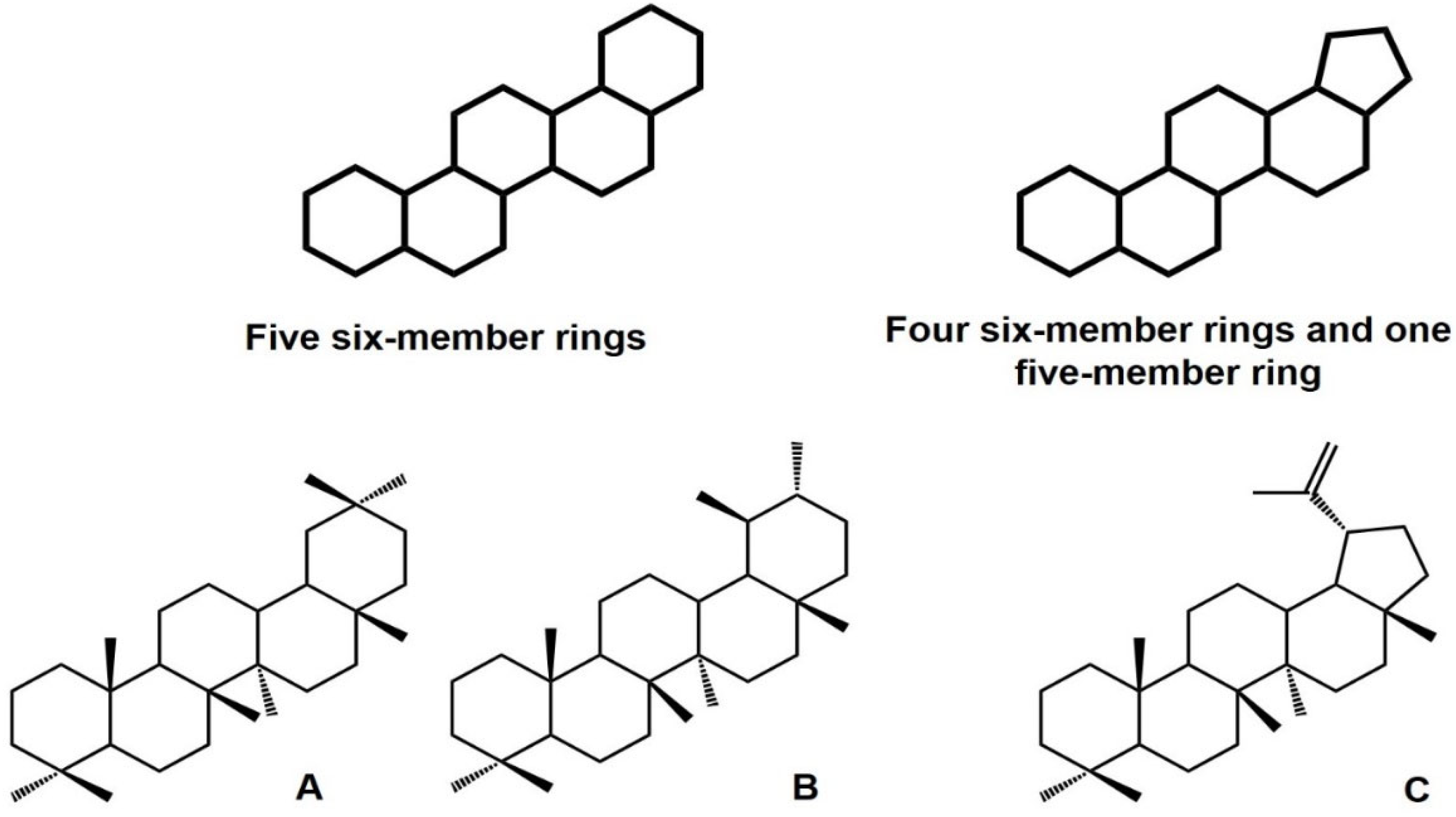
1.1.2. Biosynthesis
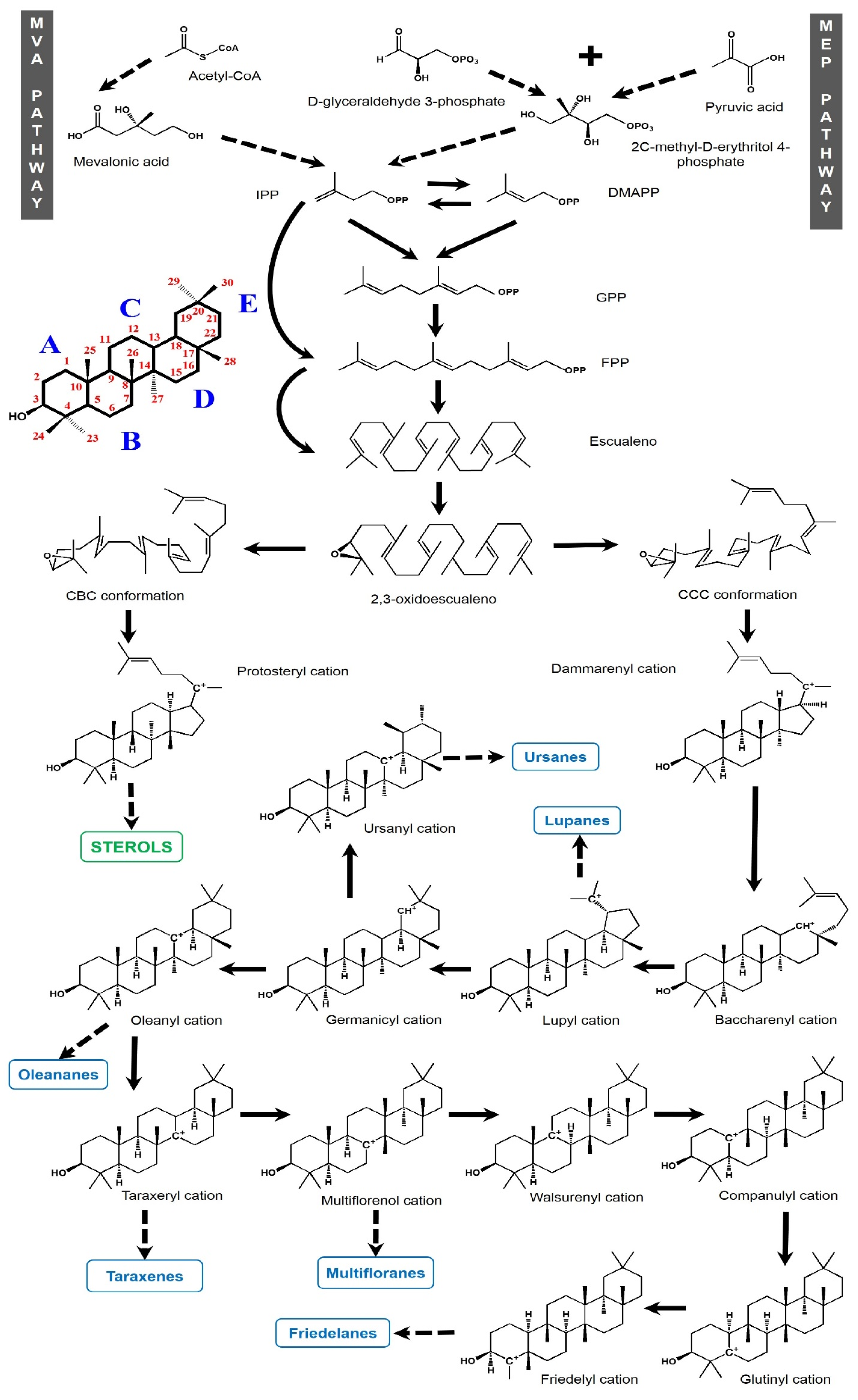
2. Pentacyclic Triterpenes from Mexican Plants
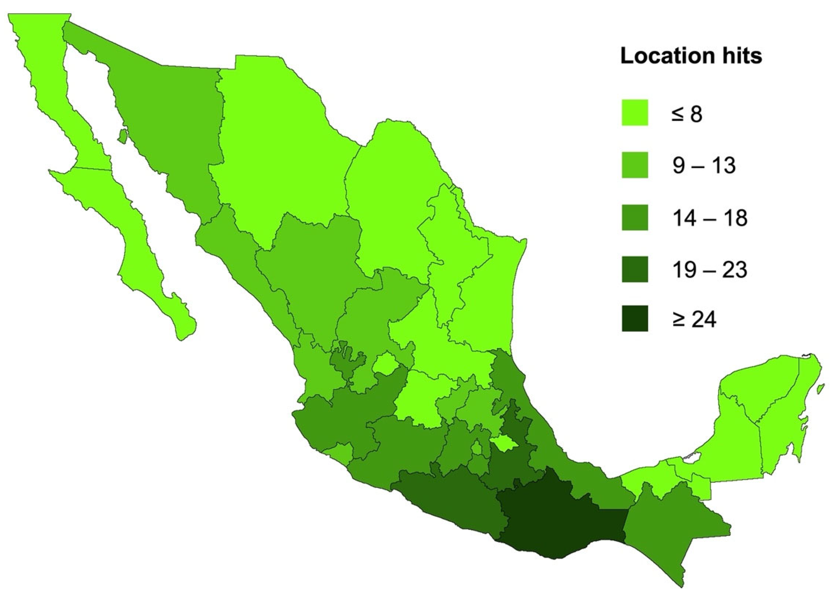
Biological Activity Survey of PCTs Found in Mexican Plants
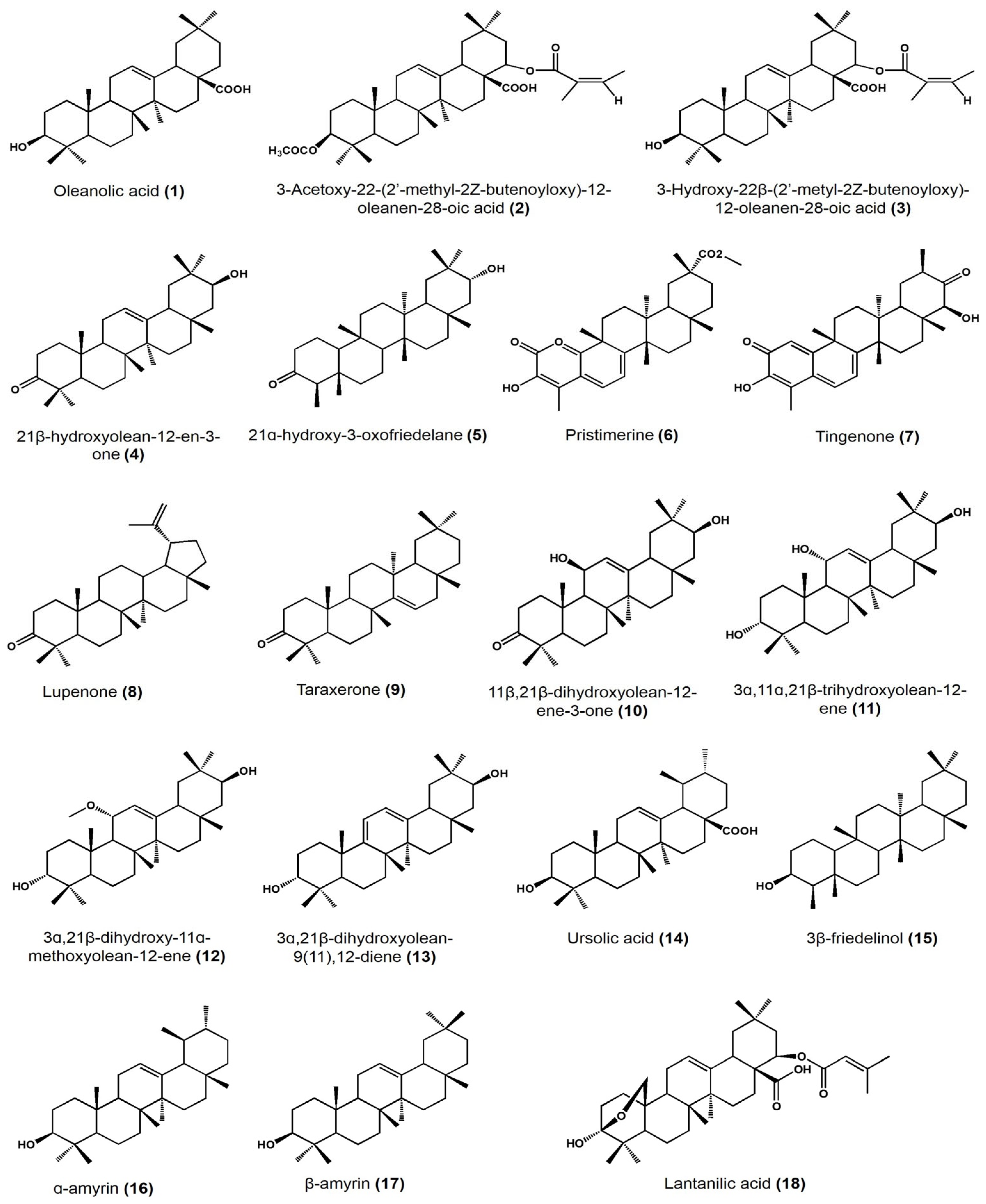
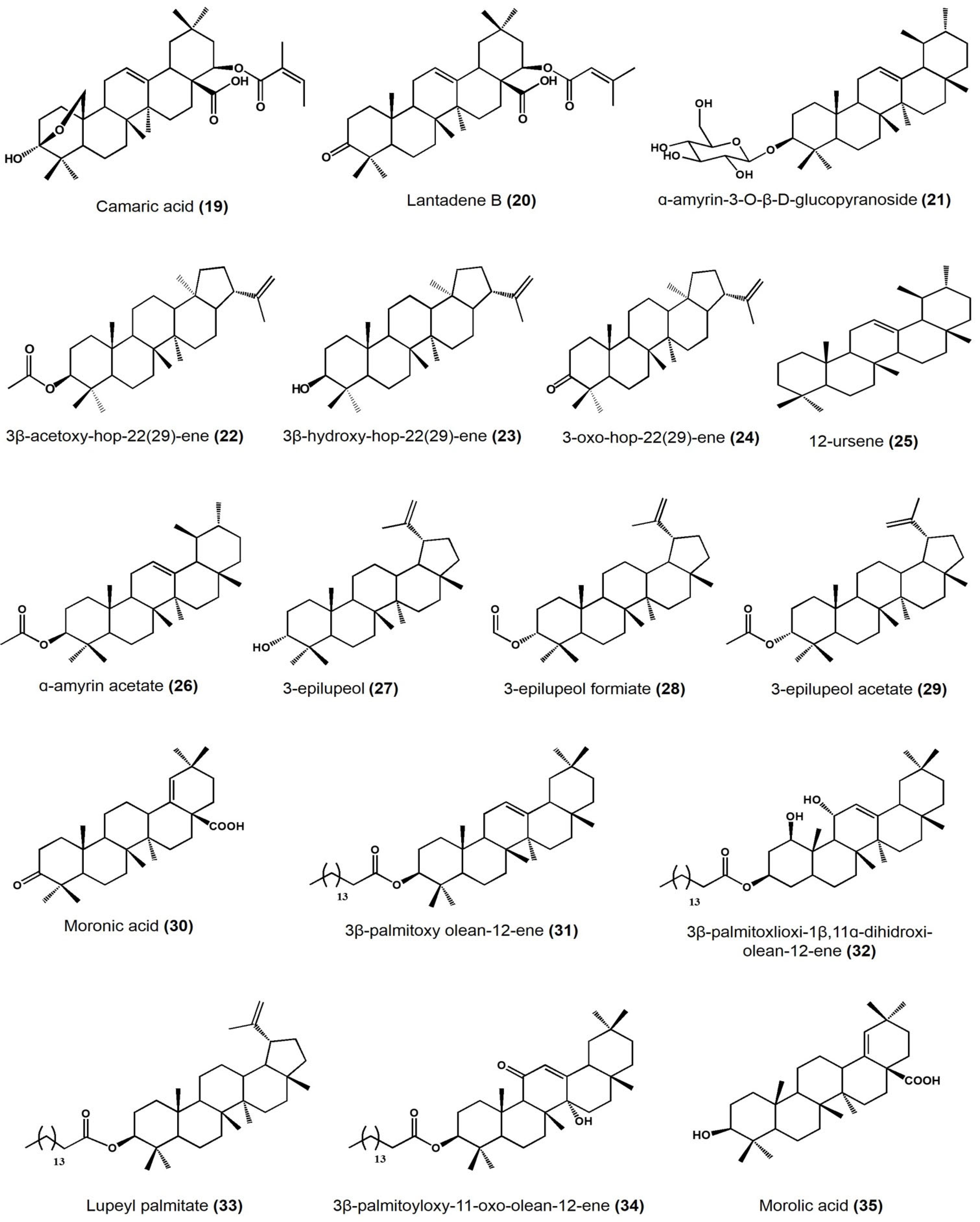
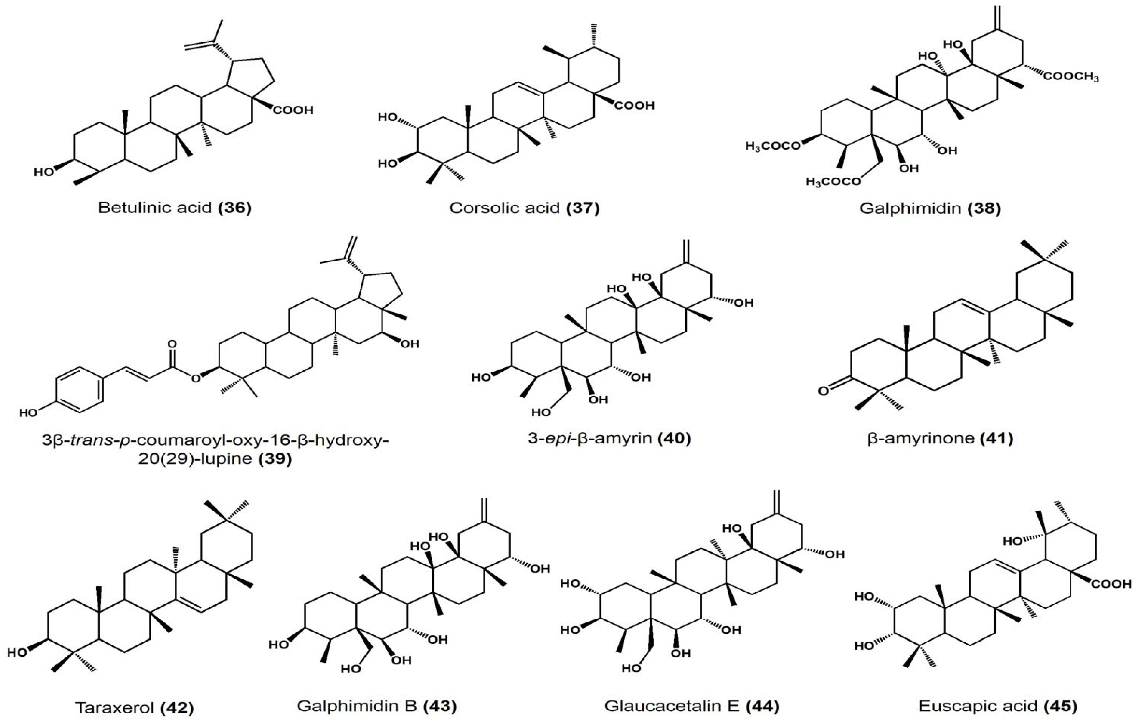
| Plant Species | Identified Compounds | Subgroup | Activities | References |
|---|---|---|---|---|
| Lantana hispida | Oleanolic acid | Oleanane | Antimicrobial activity (MIC) on Mycobacterium tuberculosis: • H37Rv: 25 μg/mL • STR-R, RIF-R, INH-R, and EMB-R: 50 μg/mL |
[29] |
| 3-acetoxy-22-(2′-methyl-2Z-butenyloxy)-12-oleanene | Oleanane | Antimicrobial activity (MIC) on M. tuberculosis: • H37Rv, STR-R, and INH-R: 25 μg/mL • RIF-R and EMB-R: 50 μg/mL |
||
| 3-hydroxy-22-(2′-methyl-2Z-butenoyloxy)-12-oleanen-28-oic acid (reduced lantadene A) | Oleanane | Antimicrobial activity (MIC) on M. tuberculosis: • H37Rv, STR-R, RIF-R, INH-R, and EMB-R: 50 μg/mL |
||
| Hippocratea excelsa | 21β-hydroxyolean-12-en-3-one | Oleanane | Antiprotozoal activity (IC50) on Giardia intestinalis: 27.4 μM | [30] |
| 21α-hydroxy-3-oxofriedelane | Friedelane | Antiprotozoal activity (IC50) on G. intestinalis: 19.8 μM | ||
| Pristimerine | Friedelane | Antiprotozoal activity (IC50) on G. intestinalis: 0.11 μM | ||
| Tingenone | Friedelane | Antiprotozoal activity (IC50) on G. intestinalis: 0.74 μM | ||
| Acacia cochliacantha | Lupenone | Lupane | Antimicrobial activity (MIC) on: • Staphylococcus aureus: 2.8 mg/mL • Bacillus subtilis: 1.4 mg/mL • Enterococcus faecium: 5.6 mg/mL • Lactobacillus plantarum: 11.3 mg/mL • Escherichia coli: 2.8 mg/mL • Salmonella typhirium: 22.5 mg/mL • Klebsiella pneumoniae: 22.5 mg/mL |
[31] |
| Taraxenone | Taraxerane | Antimicrobial activity (MIC) on: • S. aureus: 0.4 mg/mL • B. subtilis: 1.4 mg/mL • L. plantarum: 1.4 mg/mL • E. coli: 0.2 mg/mL • S. typhirium: 2.8 mg/mL • K. pneumoniae: 1.4 mg/mL • Pseudomonas aeruginosa: 2.8 mg/mL |
||
| Hippocratea excelsa | 11β,21β-dihydroxy-olean-12-ene-3-one | Oleanane | Antiprotozoal activity on Giardia intestinalis: IC50: 184.6 μM | [32] |
| 3α,11α,21β-trihydroxy-olean-12-ene | Oleanane | Antiprotozoal activity on G. intestinalis: IC50: 96.8 μM | ||
| 3α,21β-dihydroxy-11α-methoxy-olean-12-ene | Oleanane | Antiprotozoal activity on G. intestinalis: IC50: 690.7 μM | ||
| 3α,21β-dihydroxy-olean-9(11),12-diene | Oleanane | Antiprotozoal activity on G. intestinalis: IC50:78 μM | ||
| Chamaedora tepejiliote | Ursolic acid | Ursane | Antimicrobial activity (MIC) against M. tuberculosis: • H37Rv, INH-R, RIF-R, EMB-R, MMDO, and MTY147: 25 μg/mL • STR-R: 12.5 μg/mL In vitro intracellular load of M. tubrculosis MDR and H37Rv in macrophages at: • 6.25 μm/mL (14) + 12.5 μm/mL (1): (after 48 h) approximately 101 CFU/mL (both strains) • 0.625 μm/mL (14) + 1.25 μm/mL (1): (after 48 h) approximately 101 CFU/mL (H37Rv) Bacilli load per lung in mice model after 60 days of administration of 1:3 mixture of (1):(14) at 5 mg/kg • 0.1 × 10⁶ bacilli/lung approximately • Control: approximately 0.9 × 10⁶ bacilli/lung |
[33] |
| Lantana hispida | Oleanolic acid | Oleanane | Antimicrobial activity (MIC) against M. tuberculosis: • H37Rv, STR-R, MMDO, and MTY147: 50 μg/mL • INH-R, RIF-R and EMB-R: 25 μg/mL In vitro intracellular load of M. tubrculosis MDR and H37Rv in macrophages at: • 6.25 μm/mL: (after 24 and 48 h) approximately 105 CFU/mL. • 0.625 μm/mL: (after 48 h) approximately 105 CFU/mL • Control: approximately 106 CFU/mL Bacilli load per lung in mice model after 60 days of administration of 1:3 mixture of (1):(14) at 5 mg/kg • 0.1 × 10⁶ bacilli/lung approximately• Control: approximately 0.9 × 10⁶ bacilli/lung |
|
| Heterotheca inuloides | 3β-friedelinol | Friedelane | Antimicrobial activity (MIC) on Helicobacter pylori: >31.25 μg/mL | [34] |
| α-amyrin and β-amyrin | Ursane | |||
| Lantana camara | Lantanilic acid | Lantadene | Lehismaniacidal activity on L. mexicana: 9.50 ± 0.28 μM | [35] |
| Camaric acid | Lantadene | Lehismaniacidal activity on L. mexicana: 2.52 ± 0.08 μM | ||
| Lantadene B | Lantadene | Lehismaniacidal activity on L. mexicana: 23.45 ± 2.15 μM | ||
| Cisus incisa | α-amyrin-3-O-β-D-gluco pyranoside | Ursane | Antimicrobial activity (MIC) against carbapenems-resistant P. aeruginosa: 100 μg/mL | [36] |
| Cnidosculus spinosus | 3 β-acetoxy-hop-22(29)-ene | Hopane | Inhibitory activity on mouse edema at 0.31 μmol/ear: 57.27 ± 16.99% Inhibition of yeast α-glucosidase at 100 μM: 98.79% |
[37] |
| 3β-hydroxy-hop-22(29)-ene [Hopenol B] | Hopane | Inhibitory activity on mouse edema at 0.31 μmol/ear: 27.05 ± 7.38% Inhibition of yeast α-glucosidase at 100 μM: 23.47% Antiparasitic activity on Trypanosoma cruzi at 50 μM: <20% |
||
| 3-oxo-hop-22(29)-ene | Hopane | Inhibitory activity on mouse edema at 0.31 μmol/ear: 17.5 ± 4.11% Inhibition of yeast α-glucosidase at 100 μM:7.39% Antiparasitic activity on Trypanosoma cruzi at 50 μM: <20% |
||
| Agarista mexicana | 12-ursene | Ursane | Hypoglycemic activity on mice models after at 50 mg/kg: • 25.9 ± 6.1% glucose reduction |
[38] |
| Bursera copallifera | lupenone | Lupane | Anti-inflammatory activity in mouse edema inhibition at 1 mg/ear: 57.25 ± 1.36%, ID50: 1.052 μmol/ear Reduces NO production in LPS-stimulated macrophages: IC50: 20.08 ± 1.07 μM |
[39] |
| Bursera copallifera | α-amyrin | Ursane | Anti-inflammatory activity in mouse edema inhibition at 1 mg/ear: 25 ± 1.81%, ID50: >2.34 μmol/ear Reduces NO production in LPS-stimulated macrophages: IC50: 8.98 ± 1.73 μM |
[39] |
| α-amyrin acetate | Ursane | Anti-inflammatory activity in mouse edema inhibition at 1 mg/ear: 69.45 ± 1.8%, ID50: 1.17 μmol/ear Reduces NO production in LPS-stimulated macrophages: IC50: 22.47 ± 1.19 μM |
||
| 3-epilupeol | Lupane | Anti-inflammatory activity in mouse edema inhibition at 1 mg/ear: 66.39 ± 4.38%, ID50: 0.83 μmol/ear Reduces NO production in LPS-stimulated macrophages 15.50 ± 1.14 μM |
||
| 3-epilupeol formiate | Lupane | Anti-inflammatory activity in mouse edema inhibition at 1 mg/ear: 62.16 ± 1.8%, ID50: 0.96 μmol/ear Reduces NO production in LPS-stimulated macrophages 43.31 ± 2.60 μM |
||
| 3-epilupeol acetate | Lupane | Anti-inflammatory activity in mouse edema inhibition at 1 mg/ear: 49.35 ± 3.6%, ID50: >2.13 μmol/ear Reduces NO production in LPS-stimulated macrophages: IC50: 31.13 ± 1.25 μM |
||
| Bursera cuneata | α-amyrin | Ursane | Anti-inflammatory activity in mouse edema inhibition at 0.1 mg/ear: 44.9 ± 1.2% | [40] |
| Moronic acid | Oleanane | Anti-inflammatory activity in mouse edema inhibition at 0.1 mg/ear: 68.1 ± 1.3% Histamine inhibition in a mouse model at 0.1 mg/ear: 73.3 ± 1.1% Macrophages viability reduction at 60 μg/mL: 43% |
||
| Ursolic acid | Ursane | Anti-inflammatory activity in mouse edema inhibition at 0.1 mg/ear: 55.6 ± 2.1% |
||
| Sapium lateriflorum | 3β-palmitoyloxy olean-12-ene | Oleanane | In vitro cytotoxicity: inhibitory effect (%) at 50 μM • PC-3: 1.81 • K562: 6.88 • HCT-15: 10.46 •MCF-7: 3.24 • SKLU-1: 21.95 Anti-inflammatory activity in mouse edema inhibition at 1 μmol/ear: 48.26% |
[41] |
| Sapium lateriflorum | 3β-palmitoxlioxi-1β,11α-dihydroxi-olean-12-ene | Oleanane | In vitro cytotoxicity: inhibitory effect (%) at 50 μM • U251: 29.56 • PC-3: 20.72 • K562: 9.86 • HCT-15: 7.24 • MCF-7: 11.53 • SKLU-1: 12.89 Anti-inflammatory activity in mouse edema inhibition at 1 μmol/ear: 68.76% |
[41] |
| lupeyl palmitate | Lupane | In vitro cytotoxicity: inhibitory effect (%) at 50 μM • PC-3: 3.05 • K562: 3.46 • HCT-15: 10.24 •MCF-7: 13.43 • SKLU-1: 19.59 Anti-inflammatory activity in mouse edema inhibition at 1 μmol/ear: 22.31% |
||
| 3β-palmitoyloxy-11-oxo- olean-12-ene | Oleanane | In vitro cytotoxicity: inhibitory effect (%) at 50 μM • PC-3: 0.08 • HCT-15: 18.69 •MCF-7: 5.15 • SKLU-1: 20.06 Anti-inflammatory activity in mouse ear edema inhibition at 1 μmol/ear: 31.49%, ID50: 0.60 μmol/ear Anti-inflammatory activity in carrageenan-induced mouse plantar edema at 31.6 mg/kg: 48.2% after 3 h |
||
| Phoradendron reichenbachianum (Viscaceae) | Ursolic acid | Ursane | Vasorelaxant effect on rat aorta: EC50 11.7 μM, Emax 72.59% |
[42] |
| Moronic acid | Oleanane | Vasorelaxant effect on rat aorta: EC50 11.7 μM, Emax 92.01% |
||
| Morolic acid | Oleanane | Vasorelaxant effect on rat aorta: EC50 94.19 μM, Emax 73.75% |
||
| Betulinic acid | Lupane | Vasorelaxant effect on rat aorta: EC50 58.46 μM, Emax 79.01% |
||
| Cratageus gracilior | Corsolic acid | Ursane | Vasodilatory effect on rat aorta: EC50 108.9 ± 6.7 μM, Emax 96.4 ± 4.2% |
[43] |
| Galphimia glauca | Galphimidin | Nor-seco friedelane | Vasodilatory effect on rat aorta: EC50 145.9 ± 6.7 μM, Emax 99.5 ± 5.3% |
|
| Jatropha neupaciflora | 3β-trans-p-coumaroyl-oxy-16-β-hydroxy-20(29)-lupene | Lupane | Vasodilatory effect on rat aorta: 63.2 ± 5.8 μM EC50 and 27.5 ± 1.9% |
|
| Sebastiania adenophora | 3-epi-β-amyrin | Ursane | Approximate effect on root growth at 250 μg/mL • Lycopersicon esculentum: −21% • Echinocloa crus-galli: −36% • Amaranthus hypochondriacus: +55% |
[44] |
| β-amyrinone | Ursane | Approximate effect on root growth at 250 μg/mL • L. esculentum: −28% • E. crus-galli: −28% • A. hypochondriacus: +39% |
||
| 3-epi-lupeol | Lupane | Approximate effect on root growth at 250 μg/mL • L. esculentum: −32% • E. crus-galli: −9% • A. hypochondriacus: +47% |
||
| Lupenone | Lupane | Approximate effect on root growth at 250 μg/mL • L. esculentum: −36% • E. crus-galli: −5% • A. hypochondriacus: +27% |
||
| Taraxerol | Taraxerane | Approximate effect on root growth at 250 μg/mL • L. esculentum: −41% • E. crus-galli: −77% • A. hypochondriacus: +39% |
||
| Taraxerone | Taraxerane | Approximate effect on root growth at 250 μg/mL • L. esculentum: −49% • E. crus-galli: −44% • A. hypochondriacus: +23% |
||
| Galphimia glauca | Glaucacetalin E | Nor-seco friedelane | Anxiolytic effect on mice at 1, 10, and 30 mg/kg dose | [45] |
| Galphimidin B | Anxiolytic effect on mice at 1, 10, and 30 mg/kg dose | |||
| Galphimidin | Anxiolytic effect on mice at 1, 10, and 30 mg/kg dose |
References
- Comisión Nacional para el Conocimiento y Uso de la Biodiversidad (CONABIO). Biodiversidad Mexicana. Available online: https://www.biodiversidad.gob.mx/pais/quees (accessed on 15 April 2022).
- Mittermeier, R.A.; Turner, W.R.; Larsen, F.W.; Brooks, T.M.; Gascon, C. Global biodiversity conservation: The critical role of hotspots. In Biodiversity Hotspots: Distribution and Protection of Conservation Priority Areas; Zachos, F.E., Habel, J.C., Eds.; Springer: Berlin/Heidelberg, Germany, 2011; pp. 3–22.
- Llorente-Bousquets, J.; Ocegueda, S. Estado del conocimiento de la biota In Capital Natural de México, Volume I: Conocimiento Actual de la Biodiversidad; CONABIO: México City, México, 2008; pp. 283–322.
- Laszczyk, M. Pentacyclic Triterpenes of the Lupane, Oleanane and Ursane Group as Tools in Cancer Therapy. Planta Med. 2009, 75, 1549–1560.
- Malinowska, M.; Sikora, E.; Ogonowski, J. Production of triterpenoids with cell and tissue cultures. Acta Biochim. Pol. 2013, 60, 731–735.
- Menaa, F.; Badole, S.; Menaa, B.; Menaa, A.; Bodhankar, S. Anti-Inflammatory Benefits of Pentacyclic Triterpenes. In Bioactive Food as Dietary Interventions for Arthritis and Related Inflammatory Diseases; Ross Watson, R., Preedy, V.R., Eds.; Academic Press: San Diego, CA, USA, 2012; pp. 413–419.
- Sharma, H.; Kumar, P.; Deshmukh, R.R.; Bishayee, A.; Kumar, S. Pentacyclic triterpenes: New tools to fight metabolic syndrome. Phytomedicine 2018, 50, 166–177.
- Dev, S. General Introduction. In Handbook of Terpenoids. Volume I: Triterpenoids, Acyclic, Monocyclic, Bicyclic, Tricyclic and Tetracyclic Terpenoids; Dev, S., Ed.; CRC Press: Boca Raton, FL, USA, 2018; p. 72.
- Leyva-López, N.; Gutiérrez-Grijalva, E.P.; Vazquez Olivo, G.; Contreras Angulo, L.A.; Emus Medina, A.; Basilio Heredia, J. Terpenes in oregano: Constituents, extraction, analysis and biological properties. In Terpenes: Biosynthesis, Applications and Research; Bjarke, A., Ed.; Nova Science Publishers: Hauppauge, NY, USA, 2018; pp. 1–63.
- Razdan, S. An update towards the production of plant secondary metabolites. In Recent Trends and Techniques in Plant Metabolic Engineering; Yadav, S.K., Kumar, V., Singh, S.P., Eds.; Springer: Singapore, 2018; pp. 1–17.
- Hanson, J.R. The Pentacyclic Triterpenes. In Pentacyclic Triterpenes as Promising Agents in Cancer; Salvador, J.A.R., Ed.; Nova Science Publishers: New York, NY, USA, 2010; pp. 1–11.
- Ghosh, S.U.M.I.T. Biosynthesis of structurally diverse triterpenes in plants: The role of oxidosqualene cyclases. Proc. Indian Natl. Sci. Acad. 2016, 82, 1189–1210.
- Jäger, S.; Trojan, H.; Kopp, T.; Laszczyk, M.N.; Scheffler, A. Pentacyclic triterpene distribution in various plants–rich sources for a new group of multi-potent plant extracts. Molecules 2009, 14, 2016–2031.
- Patocka, J. Biologically active pentacyclic triterpenes and their current medicine signification. J. Appl. Biomed. 2003, 1, 7–12.
- Parmar, S.K.; Sharma, T.P.; Airao, V.B.; Bhatt, R.; Aghara, R.; Chavda, S.; Rabadiya, S.O.; Gangwal, A.P. Neuropharmacological effects of triterpenoids. Phytopharmacology 2013, 4, 354–372.
- Harun, N.H.; Septama, A.W.; Ahmad, W.A.N.W.; Suppian, R. Immunomodulatory effects and structure-activity relationship of botanical pentacyclic triterpenes: A review. Chin. Herb. Med. 2020, 12, 118–124.
- Rufino-Palomares, E.E.; Perez-Jimenez, A.; Reyes-Zurita, F.J.; Garcia-Salguero, L.; Mokhtari, K.; Herrera-Merchán, A.; Medina, P.P.; Peragón, J.; Lupianez, J.A. Anti-cancer and anti-angiogenic properties of various natural pentacyclic tri-terpenoids and some of their chemical derivatives. Curr. Org. Chem. 2015, 19, 919–947.
- Yang, H.; Kim, H.; Kim, Y.; Sung, S. Cytotoxic activities of naturally occurring oleanane-, ursane-, and lupane-type triterpenes on HepG2 and AGS cells. Pharmacogn. Mag. 2017, 13, 118–122.
- Ludwiczuk, A.; Skalicka-Woźniak, K.; Georgiev, M.I. Terpenoids. In Pharmacognosy; Badal, S., Delgoda, R., Eds.; Academic Press: Amsterdam, The Netherlands, 2017; pp. 233–266.
- Gleason, F.; Chollet, R. Plant Biochemistry; Jones & Bartlett Learning: Sudbury, MA, USA, 2012; pp. 100–118.
- Cheng, A.X.; Lou, Y.G.; Mao, Y.B.; Lu, S.; Wang, L.J.; Chen, X.Y. Plant Terpenoids: Biosynthesis and Ecological Functions. J. Integr. Plant Biol. 2007, 49, 179–186.
- Buchanan, B.B.; Gruissem, W.; Jones, R.L. (Eds.) Biochemistry and Molecular Biology of Plants; John Wiley & Sons: Hoboken, NJ, USA, 2015; pp. 1138–1141.
- Thimmappa, R.; Geisler, K.; Louveau, T.; O’Maille, P.; Osbourn, A. Triterpene biosynthesis in plants. Annu. Rev. Plant Biol. 2014, 65, 225–257.
- Forestier, E.; Romero-Segura, C.; Pateraki, I.; Centeno, E.; Compagnon, V.; Preiss, M.; Berna, A.; Boronat, A.; Bach, T.J.; Darnet, S.; et al. Distinct triterpene synthases in the laticifers of Euphorbia lathyris. Sci. Rep. 2019, 9, 4840.
- Stephenson, M.J.; Field, R.A.; Osbourn, A. The protosteryl and dammarenyl cation dichotomy in polycyclic triterpene biosynthesis revisited: Has this ‘rule’finally been broken? Nat. Prod. Rep. 2019, 36, 1044–1052.
- Hu, D.; Gao, H.; Yao, X.S. Biosynthesis of Triterpenoid Natural Products. In Comprehensive Natural Products III. Chemistry and Biology; Townsend, C.A., Abe, I., Tang, Y., Liu, H.-W., Begley, T.-P., Eds.; Elsevier: Amsterdam, The Netherlands, 2020; Volume 1, pp. 1–33.
- Gutiérrez, G.; Valencia, L.M.; Giraldo-Dávila, D.; Combariza, M.Y.; Galeano, E.; Balcazar, N.; Panay, A.J.; Jerez, A.M.; Montoya, G. Pentacyclic Triterpene Profile and Its Biosynthetic Pathway in Cecropia telenitida as a Prospective Dietary Supplement. Molecules 2021, 26, 1064.
- Ghosh, S. Triterpene structural diversification by plant cytochrome P450 enzymes. Front. Plant Sci. 2017, 8, 1886.
- Jiménez-Arellanes, A.; Meckes, M.; Torres, J.; Luna-Herrera, J. Antimycobacterial triterpenoids from Lantana hispida (Verbenaceae). J. Ethnopharmacol. 2007, 111, 202–205.
- Mena-Rejón, G.J.; Pérez-Espadas, A.R.; Moo-Puc, R.E.; Cedillo-Rivera, R.; Bazzocchi, I.L.; Jiménez-Diaz, I.A.; Quijano, L. Antigiardial activity of triterpenoids from root bark of Hippocratea excelsa. J. Nat. Prod. 2007, 70, 863–865.
- Manríquez-Torres, J.J.; Zúñiga-Estrada, A.; González-Ledesma, M.; Torres-Valencia, J.M. The antibacterial metabolites and proacacipetalin from Acacia cochliacantha. J. Mex. Chem. Soc. 2007, 51, 228–231.
- Cáceres-Castillo, D.; Mena-Rejón, G.J.; Cedillo-Rivera, R.; Quijano, L. 21β-Hydroxy-oleanane-type triterpenes from Hippocratea excelsa. Phytochemistry 2008, 69, 1057–1064.
- Jiménez-Arellanes, A.; Luna-Herrera, J.; Cornejo-Garrido, J.; López-García, S.; Castro-Mussot, M.E.; Meckes-Fischer, M.; Mata-Espinosa, D.; Marquina, B.; Torres, J.; Hernández-Pando, R. Ursolic and oleanolic acids as antimicrobial and immunomodulatory compounds for tuberculosis treatment. BMC Complement. Altern. Med. 2013, 13, 258.
- Egas, V.; Salazar-Cervantes, G.; Romero, I.; Méndez-Cuesta, C.A.; Rodríguez-Chávez, J.L.; Delgado, G. Anti-Helicobacter pylori metabolites from Heterotheca inuloides (Mexican arnica). Fitoterapia 2018, 127, 314–321.
- Delgado-Altamirano, R.; López-Palma, R.I.; Monzote, L.; Delgado-Domínguez, J.; Becker, I.; Rivero-Cruz, J.F.; Esturau-Escofet, N.; Vázquez-Landaverde, P.A.; Rojas-Molina, A. Chemical constituents with leishmanicidal activity from a pink-yellow cultivar of Lantana camara var. Aculeata (L.) collected in Central Mexico. Int. J. Mol. Sci. 2019, 20, 872.
- Nocedo-Mena, D.; Rivas-Galindo, V.M.; Navarro, P.; Garza-González, E.; González-Maya, L.; Ríos, M.Y.; García, A.; Ávalos-Alanís, F.J.; Rodríguez-Rodríguez, J.; Camacho-Corona, M.R. Antibacterial and cytotoxic activities of new sphingolipids and other constituents isolated from Cissus incisa leaves. Heliyon 2020, 6, e04671.
- López-Huerta, F.A.; Nieto-Camacho, A.; Morales-Flores, F.; Hernández-Ortega, S.; Chávez, M.I.; Cuesta, C.A.M.; Martínez, I.; Espinoza, B.; Espinosa-García, F.J.; Delgado, G. Hopane-type triterpenes from Cnidoscolus spinosus and their bioactivities. Bioorg. Chem. 2020, 100, 103919.
- Perez, G.R.M.; Vargas, S.R. Triterpenes from Agarista mexicana as potential antidiabetic agents. Phytother. Res. 2002, 16, 55–58.
- Romero-Estrada, A.; Maldonado-Magaña, A.; González-Christen, J.; Bahena, S.M.; Garduño-Ramírez, M.L.; Rodríguez-López, V.; Alvarez, L. Anti-inflammatory and antioxidative effects of six pentacyclic triterpenes isolated from the Mexican copal resin of Bursera copallifera. BMC Complement. Altern. Med. 2016, 16, 422.
- Figueroa-Suárez, M.Z.; Christen, J.G.; Cardoso-Taketa, A.T.; Villafuerte, M.D.C.G.; Rodríguez-López, V. Anti-inflammatory and antihistaminic activity of triterpenoids isolated from Bursera cuneata (Schldl.) Engl. J. Ethnopharmacol. 2019, 238, 111786.
- Novillo, F.; Velasco-Barrios, E.; Nieto-Camacho, A.; López-Huerta, F.A.; Cuesta, C.A.M.; Ramírez-Apan, M.T.; Chávez, M.I.; Martínez, E.M.; Hernández-Delgado, T.; Espinosa-García, F.J.; et al. 3β-Palmitoyloxy-olean-12-ene analogs from Sapium lateriflorum (Euphorbiaceae): Their cytotoxic and anti-inflammatory properties and docking studies. Fitoterapia 2021, 155, 105067.
- Rios, M.Y.; López-Martínez, S.; López-Vallejo, F.; Medina-Franco, J.L.; Villalobos-Molina, R.; Ibarra-Barajas, M.; Navarrete-Vázquez, G.; Hidalgo-Figueroa, S.; Hernández-Abreu, O.; Estrada-Soto, S. Vasorelaxant activity of some structurally related triterpenic acids from Phoradendron reichenbachianum (Viscaceae) mainly by NO production: Ex vivo and in silico studies. Fitoterapia 2012, 83, 1023–1029.
- Luna-Vázquez, F.J.; Ibarra-Alvarado, C.; Camacho-Corona, M.D.R.; Rojas-Molina, A.; Rojas-Molina, J.I.; García, A.; Bah, M. Vasodilator activity of compounds isolated from plants used in Mexican traditional medicine. Molecules 2018, 23, 1474.
- Macías-Rubalcava, M.L.; Hernández-Bautista, B.E.; Jiménez-Estrada, M.; Cruz-Ortega, R.; Anaya, A.L. Pentacyclic triterpenes with selective bioactivity from Sebastiania adenophora leaves, Euphorbiaceae. J. Chem. Ecol. 2007, 33, 147–156.
- Rios, M.Y.; Ortega, A.; Domínguez, B.; Déciga, M.; De la Rosa, V. Glaucacetalin E and galphimidin B from Galphimia glauca and their anxiolytic activity. J. Ethnopharmacol. 2020, 259, 112939.


