Submitted Successfully!
To reward your contribution, here is a gift for you: A free trial for our video production service.
Thank you for your contribution! You can also upload a video entry or images related to this topic.
Version Summary Created by Modification Content Size Created at Operation
1 -- 1379 2023-02-09 06:47:41 |
2 format correct Meta information modification 1379 2023-02-09 07:10:28 | |
3 format correct Meta information modification 1379 2023-02-09 07:12:26 |

Video Upload Options

Do you have a full video?

Confirm

Are you sure to Delete?
Cite
If you have any further questions, please contact Encyclopedia Editorial Office.
Hasegawa, T.;  Hongo, H.;  Yamamoto, T.;  Muneyama, T.;  Miyamoto, Y.;  Amizuka, N. Histological Aspects on Endochondral Ossification. Encyclopedia. Available online: https://encyclopedia.pub/entry/41012 (accessed on 06 May 2024).
Hasegawa T,  Hongo H,  Yamamoto T,  Muneyama T,  Miyamoto Y,  Amizuka N. Histological Aspects on Endochondral Ossification. Encyclopedia. Available at: https://encyclopedia.pub/entry/41012. Accessed May 06, 2024.
Hasegawa, Tomoka, Hiromi Hongo, Tomomaya Yamamoto, Takafumi Muneyama, Yukina Miyamoto, Norio Amizuka. "Histological Aspects on Endochondral Ossification" Encyclopedia, https://encyclopedia.pub/entry/41012 (accessed May 06, 2024).
Hasegawa, T.,  Hongo, H.,  Yamamoto, T.,  Muneyama, T.,  Miyamoto, Y., & Amizuka, N. (2023, February 09). Histological Aspects on Endochondral Ossification. In Encyclopedia. https://encyclopedia.pub/entry/41012
Hasegawa, Tomoka, et al. "Histological Aspects on Endochondral Ossification." Encyclopedia. Web. 09 February, 2023.
Histological Aspects on Endochondral Ossification
Edit

Bone is a mineralized tissue composed of calcium phosphates and organic materials such as collagen and proteoglycans. There are two phases of bone mineralization: primary and secondary. Primary mineralization is achieved by osteoblasts. Osteoblasts also produce a large amount of matrix vesicles, which mineralize in nodules (globular assemblies of hydroxyapatite crystals) and then extend into the collagen fibrils secreted by the osteoblasts. In contrast to primary mineralization, secondary mineralization is the process whereby the mineral density of bone increases after primary mineralization. It is postulated that secondary mineralization is regulated through physical crystal maturation, and by the cellular activities of osteocytes embedded in the bone matrix.

matrix vesicle osteoblast bone

1. Introduction

The growth of long bone depends on endochondral ossification, which can be sequentially divided into cartilage matrix mineralization, vascular invasion into the epiphyseal cartilage to expose the mineralized cartilage matrix, osteoblastic migration into the mineralized cartilage cores, and bone deposition to form the primary trabeculae. Hypertrophic chondrocytes play a key role in normal cartilage mineralization, and subsequently in endochondral ossification. These hypertrophic chondrocytes secrete matrix vesicles, extracellular small vesicles that initiate mineralization, and also produce vascular endothelial growth factor (VEGF) allowing the vascular endothelial cells to invade the epiphyseal cartilage. Cartilage mineralization is involved in the modeling of long bones and their changes of shape and size, i.e., the development and growth of the metaphyseal trabeculae. Finely tuned interplays among chondrocytes, vascular endothelial cells, osteoclasts (chondroclasts), and osteoblasts is apparently necessary for adequate endochondral ossification [1].
Histological processes of primary mineralization in the bones can be divided into two phases: matrix vesicle-mediated mineralization and collagen mineralization. In matrix vesicle-mediated mineralization, osteoblasts appear to regulate the secretion speed and the amount of matrix vesicle according to the synthesis of bone matrix. The discovery of matrix vesicles was a breakthrough in the field of bone mineralization [2][3][4][5][6][7][8], and many membrane transporters and enzymes related to matrix vesicle-mediated mineralization have recently been discovered. In addition to matrix vesicle-mediated mineralization, recent reports have suggested that osteocytes putatively regulate the mineralization in the periphery. As osteoblasts and osteocytes are directly connected to each other by means of their cytoplasmic processes, bone mineralization may be regulated by the interplay of osteoblasts and osteocytes. Updated knowledge of the matrix vesicles and osteocytic network in bone mineralization may deepen the understanding of mineral metabolism in bones.

2. Cartilage Mineralization by Hypertrophic Chondrocytes

Epiphyseal cartilage can be divided into three distinctive zones: resting, proliferating, and hypertrophic zones. Chondrocytes form the longitudinal columns in the proliferative and hypertrophic zones, but the proliferative chondrocytes synchronously enlarge in the hypertrophic phenotype [1]. Parathyroid hormone (PTH)-related peptide (PTHrP) has been reported to regulate hypertrophic differentiation of chondrocytes by mediating the Indian hedgehog (IHH)/PTHrP negative feedback [9]. IHH expressed in the prehypertrophic zone (the upper region of the hypertrophic zone) stimulates PTHrP expression in the early differentiation stage of chondrocytes. PTHrP promotes the proliferation activity of chondrocytes by binding to the common receptor of PTH and PTHrP (PTH/PTHrP receptor) in the proliferative zone. PTHrP alternatively inhibits the hypertrophic phenotype of chondrocytes, and IHH expression is then turned off. In addition to IHH/PTHrP negative feedback, another important regulatory factor in chondrocyte proliferation is fibroblast growth factor receptor 3 (FGFR3). Point mutations in FGFR3 cause achondroplasia and thanatophoric dysplasia by continuous activation of the transcription factor Stat1 [10][11]. FGFR3 signaling has also been proposed to increase the pool of proliferating cells by stimulating chondrocytes in the resting zone and promoting their transit to the proliferative zone [12][13]. Thus, the action of IHH/PTHrP and FGFR3 may be essential for chondrocyte proliferation and differentiation [14]. Hypertrophic chondrocytes have large and translucent cell bodies and produce type I and X collagens, tissue nonspecific alkaline phosphatase (TNAP), proteoglycan, and osteopontin [15][16][17][18][19]. Hypertrophic chondrocytes do not proliferate but acquire mineralization ability in the cartilage matrix. Hypertrophic chondrocytes also reportedly secrete VEGF, an angiogenic molecule that has been implicated in matrix metabolism enabling vascular invasion of the epiphyseal cartilage [20]. Hypertrophic chondrocytes of the epiphyseal cartilage secrete matrix vesicles, in which crystalline calcium phosphates appear, forming hydroxyapatite crystals that grow and eventually break through the membrane to form mineralized nodules in the cartilage matrix. Hypertrophic chondrocytes deposit matrix vesicles in the intercolumnar septae but not in the transverse partitions, consequently forming mineralized longitudinal septae and unmineralized transverse partitions. The regular distribution of mineralized cartilage matrix in the longitudinal intercolumnar septum allows the vertical invasion of vascular endothelial cells, which infiltrate into the cartilage by penetrating the unmineralized transverse partitions. After the formation of these calcified cartilage cores exposed to bone tissues, many osteoblast precursors migrate and attach to the mineralized cartilage cores to deposit abundant organic bone matrices including type I collagen, osteocalcin, osteopontin, and so forth, thereby forming the primary trabeculae. Thus, the process of endochondral ossification involves a well-defined series of events which include the invasion of vascular endothelial cells, osteogenic cell migration, new bone deposition onto the cartilage core, and the formation of primary trabeculae.

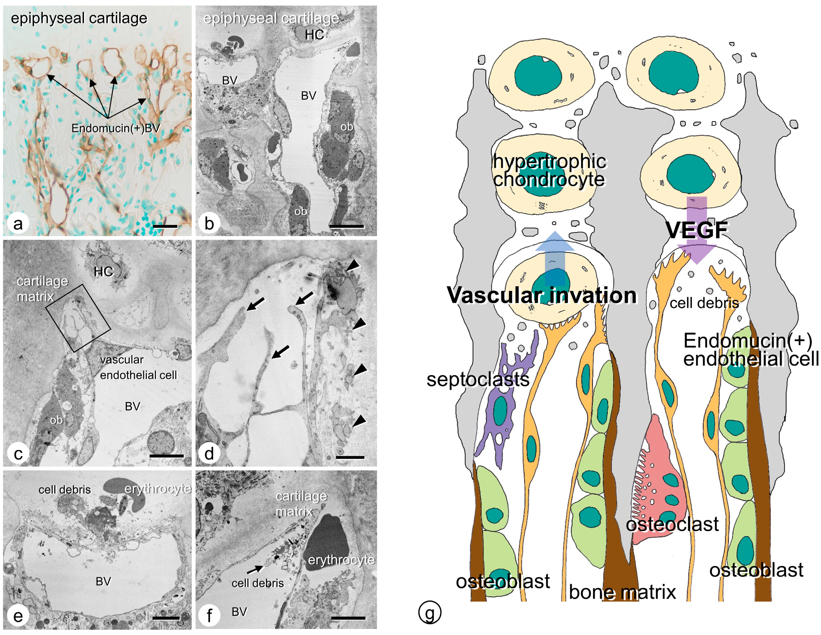
3. Vascular Invasion at the Chondro-Osseous Junction

Vascular endothelial cells can invade the epiphyseal cartilage by piercing the incompletely calcified transverse partition of the columns. Researchers demonstrated endomucin-reactive blood vessels invading the chondrocyte lacunae at the chondro-osseous junction [21]. Transmission electron microscopic (TEM) observation verified that the vascular endothelial cells, present in blood vessels close to the cartilaginous matrix, extend their fine cytoplasmic processes into the matrix. The tips of the extended cytoplasmic processes showed fine finger-like structures facing the cartilaginous matrix, suggesting that the apical region of the invading endothelial cells may be partially open. In some observations, cell debris was present inside the blood vessels facing the cartilaginous columns at the chondro-osseous junction, while erythrocytes were found outside the blood vessels. Since the apical region of invading blood vessels might be open, blood vessels could presumably invade the cartilaginous matrix and exclude unnecessary impeditive materials (mainly cellular debris) to avoid accumulation at the junction (Figure 1).
Figure 1. Vascular endothelial cells at the chondro-osseous junction. (a) Endomucin-immunoreactive (brown color) blood vessels at the chondro-osseous junction under light microscope. (bf) TEM images of blood vessels at the chondro-osseous junction. Invading blood vessels are seen beneath the chondrocytic lacunae. (c,d) When observed under higher magnification as shown in panel c, fine cytoplasmic processes (arrows) are seen extending from the vascular endothelial cell, with invaginations of the cell membranes in the superficial layer of the cartilaginous matrix. (e,f) Panel e demonstrates cell debris, including erythrocytes from the blood vessels, and panel f reveals an erythrocyte outside the vessel and cell debris in the vessels. (g) Schematic design of vascular invasion at the chondro-osseous junction. HP: hypertrophic chondrocyte; BV: blood vessel, ob: osteoblast. Bar, (a) 20 µm, (b) 10 µm, (c,e,f) 5 µm, (d) 1 µm.

4. Osteoclasts’ Function at the Chondro-Osseous Junction

It is well known that osteoclasts, also referred to as chondroclasts, accumulate in the chondro-osseous junction. Osteoclasts at the chondro-osseous junction show intense matrix metalloproteinase (MMP)-9 immunoreactivity [22]. Additionally, MMP-9 immunoreactivity is exhibited in the tips of the vascular endothelial cells facing the cartilaginous matrix, unlike the other areas distant from the chondro-osseous junction [20]. Therefore, osteoclasts and vascular endothelial cells apparently synthesize MMP-9, which dissolves the cartilaginous matrix [23][24]. Vascular invasion rather than osteoclastic resorption seems essential during endochondral ossification. Studies have found that op/op mice, c-fos−/− mice, and receptor activator of nuclear factor κβ ligand (Rankl)−/− mice preserve similar lengths of long bones to those seen in their wild-type counterparts in murine models that lack osteoclasts. However, without osteoclasts, the primary trabeculae form a disorganized but highly connected meshwork in the long bones. As described by Marks and Odgren [25], it seems likely that osteoclastic activity during endochondral ossification resorbs the excess mineralized cartilage matrices and scattered islets of mineralized cartilage in the chondro-osseous junction, enabling the longitudinal arrangement of primary trabeculae. Furthermore, another cell type, septoclasts, also referred to as perivascular cells, may also be involved in vascular invasion during endochondral ossification [26][27][28]. Septoclasts are positive for Dolichos biflorus agglutinin lectin histochemistry [26] and E-FABP [29][30], featuring well-developed Golgi apparatus and several cytoplasmic lysosomes filled with abundant cathepsin B [27]. Researchers speculate that one major function of septoclasts is to remove excess extracellular organic (non-mineralized) debris that would otherwise interrupt the vascular invasion path into the cartilage, and it is unlikely that osteoclasts are designated to resorb the excess mineralized matrices in the cartilage.

References

  1. Amizuka, N.; Hasegawa, T.; Oda, K.; Freitas, P.H.L.; Hoshi, K.; Li, M.; Ozawa, H. Histology of epiphyseal cartilage calcification and endochondral ossification. Front. Biosci. 2012, 4, 2085–2100.
  2. Ali, S.Y.; Sajdera, S.W.; Anderson, H.C. Isolation and characterization of calcifying matrix vesicles from epiphyseal cartilage. Proc. Natl. Acad. Sci. USA 1970, 67, 1513–1520.
  3. Anderson, H.C. Vesicles associated with calcification in the matrix of epiphyseal cartilage. J. Cell Biol. 1969, 41, 59–72.
  4. Bonucci, E. Fine structure of early cartilage calcification. J. Ultrastruct. Res. 1967, 20, 33–50.
  5. Bonucci, E. Fine structure and histochemistry of “calcifying globules” in epiphyseal cartilage. Z. Zellforsch. Mikrosk. Anat. 1970, 103, 192–217.
  6. Ozawa, H.; Yamada, M.; Yajima, T. The ultrastructural and cytochemical aspects of matrix vesicles and calcification processes. In Formation and Calcification of Hard Tissues; Talmage, R.V., Ozawa, H., Eds.; Shakai Hoken Pub: Tokyo, Japan, 1978; pp. 9–57.
  7. Ozawa, H.; Yamada, M.; Yamamoto, T. Ultrastructural observations on the location of lead and calcium in the mineralizing dentine of rat incisor. In Matrix Vesicles; Ascenzi, A., Bonucci, E., de Bernard, B., Eds.; Wiching Editore srl: Milano, Italy, 1981; pp. 179–187.
  8. Wuthier, R.E. Lipid composition of isolated epiphyseal cartilage cells, membranes and matrix vesicles. Biochim. Biophys. Acta 1975, 409, 128–143.
  9. Ohba, S. Hedgehog Signaling in Skeletal Development: Roles of Indian Hedgehog and the Mode of Its Action. Int. J. Mol. Sci. 2020, 21, 6665.
  10. Tavormina, P.L.; Shiang, R.; Thompson, L.M.; Zhu, Y.Z.; Wilkin, D.J.; Lachman, R.S.; Wilcox, W.R.; Rimoin, D.L.; Cohn, D.H.; Wasmuth, J.J. Thanatophoric dysplasia (types I and II) caused by distinct mutations in fibroblast growth factor receptor 3. Nat. Genet. 1995, 9, 321–328.
  11. Su, W.C.; Kitagawa, M.; Xue, N.; Xie, B.; Garofalo, S.; Cho, J.; Deng, C.; Horton, W.A.; Fu, X.Y. Activation of Stat1 by mutant fibroblast growth-factor receptor in thanatophoric dysplasia type II dwarfism. Nature 1997, 386, 288–292.
  12. Peters, K.; Ornitz, D.; Werner, S.; Williams, L. Unique expression pattern of the FGF receptor 3 gene during mouse organogenesis. Dev. Biol. 1993, 155, 423–430.
  13. Deng, C.; Wynshaw-Boris, A.; Zhou, F.; Kuo, A.; Leder, P. Fibroblast growth factor receptor 3 is a negative regulator of bone growth. Cell 1996, 84, 911–921.
  14. Amizuka, N.; Davidson, D.; Liu, H.; Valverde-Franco, G.; Chai, S.; Maeda, T.; Ozawa, H.; Hammond, V.; Ornitz, D.M.; Goltzman, D.; et al. Signalling by fibroblast growth factor receptor 3 and parathyroid hormone-related peptide coordinate cartilage and bone development. Bone 2004, 34, 13–25.
  15. Greenspan, J.S.; Blackwood, H.J. Histochemical studies of chondrocyte function in the cartilage of the mandibular codyle of the rat. J. Anat. 1966, 100, 615–626.
  16. Ikeda, T.; Nomura, S.; Yamaguchi, A.; Suda, T.; Yoshiki, S. In situ hybridization of bone matrix proteins in undecalcified adult rat bone sections. J. Histochem. Cytochem. 1992, 40, 1079–1088.
  17. Oshima, O.; Leboy, P.S.; McDonald, S.A.; Tuan, R.S.; Shapiro, I.M. Developmental expression of genes in chick growth cartilage detected by in situ hybridization. Calcif. Tissue Int. 1989, 45, 182–192.
  18. Poole, A.R.; Pidoux, I.; Rosenberg, L. Role of proteoglycans in endochondral ossification: Immunofluorescent localization of link protein and proteoglycan monomer in bovine fetal epiphyseal growth plate. J. Cell Biol. 1982, 92, 249–260.
  19. Schmid, T.M.; Linsenmayer, T.F. Immunohistochemical localization of short chain cartilage collagen (type X) in avian tissues. J. Cell Biol. 1985, 100, 598–605.
  20. Gerber, H.P.; Vu, T.H.; Ryan, A.M.; Kowalski, J.; Werb, Z.; Ferrara, N. VEGF couples hypertrophic cartilage remodeling, ossification and angiogenesis during endochondral bone formation. Nat. Med. 1999, 5, 623–628.
  21. Tsuchiya, E.; Hasegawa, T.; Hongo, H.; Yamamoto, T.; Abe, M.; Yoshida, T.; Zhao, S.; Tsuboi, K.; Udagawa, N.; Freitas, P.H.L.; et al. Histochemical assessment on the cellular interplay of vascular endothelial cells and septoclasts during endochondral ossification in mice. Microscopy 2021, 70, 201–214.
  22. Kojima, T.; Hasegawa, T.; Freitas, P.H.L.; Yamamoto, T.; Sasaki, M.; Horiuchi, K.; Hongo, H.; Yamada, T.; Sakagami, N.; Saito, N.; et al. Histochemical aspects of the vascular invasion at the erosion zone of the epiphyseal cartilage in MMP-9-deficient mice. Biomed. Res. 2013, 34, 119–128.
  23. Vu, T.H.; Shipley, J.M.; Bergers, G.; Berger, J.E.; Helms, J.A.; Hanahan, D.; Shapiro, S.D.; Senior, R.M.; Werb, Z. MMP-9/gelatinase B is a key regulator of growth plate angiogenesis and apoptosis of hypertrophic chondrocytes. Cell 1998, 93, 411–422.
  24. Engsig, M.T.; Chen, Q.J.; Vu, T.H.; Pedersen, A.C.; Therkidsen, B.; Lund, L.R.; Henriksen, K.; Lenhard, T.; Foged, N.T.; Werb, Z.; et al. Matrix metalloproteinase 9 and vascular endothelial growth factor are essential for osteoclast recruitment into developing long bones. J. Cell Biol. 2000, 151, 879–889.
  25. Marks, S.C., Jr.; Odgren, P.R. The structure and development of the skeleton. In Principles of Bone Biology; Bilezikian, J.P., Raisz, L.G., Rodan, G.A., Eds.; Academic Press: New York, NY, USA, 2002; pp. 3–15.
  26. Nakamura, H.; Ozawa, H. Ultrastructural, enzyme-, lectin, and immunohistochemical studies of the erosion zone in rat tibiae. J. Bone Miner. Res. 1996, 11, 1158–1164.
  27. Lee, E.R.; Lamplugh, L.; Shepard, N.L.; Mort, J.S. The septoclast, a cathepsin B-rich cell involved in the resorption of growth plate cartilage. J. Histochem. Cytochem. 1995, 43, 525–536.
  28. Gartland, A.; Mason-Savas, A.; Yang, M.; MacKay, C.A.; Birnbaum, M.J.; Odgren, P.R. Septoclast deficiency accompanies postnatal growth plate chondrodysplasia in the toothless (tl) osteopetrotic, colony-stimulating factor-1 (CSF-1)-deficient rat and is partially responsive to CSF-1 injections. Am. J. Pathol. 2009, 175, 2668–2675.
  29. Bando, Y.; Yamamoto, M.; Sakiyama, K.; Inoue, K.; Takizawa, S.; Owada, Y.; Iseki, S.; Kondo, H.; Amano, O. Expression of epidermal fatty acid binding protein (E-FABP) in septoclasts in the growth plate cartilage of mice. J. Mol. Histol. 2014, 45, 507–518.
  30. Bando, Y.; Yamamoto, M.; Sakiyama, K.; Sakashita, H.; Taira, F.; Miyake, G.; Iseki, S.; Owada, Y.; Amano, O. Retinoic acid regulates cell-shape and -death of E-FABP (FABP5)-immunoreactive septoclasts in the growth plate cartilage of mice. Histochem. Cell Biol. 2017, 148, 229–238.
More
Information
Contributors MDPI registered users' name will be linked to their SciProfiles pages. To register with us, please refer to https://encyclopedia.pub/register : , , , , ,
View Times: 455
Revisions: 3 times (View History)
Update Date: 09 Feb 2023
1000/1000