Your browser does not fully support modern features. Please upgrade for a smoother experience.
Submitted Successfully!
 +1 credit
Thank you for your contribution! You can also upload a video entry or images related to this topic.
For video creation, please contact our Academic Video Service.
| Version | Summary | Created by | Modification | Content Size | Created at | Operation |
|---|---|---|---|---|---|---|
| 1 | Ghassan Bkaily | -- | 1804 | 2023-02-01 02:23:39 | | | |
| 2 | Catherine Yang | Meta information modification | 1804 | 2023-02-01 02:25:47 | | |
Video Upload Options
We provide professional Academic Video Service to translate complex research into visually appealing presentations. Would you like to try it?
Cite
If you have any further questions, please contact Encyclopedia Editorial Office.
Bkaily, G.; Jacques, D. Vascular Endothelium in Cardiovascular Diseases. Encyclopedia. Available online: https://encyclopedia.pub/entry/40695 (accessed on 23 July 2026).
Bkaily G, Jacques D. Vascular Endothelium in Cardiovascular Diseases. Encyclopedia. Available at: https://encyclopedia.pub/entry/40695. Accessed July 23, 2026.
Bkaily, Ghassan, Danielle Jacques. "Vascular Endothelium in Cardiovascular Diseases" Encyclopedia, https://encyclopedia.pub/entry/40695 (accessed July 23, 2026).
Bkaily, G., & Jacques, D. (2023, February 01). Vascular Endothelium in Cardiovascular Diseases. In Encyclopedia. https://encyclopedia.pub/entry/40695
Bkaily, Ghassan and Danielle Jacques. "Vascular Endothelium in Cardiovascular Diseases." Encyclopedia. Web. 01 February, 2023.
Copy Citation
The vascular endothelium plays a vital role during embryogenesis and aging and is a cell monolayer that lines the blood vessels. The immune system recognizes the endothelium as its own. Therefore, an abnormality of the endothelium exposes the tissues to the immune system and provokes inflammation and vascular diseases such as atherosclerosis. Its secretory role allows it to release vasoconstrictors and vasorelaxants as well as cardio-modulatory factors that maintain the proper functioning of the circulatory system. The sealing of the monolayer provided by adhesion molecules plays an important role in cardiovascular physiology and pathology.
endothelium
endothelium physiology
endothelium pathology
endothelium dysfunction
2. The Vascular Endothelium
The vascular endothelium is a simple tissue in its morphology but complex in its function. Although it is formed as a single monolayer, it is capable of sensing hemodynamic and rheologic changes as well as responding to these modifications of its environment. Vascular tone is maintained by balancing vasodilating and vasoconstricting factors released by the endothelium. In addition, the endothelium plays a key role in controlling the migration and proliferation of VSMCs [1].
The integrity of the endothelial monolayer is essential to regulate vascular permeability and protect the vessel against platelet deposition and thrombus formation. Furthermore, the integrity of this monolayer requires that the morphology and the contacts between ECs do not change.
3. Origin and Differentiation of the Vascular Endothelium
The endothelium is the first cell type to constitute blood vessels. The formation of blood vessels and vasculogenesis result from the differentiation of the mesodermal cells into angioblasts due to the presence of specific proteins. These angioblasts are the precursors of endothelial and blood cells. The physiological primitive angiogenesis takes place to form the vascular tree, which gives birth to buds of the branches that give birth to the heart, including its endocardial endothelial cells. Then, remodeling of the vascular tree takes place to form capillaries and veins, including large arteries [1][2][3]. The endothelium of these newly formed blood vessels differs depending on the type of vessels. For example, fibroblast growth factor (FGF) receptors seem to be expressed only in large vessels [4]. Several ligands and their corresponding receptors are implicated in the differentiation and formation of the endothelium, including vascular endothelial growth factor (VEGF) and its receptors 1 and 2 [2].
The heart’s formation is more than a deformation of blood vessels, and differentiation of vascular endothelium into endocardial endothelium forms the left (arterial) and right (venous) endocardial endothelium layers. Both endocardial and vascular endothelium are separated from their muscle cells by a basal lamina membrane [1]. Vasculogenesis occurs during early embryonic development, whereas angiogenesis happens during adulthood. Angiogenesis is usually associated with diseases [3]. Several endothelial markers exist, such as VE-cadherin, PECAM-1, Tie-1 and 2, and flk1. Notch family activation plays an essential role in defining the characteristics and identities of arterial endothelial cells [5]. Although the molecular aspect of the arterial specification is more precise, little is known concerning the venous specification [5]. For example, the vascular endothelium can adapt its function depending on the environment. Still, it does not change its phenotype, such as in transplanted arterial and venous vessel grafts, where the graft vessel’s endothelium matches the host vessels’ characteristics [6][7].
4. Role of the Endothelium in Vascular Physiology
All blood vessels contain endothelial cells that form the intima. Two types of blood vessels only have endothelial cells: capillaries and venules. The intima is formed by continuous and discontinuous (fenestrated) layers of endothelial cells. Examples of continuous endothelium are arteries and veins. Tight, adherent junctions connect the continuous layer of endothelium side by side. Transport molecules go through this sealed monolayer of the endothelium via a transcytosis mechanism, such as caveolae (caveolin-1) and vesiculo-vacuolar organelles [8]. A fenestrated, discontinuous layer of endothelium permits extensive transport of molecules toward tissues such as the liver.
Several physiological functions are attributed to vascular endothelium, independent of their localization in the vascular tree, such as tuning the level of vascular endothelial cells (VECs) vasoconstrictors and vasorelaxers [1][9], regulation of coagulation and inflammation [10][11], and playing an essential role as a gatekeeper of fatty acids transport [12][13][14][15][16][17].
Among the vasoconstrictive factors released by the vascular endothelium are endothelin-1 (ET-1), thromboxane A2 (TxA2), as well as angiotensin-II (AngII) [1][8][9][18][19]. On the other hand, the most vasorelaxant factors released by the endothelium are nitric oxide (NO), prostacyclin (PGI2), and endothelium-derived hyperpolarizing factor (EDHF) [1][20].
The blood vascular system consists of a circuit of vessels in which the continuous movement of the heart pump maintains the blood flow. Blood vessels distribute nutrients, oxygen, and hormones to all organs and tissues and transport the products of cellular metabolism. The walls of arteries and veins, such as the thoracic aorta, used commonly in the literature, consist of three concentric tunics that are firmly joined from the inside out [1] (Figure 1): (1) The intima is the thin innermost layer that lines the various vascular walls, including those of the capillaries and venules. It is composed of a monolayer of endothelial cells (ECs) in direct contact with the blood and forming the vascular endothelium. The ECs provide a smooth inner surface that minimizes friction, which facilitates blood flow. The vascular endothelium is supported by a basal lamina and a thin connective tissue formed by collagen and some elastic fibers;
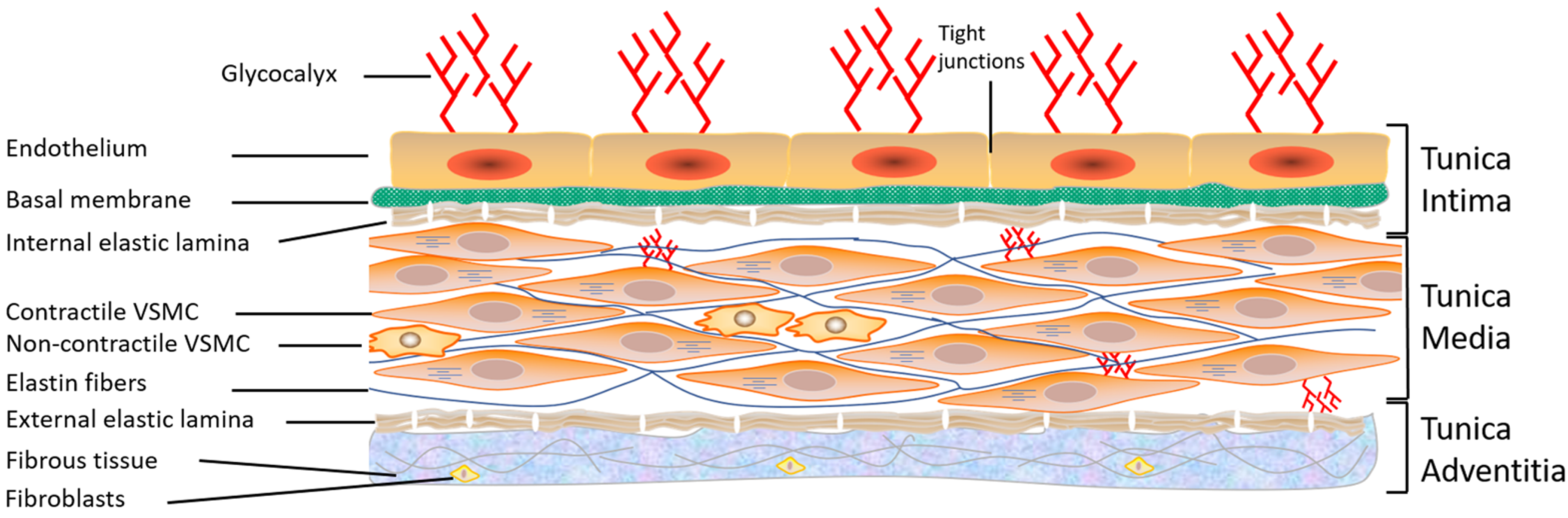
Figure 1. Structure of the vascular wall. Schematic representation showing the three layers of the vascular wall: tunica intima, tunica media, and tunica adventitia, as well as the components of each layer. VSMC: vascular smooth muscle cells (from Bkaily et al., 2021 [1]).
(2) The media is the thickest intermediate layer of the vascular wall. It consists of vascular smooth muscle cells (VSMCs), collagen, and elastin. This layer is absent in the capillaries and venules; (3) The adventitia is the outermost layer of the vascular wall. It is absent in capillaries and venules. This layer is formed of supporting connective tissue consisting mainly of collagen. It is also crossed by numerous nerve endings controlling the activity of the muscle fibers as well as the blood vessels feeding the vascular wall, called vasa vasorum (vessels of the vessels). The relative importance of these three layers varies according to the type of blood vessel [1]. In conclusion, all blood vessels have an endothelium but not necessarily adventitia or VSMCs, hence the importance of studying and learning more about the vascular endothelium.
5. Structure of the Vascular Endothelium of Arteries and Veins
A monolayer of flat cells forms the vascular endothelium of arteries and veins, with a central nucleus measuring 10–20 µm in diameter. VECs are characterized by extensive intercellular overlap and long, deep slits that contribute to the integrity of the vascular endothelium [1]. The integrity of this monolayer is ensured by a dynamic cytoskeleton [1][21][22][23] as well as by contacts between cells and between these cells and the extracellular matrix [1][24][25]. In vivo and in situ morphology studies have shown the presence of tight junctions, adhesion junctions, and gap junctions between adjacent VECs (including aortic VECs) [25][26][27]. In addition, several roles have been attributed to junctional communication at the vascular endothelium level, including intercellular nutrient exchange, regulation of growth and differentiation, coordination of cellular response to exogenous and endogenous stimuli, and maintenance of vascular tissue homeostasis [28][29][30][31].
The cytoskeleton is well-developed in ECs. It contributes to vascular homeostasis and seems to play an essential role in the repair and integrity of these cells [21][22][23]. VECs contain the actin protein in its filamentous polymeric form, called F-actin, and in its globular monomeric form, called G-actin [32][33]. Therefore, F- and G-actin play a role in the shape of ECs. The balance between the monomeric and polymeric forms could be altered during stimulation of ECs and contribute to the modulation of intercellular junctions that affect the vascular permeability of the endothelial layer. Indeed, during the migration of VECs, G-actins increase compared to F-actins [34]. The migration of these cells also involves the redistribution of centrosomes [21]. Actin microfilaments are localized within the cell as short, thin stress fibers and form a continuous band at the periphery [21][32]. In situ studies have also demonstrated the presence of the protein myosin at the level of these microfilaments [35][36], which plays an essential role in cell adhesion, and facilitates the adaptation of the vascular wall to variations in blood flow pressure [21]. The presence or absence of an actin isoform allows the identification of ECs. Therefore, the presence of α-actin in VECs is considered to be a marker for this type of cell [37].
6. Role of the Endothelium in Vascular Activity
ECs respond to chemical and physical stimuli by synthesizing and releasing various vasoactive and growth factors [1] (Figure 2). The endothelium possesses anti-adhesive substances that prevent blood from clotting. The anticoagulant and antithrombotic properties of the vascular endothelium, which are essential for vascular homeostasis, are due to the synthesis of vasodilatory factors such as nitric oxide (NO) and prostacyclin [8][35][38][39][40] (Figure 2). On the other hand, the vascular endothelium secretes several vasoconstrictor substances (Figure 2), including endothelin-1 (ET-1), prostaglandins, and several components of the renin-angiotensin system (RAS), such as angiotensin II (Ang II). Ang II [18][19][41][42] and ET-1 [43][44][45] act at the plasma and nuclear membranes of ECs and induce an increase in the intracellular calcium level via activation of their respective receptors, AT1/AT2 and ETA /ETB receptors. This increase in [Ca2+]i may, in turn, modulate the secretory function of ECs [1][46] and survival [19][44]. Furthermore, a balance between the different factors secreted by the EV is essential for maintaining intracellular homeostasis and wall integrity. Any disturbance in this balance leads to endothelial dysfunction, characterized by a decreased capacity for relaxation of the vessel, an increase in the adhesion of blood cells to the vascular wall, and a disturbance in the tunica medial [13][38][47][48][49][50][51][52]. This endothelial dysfunction is generally observed during aging and in several vascular pathologies, such as hypertension, hypotension, atherosclerosis, and heart failure [19][53][54]. All VECs synthesize and secrete von Willebrand factor (vWF), a multifunctional protein involved in the typical arrest of hemorrhage [55]. Indeed, through its interaction with extracellular matrix proteins and membrane receptors, vWF plays a prominent role in blood coagulation, platelet aggregation, and platelet adhesion to the extracellular matrix [56][57]. vWF can also bind to the pro-coagulant co-enzyme, factor VIII, contributing to its stability and, indirectly, to the production of fibrin [56][57]. vWF is stored in small vesicles characteristic of endothelial cells, the Weibel–Palade bodies [56][57][58]. The latter contain other proteins, such as ET-1 [58][59] and interleukin-8 [60]. In addition, vWF is used as a marker of ECs in vitro [61].
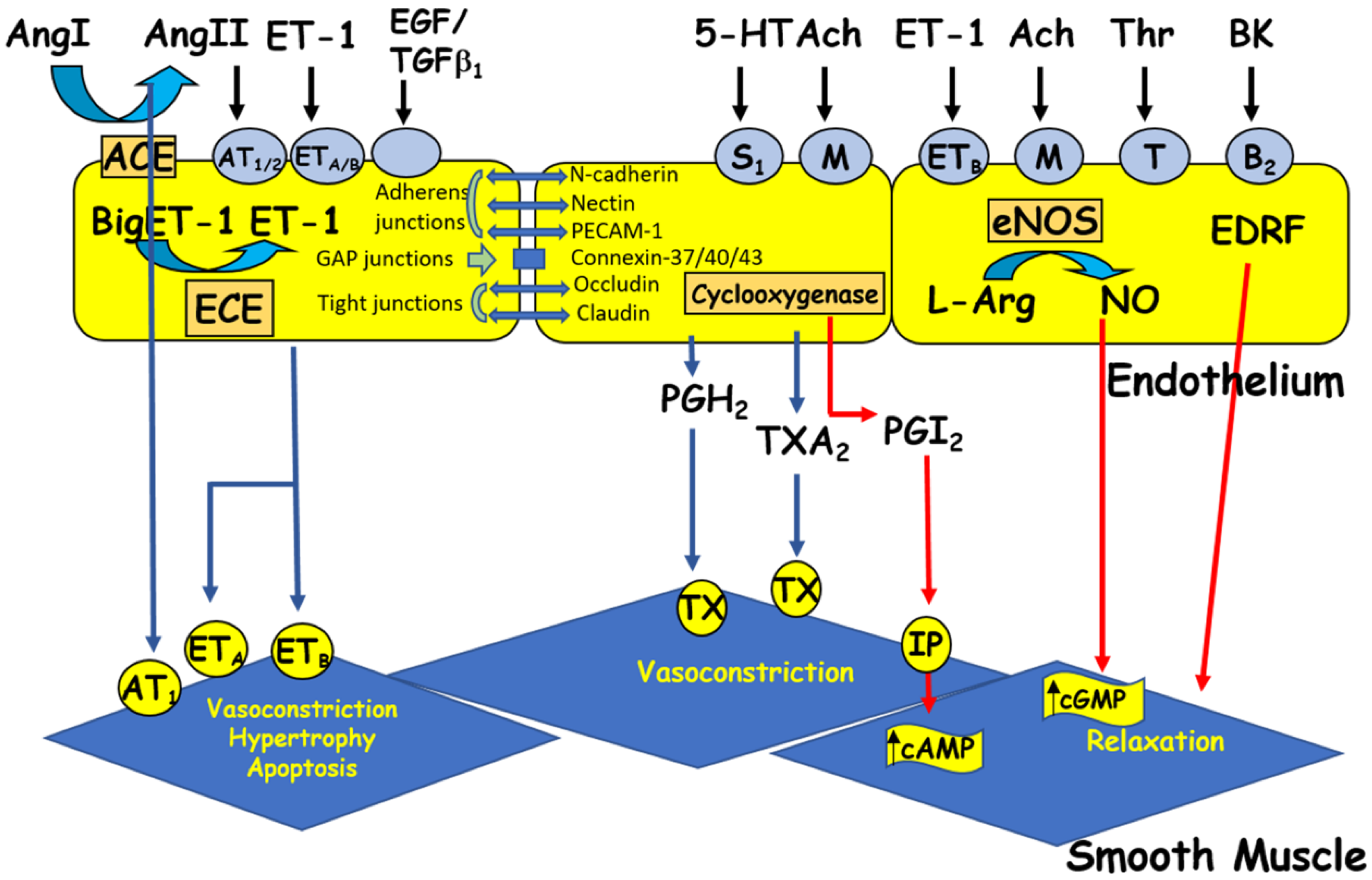
Figure 2. The endothelium produces vasoactive factors that cause either relaxation or contraction of the vascular smooth muscle. Ang I and II: angiotensin I and II, ACE: angiotensin-converting enzyme, Ach: acetylcholine, BK: bradykinin, cAMP/cGMP: cyclic adenosine/guanosine monophosphate, ECE: endothelin-converting enzyme, EDRF: endothelium-derived relaxing factor, ET-1: endothelin-1, 5HT: 5-hydroxytryptamine (serotonin), L-Arg: L-arginine, NO: nitric oxide, NOS: nitric oxide synthase, PGH2: prostaglandin H2, PGI2: prostacyclin, TGFβ1: transforming growth factor beta 1, Thr: thrombin, and TXA2: thromboxane A2. Circles represent receptors (AT: angiotensin receptor, B: bradykinin receptor, ET: endothelin receptor, M: muscarinic receptor, IP: purinergic receptor, S: serotonin receptor, T: thrombin receptor, and TX: thromboxane receptor).
References
- Bkaily, G.; Abou Abdallah, N.; Simon, Y.; Jazzar, A.; Jacques, D. Vascular smooth muscle remodeling in health and disease. Can. J. Physiol. Pharmacol. 2021, 99, 171–178.
- Ratajska, A.; Jankowska-Steifer, E.; Czarnowska, E.; Olkowski, R.; Gula, G.; Niderla-Bielińska, J.; Flaht-Zabost, A.; Jasińska, A. Vasculogenesis and Its Cellular Therapeutic Applications. Cells Tissues Organs 2017, 203, 141–152.
- Ferguson, J.E., 3rd; Kelley, R.W.; Patterson, C. Mechanisms of endothelial differentiation in embryonic vasculogenesis. Arterioscler. Thromb. Vasc. Biol. 2005, 25, 2246–2254.
- Risau, W. Differentiation of endothelium. FASEB J. 1995, 9, 926–933.
- Atkins, G.B.; Jain, M.K. Role of Krüppel-like transcription factors in endothelial biology. Circ. Res. 2007, 100, 1686–1695.
- Dyer, L.A.; Patterson, C. Development of the endothelium: An emphasis on heterogeneity. Semin. Thromb. Hemost. 2010, 36, 227–235.
- Ladak, S.S.; McQueen, L.W.; Layton, G.R.; Aujla, H.; Adebayo, A.; Zakkar, M. The Role of Endothelial Cells in the Onset, Development and Modulation of Vein Graft Disease. Cells 2022, 11, 3066.
- Gratton, J.P.; Bernatchez, P.; Sessa, W.C. Caveolae and caveolins in the cardiovascular system. Circ. Res. 2004, 94, 1408–1417.
- Sandoo, A.; Veldhuijzen van Zanten, J.J.; Metsios, G.S.; Carroll, D.; Kitas, G.D. The endothelium and its role in regulating vascular tone. Open Cardiovasc. Med. J. 2010, 4, 302–312.
- Kotlyarov, S. Immune Function of Endothelial Cells: Evolutionary Aspects, Molecular Biology and Role in Atherogenesis. Int. J. Mol. Sci. 2022, 23, 9770.
- Van Hinsbergh, V.W. Endothelium—Role in Regulation of Coagulation and Inflammation; Springer: Amsterdam, The Netherlands, 2012; pp. 93–106.
- Mehrotra, D.; Wu, J.; Papangeli, I.; Chun, H.J. Endothelium as a gatekeeper of fatty acid transport. Trends Endocrinol. Metab. 2014, 25, 99–106.
- Ghosh, A.; Gao, L.; Thakur, A.; Siu, P.M.; Lai, C.W. Role of free fatty acids in endothelial dysfunction. J. Biomed. Sci. 2017, 24, 50.
- Northcott, J.M.; Czubryt, M.P.; Wigle, J.T. Vascular senescence and ageing: A role for the MEOX proteins in promoting endothelial dysfunction. Can. J. Physiol. Pharmacol. 2017, 95, 1067–1077.
- Mallick, R.; Duttaroy, A.K. Modulation of endothelium function by fatty acids. Mol. Cell. Biochem. 2022, 477, 15–38.
- Stanek, A.; Fazeli, B.; Bartuś, S.; Sutkowska, E. The role of endothelium in physiological and pathological states: New data. BioMed. Res. Int. 2018, 2018, 1–3.
- Pi, X.; Xie, L.; Patterson, C. Emerging roles of vascular endothelium in metabolic homeostasis. Circ. Res. 2018, 123, 477–494.
- Kamal, M.; Jacques, D.; Bkaily, G. Angiotensin II receptors’ modulation of calcium homeostasis in human vascular endothelial cells. Can. J. Physiol. Pharmacol. 2017, 95, 1289–1297.
- Jacques, D.; Abdel-Karim Abdel-Malak, N.; Abou Abdallah, N.; Al-Khoury, J.; Bkaily, G. Difference in response to angiotensin II between left and right ventricular endocardial endothelial cells. Can. J. Physiol. Pharmacol. 2017, 95, 1271–1282.
- Bkaily, G. Biophysical and pharmacological properties of T-, L- and R-type Ca2+ channels. In Ionic Channels in Vascular Smooth Muscle; R.G. Landers Company: Georgetown, TX, USA, 1994.
- Gottlieb, A.I.; Langille, B.L.; Wong, M.K.; Kim, D.W. Structure and function of the endothelial cytoskeleton. Lab. Investig. 1991, 65, 123–137.
- Gaudette, S.; Hughes, D.; Boller, M. The endothelial glycocalyx: Structure and function in health and critical illness. J. Vet. Emerg. Crit. Care 2020, 30, 117–134.
- Rajendran, P.; Rengarajan, T.; Thangavel, J.; Nishigaki, Y.; Sakthisekaran, D.; Sethi, G.; Nishigaki, I. The vascular endothelium and human diseases. Int. J. Biol. Sci. 2013, 9, 1057–1069.
- Koçer, G.; Albino, I.M.C.; Verheijden, M.L.; Jonkheijm, P. Endothelial cell spreading on lipid bilayers with combined integrin and cadherin binding ligands. Bioorg. Med. Chem. 2022, 68, 116850.
- Lampugnan, A.G.; Dejana, E. Interendothelial junctions: Structure, signaling and functional roles. Curr. Opin. Cell Biol. 1997, 9, 674–682.
- Francke, W.W.; Cowin, P.; Grund, C.; Kuhn, C.; Kapprel, H.P. The endothelium junction. The plaque and its components. In Endothelial Cell Biology in Health and Disease; Siminescu, N., Simionescu, M., Eds.; Plenum Publishing Corp.: New York, NY, USA, 1988.
- Nguyen Duong, C.; Vestweber, D. Mechanisms ensuring endothelial junction integrity beyond Ve-cadherin. Front. Physiol. 2020, 11, 1–9.
- Seebach, J.; Klusmeier, N.; Schnittler, H. Autoregulatory “Multitasking” at Endothelial Cell Junctions by Junction-Associated Intermittent Lamellipodia Controls Barrier Properties. Front. Physiol. 2021, 11, 586921.
- Charollais, A.; Gjinovci, A.; Huarte, J.; Bauquis, J.; Nadal, A.; Martin, F.; Andreu, E.; Sanchez-Andres, J.V.; Calabrese, A.; Bosco, D.; et al. Junctional communication of pancreatic beta cells contributes to the control of insulin secretion and glucose tolerance. J. Clin. Investig. 2000, 106, 235–243.
- Saez, J.C.; Branes, M.C.; Corvalan, L.A.; Eugenin, E.A.; Gonzalez, H.; Martinez, A.D.; Palisson, F. Gap junctions in cells of the immune system: Structure, regulation and possible functional roles. Braz. J. Med. Biol. Res. 2000, 33, 447–455.
- Morini, M.F.; Giampietro, C.; Corada, M.; Pisati, F.; Lavarone, E.; Cunha, S.I.; Conze, L.L.; O’Reilly, N.; Joshi, D.; Kjaer, S.; et al. VE-Cadherin-Mediated Epigenetic Regulation of Endothelial Gene Expression. Circ. Res. 2018, 122, 231–245.
- Becker, C.G.; Murphy, G.E. Demonstration of contractile protein in endothelium and cells of the heart valves, endocardium, intima, arteriosclerotic plaques, and Aschoff bodies of rheumatic heart disease. Am. J. Pathol. 1969, 55, 1–37.
- Pollard, T.D. Actin and Actin-Binding Proteins. Cold Spring Harb. Perspect. Biol. 2016, 8, a018226.
- Willingham, M.C.; Yamada, S.S.; Davies, P.J.; Rutherford, A.V.; Gallo, M.G.; Pastan, I. Intracellular localization of actin in cultured fibroblasts by electron microscopic immunocytochemistry. J. Histochem. Cytochem. 1981, 29, 17–37.
- Majolée, J.; Kovačević, I.; Hordijk, P.L. Ubiquitin-based modifications in endothelial cell-cell contact and inflammation. J. Cell. Sci. 2019, 132, jcs227728.
- Alonso, F.; Spuul, P.; Daubon, T.; Kramer, I.; Génot, E. Variations on the theme of podosomes: A matter of context. Biochim. Biophys. Acta Mol. Cell. Res. 2019, 1866, 545–553.
- Bkaily, G.; Pothier, P.; D’Orléans-Juste, P.; Simaan, M.; Jacques, D.; Jaalouk, D.; Belzile, F.; Hassan, G.; Boutin, C.; Haddad, G.; et al. The use of confocal microscopy in the investigation of cell structure and function in the heart, vascular endothelium and smooth muscle cells. Mol. Cell. Biochem. 1997, 172, 171–194.
- Burnett, J.C., Jr. Coronary endothelial dysfunction in the hypertensive patient: From myocardial ischemia to heart failure. J. Hum. Hypertens. 1997, 11, 45–49.
- Cerutti, C.; Ridley, A.J. Endothelial cell-cell adhesion and signaling. Exp. Cell. Res. 2017, 358, 31–38.
- Bkaily, G.; D’Orleans-Juste, P.; Jacques, D. A new paradigm: Calcium independent and caveolae internalization dependent release of nitric oxide by the endothelial nitric oxide synthase. Circ. Res. 2006, 99, 793–794.
- Bkaily, G.; Nader, M.; Avedanian, L.; Choufani, S.; Jacques, D.; D’Orléans-Juste, P.; Al-Khoury, J. G-protein-coupled receptors, channels, and Na+–H+ exchanger in nuclear membranes of heart, hepatic, vascular endothelial, and smooth muscle cells. Can. J. Physiol. Pharmacol. 2006, 84, 431–441.
- Bkaily, G.; Al-Khoury, J.; Jacques, D. Nuclear membranes GPCRs: Implication in cardiovascular health and diseases. Curr. Vasc. Pharmacol. 2014, 12, 15–22.
- Mikhail, M.; Vachon, P.H.; D’Orléans-Juste, P.; Jacques, D.; Bkaily, G. Role of endothelin-1 and its receptors, ETA and ETB, in the survival of human vascular endothelial cells. Can. J. Physiol. Pharmacol. 2017, 95, 1298–1305.
- Bonetti, P.O.; Lerman, L.O.; Lerman, A. Endothelial dysfunction: A marker of atherosclerotic risk. Arterioscler. Thromb. Vasc. Biol. 2003, 23, 168–175.
- Jules, F.; Avedanian, L.; Al-Khoury, J.; Keita, R.; Normand, A.; Bkaily, G.; Jacques, D. Nuclear Membranes ETB Receptors Mediate ET-1-induced Increase of Nuclear Calcium in Human Left Ventricular Endocardial Endothelial Cells. J. Cardiovasc. Pharmacol. 2015, 66, 50–57.
- Nilius, B.; Droogmans, G. Ion channels and their functional role in vascular endothelium. Physiol. Rev. 2001, 81, 1415–1459.
- Mann, M.J.; Gibbons, G.H.; Tsao, P.S.; von der Leyen, H.E.; Cooke, J.P.; Buitrago, R.; Kernoff, R.; Dzau, V.J. Cell cycle inhibition preserves endothelial function in genetically engineered rabbit vein grafts. J. Clin. Investig. 1997, 99, 1295–1301.
- Santos-Gomes, J.; Le Ribeuz, H.; Brás-Silva, C.; Antigny, F.; Adão, R. Role of Ion Channel Remodeling in Endothelial Dysfunction Induced by Pulmonary Arterial Hypertension. Biomolecules 2022, 12, 484.
- Lüscher, T.F.; Barton, M. Biology of the endothelium. Clin. Cardiol. 1997, 20, 1415–1459.
- Mombouli, J.V.; Vanhoutte, P.M. Endothelial dysfunction: From physiology to therapy. J. Mol. Cell. Cardiol. 1999, 31, 61–74.
- Mitchell, C.A.; Risau, W.; Drexler, H.C. Regression of vessels in the tunica vasculosa lentis is initiated by coordinated endothelial apoptosis: A role for vascular endothelial growth factor as a survival factor for endothelium. Dev. Dyn. 1998, 213, 322–333.
- Drexler, H. Endothelial dysfunction: Clinical implications. Prog. Cardiovasc. Dis. 1997, 39, 287–324.
- Pacinella, G.; Ciaccio, A.M.; Tuttolomondo, A. Endothelial Dysfunction and Chronic Inflammation: The Cornerstones of Vascular Alterations in Age-Related Diseases. Int. J. Mol. Sci. 2022, 23, 15722.
- Al-Khoury, J.; Jacques, D.; Bkaily, G. Hypotension in hereditary cardiomyopathy. Pflugers Arch. 2022, 474, 517–527.
- de Wit, T.R.; van Mourik, J.A. Biosynthesis, processing and secretion of von Willebrand factor: Biological implications. Best Pract. Res. Clin. Haematol. 2001, 14, 241–255.
- Ruggeri, Z.M.; Landolfi, R. Platelet function and arterial thrombosis. Ital. Heart J. 2001, 2, 809–810.
- Agostini, S.; Lionetti, V. New insights into the non-hemostatic role of von Willebrand factor in endothelial protection. Can. J. Physiol. Pharmacol. 2017, 95, 1183–1189.
- Wagner, D.D.; Marder, V.J. Biosynthesis of von Willebrand protein by human endothelial cells: Processing steps and their intracellular localization. J. Cell. Biol. 1984, 99, 2123–2130.
- Sporn, L.A.; Rubin, P.; Marder, V.J.; Wagner, D.D. Irradiation induces release of von Willebrand protein from endothelial cells in culture. Blood 1984, 64, 567–570.
- Utgaard, J.O.; Jahnsen, F.L.; Bakka, A.; Brandtzaeg, P.; Haraldsen, G. Rapid secretion of prestored interleukin 8 from Weibel-Palade bodies of microvascular endothelial cells. J. Exp. Med. 1998, 188, 1751–1756.
- Wagner, D.D.; Urban-Pickering, M.; Marder, V.J. Von Willebrand protein binds to extracellular matrices independently of collagen. Proc. Natl. Acad. Sci. USA 1984, 81, 471–475.
More
Information
Subjects:
Cardiac & Cardiovascular Systems
Contributors
MDPI registered users' name will be linked to their SciProfiles pages. To register with us, please refer to https://encyclopedia.pub/register
:
View Times:
1.6K
Revisions:
2 times
(View History)
Update Date:
01 Feb 2023
Table of Contents


Notice
You are not a member of the advisory board for this topic. If you want to update advisory board member profile, please contact office@encyclopedia.pub.
OK
Confirm
Only members of the Encyclopedia advisory board for this topic are allowed to note entries. Would you like to become an advisory board member of the Encyclopedia?
Yes
No
${ textCharacter }/${ maxCharacter }
Submit
Cancel
Back
Comments
${ item }
|
${ item.createdUser.fullName }
${ item.createdAt }
${ item.vote }
${ item.reply }
Delete
${ reply.createdUser.fullName }
${ reply.createdAt }
${ reply.vote }
Delete
There is no reply to this comment~
${ item.replyTextCharacter }/${ item.replyMaxCharacter }
Submit
Cancel
More
No more~
There is no comment~
${ textCharacter }/${ maxCharacter }
Submit
Cancel
${ selectedItem.replyTextCharacter }/${ selectedItem.replyMaxCharacter }
Submit
Cancel
Confirm
Are you sure to Delete?
Yes
No