Submitted Successfully!
To reward your contribution, here is a gift for you: A free trial for our video production service.
Thank you for your contribution! You can also upload a video entry or images related to this topic.
Version Summary Created by Modification Content Size Created at Operation
1 -- 2208 2023-01-30 20:10:00 |
2 format correct Meta information modification 2208 2023-01-31 03:20:41 | |
3 format correct Meta information modification 2208 2023-01-31 03:22:38 | |
4 format correct + 3 word(s) 2211 2023-01-31 03:28:23 |

Video Upload Options

Do you have a full video?

Confirm

Are you sure to Delete?
Cite
If you have any further questions, please contact Encyclopedia Editorial Office.
Weth, F.R.;  Peng, L.;  Paterson, E.;  Tan, S.T.;  Gray, C. Cerebral Organoid Glioma ‘GLICO’ Model for Drug Screening. Encyclopedia. Available online: https://encyclopedia.pub/entry/40619 (accessed on 29 March 2024).
Weth FR,  Peng L,  Paterson E,  Tan ST,  Gray C. Cerebral Organoid Glioma ‘GLICO’ Model for Drug Screening. Encyclopedia. Available at: https://encyclopedia.pub/entry/40619. Accessed March 29, 2024.
Weth, Freya R., Lifeng Peng, Erin Paterson, Swee T. Tan, Clint Gray. "Cerebral Organoid Glioma ‘GLICO’ Model for Drug Screening" Encyclopedia, https://encyclopedia.pub/entry/40619 (accessed March 29, 2024).
Weth, F.R.,  Peng, L.,  Paterson, E.,  Tan, S.T., & Gray, C. (2023, January 30). Cerebral Organoid Glioma ‘GLICO’ Model for Drug Screening. In Encyclopedia. https://encyclopedia.pub/entry/40619
Weth, Freya R., et al. "Cerebral Organoid Glioma ‘GLICO’ Model for Drug Screening." Encyclopedia. Web. 30 January, 2023.
Cerebral Organoid Glioma ‘GLICO’ Model for Drug Screening
Edit

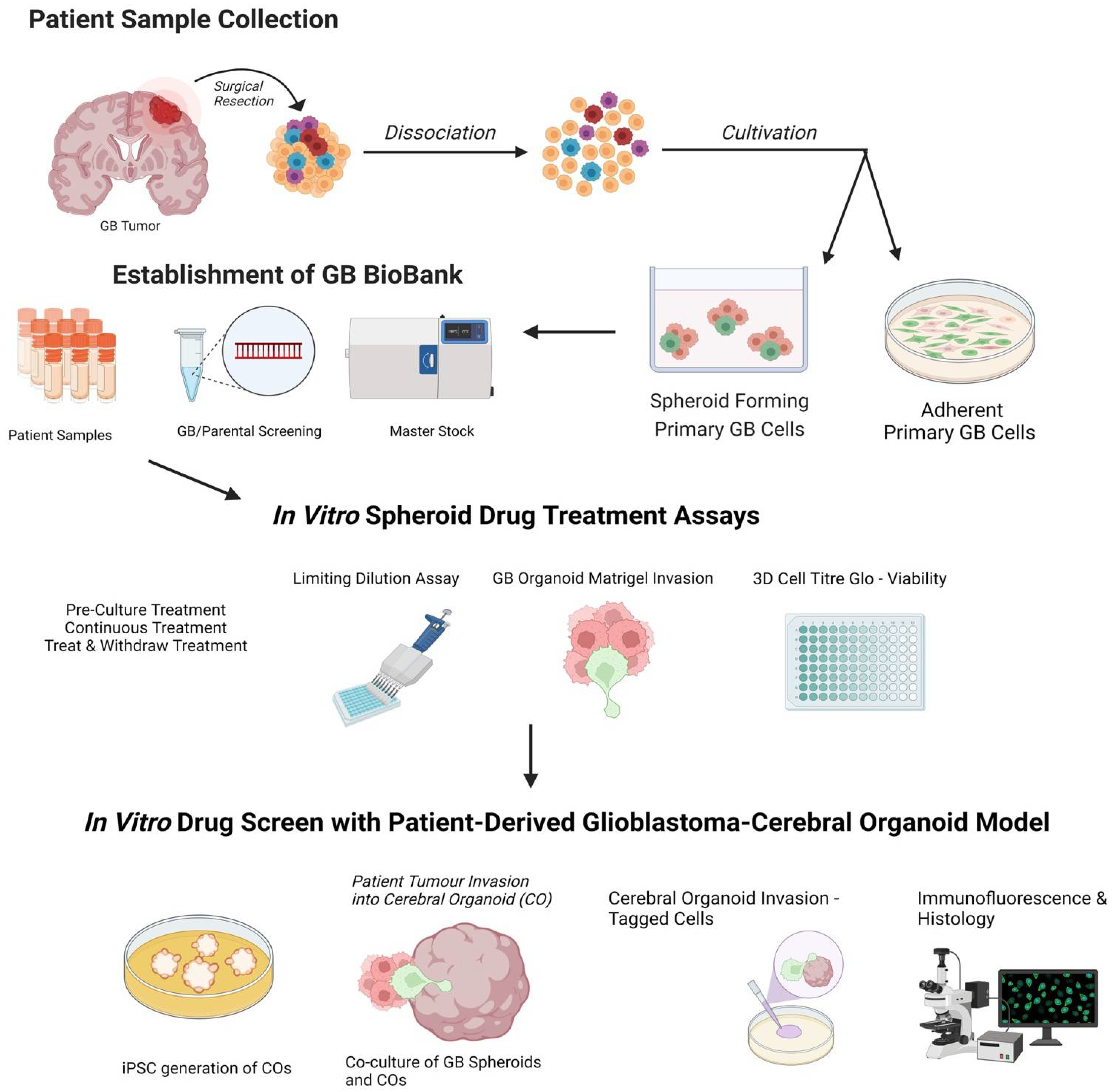
Glioblastoma, a grade IV astrocytoma, is regarded as the most aggressive primary brain tumour with an overall median survival of 16.0 months following the standard treatment regimen of surgical resection, followed by radiotherapy and chemotherapy with temozolomide. The ability to understand and manipulate complex cancers such as glioblastoma requires disease models to be clinically and translationally relevant and encompass the cellular heterogeneity of such cancers. Therefore, brain cancer research models need to aim to recapitulate glioblastoma stem cell function, whilst remaining amenable for analysis. The development of 3D cultures has overcome some of these challenges, and cerebral organoids are emerging as cutting-edge tools in glioblastoma research. The opportunity to generate cerebral organoids via induced pluripotent stem cells, and to perform co-cultures with patient-derived cancer stem cells (GLICO model), has enabled the analysis of cancer development in a context that better mimics brain tissue architecture.

glioblastoma cancer GLICO model

1. Introduction

Glioblastoma is the most common and most aggressive primary brain tumour, characterised by high recurrence rates and exceptionally poor prognosis [1]. Standard treatment involves intensive multimodal therapy including tumour resection, tumour-treating fields (TTF) radiotherapy or standard radiotherapy, and chemotherapy with Temozolomide (TMZ). The overall median survival is only 16.0 months, with tumour-treating fields (TTF)-treated patients reaching an average of 20.9 months [2]. Despite such intensive treatment, less than 30% of patients survive more than 2 years [3]. The inability to effectively treat glioblastoma is due, in part, to the ability of a subpopulation of tumorigenic cells to infiltrate normal brain tissue, preventing complete surgical removal of cancer cells, leading to subsequent recurrence [4]. Improved treatments that target the source of chemotherapy resistance and tumour recurrence are urgently needed, as the overall median survival rates have remained relatively unchanged for 30 years [5].
For glioblastoma to establish beyond the primary site within the brain, this subpopulation of cells must be able to self-renew, generate differentiated tumour cells, and spawn a heterogeneous tumour [6]. Glioblastoma, like hematopoietic malignancies and other solid cancers, has been shown to comprise a small population of cancer stem cells (CSCs) known as glioma stem cells (GSCs) [7], which possess the capacity to recapitulate the heterogeneity of the parent tumour after serial dilution and intracranial implantation into immune-compromised mice [8]. Moreover, GSCs demonstrate particularly infiltrative properties and are thought to be primary contributors to chemotherapy and radiotherapy resistance and tumour recurrence [9]. Therefore, selectively targeting GSC proliferation and invasion in combination with current therapies is seen as a viable option to improve treatment outcomes for glioblastoma patients [10][11].
Proof-of-principle genetic studies have shown that blocking the self-renewal of GSCs leads to prolonged survival in GSC patient-derived mouse efficacy studies [12]. However, identifying optimal GSC-drug targets for clinical translation remains elusive [13]. Establishing disease models that recapitulate the function, heterogeneity, and behaviour of glioblastoma within the brain is therefore a high priority for the evaluation of new therapeutics. Past research into drug treatments for glioblastoma has often relied upon 2D cell culture methods–such as adherent cell culture, in which commonly established cell lines grow in a monolayer attached to a flat surface [14]. Such methods provided a foundation for basic research but have struggled to recapitulate essential features of glioblastoma cells within the brain, such as tumour heterogeneity. Fortunately, recent developments have allowed primary patient-derived tumour cells to be cultured in both 2D and 3D conditions (as both spheroids and organoids). These 3D models can better mimic tumour growth and reflect tumour cell contact within the in vivo tumour microenvironment, features which are not present in a traditional monolayer cell culture context [15]. Additionally, 3D cultures will mimic some of the physical barriers that therapeutic agents encounter when delivered in vivo that are not present in typical 2D cultures, such as hypoxia and impeded diffusion [14][15], ameliorating some of these limitations.
Recent studies have enabled drug screening using patient-derived glioblastoma 3D spheroid cultures [16][17]. Using a 1536-well format and an ultra-high throughput proliferation assay, Quereda et al. [18] demonstrate the applicability of this assay for large-scale high-throughput drug screening. Recent studies have also enabled the generation of a patient-derived glioblastoma-cerebral organoid model in which the resultant tumours phenocopy the patient’s original glioblastoma tumour [19], allowing the study of glioblastoma biology in a human brain model.

2. Patient-Derived Glioblastoma Organoids

The GSC subpopulation has often been associated with invasion as well as radiotherapy and chemotherapy resistance [20]. The interaction of GSCs with the tumour microenvironment and the ability for quiescence and regeneration is what seems to promote survival and makes these cells difficult to target with chemotherapeutics [21]. GSCs are present throughout the entire glioblastoma tumour. They are localised in both the dense core (hypoxic microenvironment) and at the proliferating edge (increased vascularisation) of the tumour [17][22], surrounded by immune cells such as microglia, which all influence survival and the stem-like state of GSCs [17][23][24].
GSCs isolated from tumour biopsies have been shown to recapitulate the heterogeneity of the patient’s tumour when differentiated in culture or upon xenotransplantation into immune-deficient mice [16][25]. When commercialised glioblastoma cell lines are grown in adherent 2D monolayer cultures without specialised media containing relevant mitogens, they lack the intrinsic heterogeneity and 3D spatial organisation of the patient’s parent tumour [26][27]. Treatment efficacy assays performed on 2D adherent cells often do not translate well to clinical use, with drugs that initially prove effective in the context of 2D cultured cell lines seldom yield equivalent results in a clinical trial setting [14][28][29]. More refined model systems that allow the recapitulation of complex cancer phenotypes and retain the ability to perform detailed analysis are urgently needed.
Patient-derived GSCs can be grown and sustained under specific culture conditions in vitro; with media supplemented with growth factors such as epidermal growth factor and fibroblast growth factor-2 [25]. These conditions can also be used for the expansion of neural stem cells, highlighting the close relationship between these two types of cells [7]. GSCs can be expanded in 2D adherent culture with supplemented media containing relevant GSC growth factors, or as 3D neurospheres [7][30]. Neurospheres, herein referred to as glioblastoma spheroids, can be considered the first “3D model” of glioblastoma, as cells maintain a certain degree of 7 polarisation and 3D spatial organisation [31]. Glioblastoma spheroids, however, have necrotic cores due to the absence of vasculature and can achieve a maximum size of ~300–400 µm before needing disruption and replating to survive [15][32]. On their own, glioblastoma spheroids are unable to form interactions with extracellular matrices or other healthy cells required to generate the specific tumour microenvironment, so are unlikely to completely replicate in vivo GSC behaviour [16].
Formation of human cerebral organoids involves stem cells, either iPSCs or AdSCs, which are sequentially exposed to specific and appropriate exogenous signals to stimulate the developmental process. These conditions allow stem cells to differentiate and aggregate to first form an organ bud/embryoid bodies, and over longer culture, form a cerebral organoid. Mature cerebral organoids can contain differentiated astrocytes, mature neuronal cell types, and even surprisingly microglia-like cells. Ramani et al., surprisingly observed microglial cells and astrocytes in their organoids [33]. Even though it is difficult to explain the development of non-ectodermal related cell types under controlled differentiation conditions, cerebral organoids appear to have some degree of plasticity depending on the differentiation cues.
Due to their potential utility for drug discovery and development, there are many organoid-glioblastoma models that have been developed recently. Hubert et al. [16] cultured minced pieces of resected glioblastoma from patients which successfully formed more complex organoid structures composed of multiple cell types. These organoids recapitulate key glioblastoma features, such as hypoxic gradients, cellular morphology, spacial distribution, and resistance to radiation, however, in vivo GSCs are not autonomous but are heavily influenced by tumour–host cell interactions and the tumour microenvironment [5], which this model does not particularly account for.
Along a similar vein are Bioprinted glioblastoma organoids, which are generated through patient-derived dissociated glioblastoma cells combined with a decellularised porcine brain ‘bioink’ composed of extracellular matrix proteins. On top of this, a layer of human umbilical vein endothelial cells is printed in the same bioink [34]. This model shows the invasion of the organoid into the surrounding endothelial cells and other key features of glioblastoma such as a hypoxic gradient and the presence of multiple cell types. However, there is still a lack of normal brain tissue for interaction as well as the requirement for costly specialised equipment.
Genetically modified cerebral organoids such as neoplastic cerebral organoids (neoCOR) have mutations introduced to induce the expression of oncogenes to cause tumourigenesis within developing iPSC-derived organoids [35][36]. NeoCORs are composed of both healthy and tumour tissue, allowing the study of tumour–brain interactions. However, their ability to recapitulate the heterogeneity of in situ glioblastoma remains to be seen, as they are limited to known mutations of oncogenes that have been studied to date. Such tumour models are advantageous to model glioblastoma initiation, but they hardly recapitulate the genomic complexity of heterogenous patient tumours, so their utility for drug screening remains limited.
A novel approach to overcome this disadvantage has been pioneered by the laboratories of Howard Fine and Amanda Linkous. This approach involves co-culture of patient-derived GSCs in the form of tumour spheroids, with iPSC-derived human cerebral organoids [37]. The authors co-cultured patient-derived GFP-tagged GSC cell lines with mature cerebral organoids and were able to show that GSCs proliferate, form microtubules, and integrate into the organoids. This model has been termed GLIoma Cerebral Organoids (GLICO) [19][38]. The authors showed that different primary patient lines behaved in unique ways, with some showing diffuse invasion, others forming honeycomb-like structures, and some forming regional ‘nodes’ of proliferation [39]. The authors also found that co-cultured GSCs with the greatest invasiveness were more lethal when transplanted into mice [35], suggesting that the observed heterogeneity in growth and invasion in the GLICO model likely reflects certain intrinsic properties of patient-derived GSCs.
The behaviour of cancer cells in this system not only mimic the original tumour, but also maintain key genetic aberrations of the patient’s original tumour [19]. EGFR amplification was identified in two of their primary lines and was maintained in the GLICO model, but was lost in 2D adherent culture [19], indicating that this model may provide a more suitable tumour microenvironment to preserve the genetic characteristics of the tumour in vivo.
Unlike animal brains, human cerebral organoids provide a more species-specific microenvironment which is essential for GSCs to display their inherent characteristics [40]. These models are versatile to characterise various aspects of GSCs–invasion, protrusion, integration, microtubule formation, and interaction with mature neurons of cerebral organoids [41][42]. However, current glioblastoma organoid models suffer from similar weaknesses of most other organoid models, in that they commonly lack vascularisation and innate immune cells [43]. This is important as GSC’s ability to resist many treatment modalities is due in part to interactions with microglial immune cells and increased vasculature within the tumour. Fortunately, methods to produce vascularised and immune-competent organoids are being generated [44][45], with some evidence suggesting microglia innately develop within cerebral organoids generated using the protocol developed by Linkous and Fine [5][46]. Vascularised organoids can be genetically engineered by induced expression of human ETS variant 2 [45]. This allows the cerebral organoids to form a complex vascular-like network. Alternatively, embedding human endothelial cells (hECs) into 9 atrigel before cerebral organoids are incorporated allows the self-assembly of hECs into capillaries at the periphery of organoids, which invade the vascular network [47].

3. Drug Screening Using a Patient-Derived Glioblastoma-Cerebral Organoid Model

Taking advantage of the GLICO model for higher-scale compound screening has not yet been achieved [48], likely due to the prohibitive costs and time taken to generate both human cerebral organoids, and a high-throughput drug screening system suitable for these 3D cultures. Still, progress has been made towards proving the useability of the GLICO model for these applications.
Utilising a genetically engineered luciferase-based assay to measure proliferation, Linkous et al. [19] were able to show that different GLICO models utilising different patient-derived tumours have differential sensitivities to chemotherapy (TMZ, BCNU) and radiotherapy. Interestingly, when the same patient samples were cultured in 2D, they showed equal susceptibility to TMZ and BCNU which was not the case for the GLICO model where differential sensitivity was seen [19]. The use of secreted luciferase as an indirect measure for determining viable glioblastoma cells within organoids offers simplicity, sensitivity, and scalability, making it amenable for high-throughput drug screening [49]. The limitation of the luciferase system, and other cell viability assays, is the inability to address key complex features of glioblastoma such as invasion and cell morphology [48]. Therefore, researchers propose live-cell immunofluorescent imaging of GSC invasion into GLICOs alongside viability measures. Researcher's proposed workflow (Figure 1), for smaller scale screening using the GLICO model is particularly practical for use in most laboratories with accessible materials, accessible equipment, good for first pass, and no animal work required. This model is also amenable for analysis, with separate measures of invasion, growth, and viability. This workflow considers particularly important aspects of GSC function and how they may be altered upon therapeutic treatments. The reduction in cost and time taken to culture arises from the use of spheroids for preliminary screening prior to use of the GLICO model. The cost of generating and screening spheroids compared to organoids is significantly lower when using spheres which have specific and defined methods for quantification.
Figure 1. A potential workflow for drug screening using the GLICO model: 1. Generation of spheroid forming primary glioblastoma (GB) cells and biobanking of patient samples. 2. Prospective drug screen on glioblastoma spheroids. 3. Refined drug screen using patient-derived glioblastoma-cerebral organoid (GLICO) model. Created with Biorender.com accessed on 25 September 2022.

References

  1. Huse, J.T.; Holland, E.; DeAngelis, L.M. Glioblastoma: Molecular Analysis and Clinical Implications. Annu. Rev. Med. 2013, 64, 59–70.
  2. Stupp, R.; Taillibert, S.; Kanner, A.; Read, W.; Steinberg, D.M.; Lhermitte, B.; Toms, S.; Idbaih, A.; Ahluwalia, M.S.; Fink, K.; et al. Effect of Tumor-Treating Fields Plus Maintenance Temozolomide vs Maintenance Temozolomide Alone on Survival in Patients With Glioblastoma. JAMA 2017, 318, 2306–2316.
  3. Stupp, R.; Mason, W.P.; van den Bent, M.J.; Weller, M.; Fisher, B.; Taphoorn, M.J.B.; Belanger, K.; Brandes, A.A.; Marosi, C.; Bogdahn, U.; et al. Radiotherapy plus Concomitant and Adjuvant Temozolomide for Glioblastoma. New Engl. J. Med. 2005, 352, 987–996.
  4. Cloughesy, T.F.; Cavenee, W.K.; Mischel, P.S. Glioblastoma: From Molecular Pathology to Targeted Treatment. Annu. Rev. Pathol. Mech. Dis. 2014, 9, 1–25.
  5. Linkous, A.; Fine, H.A. Generating Patient-Derived Gliomas within Cerebral Organoids. STAR Protoc. 2020, 1, 100008.
  6. Andreatta, F.; Beccaceci, G.; Fortuna, N.; Celotti, M.; De Felice, D.; Lorenzoni, M.; Foletto, V.; Genovesi, S.; Rubert, J.; Alaimo, A. The Organoid Era Permits the Development of New Applications to Study Glioblastoma. Cancers 2020, 12, 3303.
  7. Kim, S.-S.; Pirollo, K.F.; Chang, E.H. Isolation and Culturing of Glioma Cancer Stem Cells. Curr. Protoc. Cell Biol. 2015, 67, 23.10.1–23.10.10.
  8. Rajcevic, U.; Petersen, K.; Knol, J.C.; Loos, M.; Bougnaud, S.; Klychnikov, O.; Li, K.W.; Pham, T.V.; Wang, J.; Miletic, H.; et al. iTRAQ-based Proteomics Profiling Reveals Increased Metabolic Activity and Cellular Cross-talk in Angiogenic Compared with Invasive Glioblastoma Phenotype. Mol. Cell Proteom. 2009, 8, 2595–2612.
  9. Bradshaw, A.; Wickremsekera, A.; Tan, S.T.; Peng, L.; Davis, P.F.; Itinteang, T. Cancer Stem Cell Hierarchy in Glioblastoma Multiforme. Front. Surg. 2016, 3, 21.
  10. Pearson, J.R.D.; Regad, T. Targeting cellular pathways in glioblastoma multiforme. Signal. Transduct. Target. 2017, 2, 17040.
  11. Alves, A.L.V.; Gomes, I.N.F.; Carloni, A.C.; Rosa, M.N.; da Silva, L.S.; Evangelista, A.F.; Reis, R.M.; Silva, V.A.O. Role of glioblastoma stem cells in cancer therapeutic resistance: A perspective on antineoplastic agents from natural sources and chemical derivatives. Stem Cell Res. 2021, 12, 206.
  12. Batlle, E.; Clevers, H. Cancer stem cells revisited. Nat. Med. 2017, 23, 1124–1134.
  13. Mao, H.; Lebrun, D.G.; Yang, J.; Zhu, V.F.; Li, M. Deregulated signaling pathways in glioblastoma multiforme: Molecular mechanisms and therapeutic targets. Cancer Investig. 2012, 30, 48–56.
  14. Melissaridou, S.; Wiechec, E.; Magan, M.; Jain, M.V.; Chung, M.K.; Farnebo, L.; Roberg, K. The effect of 2D and 3D cell cultures on treatment response, EMT profile and stem cell fea.atures in head and neck cancer. Cancer Cell Int. 2019, 19, 16.
  15. Gunti, S.; Hoke, A.T.K.; Vu, K.P.; London, N.R. Organoid and Spheroid Tumor Models: Techniques and Applications. Cancers 2021, 13, 874.
  16. Hubert, C.G.; Rivera, M.; Spangler, L.C.; Wu, Q.; Mack, S.C.; Prager, B.C.; Couce, M.; McLendon, R.E.; Sloan, A.E.; Rich, J.N. A Three-Dimensional Organoid Culture System Derived from Human Glioblastomas Recapitulates the Hypoxic Gradients and Cancer Stem Cell Heterogeneity of Tumors Found In Vivo. Cancer Res. 2016, 76, 2465–2477.
  17. Heddleston, J.M.; Li, Z.; McLendon, R.E.; Hjelmeland, A.B.; Rich, J.N. The hypoxic microenvironment maintains glioblastoma stem cells and promotes reprogramming towards a cancer stem cell phenotype. Cell Cycle 2009, 8, 3274–3284.
  18. Quereda, V.; Hou, S.; Madoux, F.; Scampavia, L.; Spicer, T.P.; Duckett, D. A Cytotoxic Three-Dimensional-Spheroid, High-Throughput Assay Using Patient-Derived Glioma Stem Cells. Slas Discov. Adv. Sci. Drug Discov. 2018, 23, 842–849.
  19. Linkous, A.; Balamatsias, D.; Snuderl, M.; Edwards, L.; Miyaguchi, K.; Milner, T.; Reich, B.; Cohen-Gould, L.; Storaska, A.; Nakayama, Y.; et al. Modeling Patient-Derived Glioblastoma with Cerebral Organoids. Cell Rep. 2019, 26, 3203–3211.e5.
  20. Auffinger, B.; Spencer, D.; Pytel, P.; Ahmed, A.U.; Lesniak, M.S. The role of glioma stem cells in chemotherapy resistance and glioblastoma multiforme recurrence. Expert Rev. Neurother. 2015, 15, 741–752.
  21. Ravi, V.M.; Will, P.; Kueckelhaus, J.; Sun, N.; Joseph, K.; Salié, H.; Vollmer, L.; Kuliesiute, U.; von Ehr, J.; Benotmane, J.K.; et al. Spatially resolved multi-omics deciphers bidirectional tumor-host interdependence in glioblastoma. Cancer Cell 2022, 40, 639–655.e13.
  22. Kilmister, E.J.; Tan, S.T. The Role of the Renin–Angiotensin System in the Cancer Stem Cell Niche. J Histochem. Cytochem. 2021, 69, 835–847.
  23. Geribaldi-Doldán, N.; Fernández-Ponce, C.; Quiroz, R.N.; Sánchez-Gomar, I.; Escorcia, L.G.; Velásquez, E.P.; Quiroz, E.N. The Role of Microglia in Glioblastoma. Front. Oncol. 2021, 10, 603495.
  24. Vinel, C.; Rosser, G.; Guglielmi, L.; Constantinou, M.; Pomella, N.; Zhang, X.; Boot, J.R.; Jones, T.A.; Millner, T.O.; Dumas, A.A.; et al. Comparative epigenetic analysis of tumour initiat.ting cells and syngeneic EPSC-derived neural stem cells in glioblastoma. Nat. Commun. 2021, 12, 6130.
  25. Lee, J.; Kotliarova, S.; Kotliarov, Y.; Li, A.; Su, Q.; Donin, N.M.; Pastorino, S.; Purow, B.W.; Christopher, N.; Zhang, W.; et al. Tumor stem cells derived from glioblastomas cultured in bFGF and EGF more closely mirror the phenotype and genotype of primary tumors than do serum-cultured cell lines. Cancer Cell 2006, 9, 391–403.
  26. Azzarelli, R. Organoid Models of Glioblastoma to Study Brain Tumor Stem Cells. Front. Cell Dev. Biol. 2020, 8, 220.
  27. Ballav, S.; Deshmukh, A.J.; Siddiqui, S.; Aich, J.; Basu, S.; Ballav, S.; Deshmukh, A.J.; Siddiqui, S.; Aich, J.; Basu, S. Two-Dimensional and Three-Dimensional Cell Culture and Their Applications; IntechOpen: London, UK, 2021; ISBN 978-1-83969-446-2.
  28. Paolillo, M.; Comincini, S.; Schinelli, S. In Vitro Glioblastoma Models: A Journey into the Third Dimension. Cancers 2021, 13, 2449.
  29. Edmondson, R.; Broglie, J.J.; Adcock, A.F.; Yang, L. Three-Dimensional Cell Culture Systems and Their Applications in Drug Discovery and Cell-Based Biosensors. Assay Drug Dev. Technol. 2014, 12, 207–218.
  30. Pollard, S.M.; Yoshikawa, K.; Clarke, I.D.; Danovi, D.; Stricker, S.; Russell, R.; Bayani, J.; Head, R.; Lee, M.; Bernstein, M.; et al. Glioma stem cell lines expanded in adherent culture have tumor-specific phenotypes and are suitable for chemical and genetic screens. Cell Stem Cell 2009, 4, 568–580.
  31. Białkowska, K.; Komorowski, P.; Bryszewska, M.; Miłowska, K. Spheroids as a Type of Three-Dimensional Cell Cultures—Examples of Methods of Preparation and the Most Important Application. Int. J. Mol. Sci. 2020, 21, 6225.
  32. Azari, H.; Millette, S.; Ansari, S.; Rahman, M.; Deleyrolle, L.P.; Reynolds, B.A. Isolation and expansion of human glioblastoma multiforme tumor cells using the neurosphere assay. J. Vis. Exp. 2011, 30, e3633.
  33. Ramani, A.; Müller, L.; Ostermann, P.N.; Gabriel, E.; Abida-Islam, P.; Müller-Schiffmann, A.; Mariappan, A.; Goureau, O.; Gruell, H.; Walker, A.; et al. SARS-CoV-2 targets neurons of 3D human brain organoids. EMBO J. 2020, 39, e106230.
  34. Yi, H.-G.; Jeong, Y.H.; Kim, Y.; Choi, Y.-J.; Moon, H.E.; Park, S.H.; Kang, K.S.; Bae, M.; Jang, J.; Youn, H.; et al. A bioprinted human-glioblastoma-on-a-chip for the identification of patient-specific responses to chemoradiotherapy. Nat. Biomed. Eng. 2019, 3, 509–519.
  35. Ogawa, J.; Pao, G.M.; Shokhirev, M.N.; Verma, I.M. Glioblastoma Model Using Human Cerebral Organoids. Cell Rep. 2018, 23, 1220–1229.
  36. Bian, S.; Repic, M.; Guo, Z.; Kavirayani, A.; Burkard, T.; Bagley, J.A.; Krauditsch, C.; Knoblich, J.A. Genetically engineered cerebral organoids model brain tumour formation. Nat. Methods 2018, 15, 631–639.
  37. Baskar, G.; Palaniyandi, T.; Viswanathan, S.; Rajendran, B.K.; Ravi, M.; Sivaji, A. Development of patient derived organoids for cancer drug screening applications. Acta Histochem. 2022, 124, 151895.
  38. Da Silva, B.; Mathew, R.K.; Polson, E.S.; Williams, J.; Wurdak, H. Spontaneous Glioblastoma Spheroid Infiltration of Early-Stage Cerebral Organoids Models Brain Tumor Invasion. SLAS Discov. 2018, 23, 862–868.
  39. Hicks, W.H.; Bird, C.E.; Gattie, L.C.; Shami, M.E.; Traylor, J.I.; Shi, D.D.; McBrayer, S.K.; Abdullah, K.G. Creation and Development of Patient-Derived Organoids for Therapeutic Screening in Solid Cancer. Curr. Stem Cell Rep. 2022, 8, 107–117.
  40. Silvia, N.; Dai, G. Cerebral organoids as a model for glioblastoma multiforme. Curr. Opin. Biomed. Eng. 2020, 13, 152–159.
  41. Mariappan, A.; Goranci-Buzhala, G.; Ricci-Vitiani, L.; Pallini, R.; Gopalakrishnan, J. Trends and challenges in modeling glioma using 3D human brain organoids. Cell Death Differ. 2021, 28, 15–23.
  42. Goranci-Buzhala, G.; Mariappan, A.; Gabriel, E.; Ramani, A.; Ricci-Vitiani, L.; Buccarelli, M.; D’Alessandris, Q.G.; Pallini, R.; Gopalakrishnan, J. Rapid and Efficient Invasion Assay of Glioblastoma in Human Brain Organoids. Cell Rep. 2020, 31, 107738.
  43. Andrews, M.G.; Kriegstein, A.R. Challenges of Organoid Research. Annu. Rev. Neurosci. 2022, 45, 23–39.
  44. Zhang, W.; Jiang, J.; Xu, Z.; Yan, H.; Tang, B.; Liu, C.; Chen, C.; Meng, Q. Microglia-containing human brain organoids for the study of brain development and pathology. Mol. Psychiatry 2022.
  45. Cakir, B.; Xiang, Y.; Tanaka, Y.; Kural, M.H.; Parent, M.; Kang, Y.-J.; Chapeton, K.; Patterson, B.; Yuan, Y.; He, C.-S.; et al. Engineering of human brain organoids with a functional vascular-like system. Nat. Methods 2019, 16, 1169–1175.
  46. Ormel, P.R.; Vieira de Sá, R.; van Bodegraven, E.J.; Karst, H.; Harschnitz, O.; Sneeboer, M.A.M.; Johansen, L.E.; van Dijk, R.E.; Scheefhals, N.; Berdenis van Berlekom, A.; et al. Microglia innately develop within cerebral organoids. Nat. Commun. 2018, 9, 4167.
  47. Pham, M.T.; Pollock, K.M.; Rose, M.D.; Cary, W.A.; Stewart, H.R.; Zhou, P.; Nolta, J.A.; Waldau, B. Generation of human vascularized brain organoids. Neuroreport 2018, 29, 588–593.
  48. Rybin, M.J.; Ivan, M.E.; Ayad, N.G.; Zeier, Z. Organoid Models of Glioblastoma and Their Role in Drug Discovery. Front. Cell. Neurosci. 2021, 15, 605255.
  49. Coombe, D.R.; Nakhoul, A.M.; Stevenson, S.M.; Peroni, S.E.; Sanderson, C.J. Expressed luciferase viability assay (ELVA) for the measurement of cell growth and viability. J. Immunol. Methods 1998, 215, 145–150.
More
Information
Contributors MDPI registered users' name will be linked to their SciProfiles pages. To register with us, please refer to https://encyclopedia.pub/register : , , , ,
View Times: 693
Revisions: 4 times (View History)
Update Date: 31 Jan 2023
1000/1000