Submitted Successfully!
To reward your contribution, here is a gift for you: A free trial for our video production service.
Thank you for your contribution! You can also upload a video entry or images related to this topic.
Version Summary Created by Modification Content Size Created at Operation
1 -- 4703 2023-01-30 11:44:31 |
2 update references and layout Meta information modification 4703 2023-01-31 06:21:40 |

Video Upload Options

Do you have a full video?

Confirm

Are you sure to Delete?
Cite
If you have any further questions, please contact Encyclopedia Editorial Office.
Pacinella, G.;  Ciaccio, A.M.;  Tuttolomondo, A. Endothelial Dysfunction and Chronic Inflammation. Encyclopedia. Available online: https://encyclopedia.pub/entry/40578 (accessed on 16 April 2024).
Pacinella G,  Ciaccio AM,  Tuttolomondo A. Endothelial Dysfunction and Chronic Inflammation. Encyclopedia. Available at: https://encyclopedia.pub/entry/40578. Accessed April 16, 2024.
Pacinella, Gaetano, Anna Maria Ciaccio, Antonino Tuttolomondo. "Endothelial Dysfunction and Chronic Inflammation" Encyclopedia, https://encyclopedia.pub/entry/40578 (accessed April 16, 2024).
Pacinella, G.,  Ciaccio, A.M., & Tuttolomondo, A. (2023, January 30). Endothelial Dysfunction and Chronic Inflammation. In Encyclopedia. https://encyclopedia.pub/entry/40578
Pacinella, Gaetano, et al. "Endothelial Dysfunction and Chronic Inflammation." Encyclopedia. Web. 30 January, 2023.
Endothelial Dysfunction and Chronic Inflammation
Edit

Vascular diseases of the elderly are a topic of enormous interest in clinical practice, as they have great epidemiological significance and lead to ever-increasing healthcare expenditures. The mechanisms underlying these pathologies have been increasingly characterized. It has emerged that endothelial dysfunction and chronic inflammation play a diriment role among the most relevant pathophysiological mechanisms. As one can easily imagine, various processes occur during aging, and several pathways undergo irreversible alterations that can promote the decline and aberrations that trigger the diseases above. Endothelial dysfunction and aging of circulating and resident cells are the main characteristics of the aged organism; they represent the framework within which an enormous array of molecular abnormalities occur and contribute to accelerating and perpetuating the decline of organs and tissues.

endothelial dysfunction cellular senescence inflamm-ageing vascular alterations chronic inflammation

1. Introduction

The pathologies with the most significant epidemiological impact nowadays, not least because of longer life spans and a rise in the average age, are vascular diseases that affect elderly individuals. Therefore, the characterization of the physiopathological mechanisms underlying them is of primary importance, intending to slow down and, where possible, counteract the onset of these illnesses, which is why scientific research, in recent decades, has increasingly attempted to recognize the predisposing and precipitating factors of age-related pathologies: among these, endothelial dysfunction and chronic age-related inflammation certainly stand out.
The work reviews scientific literature and delves into the most exciting aspects of vascular diseases in the elderly. Researchers focused, mainly, on those mechanisms that are the object of study for the development of effective therapies. These represent the only weapon to counter the inexorable decline of the human body, trying to increase the quality of life as well as the quantity, and preventing many of those adverse outcomes linked to cardio and cerebrovascular accidents.
Before discussing its dysfunction and the consequent alterations, it is necessary to digress into the structural and functional characteristics of the endothelium, formerly known as the “cellophane wrapper”, of the vascular system, consisting of a monolayer of specialized endothelial cells which line the luminal surface of the whole circulatory system [1].
Beyond its well-known role as a physical barrier between blood and tissues, it is regarded as a dynamic and multifunctional endocrine organ [2]. It plays pivotal functions in maintaining homeostasis, such as blood filtration, blood vessel tone modulation, hemostasis, immune response regulation, angiogenesis, and hormone trafficking [3].
Specifically, the endothelium is a crucial player in orchestrating vascular function; consequently, endothelial dysfunction represents the first step in vascular alteration. The latter is characterized by well-defined phenotypic alterations, including a vasoconstrictive, prothrombotic, and proinflammatory state, as well as changes in the extracellular matrix influencing vascular stability [4][5][6].
The impaired endothelial vasodilatation is mainly the result of the reduced activity of endothelial oxide nitric synthase (eNOS), leading to reduce levels of oxide nitric (NO), which represents the most important regulator of vasodilatation. It also exerts vasoprotective and cardioprotective effects. Robust evidence on the fundamental role of NO in cardiovascular protection is derived from experimental studies on eNOS knock-out mice that develop premature aging phenotypes, leading to early mortality [6].
The progressive decline of endothelial cells is a hallmark of physiological ageing. Several animal and human experimental studies showed that inflammation is one of the essential pathological processes underpinning endothelial dysfunction [7]. In addition, a chronic low-grade inflammation, termed “inflamm-ageing”, is, in fact, another feature of ageing [1][8]. Inflamm-ageing is defined as a sterile and non-resolving inflammation that progressively increases with biological aging. It was initially thought to result from “continuous antigenic load and stress”, but in the last decades, much evidence has contributed to depicting a complex phenomenon involving several mechanisms which researchers will characterize in this work.
Certain mechanisms involved in the physiological aging of the organism, and thus in vascular aging, such as telomere shortening, loss of proteostasis, accumulation of mutations to mitochondrial DNA, and alteration of nuclear DNA damage repair mechanisms, are also responsible, under specific circumstances, for the onset of pathologies characterized by premature aging like progeroid syndromes, in which the pathophysiological trigger is represented by an alteration that results in a premature defect in the functioning of the mechanisms mentioned above. All this suggests that aging is promoted by a modification of molecular processes at different levels, the accumulation of such alterations promotes its rapid progression, and the resulting inflammation deteriorates organs and tissues the more rapidly the more it is represented [9].
Confirming the above, studies in animal models have revealed that vascular aging is earlier the more some of the mechanisms that regulate the response to vasomodulating substances are altered: in knockout animals for some genes involved in nuclear DNA damage repair mechanisms of vascular smooth muscle cells, nonatherosclerotic decay was seen due to altered endothelial responses [10][11].
Endothelial dysfunction and inflamm-ageing, as mentioned above, represent two critical mutually linked mechanisms underlying vascular alterations leading to age-related diseases. In the 17th century, the physician Thomas Sydenham, called the “English Hippocrates”, stated that “a man is as old as his arteries[12]. Indeed, vascular alterations play a crucial causal role in cardio- and cerebrovascular diseases, including stroke, atherosclerosis, and coronary artery disease, representing the leading causes of disability and mortality among older individuals. Additionally, vascular alterations significantly contribute to the development of common age-related disorders, including erectile dysfunction, renal dysfunction, Alzheimer’s disease, circadian cycle alterations, Myocardial Infarction With Nonobstructive Coronary Arteries (MINOCA), HFpEF, and osteoporosis [5].

2. Endothelial Dysfunction and Impaired Angiogenesis in the Elderly

Age-related endothelial dysfunction is associated with the structural impairment of the circulatory system. Microvascular homeostasis results from the dynamic balance between pro-angiogenic and anti-angiogenic factors. Accumulating evidence suggests that aging is characterized by altered angiogenic processes leading to a progressive deterioration of microvascular homeostasis and thus is predisposed to the development of cardiovascular diseases, such as hypertension, diabetes, atherosclerosis, and cerebrovascular diseases.
Angiogenesis, defined as forming new vessels from pre-existing functional vessels, is necessary for physiological and pathological conditions. It is essential during embryogenesis, cardiovascular maturation, and tissue development and repair [13]. It also involves many pathological processes, such as tumor progression and chronic inflammation. Angiogenesis is a dynamic, complex mechanism involving several actors, such as endothelial cells, pericytes, and growth factors; it occurs in tiny capillaries and implicates the sprouting of existing endothelial cells. Specifically, the driving mechanism is the arrangement of endothelial cells in tip and stalk cells. Tip cells make up filopodia, which invade the surrounding tissue guiding the formation of new vessels [14]. This process is regulated by the interplay between the Vascular Endothelial Growth Factor (VEGF) and Notch signaling [15]. Tip cells abundantly express receptors for VEGF (VEGFR), and the activation of the VEGF pathway leads to filopodia formation, conferring the sprouting phenotype to tip cells. However, the VEGF pathway also induces the activation of the Notch signaling in the neighbouring stalk cells, which inhibits the VEGFR expression, conferring them the non-sprouting phenotype [16]. Thus, tip cells are characterized by high VEGF and low Notch expression, while the stalk cells have low VEGF and high Notch expression. Notch also has a pivotal role in lumen morphogenesis and the acquisition of barrier characteristics. Under physiological conditions, the balance between VEGF and Notch signaling is strictly regulated, forming organized structures. Beyond VEGF and Notch pathways, several factors, directly or indirectly, control endothelial proliferation, sprouting, and maturation. Among these, the most important are Ang-Tie, BMP/TGF-β, EphrinB2-EphB4, Cxcr4-Cxcl12, and Wnt [17].
Pericytes also represent important players in angiogenesis by modulating endothelial cell proliferation and migration [18]. Additionally, they have a crucial role in regulating endothelial dynamics and capillary function.
Noteworthy, the angiogenic function is progressively lost during aging, as evidenced in animal trials and clinical studies. For example, experimental models showed that old animals have a reduced capability for collateral vessel development in response to ischemia [19]. Similarly, older patients with coronary artery disease (especially arterial obstructive vascular disease) and consequent congestive heart failure have a reduced capacity to form collateral arteries [20].
Additionally, in vitro studies showed that endothelial cells from old mice have reduced proliferating and migrating capacity [21]. Another exciting piece of evidence is that, although cancer incidence increases with age, the progression is slowed down in older individuals compared to younger individuals. This could be partly explained by the impairment of angiogenesis, which represents the driving process for tumor growth and metastasis, as confirmed in experimental studies.
Several mechanisms have been hypothesized to underpin the age-related impairment of angiogenesis. First, the senescence of endothelial cells is associated with significant changes in gene expression, cellular replication, and morphological phenotypes, compromising the endothelium integrity and, consequently, the angiogenesis. The molecular mechanisms underlying endothelial cell senescence have yet to be fully understood. However, cell cycle deregulation, oxidative stress, impaired calcium signaling, and inflammation could be involved. Robust evidence highlights a prominent role, specifically in oxidative stress. It is defined as the consequence of the imbalance between the production and removal of reactive oxygen and nitrogen species (ROS and RNS, respectively) [22]. Much evidence has confirmed that free radicals are dangerous, and many mechanisms have been proposed to elucidate the relationship between oxidative stress and aging [23]. ROS and RNS include unstable free radicals, such as hydroxyl (OH), superoxide anions (O2), nitric oxide (NO) radicals, and non-free radicals, such as hydrogen peroxide (H2O2). They are highly reactive, unstable species and can modify several substrates, including DNA, proteins, and lipids. The primary source of ROS is mitochondria, but peroxisomes, microsomes, and immune system cells also contribute to their production. Under physiological conditions, ROS plays an essential biological function within different cellular processes. However, the increase in their concentrations has several adverse effects when inducing impaired cell structure and functions, resulting in senescence [24]. The increase in ROS is generally neutralized by antioxidants, which can be classified as enzymatic, such as superoxide dismutase (SOD), glutathione peroxidase (GPX), catalase (CAT), and thioredoxin (Trx), and non-enzymatic, such as carotenoids, ubiquinol, and flavonoids. The imbalance between ROS and antioxidants leads to oxidative stress.
Another important mechanism involved in age-related impaired angiogenesis is the alteration of growth factors. Experimental studies showed that old mice had reduced levels of VEGF and capillary-to-fiber perimeter exchange than young mice [25]. The restoration of VEGF signaling in aged animals by transfecting the VEGF121 gene could rescue the age-dependent decline in angiogenesis [26]. Overall, cumulating literature evidence shows that aging is characterized by the reduction of VEGF levels, which could be due to the impaired activation of the transcription factor for VEGF, namely hypoxia-inducible factor-1α [25].
Interestingly, pericytes could also contribute to the impairment of angiogenesis. During aging, pericytes undergo ultrastructural changes that could compromise their function [27]. Some authors also described a reduction in pericytes in aged monkeys and rats. In contrast, others reported increased pericytes or no changes [27][28]. Thus, findings on the possible role of pericytes in age-related endothelial and angiogenic dysfunction are still controversial, and further studies are required to elucidate them.
The decrease of nitric oxide (NO) is another hallmark of aging, with a pivotal role in endothelial dysfunction and angiogenic alteration. NO is a crucial regulator of endothelial homeostasis and has multiple biological functions, including stimulating VEGF secretion from endothelial cells and macrophages [29]. Chin et al., in in vivo murine models, showed that the inhibition of endothelial nitric oxide synthase (eNOS) was associated with the impairment of angiogenesis not because of aging per se but because of all the mechanisms (including endothelial dysfunction, smooth muscle cell disruption, increased oxidative stress, and DNA damage) that mark it [30][31]. Although eNOS represents the primary source of NO in the vascular endothelium, neuronal NOS (nNOS) also contributes to endothelial cells-derived NO. Indeed, the constitutive expression of nNOS has also been described in vascular endothelial cells [32][33]. Similarly to eNOS, nNOS decreases during aging, contributing to the age-related reduction of NO.
In summary, several mechanisms have been proposed to explain the relationship among endothelial dysfunction, impaired angiogenesis, and aging, but there is a long way to go. The discovery of mechanisms underpinnings age-related angiogenesis impairment is hampered by the difficulty in obtaining reliable in vivo and in vitro that can reflect the authentic setting of the elderly [34]. Indeed, there is no gold standard for angiogenic models in the field of aging research. Many questions remain to be answered, as brilliantly described in the review by Hodges et al.

2.1. Endothelial Dysfunction in Hypertension

Arterial hypertension, defined as a persistent increase in arterial pressure, represents a leading modifiable risk factor for cardiovascular diseases and premature death worldwide. Furthermore, the global burden of hypertension is growing due to population aging, which is expected to affect one-third of the population worldwide by 2025 [35].
Hypertension is a multifactorial disease, and the pathogenesis results from the interaction have genetic, epigenetic, and environmental factors: among these, age is the most critical risk factor. Epidemiological studies revealed that the prevalence of hypertension increases from 27% in patients younger than sixty years old to 74% in those older than eighty years old [36]. Ground-breaking discoveries on the molecular mechanisms of ageing have unveiled several mechanisms involved in age-related hypertension: endothelial dysfunction has a key mechanistic role in initiating and maintaining arterial hypertension.
The endothelial function is mainly regulated by molecular mediators, such as NO and prostaglandins, and mechanical stimuli, such as fluid shear stress (FSS). Although they have been less investigated, mechanical stimuli have a fundamental role in modulating the delicate equilibrium between endothelial function and dysfunction. FSS varies greatly based on the magnitude, direction, and flow speed. Accordingly, FSS can be distinguished in steady laminar, disturbed laminar, oscillatory, and turbulent [37]. Endothelial cells are critical sensors of FSS, and they undergo morpho-functional modifications as a physiological response. However, turbulent FSS can stimulate pathological endothelial remodeling, leading to an increased risk of developing cardiovascular diseases, such as atherosclerotic plaque formation and hypertension [38].
Aging is associated with significant structural and mechanical alterations of vascular endothelium, including the increase of intimal-to-media thickness, aortic length and circumference, and stiffness as well as the reduction of elasticity and distensibility and endothelial capacity to respond to mechanical stresses. All these mechanisms contribute to age-related hypertension. Chala et al., in an in vitro model of endothelial senescence, showed that endothelial cells have a reduced capacity to adapt to the local hemodynamic conditions [39] functionally. Interestingly, recent experimental studies showed that disturbed flow induces endothelial senescence. Thus, vascular FSS could be a critical determinant in regulating vascular senescence [40].

Causative Mechanisms of Endothelial Alterations in Hypertension

Several molecular pathways underpinning the structural and functional endothelial alterations have been described in age-related hypertension, including Sirtuins, oxidative stress, and the renin-angiotensin-aldosterone system (RAAS).
Sirtuins are a family of proteins with mono-ADP-ribosyltransferase and deacetylase activity, initially described as class III histone deacetylases [41]. It consists of seven members with different cellular localization and functions. Sirtuins have a protective effect against the decline in cell function. Specifically, SIRT1 is highly expressed in the endothelial cells of arteries, veins, and capillaries, and plays a critical role in preventing endothelial dysfunction [42]. It has been shown that the SIRT1 gene is downregulated during aging. In vivo and in vitro studies showed that the overexpression of SIRT1 slows down endothelial senescence [43].
As stated above, oxidative stress is a hallmark of aging and plays a prominent role in orchestrating age-related endothelial dysfunction. The primary source of ROS is mitochondrial oxidases, NADPH oxidases (NOX), and eNOS. Data from in vivo and in vitro indicate a leading role of NOX [44][45]. NOX is a family of transmembrane enzymes consisting of seven members. Among these, Nox1, Nox2, Nox4, and Nox5 are mainly expressed in the vascular system. The increase in NOX activity has been described in the experimental model of ageing [46]. Noteworthy, the age-dependent increase of NOX is accompanied by the alteration of several molecular pathways, including the improved activation of RAAS, which represents a key regulator of blood pressure. Despite their undiscussed importance in physiological and pathological vascular function, a few studies explored mechanistic insight into the role of NOX in aging. Age-related oxidative stress results from the imbalance between ROS production and degradation. Indeed, aging promotes ROS production while inhibiting antioxidant systems with the net increase of ROS levels. ROS stimulates cellular pathways, leading to aberrant cell signaling and post-translation modifications, such as phosphorylation and oxidation, which impair fundamental cellular processes [47]. The reversible post-translational modifications are critical for regulating protein function (activation or inactivation). Specifically, ROS activate molecular pathways, leading to vascular remodeling, inflammation, and hypertension.
Additionally, ROS can block the Sirt1 function and reduce NO concentration by increased quenching and eNOS inhibition. Overall, all these mechanisms contribute to endothelial dysfunction and related hypertension.
A mutualistic relationship between oxidative stress and RAAS has been described [48]. RAAS is the critical regulator of blood pressure and consists of three main components: renin, angiotensin II, and aldosterone [49]. Furthermore, in vitro studies showed that angiotensin II stimulates the production of superoxide anions, while the inhibition of RAAS is associated with a reduction of oxidative stress [50]. Thus, robust evidence supports a strict relationship between RAAS and oxidative stress, underpinning age-related hypertension.
In the last decade, a role for endothelial progenitor cells (EPC) has emerged [51]. EPCs are a heterogeneous cellular population originating and residing in different organs and tissues, including vascular endothelium. After an injury, they are involved in vascular remodeling by replacing the dysfunctional endothelial cells. It has been shown that aging and hypertension are independently associated with reducing EPCs [52]. Furthermore, cumulating literature evidence shows that oxidative stress induces the reduction of EPCs number. Thus, the interplay among oxidative stress reduced NO bioavailability and decreased EPCs number creates a vicious circle, boosting endothelial dysfunction.
Beyond the well-known role of hypertension in the increased risk of stroke, coronary heart disease, sudden death, heart failure, and peripheral artery disease, age-related hypertension predisposes the onset of cognitive and functional decline, resulting in dementia and physical frailty, respectively. It should be noted that hypertension is commonly underdiagnosed in patients older than 80 years. Indeed, the evaluation of such clinical conditions in the elderly is hampered by several factors, including the poor compliance of the patient and the presence of multiple comorbidities which could hide hypertension [53]. Thus, there is an urgent need to appropriately manage hypertension in the elderly, from diagnosis to treatment.

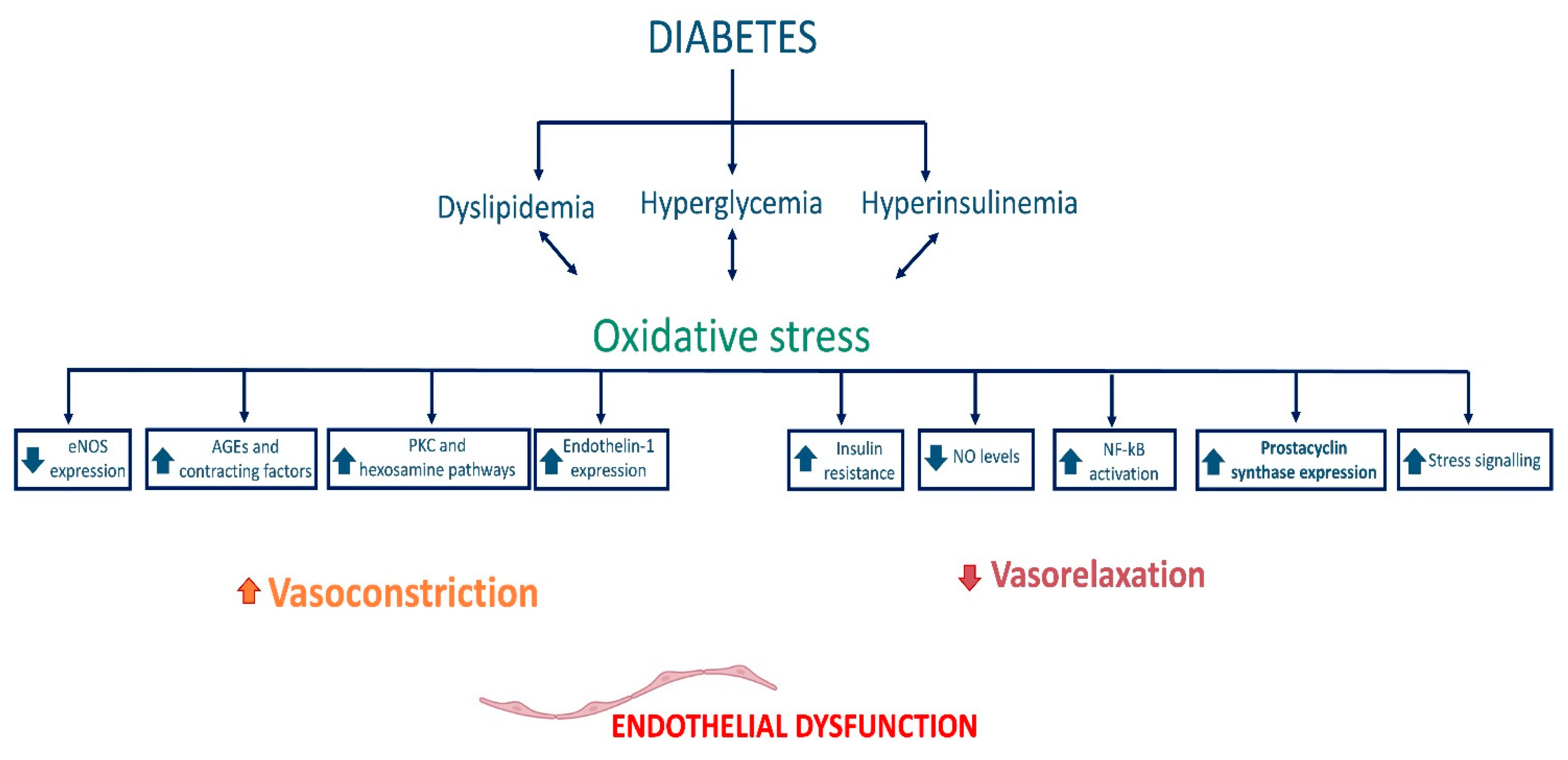
2.2. Endothelial Dysfunction in Diabetes

Type 2 diabetes mellitus (T2D) is a metabolic disease characterized by reduced insulin sensitivity by target organs or reduced insulin production by pancreatic beta cells. Whatever the initial pathophysiologic features, the reduction in insulin sensitivity leads over time to an increase in insulin production to cope, and subsequently to depletion and reduction in pancreatic beta-cell stock due to exhaustion, resulting in hyperglycemia. Certainly, given the pleiotropic effects of insulin, because of the altered glucose metabolism and the effects on lipid metabolism (insulin is an anabolic hormone), T2D is often associated with obesity. However, this association is not always present: there is the nondiabetic obese patient, and there are diabetic subjects who remain at an average weight throughout their life [54].
Undoubtedly, aging promotes the onset of metabolic disorders, and this is the fundamental reason why both T2D and obesity are more frequent in the aging population and why the two diseases often occur together: the senescence that affects other cells in the body does not spare pancreatic beta cells, which become less efficient and more likely to fail as the time passes.
In addition to this, several studies have documented the presence of senescent beta cells in the pancreatic islets of donors with T2D and subjects with high BMI, further demonstrating that several factors, including dysmetabolism, play a crucial role in aging [55].
Hyperglycemia constitutes a harmful condition for the homeostasis of cells, including endothelial cells, and consequently represents the characteristic trigger of diabetes on vascular pathologies. In addition, high serum glucose levels cause functional and metabolic changes through reduced nitric oxide bioavailability, increased oxidative stress, and induction and acceleration of cellular senescence through direct action on the NF-κB pathway.
Under hyperglycemic conditions, the induction of transcription of the gene for the NF-κB factor appears to depend on the activation of molecules of the guanosine triphosphate (GTP)-ases family, which in turn promotes the induction of transcription of inflammatory mediator genes such as IL-1 β, TNF-α, protein kinase C, and adhesion molecules of the cellular adhesion molecule (CAM) family, which facilitate the binding of circulating cells (namely monocytes and T lymphocytes) to endothelial cells.
Atherosclerosis in the diabetic subject is therefore strongly catalyzed by hyperglycemia: on the one hand, inflammation worsens plaque formation as it encourages the oxidation of low-density lipoproteins, and on the other, the reduction in nitric oxide bioavailability, due to sustained endothelial damage, causes hemodynamic alterations and increased shear stress, which are the foundations of vascular pathologies.
In healthy subjects, vasoregulation results from a delicate balance between the vasoactive substances produced by the endothelium and the shear forces exerted by the blood flow. Thanks to continuous feedback, the mechanotransduction of these shear forces allows the endothelium to produce vasodilating substances such as nitric oxide or prostacyclin (PGI2), or vasoconstrictive substances such as endothelin (ET) as required. Sulistyowati et al. documented that hyperglycemia impairs this mechanism, as it disrupts the functioning of endothelial cells by reducing endothelial nitric oxide synthase (eNOS) activity and disrupting mechanotransduction activity, generating the hemodynamic abnormalities discussed above [56].
Concerning nitric oxide, its production in endothelial cells depends on the phosphorylation of the enzyme eNOS by serine/threonine kinases acting coordinately in the insulin signaling pathway; hyperglycemia alters the binding and activation of the insulin receptor, thus leading to an impairment of this molecular mechanism and a reduction of nitroxide [57].
Hyperglycemia impairs endothelial function through a variety of mechanisms: through induction of advanced glycation end products (AGEs) formation, increased expression of receptors for AGEs (RAGE), increased polyol production, and hyperactivation of the hexosamine pathway [58].
The genesis of AGEs causes structural and functional alterations and results in receptor recognition abnormalities of matrix components. In addition, the interaction between ligand (AGE) and its receptor (RAGE) results in increased superoxide anion production that promotes macrophage-mediated inflammatory vascular damage [59][60].
Moreover, the increase in AGEs in the bloodstream reduces nitric oxide synthesis by endothelial cells by reducing the expression of the enzyme eNOS and, consequently, the bioavailability of this vasodilation mediator, conversely facilitating the synthesis of endothelin-1, which mediates vasoconstriction (Figure 1) [61][62].
Figure 1. Pathophysiological elements of vascular damage in T2D, with a focus on the dysregulation of the balance between factors regulating vasoconstriction and vasodilation. Endothelial nitric oxide synthase (eNOS), advanced glycation end products (AGEs), protein kinase C (PKC), nitric oxide (NO), and nuclear factor kappa B (NF-κB).
Regarding polyols and the hexosamine pathway, it should be remembered that hyperglycemia alters glucose metabolism by glycolysis. Therefore, an excess of this metabolite causes the activation of alternative metabolic pathways, such as those mentioned above [63].
In addition, the oxidative stress caused by hyperglycemia induces DNA damage. It promotes the production of ADP-ribose polymer through the activation of nuclear polymerase (PARP), resulting in reduced activity of glyceraldehyde-3-phosphate dehydrogenase, a key enzyme in glucose metabolism, through the cells’ preferred metabolic pathway. This increases the levels of all upstream glycolytic intermediates that trigger the damage mechanisms mentioned above. Oxidative stress, therefore, results from many metabolic alterations that, although established as a result of different preconditions, end up triggering the same abnormalities and cellular damage [58].
The glucose excess and glucotoxicity expressed so far represent part of the determinants of vascular damage, as diabetes mellitus is a dysmetabolic disease “par excellence” and leads to alterations in lipid metabolism: the excess of free fatty acids (FFAs) due to excessive release from adipose tissue and reduced utilization by skeletal striated muscle induces lipotoxicity by molecular mechanisms similar to those already discussed. FFAs induce ROS production by vascular tissue by increasing the expression of the enzyme NADPH oxidase and mitochondrial uncoupling [64].
The increase in the bioavailability of superoxide anion leads to the inactivation of important enzymes with antiatherogenic action, such as eNOS, favoring persistent vasoconstriction; moreover, the consequence of the increase in ROS is a reduction in intracellular glutathione, which constitutes a protective factor of the cell against oxidative damage, so that its reduction is both cause and consequence of oxidative stress itself. Undoubtedly, lipotoxicity has, as a consequence, the activation of the inflammatory cascade and the increase in NF-κB gene transcription, resulting in an increase in inflammation mediators that impacts the vascular district [65].
All the conditions described so far constitute the cornerstone for understanding the relevance of diabetes mellitus as a vascular pathology: metabolic alterations, oxidative stress, and the increased inflammatory response are the pivot and the tip of the iceberg in the pathophysiological cascade of vascular pathologies in the elderly, but the trigger of damage and its perpetuation as an expression of dysmetabolism is the submerged part of the enormous glacier, the element on which to reflect in order to introduce therapies aimed at correcting individual pathways to have a formidable impact on cardiovascular pathology downstream.

2.3. Endothelial Dysfunction in Cerebrovascular Diseases

Endothelial dysfunction is considered to be one of the most relevant pathogenetic factors for vascular diseases, and it certainly is for cerebrovascular diseases. Indeed, atherosclerotic vascular diseases of the brain district are a paradigmatic example of this.
The mechanisms that cause the development of the cerebrovascular disease have similar characteristics to those in other districts: oxidative stress due to increased production of reactive oxygen species, low-density lipoprotein (LDL) oxidation and the evolution of incipient atherosclerotic lesion, chronic inflammation, and alterations in vascular tone play a crucial role. Nevertheless, a morbid condition involving and damaging small cerebral vessels is of recent interest. This condition recognizes endothelial dysfunction as one of its most significant causative factors: age-related cerebral small vessel disease (ArCSVD). Cellular senescence and chronic inflammation are crucial elements in the ageing of the organism and are certainly so in cerebral damage; the deterioration of the central nervous system is continuous and progressive in the elderly, and the decline of the cerebral vascular circulation constitutes an essential element of this disease.
Several factors contribute to the genesis of brain damage in the elderly subject, among them immunosenescence and chronic inflammation. However, at the cellular level, the dysfunctional endothelial cells determine the beginning of those abnormalities that will catalyze neurodegeneration.
Senescent endothelium plays a part in regulating vascular tone, remodeling, and the consequent abnormal activation of the coagulation cascade that characterizes ischemic brain damage [66].
The loss of vascular flow regulation occurs as a consequence of inflammatory damage and excessive oxidative stress, which are critical actors of the ageing organism. Furthermore, there is a reduction in the bioavailability of nitric oxide because various factors (such as arterial hypertension and increased levels of angiotensin II) induce transcriptional and post-transcriptional modifications of the eNOS enzyme by reducing its biological activity; all of this favors an imbalance towards permanent vasoconstriction and thus induces a loss of self-regulation of the cerebral circulation, which is fundamental for brain homeostasis [67].
Additionally, in the brain district, chronic inflammation causes the overexpression of inflammatory mediators such as IL-1 β, IL-6, and TNF-α due to the increased expression of the NF-κB gene, leading to the promotion of oxidative stress and the resulting cellular damage: the accentuated oxidative stress further reduces the expression of the eNOS enzyme and the bioavailability of NO, triggering a vicious circle that worsens and amplifies the cerebral hemodynamic alterations [68].
The macroscopic consequence of what has been said so far is the rarefaction of the cerebral vascular tree. Therefore, the reduction in the perfusion of some regions of the cerebral parenchyma is responsible for the increase in perivascular spaces and the genesis of lacunar infarcts that lead to those ischemic abnormalities that result from vascular remodeling.
In the context of cerebral vascular pathologies, endothelial dysfunction plays another fundamental role: the endotheliocyte is a constituent of the blood–brain barrier (BBB), and the structural and functional damage to this cell results in irreversible damage to the BBB and thus to cerebral homeostasis [69].
Indeed, the pathogenesis of BBB disruption is complex. However, some studies have documented that chronic exposure of the endotheliocyte to increased shear stress, in subjects with hypertension, can result in a loosening of tight junctions and thus an increase in barrier permeability by cells of the bloodstream, especially cells of the immune system (which are also senescent) that can infiltrate the brain and trigger and prolong that state of chronic inflammation that can promote vascular remodeling by the mechanisms described above, since increased permeability allows the passage of antigens and harmful stimuli that should not typically reach the brain district [70][71].

References

  1. Vatner, S.F.; Zhang, J.; Vyzas, C.; Mishra, K.; Graham, R.M.; Vatner, D.E. Vascular Stiffness in Aging and Disease. Front. Physiol. 2021, 12, 762437.
  2. Donato, A.J.; Machin, D.R.; Lesniewski, L.A. Mechanisms of Dysfunction in the Aging Vasculature and Role in Age-Related Disease. Circ. Res. 2018, 123, 825–848.
  3. Filippini, A.; Tamagnone, L.; D’Alessio, A. Endothelial Cell Metabolism in Vascular Functions. Cancers 2022, 14, 1929.
  4. Esper, R.J.; Nordaby, R.A.; Vilariño, J.O.; Paragano, A.; Cacharrón, J.L.; Machado, R.A. Endothelial Dysfunction: A Comprehensive Appraisal. Cardiovasc. Diabetol. 2006, 5, 4.
  5. Herrera, M.D.; Mingorance, C.; Rodríguez-Rodríguez, R.; Alvarez de Sotomayor, M. Endothelial Dysfunction and Aging: An Update. Ageing Res. Rev. 2010, 9, 142–152.
  6. Ungvari, Z.; Tarantini, S.; Kiss, T.; Wren, J.D.; Giles, C.B.; Griffin, C.T.; Murfee, W.L.; Pacher, P.; Csiszar, A. Endothelial Dysfunction and Angiogenesis Impairment in the Ageing Vasculature. Nat. Rev. Cardiol. 2018, 15, 555–565.
  7. Tesauro, M.; Mauriello, A.; Rovella, V.; Annicchiarico-Petruzzelli, M.; Cardillo, C.; Melino, G.; Di Daniele, N. Arterial Ageing: From Endothelial Dysfunction to Vascular Calcification. J. Intern. Med. 2017, 281, 471–482.
  8. Franceschi, C.; Campisi, J. Chronic Inflammation (Inflammaging) and Its Potential Contribution to Age-Associated Diseases. J. Gerontol. A Biol. Sci. Med. Sci. 2014, 69 (Suppl. S1), S4–S9.
  9. López-Otín, C.; Blasco, M.A.; Partridge, L.; Serrano, M.; Kroemer, G. The Hallmarks of Aging. Cell 2013, 153, 1194–1217.
  10. Ataei Ataabadi, E.; Golshiri, K.; Jüttner, A.A.; de Vries, R.; Van den Berg-Garrelds, I.; Nagtzaam, N.M.A.; Khan, H.N.; Leijten, F.P.J.; Brandt, R.M.C.; Dik, W.A.; et al. Soluble Guanylate Cyclase Activator BAY 54-6544 Improves Vasomotor Function and Survival in an Accelerated Ageing Mouse Model. Aging Cell. 2022, 21, e13683.
  11. Golshiri, K.; Ataei Ataabadi, E.; Rubio-Beltran, E.; Dutheil, S.; Yao, W.; Snyder, G.L.; Davis, R.E.; van der Pluijm, I.; Brandt, R.; Van den Berg-Garrelds, I.M.; et al. Selective Phosphodiesterase 1 Inhibition Ameliorates Vascular Function, Reduces Inflammatory Response, and Lowers Blood Pressure in Aging Animals. J. Pharmacol. Exp. Ther. 2021, 378, 173–183.
  12. Brew, B.J.; McArthur, J.C. “A Man Is as Old as His Arteries” (Attributed to Thomas Sydenham, the English Hippocrates). AIDS 2020, 34, 637–639.
  13. Sewduth, R.; Santoro, M.M. “Decoding” Angiogenesis: New Facets Controlling Endothelial Cell Behavior. Front. Physiol. 2016, 7, 306.
  14. Eilken, H.M.; Adams, R.H. Dynamics of Endothelial Cell Behavior in Sprouting Angiogenesis. Curr. Opin. Cell. Biol. 2010, 22, 617–625.
  15. Adams, R.H.; Alitalo, K. Molecular Regulation of Angiogenesis and Lymphangiogenesis. Nat. Rev. Mol. Cell. Biol. 2007, 8, 464–478.
  16. Ramasamy, S.K.; Kusumbe, A.P.; Wang, L.; Adams, R.H. Endothelial Notch Activity Promotes Angiogenesis and Osteogenesis in Bone. Nature 2014, 507, 376–380.
  17. Mühleder, S.; Fernández-Chacón, M.; Garcia-Gonzalez, I.; Benedito, R. Endothelial Sprouting, Proliferation, or Senescence: Tipping the Balance from Physiology to Pathology. Cell. Mol. Life Sci. 2021, 78, 1329–1354.
  18. Gerhardt, H.; Betsholtz, C. Endothelial-Pericyte Interactions in Angiogenesis. Cell. Tissue Res. 2003, 314, 15–23.
  19. Rivard, A.; Fabre, J.E.; Silver, M.; Chen, D.; Murohara, T.; Kearney, M.; Magner, M.; Asahara, T.; Isner, J.M. Age-Dependent Impairment of Angiogenesis. Circulation 1999, 99, 111–120.
  20. Chigogidze, M.; Sharashidze, N.; Pagava, Z.; Taboridze, I. The Correlation of Collateral Circulation and Age during Acute Ischemic Heart Disease. Eur. Sci. J. ESJ 2020, 16, 335.
  21. Sadoun, E.; Reed, M.J. Impaired Angiogenesis in Aging Is Associated with Alterations in Vessel Density, Matrix Composition, Inflammatory Response, and Growth Factor Expression. J. Histochem. Cytochem. 2003, 51, 1119–1130.
  22. Theories and Molecular Basis of Vascular Aging: A Review of the Literature from VascAgeNet Group on Pathophysiological Mechanisms of Vascular Aging—PubMed. Available online: https://pubmed.ncbi.nlm.nih.gov/35955804/ (accessed on 9 November 2022).
  23. Iakovou, E.; Kourti, M. A Comprehensive Overview of the Complex Role of Oxidative Stress in Aging, The Contributing Environmental Stressors and Emerging Antioxidant Therapeutic Interventions. Front. Aging Neurosci. 2022, 14, 827900.
  24. Leyane, T.S.; Jere, S.W.; Houreld, N.N. Oxidative Stress in Ageing and Chronic Degenerative Pathologies: Molecular Mechanisms Involved in Counteracting Oxidative Stress and Chronic Inflammation. Int. J. Mol. Sci. 2022, 23, 7273.
  25. Wagatsuma, A. Effect of Aging on Expression of Angiogenesis-Related Factors in Mouse Skeletal Muscle. Exp. Gerontol. 2006, 41, 49–54.
  26. Wang, H.; Keiser, J.A.; Olszewski, B.; Rosebury, W.; Robertson, A.; Kovesdi, I.; Gordon, D. Delayed Angiogenesis in Aging Rats and Therapeutic Effect of Adenoviral Gene Transfer of VEGF. Int. J. Mol. Med. 2004, 13, 581–587.
  27. Peters, A.; Josephson, K.; Vincent, S.L. Effects of Aging on the Neuroglial Cells and Pericytes within Area 17 of the Rhesus Monkey Cerebral Cortex. Anat. Rec. 1991, 229, 384–398.
  28. Burns, E.M.; Kruckeberg, T.W.; Gaetano, P.K. Changes with Age in Cerebral Capillary Morphology. Neurobiol. Aging 1981, 2, 283–291.
  29. Yamamoto, N.; Oyaizu, T.; Enomoto, M.; Horie, M.; Yuasa, M.; Okawa, A.; Yagishita, K. VEGF and BFGF Induction by Nitric Oxide Is Associated with Hyperbaric Oxygen-Induced Angiogenesis and Muscle Regeneration. Sci. Rep. 2020, 10, 2744.
  30. Jüttner, A.A.; Danser, A.H.J.; Roks, A.J.M. Pharmacological Developments in Antihypertensive Treatment through Nitric Oxide-CGMP Modulation. Adv. Pharmacol. 2022, 94, 57–94.
  31. Chin, L.C.; Kumar, P.; Palmer, J.A.; Rophael, J.A.; Dolderer, J.H.; Thomas, G.P.L.; Morrison, W.A.; Penington, A.J.; Stewart, A.G.; Mitchell, G.M. The Influence of Nitric Oxide Synthase 2 on Cutaneous Wound Angiogenesis. Br. J. Dermatol. 2011, 165, 1223–1235.
  32. Bachetti, T.; Comini, L.; Curello, S.; Bastianon, D.; Palmieri, M.; Bresciani, G.; Callea, F.; Ferrari, R. Co-Expression and Modulation of Neuronal and Endothelial Nitric Oxide Synthase in Human Endothelial Cells. J. Mol. Cell. Cardiol. 2004, 37, 939–945.
  33. Rocha, B.S. The Nitrate-Nitrite-Nitric Oxide Pathway on Healthy Ageing: A Review of Pre-Clinical and Clinical Data on the Impact of Dietary Nitrate in the Elderly. Front. Aging 2021, 2, 778467.
  34. Hodges, N.A.; Suarez-Martinez, A.D.; Murfee, W.L. Understanding Angiogenesis during Aging: Opportunities for Discoveries and New Models. J. Appl. Physiol. (1985) 2018, 125, 1843–1850.
  35. Oliveros, E.; Patel, H.; Kyung, S.; Fugar, S.; Goldberg, A.; Madan, N.; Williams, K.A. Hypertension in Older Adults: Assessment, Management, and Challenges. Clin. Cardiol. 2020, 43, 99–107.
  36. Lloyd-Jones, D.M.; Evans, J.C.; Levy, D. Hypertension in Adults across the Age Spectrum: Current Outcomes and Control in the Community. JAMA 2005, 294, 466–472.
  37. Baeyens, N.; Bandyopadhyay, C.; Coon, B.G.; Yun, S.; Schwartz, M.A. Endothelial Fluid Shear Stress Sensing in Vascular Health and Disease. J. Clin. Investig. 2016, 126, 821–828.
  38. Souilhol, C.; Serbanovic-Canic, J.; Fragiadaki, M.; Chico, T.J.; Ridger, V.; Roddie, H.; Evans, P.C. Endothelial Responses to Shear Stress in Atherosclerosis: A Novel Role for Developmental Genes. Nat. Rev. Cardiol. 2020, 17, 52–63.
  39. Chala, N.; Moimas, S.; Giampietro, C.; Zhang, X.; Zambelli, T.; Exarchos, V.; Nazari-Shafti, T.Z.; Poulikakos, D.; Ferrari, A. Mechanical Fingerprint of Senescence in Endothelial Cells. Nano Lett. 2021, 21, 4911–4920.
  40. Warboys, C.M.; de Luca, A.; Amini, N.; Luong, L.; Duckles, H.; Hsiao, S.; White, A.; Biswas, S.; Khamis, R.; Chong, C.K.; et al. Disturbed Flow Promotes Endothelial Senescence via a P53-Dependent Pathway. Arterioscler. Thromb. Vasc. Biol. 2014, 34, 985–995.
  41. Zhu, S.; Dong, Z.; Ke, X.; Hou, J.; Zhao, E.; Zhang, K.; Wang, F.; Yang, L.; Xiang, Z.; Cui, H. The Roles of Sirtuins Family in Cell Metabolism during Tumor Development. Semin. Cancer Biol. 2019, 57, 59–71.
  42. Factors and Pathways Modulating Endothelial Cell Senescence in Vascular Aging—PubMed. Available online: https://pubmed.ncbi.nlm.nih.gov/36077539/ (accessed on 9 November 2022).
  43. Zhou, S.; Chen, H.-Z.; Wan, Y.-Z.; Zhang, Q.-J.; Wei, Y.-S.; Huang, S.; Liu, J.-J.; Lu, Y.-B.; Zhang, Z.-Q.; Yang, R.-F.; et al. Repression of P66Shc Expression by SIRT1 Contributes to the Prevention of Hyperglycemia-Induced Endothelial Dysfunction. Circ. Res. 2011, 109, 639–648.
  44. Takaishi, K.; Kinoshita, H.; Kawashima, S.; Kawahito, S. Human Vascular Smooth Muscle Function and Oxidative Stress Induced by NADPH Oxidase with the Clinical Implications. Cells 2021, 10, 1947.
  45. Endothelial Dysfunction: Is There a Hyperglycemia-Induced Imbalance of NOX and NOS?—PubMed. Available online: https://pubmed.ncbi.nlm.nih.gov/31382355/ (accessed on 9 November 2022).
  46. Wang, M.; Zhang, J.; Walker, S.J.; Dworakowski, R.; Lakatta, E.G.; Shah, A.M. Involvement of NADPH Oxidase in Age-Associated Cardiac Remodeling. J. Mol. Cell. Cardiol. 2010, 48, 765–772.
  47. Endothelial Dysfunction in Hypertension: Current Concepts and Clinical Implications—PubMed. Available online: https://pubmed.ncbi.nlm.nih.gov/35127755/ (accessed on 9 November 2022).
  48. Sahoo, S.; Meijles, D.N.; Pagano, P.J. NADPH Oxidases: Key Modulators in Aging and Age-Related Cardiovascular Diseases? Clin. Sci. 2016, 130, 317–335.
  49. Fountain, J.H.; Lappin, S.L. Physiology, Renin Angiotensin System. In StatPearls; StatPearls Publishing: Treasure Island, FL, USA, 2022.
  50. Jaimes, E.A.; Galceran, J.M.; Raij, L. Angiotensin II Induces Superoxide Anion Production by Mesangial Cells. Kidney Int. 1998, 54, 775–784.
  51. Yang, J.-X.; Pan, Y.-Y.; Wang, X.-X.; Qiu, Y.-G.; Mao, W. Endothelial Progenitor Cells in Age-Related Vascular Remodeling. Cell. Transplant. 2018, 27, 786–795.
  52. Umemura, T.; Higashi, Y. Endothelial Progenitor Cells: Therapeutic Target for Cardiovascular Diseases. J. Pharmacol. Sci. 2008, 108, 1–6.
  53. Benetos, A.; Petrovic, M.; Strandberg, T. Hypertension Management in Older and Frail Older Patients. Circ. Res. 2019, 124, 1045–1060.
  54. Eckel, R.H.; Kahn, S.E.; Ferrannini, E.; Goldfine, A.B.; Nathan, D.M.; Schwartz, M.W.; Smith, R.J.; Smith, S.R. Obesity and Type 2 Diabetes: What Can Be Unified and What Needs to Be Individualized? J. Clin. Endocrinol. Metab. 2011, 96, 1654–1663.
  55. Aguayo-Mazzucato, C.; Andle, J.; Lee, T.B.; Midha, A.; Talemal, L.; Chipashvili, V.; Hollister-Lock, J.; van Deursen, J.; Weir, G.; Bonner-Weir, S. Acceleration of β Cell Aging Determines Diabetes and Senolysis Improves Disease Outcomes. Cell. Metab. 2019, 30, 129–142.e4.
  56. Sulistyowati, E.; Permatasari, N.; Aris Widodo, M. Combined Effects of Shear Stress and Glucose on the Morphology, Actin Filaments, and VE-Cadherin of Endothelial Cells in Vitro. Int. J. Cardiol. Heart Vasc. 2017, 15, 31–35.
  57. Saltiel, A.R.; Kahn, C.R. Insulin Signalling and the Regulation of Glucose and Lipid Metabolism. Nature 2001, 414, 799–806.
  58. Brownlee, M. The Pathobiology of Diabetic Complications: A Unifying Mechanism. Diabetes 2005, 54, 1615–1625.
  59. Schmidt, A.M.; Yan, S.D.; Wautier, J.L.; Stern, D. Activation of Receptor for Advanced Glycation End Products: A Mechanism for Chronic Vascular Dysfunction in Diabetic Vasculopathy and Atherosclerosis. Circ. Res. 1999, 84, 489–497.
  60. Chavakis, T.; Bierhaus, A.; Nawroth, P.P. RAGE (Receptor for Advanced Glycation End Products): A Central Player in the Inflammatory Response. Microbes Infect. 2004, 6, 1219–1225.
  61. Chakravarthy, U.; Hayes, R.G.; Stitt, A.W.; McAuley, E.; Archer, D.B. Constitutive Nitric Oxide Synthase Expression in Retinal Vascular Endothelial Cells Is Suppressed by High Glucose and Advanced Glycation End Products. Diabetes 1998, 47, 945–952.
  62. Xu, B.; Chibber, R.; Ruggiero, D.; Kohner, E.; Ritter, J.; Ferro, A.; Ruggerio, D. Impairment of Vascular Endothelial Nitric Oxide Synthase Activity by Advanced Glycation End Products. FASEB J. 2003, 17, 1289–1291.
  63. Brownlee, M. Biochemistry and Molecular Cell Biology of Diabetic Complications. Nature 2001, 414, 813–820.
  64. Inoguchi, T.; Li, P.; Umeda, F.; Yu, H.Y.; Kakimoto, M.; Imamura, M.; Aoki, T.; Etoh, T.; Hashimoto, T.; Naruse, M.; et al. High Glucose Level and Free Fatty Acid Stimulate Reactive Oxygen Species Production through Protein Kinase C—Dependent Activation of NAD(P)H Oxidase in Cultured Vascular Cells. Diabetes 2000, 49, 1939–1945.
  65. Boden, G.; She, P.; Mozzoli, M.; Cheung, P.; Gumireddy, K.; Reddy, P.; Xiang, X.; Luo, Z.; Ruderman, N. Free Fatty Acids Produce Insulin Resistance and Activate the Proinflammatory Nuclear Factor-KappaB Pathway in Rat Liver. Diabetes 2005, 54, 3458–3465.
  66. Buford, T.W. Hypertension and Aging. Ageing Res. Rev. 2016, 26, 96–111.
  67. Del Cuore, A.; Pacinella, G.; Riolo, R.; Tuttolomondo, A. The Role of Immunosenescence in Cerebral Small Vessel Disease: A Review. Int. J. Mol. Sci. 2022, 23, 7136.
  68. Pierce, G.L.; Lesniewski, L.A.; Lawson, B.R.; Beske, S.D.; Seals, D.R. Nuclear Factor-κB Activation Contributes to Vascular Endothelial Dysfunction via Oxidative Stress in Overweight/Obese Middle-Aged and Older Humans. Circulation 2009, 119, 1284–1292.
  69. Wardlaw, J.M.; Sandercock, P.a.G.; Dennis, M.S.; Starr, J. Is Breakdown of the Blood-Brain Barrier Responsible for Lacunar Stroke, Leukoaraiosis, and Dementia? Stroke 2003, 34, 806–812.
  70. Henning, E.C.; Warach, S.; Spatz, M. Hypertension-Induced Vascular Remodeling Contributes to Reduced Cerebral Perfusion and the Development of Spontaneous Stroke in Aged SHRSP Rats. J. Cereb. Blood Flow Metab. 2010, 30, 827–836.
  71. Montagne, A.; Barnes, S.R.; Sweeney, M.D.; Halliday, M.R.; Sagare, A.P.; Zhao, Z.; Toga, A.W.; Jacobs, R.E.; Liu, C.Y.; Amezcua, L.; et al. Blood-Brain Barrier Breakdown in the Aging Human Hippocampus. Neuron 2015, 85, 296–302.
More
Information
Contributors MDPI registered users' name will be linked to their SciProfiles pages. To register with us, please refer to https://encyclopedia.pub/register : , ,
View Times: 257
Revisions: 2 times (View History)
Update Date: 31 Jan 2023
1000/1000