Submitted Successfully!
To reward your contribution, here is a gift for you: A free trial for our video production service.
Thank you for your contribution! You can also upload a video entry or images related to this topic.
Version Summary Created by Modification Content Size Created at Operation
1 -- 2852 2023-01-20 15:17:31 |
2 update references and layout Meta information modification 2852 2023-01-28 03:27:55 |

Video Upload Options

Do you have a full video?

Confirm

Are you sure to Delete?
Cite
If you have any further questions, please contact Encyclopedia Editorial Office.
Barreto, M.E.V.;  Medeiros, R.P.;  Shearer, A.;  Fook, M.V.L.;  Montazerian, M.;  Mauro, J.C. Gelatin and Bioactive Glass Composites. Encyclopedia. Available online: https://encyclopedia.pub/entry/40456 (accessed on 29 March 2024).
Barreto MEV,  Medeiros RP,  Shearer A,  Fook MVL,  Montazerian M,  Mauro JC. Gelatin and Bioactive Glass Composites. Encyclopedia. Available at: https://encyclopedia.pub/entry/40456. Accessed March 29, 2024.
Barreto, Maria E. V., Rebeca P. Medeiros, Adam Shearer, Marcus V. L. Fook, Maziar Montazerian, John C. Mauro. "Gelatin and Bioactive Glass Composites" Encyclopedia, https://encyclopedia.pub/entry/40456 (accessed March 29, 2024).
Barreto, M.E.V.,  Medeiros, R.P.,  Shearer, A.,  Fook, M.V.L.,  Montazerian, M., & Mauro, J.C. (2023, January 20). Gelatin and Bioactive Glass Composites. In Encyclopedia. https://encyclopedia.pub/entry/40456
Barreto, Maria E. V., et al. "Gelatin and Bioactive Glass Composites." Encyclopedia. Web. 20 January, 2023.
Gelatin and Bioactive Glass Composites
Edit

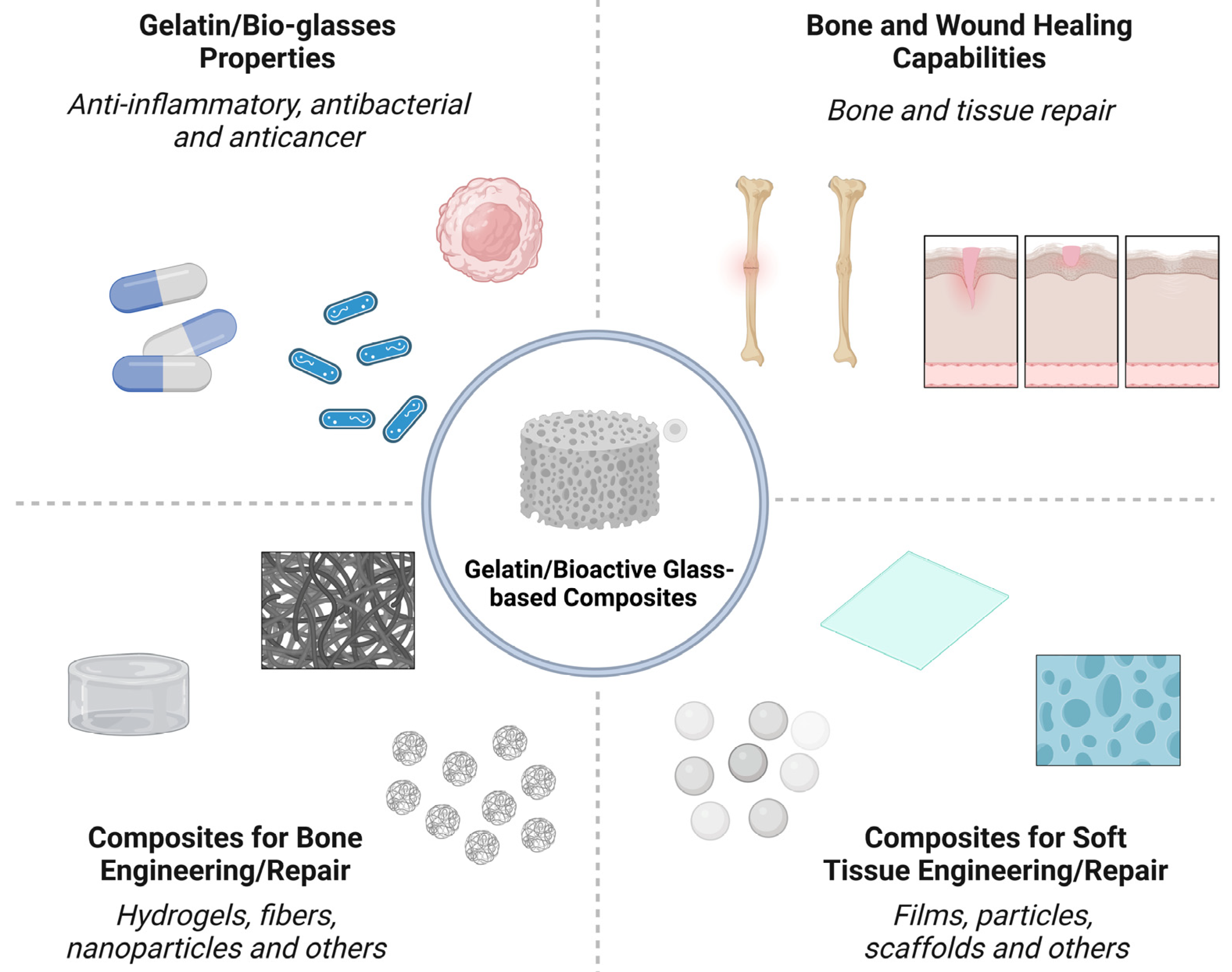
Nano-/micron-sized bioactive glass (BG) particles are attractive candidates for both soft and hard tissue engineering. They can chemically bond to the host tissues, enhance new tissue formation, activate cell proliferation, stimulate the genetic expression of proteins, and trigger unique anti-bacterial, anti-inflammatory, and anti-cancer functionalities. Composites based on biopolymers and BG particles have been developed with various state-of-the-art techniques for tissue engineering. Gelatin, a semi-synthetic biopolymer, has attracted the attention of researchers because it is derived from the most abundant protein in the body, viz., collagen. It is a polymer that can be dissolved in water and processed to acquire different configurations, such as hydrogels, fibers, films, and scaffolds.

bioactive glass gelatin tissue engineering bone

1. Introduction

Composites based on gelatin and bioactive glass (BG) with different morphologies and compositions have been designed to assist in the treatment of tissue injuries, aiming at the aesthetic and functional recovery of damaged limbs [1][2]. In general, these materials must be biocompatible and/or biodegradable, have mechanical strength comparable to that of the host tissue, and allow cellular activity at the implant site [3]. In addition, it is desirable that gelatin/BG composites overcome the clinical and socioeconomic limitations associated with the use of conventional applications [4]. The properties, applications, and processing of gelatin/BG composites are summarized in Figure 1.
Figure 1. Properties, applications, and processing of gelatin/BG composites.
An intrinsic characteristic of soft and hard tissues is the ability to induce regeneration mechanisms [5][6]. In bone, osteoblastic cells initiate the process from the secretion of collagen and, subsequently, the crystallization of hydroxyapatite [7]. Skin, which is the largest organ in the body, induces wound repair through the recruitment of cells that perform angiogenesis and form granulation issue, culminating in wound re-epithelialization [8].
However, the natural mechanisms of tissue recovery are restricted by the type, extent, and/or depth of the lesion, as well as by the interference of microorganisms that cause infections [9][10]. In the first case, the severity of the injury would require extensive time for tissue remodeling to occur only by induction of the organism. This could lead to residual defects, such as the formation of fibrous tissue where tissues should grow [11].
In the second hypothesis, the invasion of infectious agents during or after the implantation would impair the patient’s recovery and increase the costs associated with hospitalization and clinical procedures [12]. In the United States, annual wound repair costs exceed $50 billion [13]. Worldwide, the impacts reach enormous proportions. These numbers reinforce the demand for new products and therapeutic approaches. Added to this is the evident expansion of the biomaterials market, estimated at $106.5 billion in a 2019 evaluation [14].
The development of composites and hybrids based on gelatin and bio-glass to solve tissue engineering issues has been reported since the early 2000s [15][16][17][18]. For bone tissue, there is evidence that these materials promote more effective mineralization compared to other types of treatment [19][20]. The main justification reported for this is the chemical similarity between the composite and the organic and inorganic phases in the bone structure; thus making possible a greater reactivity with the host tissue when the composite is implanted [21]. In addition, the mechanical performance achieved with the union of the polymer with the ceramic approximates the degradation time of the material to the speed of bone formation [22].
Regarding soft tissue applications, the gelatin/BG combination has accelerated wound closure, stimulating increased angiogenesis and granulation [23]. This is due to the presence of bioactive ions capable of reducing the inflammatory response and stimulating the secretion of proteins and growth factors [24]. For example, the presence of Si promotes an upregulation in the expression of vascular endothelial growth factor (VEGF), and the difference between the results achieved only with the polymeric matrix after the addition of BG is evident [25]. In this scenario, there are many possibilities for the application of products based on gelatin and BG including: treatment of subcutaneous and cutaneous lesions [23][26], chronic wounds [27], nerve regeneration [28], muscles [29], cartilage [30] and others.
Although both gelatin and bioactive glasses have been studied extensively for their impact on both hard and soft tissue repair, many limitations remain to be addressed. Gelatin requires crosslinking of its chains to obtain the required stability in physiological environments [31][32]. For example, the thermostability of gelatin without crosslinking can change the structure from a solid to a gel which can lead to adverse effects when implanted in vivo [33][34]. The field of bioactive glass has received an overwhelming amount of attention, with hundreds of publications being published per year. All aspects including chemistry, processing, and application, have been studied, but such a broad field has left many gaps to be explored. Compositional design requires optimization depending on the desired application. For example, SiO2-based compositions are less suited for soft tissue regeneration than B2O3 or P2O5 [35]. Processing methods such as sol-gel synthesis have yet to be commercialized and less than 26 BG-based medical devices have been approved for clinical use [36].

2. Bone Engineering

Bone tissue is one of the largest systems present in living organisms [37]. It differs from other tissues in that it is in a constant process of reconstruction, as some parts of the bone are absorbed, others are excreted and/or remodeled as a result of the dynamics of osteoblastic, osteolytic, and osteoclastic cells [7][11][37].
Bone is a natural composite, with about 70% of its composition consisting of inorganic phases based on calcium and phosphate salts. The other fraction is organic, predominately composed of type I collagen, but proteins such as proteoglycans and glycoproteins are also present. The tissue morphology is also heterogenous as it consists of some compact/dense regions (cortical bone) and other porous/spongy (trabecular bone) regions [1][38][39].

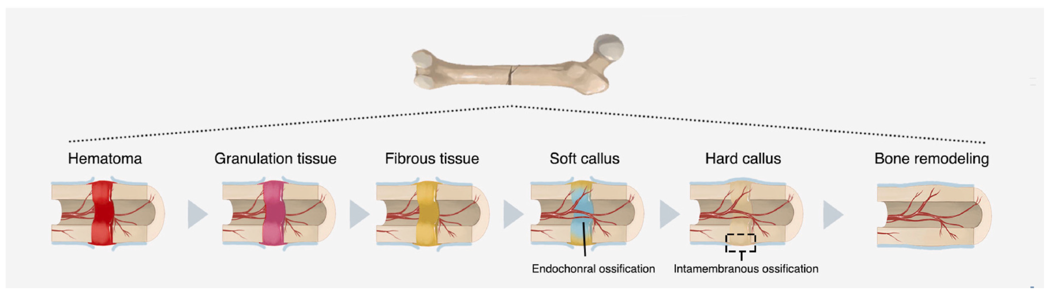
2.1. Bone Healing Mechanisms

When bone is damaged, whether as a result of bone loss, fractures, disease, or any other type of injury, phenomena such as hemorrhage, matrix destruction and cell death occur. From this, the regeneration process can be summarized in three continuous and simultaneous phases: inflammation, regeneration, and remodeling [5][11].
Initially, macrophages eliminate cellular and tissue debris. Then, new osteoprogenitor cells begin to proliferate, forming connective tissue, “glue”, between the ends of the injured region [7]. Gradually, a “bone callus” is formed at the site (Figure 2), which is replaced by a secondary structure similar in shape to the one that previously existed [37].
Figure 2. Natural process of bone repair in the fracture zone. Adapted from Zhu et al. [7].
The first phase of bone formation itself occurs when osteoblasts secrete collagen molecules and proteoglycans [7]. It is assumed that after these steps, salt deposition begins, culminating in the final product known as hydroxyapatite, with the chemical formula Ca10(PO4)6(OH)2 [39][40].
The subsequent stage is the polymerization of the excreted monomers, resulting in an osteoid, which consists of a non-mineralized matrix whose texture is similar to cartilage. Gradually, the calcium and phosphate particles deposited on the collagen matrix multiply and are distributed throughout the tissue, converting into hydroxyapatite crystals over the course of a few days or weeks. This structural characteristic confers bone tissue’s high tenacity and compressive strength [37][39][41].
The regeneration processes described above are expected to occur without the intervention of fibrous tissue and undesirable microorganisms such as bacteria. This is an important issue to be considered, given that the injuries caused to hard tissue also directly impact the socioeconomic system due to the costs of hospitalization, clinical procedures, surgeries and work disability in some cases [42].

2.2. Orthopedic Clinical Challenges

For hundreds of years, prosthetic implants utilized metals and their alloys with a primary emphasis on titanium, cobalt-chromium, and stainless steel. These metals had good mechanical performance but were subject to corrosion and lacked osteointegration [10][14][43][44]. From the end of the 20th century, with the creation of the tissue engineering concept, studies were directed towards the search for materials that exhibit chemical similarity with the tissue, maintain the mechanical stability of the host and lead the tissue regeneration process [43][45]. However, orthopedic problems still represent an emerging and global issue. In the 2019 World Health Organization report, injuries caused by trauma occupy the second position in the ranking of the main causes of death in the world [46].
Critical-sized bone defect healing represents one of the most significant unmet obstacles in bone regeneration. Originally, bone grafting was used to repair defects caused by tumors, traumatic fractures, and other types of injuries [41]. However, the technique has limitations associated with prohibitive costs and potential damage to health, resulting from infection, inflammation, or immunological rejection at the implant site [5][47].
Biomaterials are used to repair these defects and restore structure and function, often by acting as a substitute for the missing bone. The optimal characteristics for such biomaterials may differ significantly depending on the location of the bone defect and the kind of bone loss (cortical versus cancellous). If a soft biomaterial (e.g., gelatin/BGs composites) is used to fill the cortical lesion instead, a stable plate fixation is necessary to provide mechanical stability. In such a circumstance, the patient will need to be able to move around freely, which requires a rapid change of the softer biomaterial into cortical bone. In most cases, implant loosening or fatigue failure should not occur until after bone growth and consolidation have occurred. If this race is lost, incomplete osteosynthesis leads to nonunion and implant failure [48][49][50].
Bone loss or resection due to a tumor or infection can also result in critical-sized defects. Bone replacement is an integral aspect of treatment in these scenarios. It would be beneficial if a biomaterial could deliver substances that cure the underlying disease that causes bone loss. This functionalization of biomaterials may become one of the most important progresses in biomaterials research. Treatment for bone abnormalities following infection typically entails two or more phases of revision surgery, with antibiotic-loaded bone cement spacers used between procedures. In this case, a vascularized fibular graft is used to bypass the donor site morbidity of the autologous bone graft by using a biomaterial with bone regeneration capabilities for large defects and the elution of antibiotics [51][52][53].
Another problem is bone abnormalities in seniors because of low-impact fractures. Significant deformities sometimes result from several fractures in these people, with the underlying cause often being an osteopenic bone weakness. A commuted fracture most often occurs in the proximal femur, proximal humerus, or vertebral body. Limited bone quality in the remaining bone makes rigid fracture fixation by standard instrumentation difficult. Bone grafting, either autologous or allogeneic, is frequently used to repair these types of abnormalities, which can lead to arthroplasty in the future. Methods of enhancing bone regeneration are desperately needed considering the aging of the population and the rise in late-life activity. Given this, it is easy to appreciate the pressing need for novel therapies that give surgeons the tools they need to facilitate rapid and reliable bone regeneration in their patients [54][55][56].

3. Soft Tissues Engineering

Soft tissues are present in all organs that make up the body, being distinguished into four types: epithelial, connective, muscular and nervous. The epithelial tissue (or epithelium) lines the surfaces and body cavities and has the function of secreting substances. Connective tissue is located below the others, acting to support and sustain them. Muscle tissue, in turn, is responsible for body movements induced by cells capable of contracting. Nervous tissue establishes the connection between external and internal stimuli to the organism, enabling the performance of activities with different levels of complexity [37][40]. In this section, skin lesions, which predominantly consider the epithelial and connective tissues, will be discussed in greater depth.
The skin is considered the largest organ in the body. It plays an immunological role, as it acts as a mechanical, physical and chemical barrier, protecting internal structures against infections and injuries of different nature, such as cuts, traumas, burns and ulcers [57]. In addition to functioning as an “envelope” for the body, the skin regulates moisture loss and changes in body temperature while also acting as natural mechanism to promote the reconstitution of its structure when damaged, which makes up the wound healing cascade [6][58].
The skin’s immune mechanism can be subdivided into two parts that are connected to each other and synchronized with the body’s immune system as a whole: the epidermal region and the dermal region. Both generate a favorable environment for the performance of immune cells, but also coexist cells responsible for continuous tissue maintenance and regeneration. Fibroblasts stand out as a predominant lineage in connective tissues in general, whose functions include locomotion capacity, collagen fiber production and extracellular matrix (ECM) renewal [59].

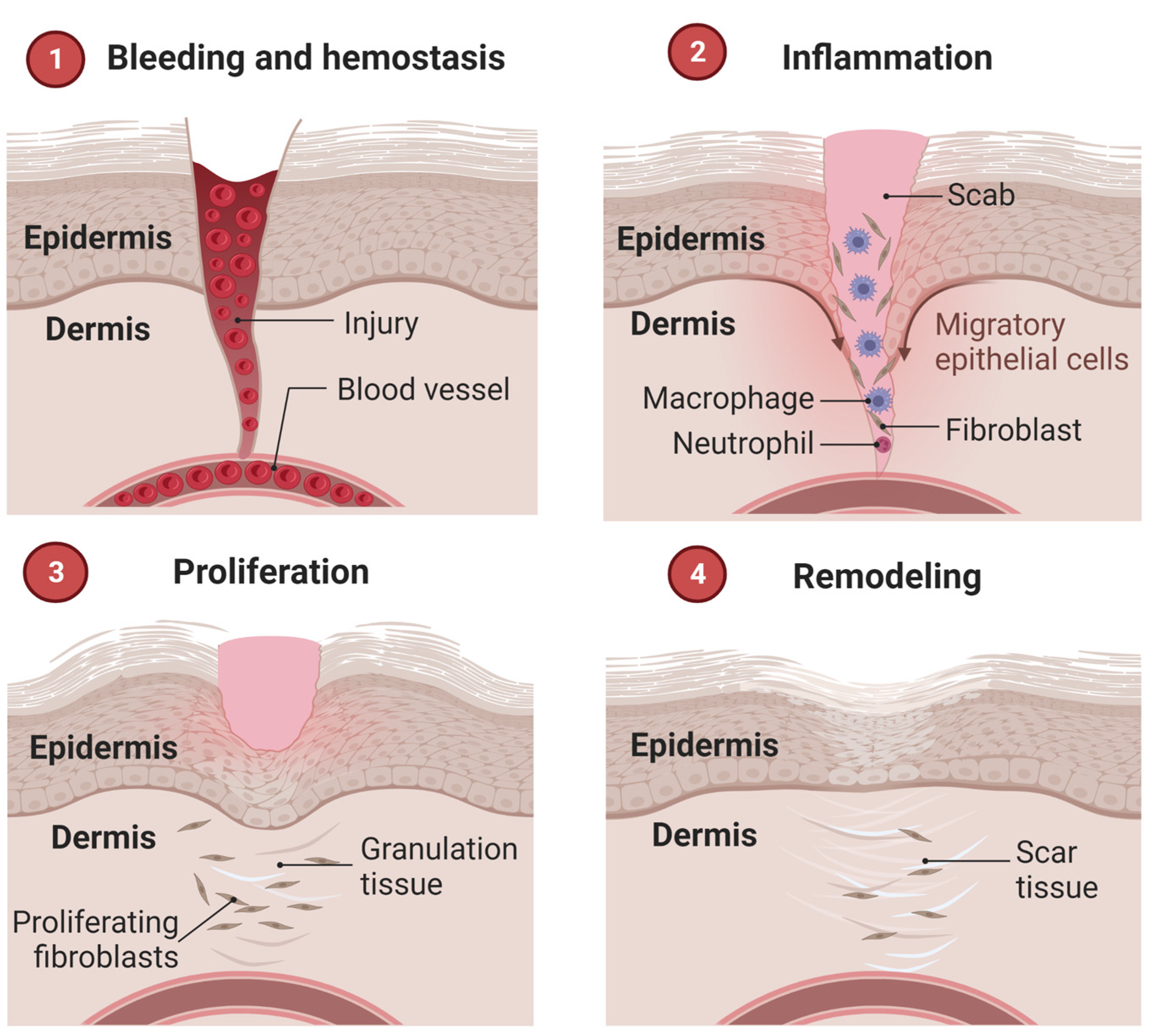
3.1. Wound Healing Mechanisms

The wound healing process occurs in well-defined phases, involving different cell types and metabolisms. Three overlapping steps are known: inflammation, proliferation, and remodeling [8][60]. Some classifications consider separately a hemostasis stage, totaling four, briefly described below and illustrated in Figure 3. The initial stage precedes inflammation and results in bleeding interruption from clot formation (hemostasis). In this scenario, activated platelets secrete cytokines that attract inflammatory cells and other populations to the wound site [58].
Figure 3. Stages of the wound healing cascade.
The inflammatory phase occurs during the initial stages of recovery after an injury, protecting it from pathogens. It is characterized by the secretion of growth factors from inflammatory cells, which stimulate the proliferation of vascular endothelial cells and fibroblasts. The latter produces type III collagen that replaces the fibrin matrix. Other cells are recruited to the site of injury at the same time, including neutrophils, monocytes, mast cells, and other non-inflammatory categories that actively contribute to the healing flow and protection against bacteria and antigens [61][62].
From cell proliferation, which consists of the second stage, angiogenesis and granulation tissue formation begin, followed by wound re-epithelialization. When there is a deficiency in the speed of cell proliferation and, consequently, in the deposition of collagen, the healing process exceeds the expected period. According to this criterion, wounds are classified as acute when recovered between 8 and 12 weeks, and chronic when healing is delayed or does not occur [13][63].
Finally, remodeling and/or maturation occurs, which can last up to two years after the appearance of the lesion. It is characterized by the gradual replacement of type III collagen by type I collagen, which generates a more rigid structure at the wound site and forms scar tissue [64].

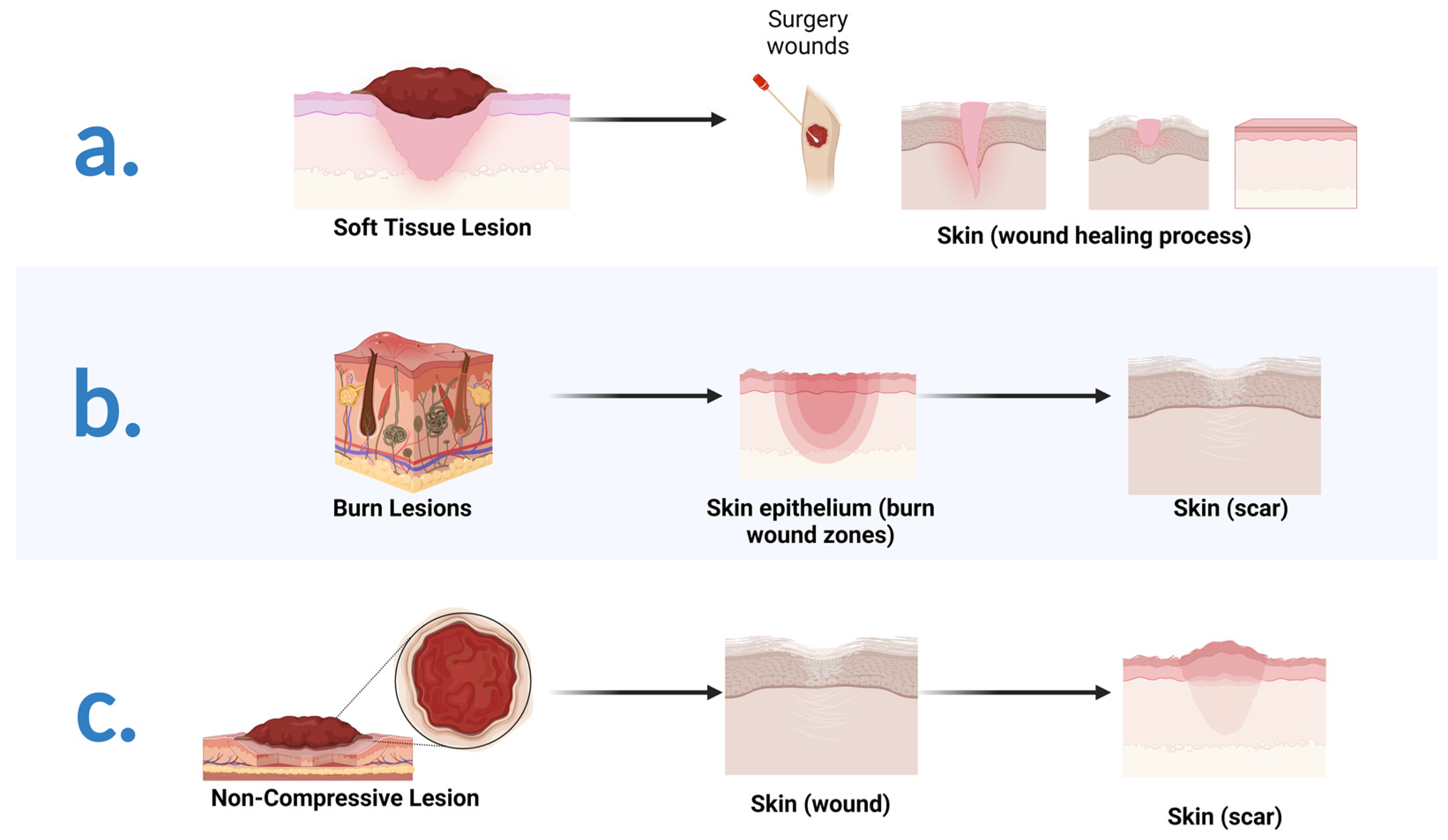
3.2. Therapeutic Approaches in Wound Repair: A Brief Introduction

To repair injuries caused to soft tissues (Figure 4a) as a result of trauma, diseases and/or accidents, one of the most used practices over time is grafting, as for hard tissue. For wound care, costs exceed $50 billion annually to serve more than 5.7 million people in the United States alone [13]. When the wound does not heal on its own, standard therapy includes debridement and skin grafting once the granulation tissue has formed [65].
Figure 4. Typical injuries caused to soft tissue and healing processes: (a) Lesions caused by traumas, diseases, and/or accidents; (b) Lesions caused by burns; (c) Non-compressive lesions caused by sharp objects and/or firearms.
However, autologous grafts can trigger complications such as infections in the postoperative phase, immunological rejection, absorption, and loss of volume. In addition, this technique is associated with a decrease in mechanical resistance, which can lead to graft failure and generate a severe scar contracture. For this reason, the scientific community in the field of tissue engineering has been dedicated to the development of systems capable of regenerating and restoring the functionality of these tissues, overcoming the limitations of practices already in use [4].
Until the 20th century, the treatment of burn wounds (Figure 4b) had many limitations, commonly resulting in the patient’s death due to poor wound care management. Pharmacotherapy strategies have advanced, but challenges in treating soft tissue injuries remain. As already stated, infection is a predominant issue, whether endogenous or exogenous. The multiplication of microorganisms, primarily facilitated by overly moist wound environments or delays in healing, prolongs the hospitalization period driving costs higher for the healthcare system [9][58].
A particular problem is deep wounds generated by trauma that cause uncontrolled bleeding. Currently, hemorrhage is the cause of more than 30% of deaths from trauma worldwide due to the difficulty of providing the patient with immediate intervention and prior to hospital care [24].
Especially in cases of non-compressible injuries (Figure 4c), such as those caused by sharp objects and/or firearms, conventional dressing methods and direct pressure are inefficient, which reinforces the demand for hemostatic agents capable of stopping acute bleeding and promoting healing, minimizing the risk of bacterial colonization at the wound site [66].
Additionally, the type of injury is also a limiting factor to allow healing by conventional methods. In the case of extensive and/or deep wounds, there is a shortage of healthy tissue available for autogenous grafting; that is, those whose source of extraction is the patient themself. In the cases of large tissue loss, the wound does not heal by primary intention, measured by approximation by edges of the suture [63]. These instances require intensive care to promote secondary intention where granulation grows at the edge of the open wound. To meet this demand, new approaches have been explored including implants, dressings, artificial organs and living tissue, which are created by growing cells in scaffolds before insertion into the body [13][61]. Among the candidate materials for repairing soft and hard tissue injuries, composites based on gelatin and bioactive glass have been extensively explored. Their potential is thoroughly discussed after briefly addressing the gelatin and BGs characteristics.

References

  1. Sonatkar, J.; Kandasubramanian, B. Bioactive glass with biocompatible polymers for bone applications. Eur. Polym. J. 2021, 160, 110801.
  2. Kargozar, S.; Mozafari, M.; Ghenaatgar-Kasbi, M.; Baino, F. Bioactive glasses and glass/polymer composites for neuroregeneration: Should we be hopeful? Appl. Sci. 2020, 10, 3421.
  3. Sergi, R.; Bellucci, D.; Cannillo, V. A review of bioactive glass/natural polymer composites: State of the art. Materials 2020, 13, 5560.
  4. Van Damme, L.; Blondeel, P.; Van Vlierberghe, S. Injectable biomaterials as minimal invasive strategy towards soft tissue regeneration—An overview. J. Phys. Mater. 2021, 4, 022001.
  5. Newman, H.; Shih, Y.V.; Varghese, S. Resolution of inflammation in bone regeneration: From understandings to therapeutic applications. Biomaterials 2021, 277, 121114.
  6. Leong, C.; Gouliouris, T. Skin and soft tissue infections. Medicine 2021, 49, 699–705.
  7. Zhu, G.; Zhang, T.; Chen, M.; Yao, K.; Huang, X.; Zhang, B.; Li, Y.; Liu, J.; Wang, Y.; Zhao, Z. Bone physiological microenvironment and healing mechanism: Basis for future bone-tissue engineering scaffolds. Bioact. Mater. 2021, 6, 4110–4140.
  8. Masson-Meyers, D.S.; Tayebi, L. Vascularization strategies in tissue engineering approaches for soft tissue repair. J. Tissue Eng. Regen. Med. 2021, 15, 747–762.
  9. Markiewicz-Gospodarek, A.; Kozioł, M.; Tobiasz, M.; Baj, J.; Radzikowska-Büchner, E.; Przekora, A. Burn Wound Healing: Clinical Complications, Medical Care, Treatment, and Dressing Types: The Current State of Knowledge for Clinical Practice. Int. J. Environ. Res. Public Health 2022, 19, 1338.
  10. Kim, T.; See, C.W.; Li, X.; Zhu, D. Orthopedic implants and devices for bone fractures and defects: Past, present and perspective. Eng. Regen. 2020, 1, 6–18.
  11. Safari, B.; Davaran, S.; Aghanejad, A. Osteogenic potential of the growth factors and bioactive molecules in bone regeneration. Int. J. Biol. Macromol. 2021, 175, 544–557.
  12. Ministério da Saúde (DATASUS). Available online: http://www2.datasus.gov.br/DATASUS/index.php?area=0203&id=6926&VObj=http://tabnet.datasus.gov.br/cgi/deftohtm.exe?sih/cnv/ni (accessed on 30 January 2022).
  13. Araujo, T.A.T.; Almeida, M.C.; Avanzi, I.; Parisi, J.; Simon Sales, A.F.; Na, Y.; Renno, A. Collagen membranes for skin wound repair: A systematic review. J. Biomater. Appl. 2020, 36, 95–112.
  14. Pratim Das, P.; Aditya Bachchan, A.; Sahu, R.; Chaudhary, V. Whole body vibration: Effects on human body and role of biomaterials in repairing fracture joints and tissues. Mater. Today Proc. 2021, 43, 141–147.
  15. Ren, L.; Wang, Y.; Chen, X.; Zhao, Y. Preparation of the porous scaffolds of chitosan gelatin/APTES modified bioglass. Fuhe Cailiao Xuebao/Acta Mater. Compos. Sin. 2009, 26, 47–52.
  16. Peter, M.; Binulal, N.S.; Nair, S.V.; Selvamurugan, N.; Tamura, H.; Jayakumar, R. Novel biodegradable chitosan-gelatin/nano-bioactive glass ceramic composite scaffolds for alveolar bone tissue engineering. Chem. Eng. J. 2010, 158, 353–361.
  17. Koudehi, M.F.; Fooladi, A.A.I.; Mansoori, K.; Jamalpoor, Z.; Amiri, A.; Nourani, M.R. Preparation and evaluation of novel nano-bioglass/gelatin conduit for peripheral nerve regeneration. J. Mater. Sci. Mater. Med. 2014, 25, 363–373.
  18. Liang, W.; Wu, X.; Dong, Y.; Shao, R.; Chen, X.; Zhou, P.; Xu, F. In vivo behavior of bioactive glass-based composites in animal models for bone regeneration. Biomater. Sci. 2021, 9, 1924–1944.
  19. Lao, J.; Dieudonné, X.; Fayon, F.; Montouillout, V.; Jallot, E. Bioactive glass-gelatin hybrids: Building scaffolds with enhanced calcium incorporation and controlled porosity for bone regeneration. J. Mater. Chem. B 2016, 4, 2486–2497.
  20. Abd El-Aziz, A.M.; Abd El-Fattah, A.; El-Maghraby, A.; Ghareeb, D.A.; Kandil, S. Viscoelasticity, mechanical properties, and in vitro bioactivity of gelatin/borosilicate bioactive glass nanocomposite hydrogels as potential scaffolds for bone regeneration. Polymers 2021, 13, 2014.
  21. Gupta, N.; Santhiya, D. In situ mineralization of bioactive glass in gelatin matrix. Mater. Lett. 2017, 188, 127–129.
  22. Jain, S.; Gujjala, R.; Abdul Azeem, P.; Ojha, S.; Samudrala, R.K. A review on mechanical and In-vitro studies of polymer reinforced bioactive glass-scaffolds and their fabrication techniques. Ceram. Int. 2022, 48, 5908–5921.
  23. Sharifi, E.; Sadati, S.A.; Yousefiasl, S.; Sartorius, R.; Zafari, M.; Rezakhani, L.; Alizadeh, M.; Nazarzadeh Zare, E.; Omidghaemi, S.; Ghanavatinejad, F.; et al. Cell loaded hydrogel containing Ag-doped bioactive glass–ceramic nanoparticles as skin substitute: Antibacterial properties, immune response, and scarless cutaneous wound regeneration. Bioeng. Transl. Med. 2022, 7, e10386.
  24. Yao, L.; Gao, H.; Lin, Z.; Dai, Q.; Zhu, S.; Li, S.; Liu, C.; Feng, Q.; Li, Q.; Wang, G.; et al. A shape memory and antibacterial cryogel with rapid hemostasis for noncompressible hemorrhage and wound healing. Chem. Eng. J. 2022, 428, 131005.
  25. Afghah, F.; Iyison, N.B.; Nadernezhad, A.; Midi, A.; Sen, O.; Saner Okan, B.; Culha, M.; Koc, B. 3D Fiber Reinforced Hydrogel Scaffolds by Melt Electrowriting and Gel Casting as a Hybrid Design for Wound Healing. Adv. Healthc. Mater. 2022, 11, 2102068.
  26. Ma, W.; Yang, X.; Ma, L.; Wang, X.; Zhang, L.; Yang, G.; Han, C.; Gou, Z. Fabrication of bioactive glass-introduced nanofibrous membranes with multifunctions for potential wound dressing. RSC Adv. 2014, 4, 60114–60122.
  27. Chen, Y.-H.; Rao, Z.-F.; Liu, Y.-J.; Liu, X.-S.; Liu, Y.-F.; Xu, L.-J.; Wang, Z.-Q.; Guo, J.-Y.; Zhang, L.; Dong, Y.-S.; et al. Multifunctional Injectable Hydrogel Loaded with Cerium-Containing Bioactive Glass Nanoparticles for Diabetic Wound Healing. Biomolecules 2021, 11, 702.
  28. Foroutan Koudehi, M.; Imani Fooladi, A.A.; Aghozbeni, E.A.H.; Nourani, M.R. Nano bioglass/gelatin scaffold enhanced by nanosilver as an antibacterial conduit for peripheral nerve regeneration. Mater. Technol. 2019, 34, 776–784.
  29. Barabadi, Z.; Azami, M.; Sharifi, E.; Karimi, R.; Lotfibakhshaiesh, N.; Roozafzoon, R.; Joghataei, M.T.; Ai, J. Fabrication of hydrogel based nanocomposite scaffold containing bioactive glass nanoparticles for myocardial tissue engineering. Mater. Sci. Eng. C 2016, 69, 1137–1146.
  30. Begines, B.; Arevalo, C.; Romero, C.; Hadzhieva, Z.; Boccaccini, A.R.; Torres, Y. Fabrication and Characterization of Bioactive Gelatin-Alginate-Bioactive Glass Composite Coatings on Porous Titanium Substrates. ACS Appl. Mater. Interfaces 2022, 14, 15008–15020.
  31. Yang, J.-A.; Yeom, J.; Hwang, B.W.; Hoffman, A.S.; Hahn, S.K. In situ-forming injectable hydrogels for regenerative medicine. Prog. Polym. Sci. 2014, 39, 1973–1986.
  32. Feng, Q.; Wei, K.; Lin, S.; Xu, Z.; Sun, Y.; Shi, P.; Li, G.; Bian, L. Mechanically resilient, injectable, and bioadhesive supramolecular gelatin hydrogels crosslinked by weak host-guest interactions assist cell infiltration and in situ tissue regeneration. Biomaterials 2016, 101, 217–228.
  33. Bello, A.B.; Kim, D.; Kim, D.; Park, H.; Lee, S.-H. Engineering and Functionalization of Gelatin Biomaterials: From Cell Culture to Medical Applications. Tissue Eng. Part B Rev. 2020, 26, 164–180.
  34. Echave, M.C.; Saenz del Burgo, L.; Pedraz, J.L.; Orive, G. Gelatin as Biomaterial for Tissue Engineering. Curr. Pharm. Des. 2017, 23, 3567–3584.
  35. Rahaman, M.N.; Day, D.E.; Sonny Bal, B.; Fu, Q.; Jung, S.B.; Bonewald, L.F.; Tomsia, A.P. Bioactive glass in tissue engineering. Acta Biomater. 2011, 7, 2355–2373.
  36. Shearer, A.; Montazerian, M.; Sly, J.J.; Hill, R.G.; Mauro, J.C. Trends and Perspectives on the Commercialization of Bioactive Glasses. Available online: https://papers.ssrn.com/sol3/papers.cfm?abstract_id=4307825 (accessed on 20 December 2022).
  37. Junqueira, L.C.; Carneiro, J. Histologia Básica: Texto e Atlas, 13th ed.; Guanabara Koogan: Rio de Janeiro, Brazil, 2018.
  38. Hutmacher, D.W. Scaffolds in tissue engineering bone and cartilage. Biomaterials 2000, 21, 2529–2543.
  39. Guyton, A.C.; Hall, J.E. Tratado de Fisiologia Médica, 13th ed.; Elsevier: Rio de Janeiro, Brazil, 2017.
  40. Ross, M.H. Histologia: Texto e Atlas, 7th ed.; Guanabara Koogan: Rio de Janeiro, Brazil, 2016.
  41. Zhang, D.; Wu, X.; Chen, J.; Lin, K. The development of collagen based composite scaffolds for bone regeneration. Bioact. Mater. 2018, 3, 129–138.
  42. World Health Organization (WHO). Available online: https://www.who.int/data/maternal-newborn-child-adolescent-ageing/advisorygroups/gama/gama-advisory-group-members (accessed on 15 October 2021).
  43. Li, L.; Lu, H.; Zhao, Y.; Luo, J.; Yang, L.; Liu, W.; He, Q. Functionalized cell-free scaffolds for bone defect repair inspired by self-healing of bone fractures: A review and new perspectives. Mater. Sci. Eng. C 2019, 98, 1241–1251.
  44. Park, J.B.; Bronzino, J.D. Biomaterials: Principles and Applications; CRC Press: Boca Raton, FL, USA, 2003.
  45. Udduttula, A.; Li, J.; Zhao, P.-Y.; Wang, G.-C.; Zhang, J.V.; Ren, P.-G. Sol-gel derived nanosized Sr5(PO4)2SiO4 powder with enhanced in vitro osteogenesis and angiogenesis for bone regeneration applications. Ceram. Int. 2019, 45, 3148–3158.
  46. World Health Organization (WHO). Available online: https://www.who.int/news-room/factsheets/detail/road-traffic-injuries (accessed on 15 October 2021).
  47. Ramlee, S.N.L.; Sharifulden, N.S.A.N.; Mohamad, H.; Noor, S.N.F.M. Sol-gel derived bioactive glass scaffolds incorporated with polyvinyl-alcohol and pluronic P123 polymers using sponge replication technique. Mater. Today Proc. 2019, 17, 966–975.
  48. Liao, X.; Wang, F.; Wang, G. Progress and challenges of bone tissue engineering scaffolds. Chin. J. Tissue Eng. Res. 2021, 25, 4553–4560.
  49. Winkler, T.; Sass, F.A.; Duda, G.N.; Schmidt-Bleek, K. A review of biomaterials in bone defect healing, remaining shortcomings and future opportunities for bone tissue engineering: The unsolved challenge. Bone Jt. Res. 2018, 7, 232–243.
  50. Kashte, S.; Jaiswal, A.K.; Kadam, S. Artificial Bone via Bone Tissue Engineering: Current Scenario and Challenges. Tissue Eng. Regen. Med. 2017, 14, 1–14.
  51. Bharathi, R.; Ganesh, S.S.; Harini, G.; Vatsala, K.; Anushikaa, R.; Aravind, S.; Abinaya, S.; Selvamurugan, N. Chitosan-based scaffolds as drug delivery systems in bone tissue engineering. Int. J. Biol. Macromol. 2022, 222, 132–153.
  52. Zhao, C.; Liu, W.; Zhu, M.; Wu, C.; Zhu, Y. Bioceramic-based scaffolds with antibacterial function for bone tissue engineering: A review. Bioact. Mater. 2022, 18, 383–398.
  53. Mohaghegh, S.; Hosseini, S.F.; Rad, M.R.; Khojasteh, A. 3D Printed Composite Scaffolds in Bone Tissue Engineering: A Systematic Review. Curr. Stem Cell Res. Ther. 2022, 17, 648–709.
  54. Kanczler, J.M.; Wells, J.A.; Gibbs, D.M.R.; Marshall, K.M.; Tang, D.K.O.; Oreffo, R.O.C. Chapter 50—Bone tissue engineering and bone regeneration. In Principles of Tissue Engineering, 5th ed.; Lanza, R., Langer, R., Vacanti, J.P., Atala, A., Eds.; Academic Press: Cambridge, MA, USA, 2020; pp. 917–935.
  55. Diba, M.; Camargo, W.A.; Brindisi, M.; Farbod, K.; Klymov, A.; Schmidt, S.; Harrington, M.J.; Draghi, L.; Boccaccini, A.R.; Jansen, J.A.; et al. Composite Colloidal Gels Made of Bisphosphonate-Functionalized Gelatin and Bioactive Glass Particles for Regeneration of Osteoporotic Bone Defects. Adv. Funct. Mater. 2017, 27, 1703438.
  56. Gilarska, A.; Hinz, A.; Bzowska, M.; Dyduch, G.; Kamiński, K.; Nowakowska, M.; Lewandowska-Łańcucka, J. Addressing the Osteoporosis Problem—Multifunctional Injectable Hybrid Materials for Controlling Local Bone Tissue Remodeling. ACS Appl. Mater. Interfaces 2021, 13, 49762–49779.
  57. Zhu, J.; Jiang, G.; Song, G.; Liu, T.; Cao, C.; Yang, Y.; Zhang, Y.; Hong, W. Incorporation of ZnO/Bioactive Glass Nanoparticles into Alginate/Chitosan Composite Hydrogels for Wound Closure. ACS Appl. Bio Mater. 2019, 2, 5042–5052.
  58. Wilkinson, H.N.; Hardman, M.J. Wound healing: Cellular mechanisms and pathological outcomes. Open Biol. 2020, 10, 200223.
  59. Tsepkolenko, A.; Tsepkolenko, V.; Dash, S.; Mishra, A.; Bader, A.; Melerzanov, A.; Giri, S. The regenerative potential of skin and the immune system. Clin. Cosmet. Investig. Dermatol. 2019, 12, 519.
  60. Yu, H.; Peng, J.; Xu, Y.; Chang, J.; Li, H. Bioglass Activated Skin Tissue Engineering Constructs for Wound Healing. ACS Appl. Mater. Interfaces 2016, 8, 703–715.
  61. Su, L.; Zheng, J.; Wang, Y.; Zhang, W.; Hu, D. Emerging progress on the mechanism and technology in wound repair. Biomed. Pharmacother. 2019, 117, 109191.
  62. Wang, Z.; Qi, F.; Luo, H.; Xu, G.; Wang, D. Inflammatory Microenvironment of Skin Wounds. Front. Immunol. 2022, 13, 789274.
  63. Demmer, W.; Sorg, H.; Steiert, A.; Hauser, J.; Tilkorn, D.J. Wound Healing and Therapy in Soft Tissue Defects of the Hand and Foot from a Surgical Point of View. Med. Sci. 2021, 9, 71.
  64. Przekora, A. A Concise Review on Tissue Engineered Artificial Skin Grafts for Chronic Wound Treatment: Can We Reconstruct Functional Skin Tissue In Vitro? Cells 2020, 9, 1622.
  65. Gentile, P.; Garcovich, S. Systematic Review: Adipose-Derived Mesenchymal Stem Cells, Platelet-Rich Plasma and Biomaterials as New Regenerative Strategies in Chronic Skin Wounds and Soft Tissue Defects. Int. J. Mol. Sci. 2021, 22, 1538.
  66. Zhao, X.; Guo, B.; Wu, H.; Liang, Y.; Ma, P.X. Injectable antibacterial conductive nanocomposite cryogels with rapid shape recovery for noncompressible hemorrhage and wound healing. Nat. Commun. 2018, 9, 2784.
More
Information
Contributors MDPI registered users' name will be linked to their SciProfiles pages. To register with us, please refer to https://encyclopedia.pub/register : , , , , ,
View Times: 336
Revisions: 2 times (View History)
Update Date: 28 Jan 2023
1000/1000