Submitted Successfully!
To reward your contribution, here is a gift for you: A free trial for our video production service.
Thank you for your contribution! You can also upload a video entry or images related to this topic.
Version Summary Created by Modification Content Size Created at Operation
1 -- 2355 2023-01-16 09:33:54 |
2 format correct Meta information modification 2355 2023-01-17 04:01:56 |

Video Upload Options

We provide professional Video Production Services to translate complex research into visually appealing presentations. Would you like to try it?

Confirm

Are you sure to Delete?
Cite
If you have any further questions, please contact Encyclopedia Editorial Office.
Varan, G.;  Unal, S. Three-Dimensional Cell Culture Methods. Encyclopedia. Available online: https://encyclopedia.pub/entry/40202 (accessed on 23 November 2024).
Varan G,  Unal S. Three-Dimensional Cell Culture Methods. Encyclopedia. Available at: https://encyclopedia.pub/entry/40202. Accessed November 23, 2024.
Varan, Gamze, Serhat Unal. "Three-Dimensional Cell Culture Methods" Encyclopedia, https://encyclopedia.pub/entry/40202 (accessed November 23, 2024).
Varan, G., & Unal, S. (2023, January 16). Three-Dimensional Cell Culture Methods. In Encyclopedia. https://encyclopedia.pub/entry/40202
Varan, Gamze and Serhat Unal. "Three-Dimensional Cell Culture Methods." Encyclopedia. Web. 16 January, 2023.
Three-Dimensional Cell Culture Methods
Edit

Cells, the basic structures of all living organisms, reside in an extracellular matrix consisting of a complex three-dimensional architecture and interact with neighboring cells both mechanically and biochemically. Cell–cell and cell–extracellular matrix interactions form a three-dimensional network that maintains tissue specificity and homeostasis. Important biological processes in a cell cycle are regulated by principles organized by the microenvironment surrounding the cell. The conventional cell culture methods failed to mimic in vivo-like structural organization and are insufficient to examine features such as connectivity of cells, cellular morphology, viability, proliferation, differentiation, gene and protein expression, response to stimuli, and drug/vaccine metabolism. Three-dimensional cell culture studies are very important in terms of reducing the need for in vivo studies and creating an intermediate step.

cell culture infectious disease three-dimensional vaccine pathogen-host interaction

1. Introduction

Cell-based assays provide a simple, rapid, and low-cost option to avoid large-scale and costly animal testing. Therefore, cell culture studies are an important step in the vaccine development process. Conventional cell culture studies are based on the development of a monolayer of adherent cells grown on flat, two-dimensional (2D) substrates such as polystyrene or glass. 2D cell culture studies play an important role in advancing viral biology, tissue morphogenesis, disease mechanisms, vaccine studies, large-scale protein production, tissue engineering, and regenerative medicine [1][2][3][4][5]. However, the need for new methods such as three-dimensional (3D) cell culture has increased, especially due to the inability to mimic in vivo conditions and provide adequate physiological compatibility.
In recent years, 3D cell culture studies have attracted great interest in vaccine development studies in terms of host-virus interaction, infection mechanisms, vaccine screening, and replication kinetics. Considering COVID-19, the 21st century pandemic, the virus–host interaction, and cellular entry of SARS-CoV-2 clarified by conventional cell culture studies [6][7]. Performing cell culture studies, which are important at every stage of vaccine development studies, in 3D models that can imitate the natural morphology of the virus and cells, provides many indisputable advantages.
Cell-based assays are the main tool used in vaccine development to evaluate the potential efficacy of a new antigen, adjuvant, delivery system, or vaccine formulation [8][9]. To obtain the most reliable results, the cell culture model used as the test platform should work similarly to in vivo models. There are several reasons for the differences between the 2D and 3D cell culture methods. One of the most important of these reasons is that the cell morphology changes according to the culture method. In 2D cell culture, cells are stretched in an unnatural state on a flat surface, while cells replicated in 3D on a biological or synthetic scaffold material maintain normal morphology. In addition, the expression level and membrane arrangement of cell surface receptors are quite different in 3D and 2D cell culture methods. This directly affects many parameters, especially virus–host interactions. Another important difference between these cell culture methods is that the gene expression levels of the cells vary according to the method used. Cells growing in 2D and monolayers are under stress, and therefore some expressed genes and proteins are altered as a result of this unnatural state [10][11][12]. These genes and proteins directly affect the efficacy and cellular response of the tested vaccine formulation. Moreover, 3D cell culture also has some advantages in vaccine development and virus–host interaction studies compared to the in vivo animal model. Although the results obtained with rodents are accepted, they cannot adequately mimic the virus-cell interaction and the pathophysiology of the disease due to the lack of human cells [13][14][15].

2. Three-Dimensional Cell Culture Methods

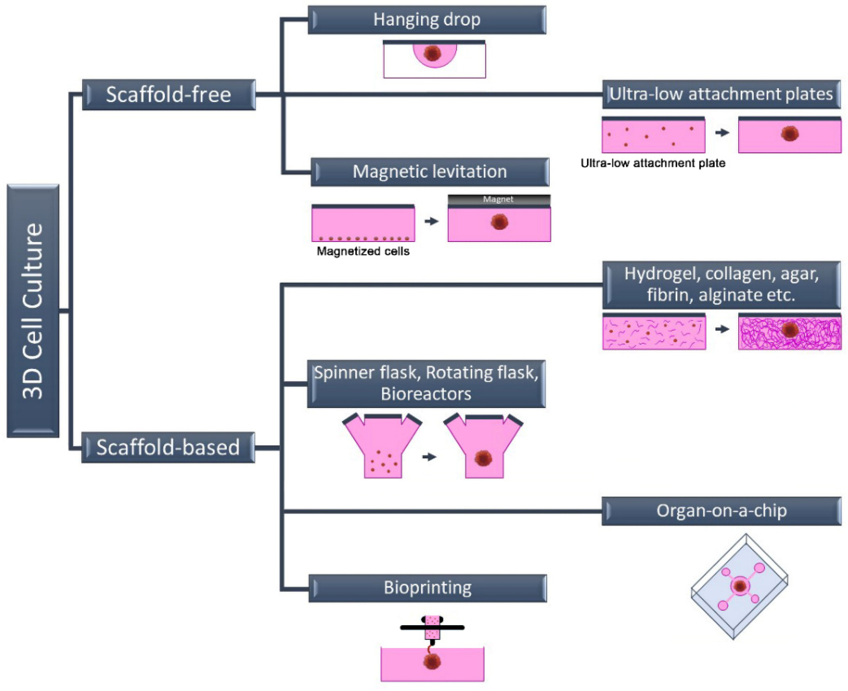
Cells, the building blocks of tissues and organs in the organism, reside in a complex 3D extracellular matrix (ECM) environment. This complex 3D architecture allows cells to interact both with each other and with the ECM. In this way, each cell acquires its own morphology and maintains the specificity and homeostasis of the tissue. Today, new methods have been developed that enable cells to grow in this complex 3D architecture in a laboratory environment. These methods are basically divided into two categories: scaffold-free methods and scaffold-based methods (Figure 1).
Figure 1. Schematic representation of three-dimensional cell culture methods.

2.1. Scaffold-Free 3D Cell Culture Method

The most commonly used scaffold-free 3D cell culture techniques are cell suspension culture on non-adhesive or ultra-low attachment plates (liquid overlay), hanging drop, magnetic levitation, and microfluidic. Scaffold-free methods are generally fast and economical. This method is a “bottom-up” approach and is based on the fact that cells come together to form a spheroid structure. The formation of 3D spheroids depends entirely on the natural abilities of the cells because there is no material that can be used as a scaffold in these techniques. The hanging drop technique gravitationally collects cells in the form of suspended drops at a spherical air–liquid interface, thus facilitating the formation of a 3D cell structure without a scaffold [16]. Similar to the cell suspension method, the cell density of the suspension can be varied depending on the desired cell aggregate size. Spheroid formation, in contrast to these approaches, can be accomplished through external physical intervention in some scaffold-free techniques such as magnetic levitation or agitation bioreactor. The biggest shortcoming of scaffold-free 3D cell culture methods is the absence of an ECM component. Since there is no ECM in the environment, only cell–cell interaction occurs. Cell–ECM interaction is missing, unlike in their natural environment [17]. The wells of the plates can also be coated with various chemicals, such as poly (2-hydroxyethyl methacrylate), agar, agarose, or pol (N-p vinyl benzyl-D-lactone amide), to produce a non-attached surface. However, this incurs additional equipment and/or costs. These approaches might not be adequate because they lack the scaffold support necessary for the cell–matrix interaction needed for cell biology and proper functioning [18][19].

2.2. Scaffold-Based 3D Cell Culture Method
In scaffold-based 3D cell culture methods, ECM components are also added to the culture to provide extracellular components that mimic the biological environment. For this purpose, commercially available, ready-to-use scaffolds as well as suitable ECM components can be used. Unlike scaffold-free methods, the ECM is a complex and dynamic structure found between cells in these methods. In addition to providing structural support, ECM plays an active role in helping cells acquire tissue-specific properties. Although its content varies according to the characteristics of tissues and cells, ECM basically consists of two components: proteins (collagen, elastin, fibronectin, laminin, fibrillin, etc.) and proteoglycans (heparan sulfate, chondroitin sulfate, etc.) [20][21]. Each of these components varies according to the task undertaken by the cell, and they also change in different physiological and biochemical events such as proliferation, genetic changes, differentiation, attachment, and migration. Therefore, the ECM is defined as a dynamic structure. In scaffold-based 3D cell culture, natural polymers such as collagen, hydroxyapatite, agar, fibrin or alginate are used as scaffolds, as well as biodegradable synthetic polymers such as poly (ethylene glycol) and poly (lactide-co-glycolide) [20][21].
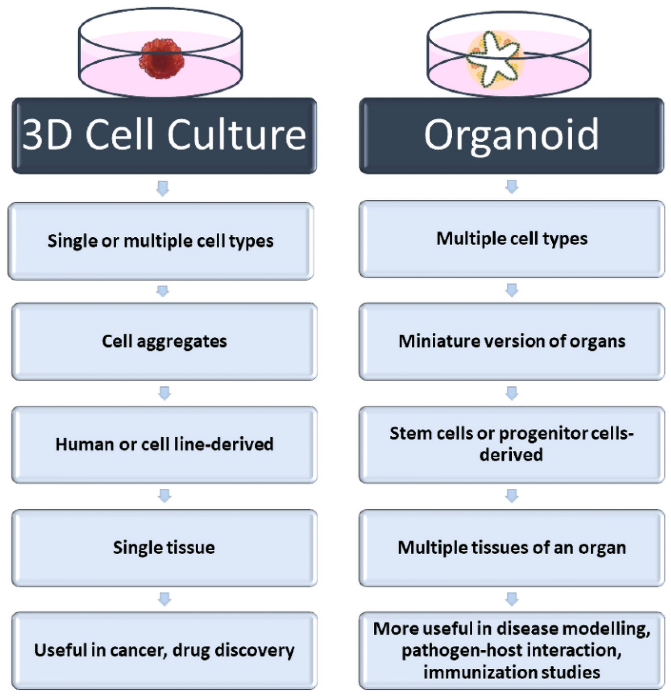
In addition to homologous 3D cell culture, heterologous 3D spheroids and organoids can be prepared by scaffold-based methods. Organoids called “Culturable Mini-Organs” are thought of as miniature versions of organs. On the other hand, spheroids can be prepared either homogeneously (from a single cell type) or heterogeneously (from different cell types) (Figure 2). Mostly, immortal cell lines are used for spheroids, while organoids are prepared from adult or embryonic stem cells. For this reason, organoids are complex structures that can better mimic organs [22][23]. On the contrary, spheroids are more economical and easy-to-prepare structures that can also be called “cell aggregates” or “organotypic culture” [24][25]. Although both approaches are used in vaccine and drug research and in in vitro disease modeling, spheroids are frequently preferred in tumor and drug development studies. Organoids are preferred in vaccine and pathogen interaction studies for assessing immune responses, especially in mimicking complex and multi-component organs such as the respiratory tract. Heterologous 3D models, in which more than one cell type is cultured together, also provide cell–cell interactions where growth factors and other biological factors can be exchanged [26][27]. The interactions of cells with each other and with the ECM are very important in terms of cell polarity. Cell polarity plays a direct role in the viral–host relationship as it affects the expression of the relevant receptor. For this reason, heterologous 3D cell culture studies attract attention as an effective in vitro study method in infection and virology studies.
Figure 2. Main characteristics of spheroids, and organoids.
Bioprinting, which is used to create a 3D cell culture model, is a very successful method of mimicking the complexity of biological structures and is a computer-based approach. Although it mostly has applications in tissue engineering studies, it also allows the production of 3D tissues by printing a solution consisting of one or more cell types and ECM, called bioink, on the desired surface layer by layer with a printer. In the bioprinting process, since the modeling and printing are carried out under computer control, the printing of the cells into the layers can be carried out in a very sensitive and controlled manner. Although it is a costly approach compared to other 3D cell culture models, it offers unique opportunities in artificial tissue production and artificial disease models. Another advantage of this method is that it allows the interaction of cells with each other and with the ECM, as well as the ability to diversify the printing pattern. The addition of bioactive components to the pattern, which can be changed by cells after printing, provides the cells with physiological conditions in the biological environment [28][29][30]. Furthermore, bioprinting offers the opportunity to make copies of tissues and even organs with different tissues in a biological environment. Since the first years of bioprinting studies, the production of artificial organs has been the most attractive field of biomedicine. However, nowadays, they are preferred not only in the production of artificial organs or tissues but also in the use of these organs in the investigation of the pathology of the disease and its relationship with the pathogen.
Bioreactors are another method used to prepare 3D cell cultures and organoid models. This method was originally designed to minimize the effect of gravity and allow cells to clump together in a liquid medium to form spheroids. Bioreactors can generally be classified into four groups: perfusion bioreactors, spinner flask bioreactors, rotating vessels, and mechanical force systems. In the spinner flask approach, one of the two most commonly used types, there is a constantly rotating impeller inside the bioreactor tank and it moves the liquid, allowing the cells to interact with each other. The second method was developed to eliminate the physical effect of the propeller on the cells. In this method, which is called a rotating vessel bioreactor, the tank containing the cells rotates. The most important advantage of bioreactors over other methods is that they provide the same physical conditions to all cells in a dynamic environment. With the continuous movement of the bioreactor tanks, nutrients can be delivered to the cells in many ways. The bioreactor approach is frequently used in studies of pathogen–host interactions. It allows the expression of cell connections and surface molecules that play a direct role in the entry of viruses into cells and thus infecting them. Bioreactors are also suitable for large-scale production when compared to other methods [31][32][33].
Organ-on-a-chip systems promise much more than current 3D cell culture studies, and they are seen as the future of these studies. Organ-on-a-chip technology, which has become more popular in the last 10 years, provides the desired artificial organ to mimic the biology of the disease [34][35]. Besides providing a 3D structure like other methods, it makes it possible to design a system with adjacent tissues. This approach allows for tissue and organ formation that more realistically mimics the biological structure in vitro. Especially thanks to the advances in nanotechnology, the diversity of applications has increased in the organ-on-chip approach. There are organ-on-a-chip models on the market that are designed separately for almost every organ and are commercially available. Moreover, there are organ-on-a-chip designs developed with sensors for imaging and biological/physiological changes designed in accordance with the experiments to be carried out. Virus–host interaction is one of the areas where organ-on-a-chip technology is used most frequently. Since this technology allows the formation of miniature tissues and organoids, studies on the examination of the interaction of pathogens with the host and the determination of the subsequent physiological changes with sensors are quite surprising and promising [36][37]. It is a very useful approach, especially in the elucidation of complex systems in which multiple biological factors play an active role, such as the development of resistance in infectious diseases and the evaluation of the immune response developing after vaccination.
As already mentioned, each of the different 3D cell culture methods has its own advantages and limitations. When all methods are compared, it can be said that organoids and organ-on-a-chip approaches are the best imitation techniques in terms of containing more than one tissue type and using stem cells. The lack of vascularization and blood vessels, interorgan communication, and immune system components prevent even organoids from fully mimicking in vivo conditions. As one of the most researched organoid models in the field of infectious illnesses, intestinal organoids include a variety of cell types from human tissues, enabling the investigation of heterocellular interactions. The major drawbacks are the absence of neural innervation, lumen content, and fluid flow, in addition to vascularization. One of the most popular techniques for the lung, another organ where pathogen–host interaction is frequently studied, is air–liquid interface (ALI) culture. The key advantage of ALI culture over other techniques is that it gives the cells an apical-basal configuration, enabling cell development towards a mucociliary phenotype. In this method, the apical surfaces of the cells are exposed to air, while the basal surfaces are in the liquid cell culture medium. Despite the fact that lung organoids better mimic the real functioning of the organ than the ALI method, they are still insufficient, particularly in terms of immune cell depletion and lack of vascularization. Furthermore, all 3D cell culture models developed for the lung are under ambient air pressure. Given the close correlation between respiratory rate and pressure in vivo, the models are insufficient to depict this relationship.

References

  1. Dolskiy, A.A.; Grishchenko, I.V.; Yudkin, D.V. Cell Cultures for Virology: Usability, Advantages, and Prospects. Int. J. Mol. Sci. 2020, 21, 7978.
  2. Han, F.; Wang, J.; Ding, L.; Hu, Y.; Li, W.; Yuan, Z.; Guo, Q.; Zhu, C.; Yu, L.; Wang, H.; et al. Tissue Engineering and Regenerative Medicine: Achievements, Future, and Sustainability in Asia. Front. Bioeng. Biotechnol. 2020, 8, 83.
  3. Hudu, S.A.; Alshrari, A.S.; Syahida, A.; Sekawi, Z. Cell Culture, Technology: Enhancing the Culture of Diagnosing Human Diseases. J. Clin. Diagn. Res. 2016, 10, DE01–DE05.
  4. Verma, A.; Verma, M.; Singh, A. Animal tissue culture principles and applications. In Animal Biotechnology; Academic Press: Cambridge, MS, USA, 2020; pp. 269–293.
  5. O’Flaherty, R.; Bergin, A.; Flampouri, E.; Mota, L.M.; Obaidi, I.; Quigley, A.; Xie, Y.; Butler, M. Mammalian cell culture for production of recombinant proteins: A review of the critical steps in their biomanufacturing. Biotechnol. Adv. 2020, 43, 107552.
  6. Ju, X.; Zhu, Y.; Wang, Y.; Li, J.; Zhang, J.; Gong, M.; Ren, W.; Li, S.; Zhong, J.; Zhang, L.; et al. A novel cell culture system modeling the SARS-CoV-2 life cycle. PLoS Pathog. 2021, 17, e1009439.
  7. Shang, J.; Wan, Y.; Luo, C.; Ye, G.; Geng, Q.; Auerbach, A.; Li, F. Cell entry mechanisms of SARS-CoV-2. Proc. Natl. Acad. Sci. USA 2020, 117, 11727–11734.
  8. Graham, B.S. Advances in antiviral vaccine development. Immunol. Rev. 2013, 255, 230–242.
  9. Josefsberg, J.O.; Buckland, B. Vaccine process technology. Biotechnol. Bioeng. 2012, 109, 1443–1460.
  10. Cacciamali, A.; Villa, R.; Dotti, S. 3D Cell Cultures: Evolution of an Ancient Tool for New Applications. Front. Physiol. 2022, 13, 836480.
  11. Jensen, C.; Teng, Y. Is It Time to Start Transitioning From 2D to 3D Cell Culture? Front. Mol. Biosci. 2020, 7, 33.
  12. Kapałczyńska, M.; Kolenda, T.; Przybyła, W.; Zajączkowska, M.; Teresiak, A.; Filas, V.; Ibbs, M.; Bliźniak, R.; Łuczewski, Ł.; Lamperska, K. 2D and 3D cell cultures - a comparison of different types of cancer cell cultures. Arch. Med. Sci. 2018, 14, 910–919.
  13. Barrila, J.; Crabbé, A.; Yang, J.; Franco, K.; Nydam, S.D.; Forsyth, R.J.; Davis, R.R.; Gangaraju, S.; Ott, C.M.; Coyne, C.B.; et al. Modeling Host-Pathogen Interactions in the Context of the Microenvironment: Three-Dimensional Cell Culture Comes of Age. Infect. Immun. 2018, 86, e00282-18.
  14. Häfner, S.J. Level up for culture models - How 3D cell culture models benefit SARS-CoV-2 research. Biomed. J. 2021, 44, 1–6.
  15. Harb, A.; Fakhreddine, M.; Zaraket, H.; Saleh, F.A. Three-Dimensional Cell Culture Models to Study Respiratory Virus Infections Including COVID-19. Biomimetics 2021, 7, 3.
  16. Hsiao, A.Y.; Tung, Y.C.; Qu, X.; Patel, L.R.; Pienta, K.J.; Takayama, S. 384 hanging drop arrays give excellent Z-factors and allow versatile formation of co-culture spheroids. Biotechnol. Bioeng. 2012, 109, 1293–1304.
  17. Alghuwainem, A.; Alshareeda, A.T.; Alsowayan, B. Scaffold-Free 3-D Cell Sheet Technique Bridges the Gap between 2-D Cell Culture and Animal Models. Int. J. Mol. Sci. 2019, 20, 4926.
  18. Foty, R. A simple hanging drop cell culture protocol for generation of 3D spheroids. J. Vis. Exp. 2011, 51, e2720.
  19. Shri, M.; Agrawal, H.; Rani, P.; Singh, D.; Onteru, S.K. Hanging Drop, A Best Three-Dimensional (3D) Culture Method for Primary Buffalo and Sheep Hepatocytes. Sci. Rep. 2017, 7, 1203.
  20. Hu, M.; Ling, Z.; Ren, X. Extracellular matrix dynamics: Tracking in biological systems and their implications. J. Biol. Eng. 2022, 16, 13.
  21. Nicolas, J.; Magli, S.; Rabbachin, L.; Sampaolesi, S.; Nicotra, F.; Russo, L. 3D Extracellular Matrix Mimics: Fundamental Concepts and Role of Materials Chemistry to Influence Stem Cell Fate. Biomacromolecules 2020, 21, 1968–1994.
  22. Koike, H.; Takebe, T. Generating Mini-Organs in Culture. Curr. Pathobiol. Rep. 2016, 4, 59–68.
  23. Dutta, D.; Clevers, H. Organoid culture systems to study host-pathogen interactions. Curr. Opin. Immunol. 2017, 48, 15–22.
  24. Gunti, S.; Hoke, A.T.K.; Vu, K.P.; London, N.R., Jr. Organoid and Spheroid Tumor Models: Techniques and Applications. Cancers 2021, 13, 874.
  25. Zanoni, M.; Cortesi, M.; Zamagni, A.; Arienti, C.; Pignatta, S.; Tesei, A. Modeling neoplastic disease with spheroids and organoids. J. Hematol. Oncol. 2020, 13, 97.
  26. Altmann, B.; Grün, C.; Nies, C.; Gottwald, E. Advanced 3D Cell Culture Techniques in Micro-Bioreactors, Part II: Systems and Applications. Processes 2021, 9, 21.
  27. Sangeeta, B.; Ankita Jaywant, D.; Shafina, S.; Jyotirmoi, A.; Soumya, B. Two-Dimensional and Three-Dimensional Cell Culture and Their Applications, in Cell Culture; Zhan, X., Ed.; IntechOpen: Rijeka, Croatia, 2021.
  28. Dey, M.; Ozbolat, I.T. 3D bioprinting of cells, tissues and organs. Sci. Rep. 2020, 10, 14023.
  29. Kabir, A.; Datta, P.; Oh, J.; Williams, A.; Ozbolat, V.; Unutmaz, D.; T Ozbolat, I. 3D Bioprinting for fabrication of tissue models of COVID-19 infection. Essays Biochem. 2021, 65, 503–518.
  30. Koban, R.; Lam, T.; Schwarz, F.; Kloke, L.; Bürge, S.; Ellerbrok, H.; Neumann, M. Simplified Bioprinting-Based 3D Cell Culture Infection Models for Virus Detection. Viruses 2020, 12, 1298.
  31. Grün, C.; Altmann, B.; Gottwald, E. Advanced 3D Cell Culture Techniques in Micro-Bioreactors, Part I: A Systematic Analysis of the Literature Published between 2000 and 2020. Processes 2020, 8, 1656.
  32. Kizilova, N.; Rokicki, J. 3D Bioreactors for Cell Culture: Fluid Dynamics Aspects. In Biomechanics in Medicine, Sport and Biology; Springer International Publishing: Cham, Switzerland, 2022.
  33. Yi, T.; Huang, S.; Liu, G.; Li, T.; Kang, Y.; Luo, Y.; Wu, J. Bioreactor Synergy with 3D Scaffolds: New Era for Stem Cells Culture. ACS Appl. Bio. Mater. 2018, 1, 193–209.
  34. Ingber, D.E. Human organs-on-chips for disease modelling, drug development and personalized medicine. Nat. Rev. Genet. 2022, 23, 467–491.
  35. Sun, W.; Luo, Z.; Lee, J.; Kim, H.-J.; Lee, K.; Tebon, P.; Feng, Y.; Dokmeci, M.R.; Sengupta, S.; Khademhosseini, A. Organ-on-a-Chip for Cancer and Immune Organs Modeling. Adv. Healthc. Mater. 2019, 8, 1801363.
  36. Tang, H.; Abouleila, Y.; Si, L.; Ortega-Prieto, A.M.; Mummery, C.L.; Ingber, D.E.; Mashaghi, A. Human Organs-on-Chips for Virology. Trends Microbiol. 2020, 28, 934–946.
  37. Wang, Y.; Wang, P.; Qin, J. Human Organoids and Organs-on-Chips for Addressing COVID-19 Challenges. Adv. Sci. 2022, 9, 2105187.
More
Information
Contributors MDPI registered users' name will be linked to their SciProfiles pages. To register with us, please refer to https://encyclopedia.pub/register : ,
View Times: 766
Revisions: 2 times (View History)
Update Date: 17 Jan 2023
1000/1000
ScholarVision Creations