Submitted Successfully!
To reward your contribution, here is a gift for you: A free trial for our video production service.
Thank you for your contribution! You can also upload a video entry or images related to this topic.
Version Summary Created by Modification Content Size Created at Operation
1 -- 2848 2023-01-10 14:19:45 |
2 Reference format revised. Meta information modification 2848 2023-01-11 02:22:39 |

Video Upload Options

We provide professional Video Production Services to translate complex research into visually appealing presentations. Would you like to try it?

Confirm

Are you sure to Delete?
Cite
If you have any further questions, please contact Encyclopedia Editorial Office.
Ramos-Martínez, I.;  Ramos-Martínez, E.;  Cerbón, M.;  Pérez-Torres, A.;  Mayoral, L.P.;  Hernández-Huerta, M.T.;  Martínez-Cruz, M.;  Pérez-Santiago, A.D.;  Sánchez-Medina, M.A.;  García-Montalvo, I.A.; et al. Role of Glycosylation in Systemic Lupus Erythematosus. Encyclopedia. Available online: https://encyclopedia.pub/entry/39977 (accessed on 16 November 2024).
Ramos-Martínez I,  Ramos-Martínez E,  Cerbón M,  Pérez-Torres A,  Mayoral LP,  Hernández-Huerta MT, et al. Role of Glycosylation in Systemic Lupus Erythematosus. Encyclopedia. Available at: https://encyclopedia.pub/entry/39977. Accessed November 16, 2024.
Ramos-Martínez, Ivan, Edgar Ramos-Martínez, Marco Cerbón, Armando Pérez-Torres, Laura Pérez-Campos Mayoral, María Teresa Hernández-Huerta, Margarito Martínez-Cruz, Alma Dolores Pérez-Santiago, Marco Antonio Sánchez-Medina, Iván Antonio García-Montalvo, et al. "Role of Glycosylation in Systemic Lupus Erythematosus" Encyclopedia, https://encyclopedia.pub/entry/39977 (accessed November 16, 2024).
Ramos-Martínez, I.,  Ramos-Martínez, E.,  Cerbón, M.,  Pérez-Torres, A.,  Mayoral, L.P.,  Hernández-Huerta, M.T.,  Martínez-Cruz, M.,  Pérez-Santiago, A.D.,  Sánchez-Medina, M.A.,  García-Montalvo, I.A.,  Zenteno, E.,  Matias-Cervantes, C.A.,  Ojeda-Meixueiro, V.,  Pérez-Campos, E., & Pérez-Campos Mayoral, L. (2023, January 10). Role of Glycosylation in Systemic Lupus Erythematosus. In Encyclopedia. https://encyclopedia.pub/entry/39977
Ramos-Martínez, Ivan, et al. "Role of Glycosylation in Systemic Lupus Erythematosus." Encyclopedia. Web. 10 January, 2023.
Role of Glycosylation in Systemic Lupus Erythematosus
Edit

Glycosylation is a post-translational modification that affects the stability, structure, antigenicity and charge of proteins. In the immune system, glycosylation is involved in the regulation of ligand–receptor interactions, such as in B-cell and T-cell activating receptors. Alterations in glycosylation have been described in several autoimmune diseases, such as systemic lupus erythematosus (SLE), in which alterations have been found mainly in the glycosylation of B lymphocytes, T lymphocytes and immunoglobulins. In immunoglobulin G of lupus patients, a decrease in galactosylation, sialylation, and nucleotide fucose, as well as an increase in the N-acetylglucosamine bisector, are observed. These changes in glycoisolation affect the interactions of immunoglobulins with Fc receptors and are associated with pericarditis, proteinuria, nephritis, and the presence of antinuclear antibodies. In T cells, alterations have been described in the glycosylation of receptors involved in activation, such as the T cell receptor; these changes affect the affinity with their ligands and modulate the binding to endogenous lectins such as galectins. In T cells from lupus patients, a decrease in galectin 1 binding is observed, which could favor activation and reduce apoptosis. Furthermore, these alterations in glycosylation correlate with disease activity and clinical manifestations, and thus have potential use as biomarkers.

autoimmune diseases glycosylation systemic lupus erythematosus (SLE)

1. Introduction

Glycosylation is a very common post-translational modification; it is present in virtually all secreted proteins and membrane proteins. It plays a key role in folding and transport through the endoplasmic reticulum and the Golgi apparatus [1][2]. Once the protein has completed its synthesis and transport phase, additional glycosylation can affect its stability, function, charge, and antigenicity. Glycosylation modifications can occur for a single monosaccharide or dozens of monosaccharides arranged in various three-dimensional structures [3].
Glycosylation is a mechanism that regulates many immune system processes, such as ligand–receptor interaction [4]. Changes in the glycosylation of the T cell receptor complex or its co-receptors affect the activation threshold of T cells by modifying the affinity for their ligands [5]. In the case of immunoglobulins, it has been reported that changes in glycosylation of heavy chain constant regions may be the difference between a pro-inflammatory or anti-inflammatory response [6]. Furthermore, glycosylation in cells of the immune system can be recognized by endogenous lectins such as galectins, selectins, and dectins that mediate functions such as cell activation, differentiation, and survival [4][7]. Therefore, alterations in glycosylation could lead to the development of autoimmune diseases.
Glycosylation abnormalities have been reported in autoimmune diseases such as IgA nephropathy, rheumatoid arthritis, inflammatory bowel disease, and multiple sclerosis [8][9]. For example, galactose deficiency in immunoglobulin G (IgG) in patients with rheumatoid arthritis correlates with disease progression [10]. Galactose deficiency in immunoglobulin G has also been reported in other diseases, such as inflammatory bowel disease [11] and multiple sclerosis [12]. In addition, decreased immunoglobulin G fucosylation is a marker of autoimmune thyroid diseases [8].

2. Systemic Lupus Erythematosus

Systemic lupus erythematosus (SLE) is an autoimmune disease that can affect every organ and system in the body. It presents a wide spectrum of clinical manifestations in acute periods and periods of remission. In mild forms, skin and joint manifestations are observed; in severe forms, it affects the kidneys, heart, and nervous system [13]. SLE develops mainly in women of reproductive age, in a ratio of nine women to one man [14].
Etiopathogenesis of SLE involves immunological, genetic, hormonal, and environmental factors that play an important role in the regulation of immunological tolerance [13][15]. B cells and their various subpopulations play a crucial role in the pathogenesis of SLE. These cells show abnormal activation through their B cell receptor and Toll-like receptors [16]. Another factor that favors the activation of B cells in SLE is altered levels of cytokines such as interferons alpha (IFNɑ), beta (IFNβ), and gamma (IFNɣ), as well as BAFF/Blys, which promote B cells survival [16][17].
Serum of SLE patients is characterized by having autoantibodies against antigens such as double-stranded DNA (dsDNA), single-stranded DNA (ssDNA), RNA, ribonucleoproteins (rRNA/Ro, La, Smith), and cytoskeletal antigens [18]. The autoantibodies in SLE patients are of a high affinity and IgG type, indicating that an isotype switch has occurred, for which the participation of T cells is required [19]. T cells from SLE patients overexpress the CD40L molecule, which allows them to provide costimulatory signals necessary for B cell differentiation, proliferation, and isotype switching [20].
The fundamental role of T cells in the etiopathogenesis of SLE is supported by studies in murine models, where mice develop lupus spontaneously. While in NZB/NZW F1(B/W) mice, T cells elimination by monoclonal antibodies prevented disease development [21]; as well as the fact that the hybrid nu/nu NZB/NZW F1 (B/W) mice, which lack a thymus, do not develop lupus [22].

3. Glycosylation

3.1. O-Glycosylation

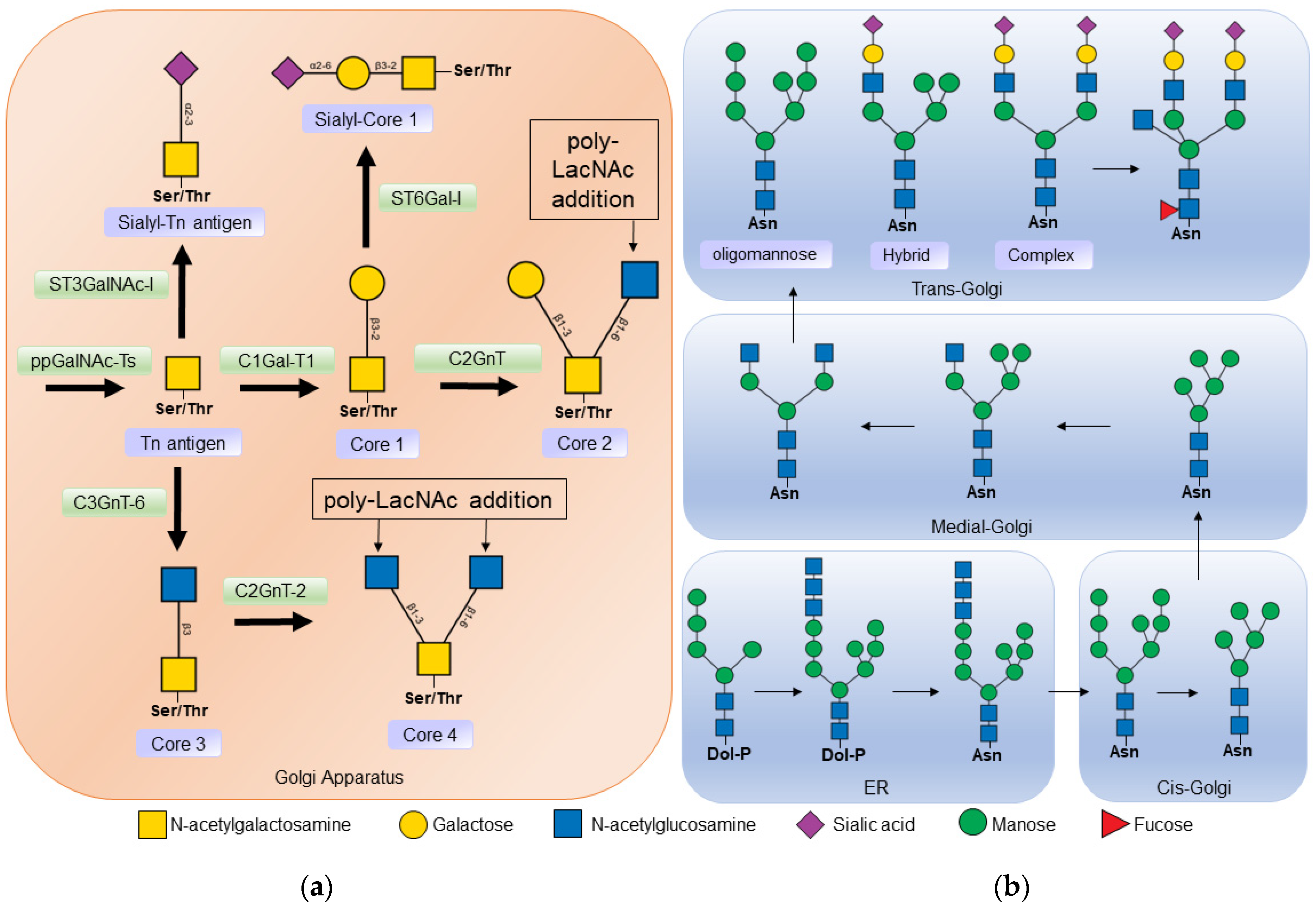
In O-glycosylation, N-acetyl-galactosamine (GalNAc) residues are added from N-acetyl-galactosamine-uridine diphosphate (UDP-GalNAc) by the action of N-acetyl-galactosaminyltransferase (ppGalNAcT). At least 21 isoforms of this enzyme have been described, which have different affinities depending on the amino acid sequence and the three-dimensional arrangement of carbohydrate residues [23][24]. The binding of N-acetyl-galactosamine (GalNAc) to the serine or threonine of a protein is known as the Tn antigen. In a second step, galactose (Gal) is added to the N-acetyl-galactosamine residue in the β1-3 bond, by the action of Core 1 β1-3 galactosyltransferase (C1GalT-1), to form the structure known as core 1 (Figure 1a) [2][25].
Figure 1. (a) Biosynthesis of O-glycans. O-glycosylation occurs in the Golgi apparatus, where N-acetyl-galactosamine is linked to a serine or threonine residue by the action of polypeptide N-acetylgalactosaminyltransferase (ppGalNAc-Ts) forming the Tn antigen. Core 1 β1-3 galactosyltransferase 1 (C1Gal-T1) then binds to a galactose to form core 1. Core 1 can be a substrate for some sialyltransferase or core 2 β1-6 N-acetylglucosaminyltransferase (C2GnT) to thus form core 2. Core 3 is formed from insufficient Tn by the action of core 3 β1-3 N-acetylglucosaminyltransferase 6 (C3GnT-6); in turn, core 3 is a substrate to form core 4 by the addition of another N -acetyl-glucosamine by Core 2/4 β1-6 N-acetylglucosaminyltransferase 2 (C2GnT-2). (b) Biosynthesis of N-glycans. Synthesis begins in the endoplasmic reticulum (ER), where a dolicholphosphate oligosaccharide is transferred to an asparagine residue by oligosaccharyltransferase (OST). Next, α-glucosidases I and II, and α-mannosidase I cleave the sugars from the oligosaccharide. The protein is then transported to the Golgi apparatus, where other mannosidase result from the processing of the N-glycan. In the middle part of the Golgi apparatus, N-acetylglucosaminyltransferase 1 (MGAT1) adds N-acetyl-glucosamine to begin the synthesis of hybrid and complex N-glycans. In the Trans Golgi part, fucoses, sialic acids, and poly-lactosamine chains, α2-6 sialyltransferase (ST3GalNAc), α2-6 sialyltransferase (ST6Gal), poly-N-acetyllactosamine (Poly-LacNAc) are added.
Core 1 may be a substrate for the enzyme core 2 β1-6 N-acetylglucosaminyltransferase (C2GnT), which catalyzes the addition of N-acetylglucosamine (GlcNAc) at the β1-6 bond with galactose [25]. Two isoforms of this enzyme have been described: the L type that synthesizes core 2, and the M type that also participates in the synthesis of other cores [2][26]. Core 2 is produced at many sites, including the intestinal mucosa, on activated T cells, and during embryonic development. The synthesis of cores 3 and 4 is mainly restricted to sites such as the mucosal epithelium of the gastrointestinal tract, the respiratory tract, and the salivary glands [2][26].

3.2. N-Glycosylation

The first sugar to be attached to eukaryotic N-glycans is N-acetyl-glucosamine, which is linked to the asparagine residue via an N-glycosidic bond in the consensus sequence Asn-X-Ser/Thr, where X is any amino acid except proline. N-glycans affect the conformation, solubility, antigenicity, activity, and recognition of proteins [1].
N-glycans have a common core sequence and are classified into three types based on the branches they contain. Oligomannose types contain only mannose residues in the branches, complex types have N-acetyl-glucosamine-initiated branches, and hybrid types have both mannose-initiated and N-acetyl-glucosamine-initiated branches (Figure 1b). N-acetyl-glucosamine-initiated branches can be lengthened by the addition of N-acetyl-lactosamine (Galβ1-4GlcNAc) repeats, which are ligands for endogenous lectins such as galectins [1][27].
The synthesis of N-glycans begins in the membrane of the endoplasmic reticulum with the addition of 14 sugars to the lipid molecule dolichol-phosphate. Then this oligosaccharide is transferred by oligosaccharyltransferase (OST) to an asparagine protein. In the lumen of the endoplasmic reticulum, the 14-sugar oligosaccharide is cleaved by α-glucosidase I (MOGS) and α-glucosidase II (GANAB) [1]. This step is important in protein folding, as chaperones in the endoplasmic reticulum recognize the cleaved N-glycans [28]

4. Functions of Glycosylation in the Immune System

Glycosylation affects the conformation, stability, charge, protease resistance, and affinity of the proteins for other proteins [3]; that is, it is part of the regulation of ligand–receptor interactions. During the inflammation process, the receptor proteins involved in migration, adhesion, and diapedesis are differentially glycosylated, thus they are recognized by their ligands [3].
In the case of T cells, protein glycosylation is a dynamic process and changes depending on the stimuli they receive; for example, the pattern of glycosylation between naïve T cells and active T cells is different [29]. When the T cell is activated by stimuli through its T cell receptor (TCR), an increase in Tn antigen on glycoproteins is observed, as recognition of these cells by Arachis hypogaea agglutinin (PNA) and Helix pomatia agglutinin, two lectins with an affinity for the Tn antigen, is increased [30][31][32].
Glycosylation also regulates T cell activation and maturation by affecting the affinity between the T cell receptor complex and the major histocompatibility complex (MHC). Glycosylation of membrane receptors such as CD4, CD8, and T cell receptors has been shown to alter the affinity of interactions with the MHC, thereby affecting positive selection (Zhou et al., 2014). In addition, the presence of sialic acid in position α2-3 in core 1 of the O-glycans of the CD8 co-receptor decreases the avidity for MHC class I molecules, which plays an important role in negative selection. Glycosylation is also involved in the control of T cell differentiation. Decreased CD25 glycosylation has been shown to prevent T cell differentiation to the Th1, Th2, and Treg-induced phenotype, but promotes Th17 cell differentiation [33].
Glycoprotein sialylation is another important factor that regulates the interaction between receptors and their ligands. The interaction between CD69 and the S100A8/S100A9 complex is important in Treg cell differentiation, but the removal of sialic acid by sialidase treatment on activated CD4 T cells prevents binding between these proteins and decreases Treg cells differentiation [34].
Therefore, alterations in glycosylation lead to changes in the T cell-mediated immune response and have been linked to inflammation and autoimmune diseases [9][35]. For example, treatment of mice with glucosamine decreases CD25 glycosylation, prevents differentiation to Th1 cells, increases islet graft survival in diabetic mice, and exacerbates the severity of experimental autoimmune encephalitis [33].
In this regard, it has been shown that naïve, memory, and germinal center B cells express triantennary and tetraantennary N-glycans with long chains of poly-N-acetyl-lactosamine; these chains show specific differences that allow them to be ligands for certain galectins [36]. Galectins are lectins secreted by different cells, such as endothelial cells, monocytes, and dendritic cells, which recognize lactosamine in O- and N-glycans expressed in membrane proteins. The binding of galectins to their ligands triggers signals related to inflammation, activation, and death of target cells [37].
In B cells, the interaction of glycans and galectins regulates processes such as activation (galectin 3 and 9), plasma cell differentiation (galectin 1 and 8), and survival (galectin 1) [7]. Alterations in glycosylation have effects on the affinity for galectins. For example, in germinal center B cells, it has been observed that the addition of the disaccharide β1,6 N-Acetyl-glucosamine-galactose to the poly-N-acetyl-lactosamine chains prevents the binding of galectin 3 and 9 [36][38].
In B-cell chronic lymphocytic leukemia, B-cell glycosylation is altered in a way that allows interaction with galectin 1, thereby lowering the B cell receptor (BCR) signaling threshold; this causes the signaling and survival of these B cells; in addition, the interaction of galectin 1 with glycans stimulates the expression of BAFF and APRIL that promote cell survival [39].

5. Alterations in Glycosylation in Systemic Lupus Erythematosus

5.1. Alterations in B Cell Glycosylation and Antibodies in SLE

In the case of B cells, glycosylation studies have been directed primarily at immunoglobulins. It has been proposed that the acquisition of N-glycosylation sites in the variable regions of immunoglobulin light or heavy chains could favor B cell survival during germinal center selection, providing B cells with an additional mechanism for the generation of autoreactive cells [40][41].

5.1.1. Glycosylation of Constant Regions

SLE patients have alterations in the glycosylation of immunoglobulin G. A greater recognition of the Aleuria aurantia lectin, which has an affinity for fucosylated residues, has been observed in comparison with healthy individuals; recognition of this lectin correlates with disease activity [42]. Antibodies with reduced fucosylation of their Fc region have a better interaction with the Fc gamma receptor IIIa (FcgRIIIa) [43][44].
Increased recognition of the Lens culinaris lectin, which is specific for the core fucosylated trimannose N-glycan, towards immunoglobulin G has been also reported in SLE patients [42]. The increased exposure of mannose residues in IgG facilitates the activation of FcγRIIIa and increases antibody-mediated cytotoxicity [43]. These mannose-exposed glycans have few fucose residues, which may also affect FcγRIIIa binding; in addition, mannose-binding lectin (MBL) can recognize these mannose-exposed glycans in IgG and activate the complement lectin pathway [43].
Endoglycosidase S is an enzyme that cleaves the N-glycans of IgG, but not those of IgM, exposing an N-acetylglucosamine (GlcNAc) from the glycosidic core and its branched fucose residue. Treatment of BXSB mice that developed a lupus-like disease with endoglycosidase S did not reduce anti-dsDNA (double-stranded) or antinuclear antibody levels, but increased mouse survival and improved blood urea levels [45]. The proposed mechanism is that treatment with endoglycosidase S reduces the affinity of antibodies towards FcγR, which could prevent activation by immune complexes [46].
In the case of SLE patients, using ultra-resolution liquid chromatography, it was found that they present a decrease in IgG galactosylation and sialylation; a decrease in the fucose nucleus and an increase in N-acetylglucosamine bisector was also observed. These glycosylation abnormalities were associated with pericarditis, proteinuria, and the presence of antinuclear antibodies [47].

5.1.2. Glycosylation of Variable Regions

The wide variety of antibodies with different affinities is due to the rearrangement of the variable (V), diversity (D), and joining (J) gene segments, as well as somatic hypermutation and class-switch recombination. During somatic hypermutation, point mutations occur in the variable regions of the heavy and light chains of immunoglobulins. These mutations can generate consensus sequences for N-glycosylation. The glycans attached to these sequences are called Fab glycans. It is estimated that 15% of IgGs have N-glycosylations in the variable regions, mainly as a result of hypermutation [48][49]. The function of these N-glycans is to regulate the affinity and stability of the antigen–antibody bond, as well as to prolong the half-life of the antibodies [40][41][50].

5.2. Alterations in the Glycosylation of the T Cells in SLE

It has been shown that T cells from SLE patients present alterations in O-glycosylation [51] using the Amaranthus leucocarpus lectin (ALL), which recognizes core 1 in coactivating T cell receptors [52]. T cells from patients with active SLE had less recognition by ALL than those with inactive SLE, and expression of receptors recognized by ALL was inversely correlated with disease activity [51].
Alterations in the expression of core 1 have also been described in murine models of lupus. T cells from MLR-lpr mice show reduced recognition of Maclura pomifera lectin compared to T cells from MLR++ mice [53]. The Maclura pomifera lectin is a Tn antigen-specific lectin [54]. MLR-lpr mice develop the disease faster and with greater clinical manifestations than MLR++ mice (Richard and Gilkeson, 2018), and show decreased O-glycosylation of their T cells similar to that seen in patients with active SLE [51].
Other lectins that recognize O-glycans, such as the Arachis hypogaea lectin or Peanut Agglutinin (PNA) and the Artocarpus integrifolia lectin (Jacalin), have not shown alterations in O-glycosylation in T cells from SLE patients [51]. These discrepancies may be due to the type of lectin used, since despite recognizing the same sugar, their affinity is variable and they recognize sugars present in different proteins [55][56].

6. Alterations in Cytoplasmic O-GlcNAcylation

O-linked N-acetylglucosamine (O-GlcNAcylation) is a post-translational modification in which the monosaccharide N-acetylglucosamine (GlcNA) is added to serine or threonine residues of cytoplasmic or nuclear proteins. This transfer is mediated by O-GlcNAc transferase (OGT) and can be removed by O-GlcNAcase (OGA). Unlike the other types of glycosylation previously discussed, O-GlcNAcylation does not occur in the endoplasmic reticulum or Golgi apparatus, but rather in the cytosol. The addition of this monosaccharide to proteins has effects on protein localization, stability, and interactions [57]. O-GlcNAcylation regulates immune system functions such as inflammatory and antiviral responses in macrophages [58], promotes activated neutrophils functions [59], and inhibits natural killer cell activity [57][60].
A decrease in O-GlcNAcylation and phosphorylation of E74-like factor 1 (ELF-1) has been observed in SLE patients. ELF-1 is a transcriptional factor that binds to the promoter of the gene that encodes the zeta (ζ) chain of the CD3 molecule, thereby increasing its transcription. For this interaction to occur, the ELF-1 factor must be glycosylated and phosphorylated, constituting the 98 KDa isoform. Decreased glycosylation and phosphorylation of ELF-1 cause a decrease in its binding to DNA and a decrease in CD3ζ expression [61].
The decrease in O-GlcNAcylation and phosphorylation of E74-like factor 1 (ELF-1) is consistent with decreased expression of CD3ζ mRNA and its protein in SLE patients. To compensate for CD3ζ deficiency, the T-cell receptor complex associates with the Fc receptor gamma chain (FcRγ), causing alterations in intracellular signaling pathways as well as increased phosphorylation of tyrosine residues, an increase in the concentration of cytoplasmic calcium, and low production of IL-2 [62][63].

References

  1. Stanley, P.; Moremen, K.; Lewis, N.; Taniguchi, N.; Aebi, M. N-Glycans. In Essentials of Glycobiology; Varki, A., Cummings, R., Esko, J., Stanley, P., Hart, G., Aebi, M., Mohnen, D., Kinoshita, T., Packer, N., Prestegard, J., et al., Eds.; Cold Spring Harbor Laboratory Press: New York, NY, USA, 2022.
  2. Brockhausen, I.; Wandall, H.; Ten Hagen, K.; Stanley, P. O-GalNAc Glycans. In Essentials of Glycobiology; Varki, A., Cummings, R.D., Esko, J.D., Freeze, H.H., Stanley, P., Bertozzi, C.R., Hart, G.W., Etzler, M.E., Eds.; Cold Spring Harbor: New York, NY, USA, 2022; ISBN 9780879697709.
  3. Sprovieri, P.; Martino, G. The role of the carbohydrates in plasmatic membrane. Physiol. Res. 2018, 67, 1–11.
  4. Maverakis, E.; Kim, K.; Shimoda, M.; Gershwin, M.E.; Patel, F.; Wilken, R.; Raychaudhuri, S.; Ruhaak, L.R.; Lebrilla, C.B. Glycans in the immune system and The Altered Glycan Theory of Autoimmunity: A critical review. J. Autoimmun. 2015, 57, 1–13.
  5. Zhou, R.W.; Mkhikian, H.; Grigorian, A.; Hong, A.; Chen, D.; Arakelyan, A.; Demetriou, M. N-glycosylation bidirectionally extends the boundaries of thymocyte positive selection by decoupling Lck from Ca2+ signaling. Nat. Immunol. 2014, 15, 1038–1045.
  6. Mihai, S.; Nimmerjahn, F. The role of Fc receptors and complement in autoimmunity. Autoimmun. Rev. 2013, 12, 657–660.
  7. Giovannone, N.; Smith, L.K.; Treanor, B.; Dimitroff, C.J. Galectin-Glycan Interactions as Regulators of B Cell Immunity. Front. Immunol. 2018, 9, 2839.
  8. Ząbczyńska, M.; Link-Lenczowski, P.; Pocheć, E. Glycosylation in Autoimmune Diseases BT—The Role of Glycosylation in Health and Disease; Lauc, G., Trbojević-Akmačić, I., Eds.; Springer International Publishing: Cham, Switzerland, 2021; pp. 205–218. ISBN 978-3-030-70115-4.
  9. Li, X.; Xu, J.; Li, M.; Zeng, X.; Wang, J.; Hu, C. Aberrant glycosylation in autoimmune disease. Clin. Exp. Rheumatol. 2020, 38, 767–775.
  10. Lacki, J.K.; Porawska, W.; Mackiewicz, U.; Mackiewicz, S.; Müller, W. Changes in agalactosyl IgG levels correlate with radiological progression in early rheumatoid arthritis. Ann. Med. 1996, 28, 265–269.
  11. Akmačić, I.T.; Ventham, N.T.; Theodoratou, E.; Vučković, F.; Kennedy, N.A.; Krištić, J.; Nimmo, E.R.; Kalla, R.; Drummond, H.; Štambuk, J.; et al. Inflammatory bowel disease associates with proinflammatory potential of the immunoglobulin G glycome. Inflamm. Bowel Dis. 2015, 21, 1237–1247.
  12. Wuhrer, M.; Selman, M.H.J.; McDonnell, L.A.; Kümpfel, T.; Derfuss, T.; Khademi, M.; Olsson, T.; Hohlfeld, R.; Meinl, E.; Krumbholz, M. Pro-inflammatory pattern of IgG1 Fc glycosylation in multiple sclerosis cerebrospinal fluid. J. Neuroinflamm. 2015, 12, 235.
  13. Abida, R.; Yeoh, S.-A.; Isenberg, D.A. Advances in systemic lupus erythematosus. Medicine 2022, 50, 7–17.
  14. Boodhoo, K.D.; Liu, S.; Zuo, X. Impact of sex disparities on the clinical manifestations in patients with systemic lupus erythematosus: A systematic review and meta-analysis. Medicine 2016, 95, e4272.
  15. Fava, A.; Petri, M. Systemic lupus erythematosus: Diagnosis and clinical management. J. Autoimmun. 2019, 96, 1–13.
  16. Bacalao, M.A.; Satterthwaite, A.B. Recent Advances in Lupus B Cell Biology: PI3K, IFNγ, and Chromatin. Front. Immunol. 2020, 11, 615673.
  17. Rönnblom, L.; Elkon, K.B. Cytokines as therapeutic targets in SLE. Nat. Rev. Rheumatol. 2010, 6, 339–347.
  18. Cozzani, E.; Drosera, M.; Gasparini, G.; Parodi, A. Serology of Lupus Erythematosus: Correlation between Immunopathological Features and Clinical Aspects. Autoimmune Dis. 2014, 2014, 321359.
  19. Pisetsky, D.S. Evolving story of autoantibodies in systemic lupus erythematosus. J. Autoimmun. 2020, 110, 102356.
  20. Yazdany, J.; Davis, J. The role of CD40 ligand in systemic lupus erythematosus. Lupus 2004, 13, 377–380.
  21. Wofsy, D.; Seaman, W.E. Reversal of advanced murine lupus in NZB/NZW F1 mice by treatment with monoclonal antibody to L3T4. J. Immunol. 1987, 138, 3247–3253.
  22. Mihara, M.; Ohsugi, Y.; Saito, K.; Miyai, T.; Togashi, M.; Ono, S.; Murakami, S.; Dobashi, K.; Hirayama, F.; Hamaoka, T. Immunologic abnormality in NZB/NZW F1 mice. Thymus-independent occurrence of B cell abnormality and requirement for T cells in the development of autoimmune disease, as evidenced by an analysis of the athymic nude individuals. J. Immunol. 1988, 141, 85–90.
  23. Hassan, H.; Reis, C.A.; Bennett, E.P.; Mirgorodskaya, E.; Roepstorff, P.; Hollingsworth, M.A.; Burchell, J.; Taylor-Papadimitriou, J.; Clausen, H. The lectin domain of UDP-N-acetyl-D-galactosamine: Polypeptide N-acetylgalactosaminyltransferase-T4 directs its glycopeptide specificities. J. Biol. Chem. 2000, 275, 38197–38205.
  24. Wandall, H.H.; Irazoqui, F.; Tarp, M.A.; Bennett, E.P.; Mandel, U.; Takeuchi, H.; Kato, K.; Irimura, T.; Suryanarayanan, G.; Hollingsworth, M.A.; et al. The lectin domains of polypeptide GalNAc-transferases exhibit carbohydrate-binding specificity for GalNAc: Lectin binding to GalNAc-glycopeptide substrates is required for high density GalNAc-O-glycosylation. Glycobiology 2007, 17, 374–387.
  25. Magalhães, A.; Duarte, H.O.; Reis, C.A. The role of O-glycosylation in human disease. Mol. Asp. Med. 2021, 79, 100964.
  26. Brockhausen, I. Pathways of O-glycan biosynthesis in cancer cells. Biochim. Biophys. Acta 1999, 1473, 67–95.
  27. Banerjee, D.K. N-glycans in cell survival and death: Cross-talk between glycosyltransferases. Biochim. Biophys. Acta 2012, 1820, 1338–1346.
  28. Trombetta, E.S. The contribution of N-glycans and their processing in the endoplasmic reticulum to glycoprotein biosynthesis. Glycobiology 2003, 13, 77R–91R.
  29. Hernandez, J.D.; Klein, J.; Van Dyken, S.J.; Marth, J.D.; Baum, L.G. T-cell activation results in microheterogeneous changes in glycosylation of CD45. Int. Immunol. 2007, 19, 847–856.
  30. Chervenak, R.; Cohen, J.J. Peanut lectin binding as a marker for activated T-lineage lymphocytes. Thymus 1982, 4, 61–67.
  31. De Petris, S.; Takacs, B. Relationship between mouse lymphocyte receptors for peanut agglutinin (PNA) and Helix pomatia agglutinin (HPA). Eur. J. Immunol. 1983, 13, 831–840.
  32. van Vliet, S.J.; Vuist, I.M.; Lenos, K.; Tefsen, B.; Kalay, H.; García-Vallejo, J.J.; van Kooyk, Y. Human T cell activation results in extracellular signal-regulated kinase (ERK)-calcineurin-dependent exposure of Tn antigen on the cell surface and binding of the macrophage galactose-type lectin (MGL). J. Biol. Chem. 2013, 288, 27519–27532.
  33. Chien, M.-W.; Lin, M.-H.; Huang, S.-H.; Fu, S.-H.; Hsu, C.-Y.; Yen, B.L.-J.; Chen, J.-T.; Chang, D.-M.; Sytwu, H.-K. Glucosamine Modulates T Cell Differentiation through Down-regulating N-Linked Glycosylation of CD25. J. Biol. Chem. 2015, 290, 29329–29344.
  34. Lin, C.-R.; Wei, T.-Y.W.; Tsai, H.-Y.; Wu, Y.-T.; Wu, P.-Y.; Chen, S.-T. Glycosylation-dependent interaction between CD69 and S100A8/S100A9 complex is required for regulatory T-cell differentiation. FASEB J. Off. Publ. Fed. Am. Soc. Exp. Biol. 2015, 29, 5006–5017.
  35. Chien, M.-W.; Fu, S.-H.; Hsu, C.-Y.; Liu, Y.-W.; Sytwu, H.-K. The Modulatory Roles of N-glycans in T-Cell-Mediated Autoimmune Diseases. Int. J. Mol. Sci. 2018, 19, 780.
  36. Giovannone, N.; Liang, J.; Antonopoulos, A.; Sweeney, J.G.; King, S.L.; Pochebit, S.M.; Bhattacharyya, N.; Lee, G.S.; Dell, A.; Widlund, H.R.; et al. Galectin-9 suppresses B cell receptor signaling and is regulated by I-branching of N-glycans. Nat. Commun. 2018, 9, 3287.
  37. Blidner, A.G.; Méndez-Huergo, S.P.; Cagnoni, A.J.; Rabinovich, G.A. Re-wiring regulatory cell networks in immunity by galectin-glycan interactions. FEBS Lett. 2015, 589, 3407–3418.
  38. Sweeney, J.G.; Liang, J.; Antonopoulos, A.; Giovannone, N.; Kang, S.; Mondala, T.S.; Head, S.R.; King, S.L.; Tani, Y.; Brackett, D.; et al. Loss of GCNT2/I-branched glycans enhances melanoma growth and survival. Nat. Commun. 2018, 9, 3368.
  39. Croci, D.O.; Morande, P.E.; Dergan-Dylon, S.; Borge, M.; Toscano, M.A.; Stupirski, J.C.; Bezares, R.F.; Avalos, J.S.; Narbaitz, M.; Gamberale, R.; et al. Nurse-like cells control the activity of chronic lymphocytic leukemia B cells via galectin-1. Leukemia 2013, 27, 1413–1416.
  40. van de Bovenkamp, F.S.; Derksen, N.I.L.; van Breemen, M.J.; de Taeye, S.W.; Ooijevaar-de Heer, P.; Sanders, R.W.; Rispens, T. Variable Domain N-Linked Glycans Acquired During Antigen-Specific Immune Responses Can Contribute to Immunoglobulin G Antibody Stability. Front. Immunol. 2018, 9, 740.
  41. van de Bovenkamp, F.S.; Derksen, N.I.L.; Heer, P.O.-D.; van Schie, K.A.; Kruithof, S.; Berkowska, M.A.; van der Schoot, C.E.; IJspeert, H.; van der Burg, M.; Gils, A.; et al. Adaptive antibody diversification through N-linked glycosylation of the immunoglobulin variable region. Proc. Natl. Acad. Sci. USA 2018, 115, 1901–1906.
  42. Sjöwall, C.; Zapf, J.; von Löhneysen, S.; Magorivska, I.; Biermann, M.; Janko, C.; Winkler, S.; Bilyy, R.; Schett, G.; Herrmann, M.; et al. Altered glycosylation of complexed native IgG molecules is associated with disease activity of systemic lupus erythematosus. Lupus 2014, 24, 569–581.
  43. Raju, T.S. Terminal sugars of Fc glycans influence antibody effector functions of IgGs. Curr. Opin. Immunol. 2008, 20, 471–478.
  44. Ferrara, C.; Stuart, F.; Sondermann, P.; Brünker, P.; Umaña, P. The carbohydrate at FcgammaRIIIa Asn-162. An element required for high affinity binding to non-fucosylated IgG glycoforms. J. Biol. Chem. 2006, 281, 5032–5036.
  45. Albert, H.; Collin, M.; Dudziak, D.; Ravetch, J.V.; Nimmerjahn, F. In vivo enzymatic modulation of IgG glycosylation inhibits autoimmune disease in an IgG subclass-dependent manner. Proc. Natl. Acad. Sci. USA 2008, 105, 15005–15009.
  46. Allhorn, M.; Olin, A.I.; Nimmerjahn, F.; Collin, M. Human IgG/Fc gamma R interactions are modulated by streptococcal IgG glycan hydrolysis. PLoS ONE 2008, 3, e1413.
  47. Vučković, F.; Krištić, J.; Gudelj, I.; Teruel, M.; Keser, T.; Pezer, M.; Pučić-Baković, M.; Štambuk, J.; Trbojević-Akmačić, I.; Barrios, C.; et al. Association of systemic lupus erythematosus with decreased immunosuppressive potential of the IgG glycome. Arthritis Rheumatol. 2015, 67, 2978–2989.
  48. Bondt, A.; Rombouts, Y.; Selman, M.H.J.; Hensbergen, P.J.; Reiding, K.R.; Hazes, J.M.W.; Dolhain, R.J.E.M.; Wuhrer, M. Immunoglobulin G (IgG) Fab glycosylation analysis using a new mass spectrometric high-throughput profiling method reveals pregnancy-associated changes. Mol. Cell. Proteom. 2014, 13, 3029–3039.
  49. Koers, J.; Derksen, N.I.L.; Heer, P.O.-D.; Nota, B.; van de Bovenkamp, F.S.; Vidarsson, G.; Rispens, T. Biased N-Glycosylation Site Distribution and Acquisition across the Antibody V Region during B Cell Maturation. J. Immunol. 2019, 202, 2220–2228.
  50. Uhrík, L.; Hernychová, L.; Vojtěšek, B. Glycosylation as an Important Regulator of Antibody Function. Klin. Onkol. 2019, 32, 46–55.
  51. Ramos-Martínez, E.; Lascurain, R.; Tenorio, E.P.; Sánchez-González, A.; Chávez-Rueda, K.; Chávez-Sánchez, L.; Jara-Quezada, L.J.; Chávez-Sánchez, R.; Zenteno, E.; Blanco-Favela, F. Differential Expression of O-Glycans in CD4+ T Lymphocytes from Patients with Systemic Lupus Erythematosus. Tohoku J. Exp. Med. 2016, 240, 79–89.
  52. Urrea, F.; Zenteno, E.; Avila-Moreno, F.; Sanchez-Garcia, F.J.; Zuñiga, J.; Lascurain, R.; Ortiz-Quintero, B. Amaranthus leucocarpus lectin (ALL) enhances anti-CD3-dependent activation of murine T cells and promotes cell survival. Immunol. Invest. 2011, 40, 113–129.
  53. Dumont, F.; Habbersett, R.G. Unusual cell surface properties of the T lymphocyte population expanding in MRL/Mp-lpr/lpr mice. Immunology 1982, 47, 271–281.
  54. Sarkar, M.; Wu, A.M.; Kabat, E.A. Immunochemical studies on the carbohydrate specificity of Maclura pomifera lectin. Arch. Biochem. Biophys. 1981, 209, 204–218.
  55. Hernández, P.; Tetaert, D.; Vergoten, G.; Debray, H.; del Carmen Jimenez, M.; Fernández, G.; Agundis, C.; Degand, P.; Zenteno, E. Specificity of Amaranthus leucocarpus syn. hypocondriacus lectin for O-glycopeptides. Biochim. Biophys. Acta 2004, 1674, 282–290.
  56. Wu, A.M. Polyvalency of Tn (GalNAcα1→Ser/Thr) glycotope as a critical factor for Vicia villosa B4 and glycoprotein interactions. FEBS Lett. 2004, 562, 51–58.
  57. Chang, Y.-H.; Weng, C.-L.; Lin, K.-I. O-GlcNAcylation and its role in the immune system. J. Biomed. Sci. 2020, 27, 57.
  58. Song, N.; Qi, Q.; Cao, R.; Qin, B.; Wang, B.; Wang, Y.; Zhao, L.; Li, W.; Du, X.; Liu, F.; et al. MAVS O-GlcNAcylation Is Essential for Host Antiviral Immunity against Lethal RNA Viruses. Cell Rep. 2019, 28, 2386–2396.e5.
  59. Madsen-Bouterse, S.A.; Xu, Y.; Petty, H.R.; Romero, R. Quantification of O-GlcNAc protein modification in neutrophils by flow cytometry. Cytom. Part A J. Int. Soc. Anal. Cytol. 2008, 73, 667–672.
  60. Božič, J.; Stoka, V.; Dolenc, I. Glucosamine prevents polarization of cytotoxic granules in NK-92 cells by disturbing FOXO1/ERK/paxillin phosphorylation. PLoS ONE 2018, 13, e0200757.
  61. Tsokos, G.C.; Nambiar, M.P.; Juang, Y.-T. Activation of the Ets Transcription Factor Elf-1 Requires Phosphorylation and Glycosylation. Ann. N. Y. Acad. Sci. 2003, 987, 240–245.
  62. Enyedy, E.J.; Nambiar, M.P.; Liossis, S.N.; Dennis, G.; Kammer, G.M.; Tsokos, G.C. Fc epsilon receptor type I gamma chain replaces the deficient T cell receptor zeta chain in T cells of patients with systemic lupus erythematosus. Arthritis Rheum. 2001, 44, 1114–1121.
  63. Nambiar, M.P.; Fisher, C.U.; Warke, V.G.; Krishnan, S.; Mitchell, J.P.; Delaney, N.; Tsokos, G.C. Reconstitution of deficient T cell receptor zeta chain restores T cell signaling and augments T cell receptor/CD3-induced interleukin-2 production in patients with systemic lupus erythematosus. Arthritis Rheum. 2003, 48, 1948–1955.
More
Information
Subjects: Rheumatology
Contributors MDPI registered users' name will be linked to their SciProfiles pages. To register with us, please refer to https://encyclopedia.pub/register : , , , , , , , , , , , , , ,
View Times: 713
Revisions: 2 times (View History)
Update Date: 11 Jan 2023
1000/1000
ScholarVision Creations