You're using an outdated browser. Please upgrade to a modern browser for the best experience.
Submitted Successfully!
Thank you for your contribution! You can also upload a video entry or images related to this topic. For video creation, please contact our Academic Video Service.
Version Summary Created by Modification Content Size Created at Operation
1 Vania Paschoalin -- 4290 2022-12-23 00:20:12 |
2 format change Peter Tang Meta information modification 4290 2022-12-23 03:02:46 |

Video Upload Options

We provide professional Academic Video Service to translate complex research into visually appealing presentations. Would you like to try it?

Confirm

Are you sure to Delete?
Yes No
Cite
If you have any further questions, please contact Encyclopedia Editorial Office.
Cardoso, R.V.;  Pereira, P.R.;  Freitas, C.S.;  Paschoalin, V.M.F. Drug Delivery Systems for Natural Bioactive Molecules. Encyclopedia. Available online: https://encyclopedia.pub/entry/39124 (accessed on 05 December 2025).
Cardoso RV,  Pereira PR,  Freitas CS,  Paschoalin VMF. Drug Delivery Systems for Natural Bioactive Molecules. Encyclopedia. Available at: https://encyclopedia.pub/entry/39124. Accessed December 05, 2025.
Cardoso, Raiane Vieira, Patricia Ribeiro Pereira, Cyntia Silva Freitas, Vania Margaret Flosi Paschoalin. "Drug Delivery Systems for Natural Bioactive Molecules" Encyclopedia, https://encyclopedia.pub/entry/39124 (accessed December 05, 2025).
Cardoso, R.V.,  Pereira, P.R.,  Freitas, C.S., & Paschoalin, V.M.F. (2022, December 23). Drug Delivery Systems for Natural Bioactive Molecules. In Encyclopedia. https://encyclopedia.pub/entry/39124
Cardoso, Raiane Vieira, et al. "Drug Delivery Systems for Natural Bioactive Molecules." Encyclopedia. Web. 23 December, 2022.
Drug Delivery Systems for Natural Bioactive Molecules
Edit

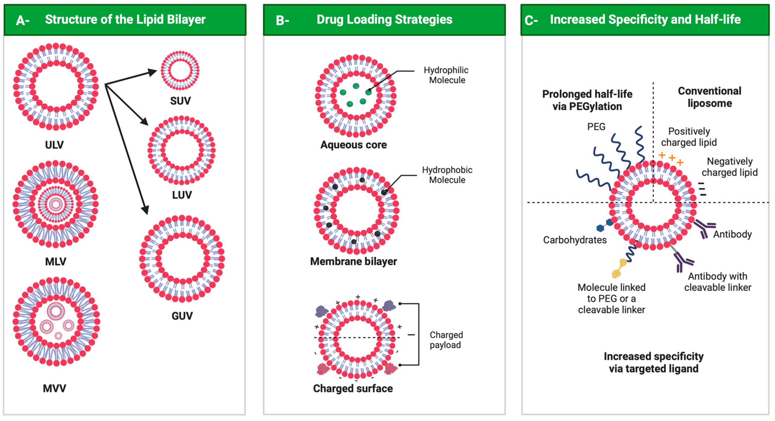
Drug delivery systems are believed to increase pharmaceutical efficacy and the therapeutic index by protecting and stabilizing bioactive molecules, such as protein and peptides, against body fluids’ enzymes and/or unsuitable physicochemical conditions while preserving the surrounding healthy tissues from toxicity. Liposomes are biocompatible and biodegradable and do not cause immunogenicity following intravenous or topical administration. Still, their most important characteristic is the ability to load any drug or complex molecule uncommitted to its hydrophobic or hydrophilic character. Selecting lipid components, ratios and thermo-sensitivity is critical to achieve a suitable nano-liposomal formulation. Nano-liposomal surfaces can be tailored to interact successfully with target cells, avoiding undesirable associations with plasma proteins and enhancing their half-life in the bloodstream. Macropinocytosis-dynamin-independent, cell-membrane-cholesterol-dependent processes, clathrin, and caveolae-independent mechanisms are involved in liposome internalization and trafficking within target cells to deliver the loaded drugs to modulate cell function. 

nano-delivery systems liposomal formulations liposomal marketed formulations liposome–cell interaction protein and peptide encapsulation

1. A General Overview of the Remarkable Role of Nature in Providing Potential Pharmacological Compounds

Nature has long proved an outstanding source of bioactive molecules with vast structural complexity and diversity. These have been used for millennia, mainly as component mixtures in crude extracts, as a unique strategy to treat several physiopathological conditions, including wounds, infectious diseases and other disorders [1][2]. In contrast, conventional drugs approved for therapeutic use are mainly represented by synthetic and small molecules ranging from 900 to 1500 Da designed and chemically synthetized to fit a pre-determined target via high-throughput screening. However, since the targets for small therapeutic molecules represent only 2–5% of the human genome products, the search for alternative therapeutics capable of expanding the number of new targets has increased the number of studies on natural therapeutic biomolecules, comprising searches in diverse classes, including natural macromolecules particularly proteins, peptides and polysaccharides [3][4]. The first efforts to identify and isolate active principles from natural extracts began in the 19th century with the isolation of the anti-malarial quinine, the opiate analgesic morphine and salicylic acid, creating the medicinal use of naturally isolated compounds. Since then, many other bioactive compounds have been isolated, mainly alkaloids, such as caffeine, nicotine, codeine, atropine, colchicine, cocaine and capsaicin [5]. The utilization of unmodified natural molecules increased by 43% during the 1930s (Figure 1). After this period, a decline in the number of unmodified natural products entering clinical practices took place, although the number of published data on natural products and bioactive molecules has increased. The past 30 years have been marked by a profile shift characterized by the replacement of unmodified natural molecules by semisynthetic and synthetic derivatives chemically designed but inspired by natural molecules (Figure 1) [6]. This profile shift was stimulated by the emergence of modern high-throughput platforms for screening and synthetic combinatorial strategies aimed at the fast discovery of new drug candidates similar to or analogs of natural molecules displaying special activities identified over the years, conducted through semi-synthetic modifications or by total synthesis, but still maintaining their pharmacophore. Moreover, the generation of synthetic/semisynthetic analogs is in contrast to time-consuming and high-cost methodologies applied to the identification of natural compounds in crude extracts, followed by their isolation and obtention in bulk amounts, impacting the emergence of new but unmodified natural pharmacological compounds for clinical use [7][8][9]. Because of this, the chemical synthesis of natural products was prioritized to optimize pharmaceutical production, improving the purity, quality and yields of bioactive compounds with reduced costs. Salicylic acid was the first natural product synthesized in 1853, copying the natural molecule [5].
Figure 1. Number of reports on bioactive molecules and natural products over the years, retrieved by an advanced search on the Pubmed database (https://pubmed.ncbi.nlm.nih.gov accessed on 5 July 2022) combining the terms “natural products” OR “bioactive molecules”. The number of publications contrast with FDA-approved unmodified natural drugs, which reached 43% in the 1930s. With the emergence of modern high-throughput platforms for screening and synthetic combinatorial strategies, synthetic/semi-synthetic drugs inspired by natural molecules reported throughout the years predominate over unmodified natural drugs in the market, increasing from 34% in 2010 to 50% currently.
The number of compounds approved by the FDA for clinical use classified as natural-based synthetic and semi-synthetic and purely natural compounds comprised 34% of the top 100 best-selling medicines in 2010, increasing to about 50% today, as a result of the cumulative knowledge reported in studies on bioactive molecules over the past 20 years, expanding the hall of new pharmaceutical molecules (Figure 1) [3][10].
Natural bioactive compounds can be found in living organisms from both edible or non-edible sources, mainly plants, including fruits, vegetables, whole grains, legumes, oils and others, but also animals and microorganisms such as bacteria and fungi, as well as algae [11]. The most studied bioactive compounds are polyphenols, such as curcumin, resveratrol, epigallocatechin gallate (EGCG), quercetin and anthocyanin, followed by polyunsaturated fatty acids such as docosahexaenoic acid (DHA), eicosapentaenoic acid (EPA) and arachidonic acid (ARA). However, native proteins or enzymes, as well as their bioactive peptides encrypted in primary structures, are being considered the next generation of pharmacological compounds. Several studies have been developed addressing plant lectins and milk proteins as non-toxic but highly effective drugs to treat several diseases. Moreover, bioactive polysaccharides, including lentinan and grifolan, vitamins, carotenoids, alkaloids and even mineral elements such as Zn, Fe, Mg, Ca, Na and P can have a positive impact on human health. These natural compounds exhibit various health-promoting effects such as antioxidant [12], antitumoral [13], immunomodulatory [14], anti-diabetic and anti-obesity [15][16], antiviral [17], anti-inflammatory [18] and neuroprotector activity [19], among others.
Therefore, natural bioactive compounds and their structural analogs—the natural-inspired molecules—have profoundly impacted the history of drug discovery, especially concerning cancer and infectious diseases, and continue providing pharmaceuticals able to treat almost all health disorders while composing the hall of new active agents in their unaltered, synthetic or semi-synthetic forms [1][8][9].

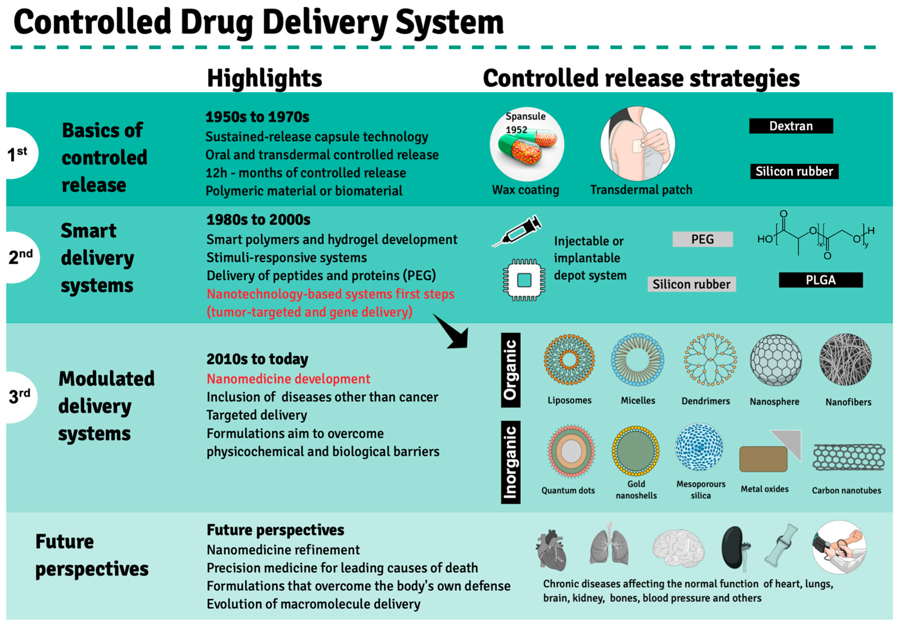
2. Evolution of Drug Delivery Systems and the Emergence of Nanotechnology in Clinical Treatments

Drug performance depends primarily on the type of delivery and release rate in order to guarantee a sustained drug amount that matches the therapeutic index between the maximum safe concentration and above the minimum effectiveness in the blood stream, avoiding abrupt peak concentrations as a result of massive drug release or the need for multiple doses to achieve drug effectiveness. These limitations encouraged the search for an ideal drug delivery system to substitute pills and capsules that release active compounds in aqueous media, termed immediate release (IR) formulations. The pioneer strategy in this regard comprised Spansules®, a first-generation drug delivery system, where micro-pellets were coated with a water-soluble wax of varied thicknesses, allowing for the controlled release of oral drugs for up to 12 h, leading to a constant concentration in blood. The first-generation drug delivery period, which ran from the 1950s to the 1970s, was also marked by the development of silicon- or dextran-based transdermal formulations based on controlled release, mainly through dissolution and diffusion (Figure 2). Both delivery systems faced several challenges, leading to advanced-generation systems [20].
Figure 2. Development of controlled drug delivery systems. Three distinct development phases—1st, 2nd and 3rd generations—are highlighted based on therapeutic products approved by the FDA since the 1950s, when Spansule®, the first drug release system, was approved for clinical use. The main highlights of each phase are depicted in the central column, while the main synthetic or biological material used in the drug delivery system preparation is indicated in the left column. The advent of nanotechnological drug delivery strategies is highlighted in red during the last decade of the second development phase. Future perspectives are exhibited at the bottom panel. Created using Mind the Graph (https://mindthegraph.com accessed on 14 August 2022).
The second generation of drug delivery formulations began in 1980 and lasted until 2010 and came with the understanding that it is not indispensable that drug concentrations be maintained at fixed levels. This period was marked by the development of smart polymers and hydrogels that evolved alongside biodegradable microparticles, solid implants and in situ gel-forming implants capable of delivering long-term release and stimuli-responsive bioactive compounds. Finally, nano-delivery systems emerged, where nanoparticles obtained from biodegradable polymers, polymeric micelles, lipids, chitosan and dendrimers are used to carry anticancer agents and gene sequences. The need to overcome biological and physicochemical barriers gave rise to the third generation of drug delivery systems, in which advanced nanomaterials enable the delivery of poorly water-soluble and/or very labile drugs or cell components, including peptides, proteins and DNA or RNA sequences. Novel delivery concepts include targeted drug delivery using nanoparticles and self-regulated drug delivery [20]. However, few drugs were approved by the FDA, even though much experimental evidence has proven that drug delivery employing nanotechnology systems could enhance the effectiveness of anticancer drugs against tumors in animal models [10].
According to the National Nanotechnology Initiative (2021), nanotechnology involves the manipulation of nanoparticles ranging from 1 to 100 nm, and their use is widespread in several areas, including engineering, physics and informatics. However, nanotechnology applications for pharmaceutical purposes may cause the most significant impact on human health and, because of this, has been considered the most promissory technology for treatments against degenerative pathologies such as cancer [21][22] and central nervous system disorders [23][24], as well as in antiviral therapy to aid in SARS-CoV-2 immunization and treatment [25][26].
The morphology of nano-scaled particles led to the optimization of treatments by enabling nano-materials to reach physiological sites that used to remain inaccessible, such as specific areas of the brain damaged by synucleinopathies or brain neoplasms, which require the ability to cross the blood–brain barrier to achieve therapeutic intervention success [24]. Moreover, nanomaterials exhibit a large surface/volume ratio, potentiating drug effects on target sites (cell, tissue or organ), comprising efficient bioactive molecule nanocarriers that can be encapsulated through adsorption, core entrapment or covalent surface binding. The nano-encapsulated bioactive molecule is released constantly and in a controlled manner, reducing adequate drug dosages to achieve pharmacological effects and minimize side effects widely attributed to conventional pharmaceuticals [27][28][29].
Nano-encapsulated compounds can be protected from degradation in the bloodstream. They can still reach intracellular compartments via endosomes through passive permeability, releasing bioactive compounds into the cytoplasm or directing them to intracellular targets with ligands associated with nano-capsule internalization [30]. In anti-tumorigenic therapies, the passive accumulation of anticancer drugs at the tumor localization can be increased by facilitating the permeability and retention effect (EPR), a phenomenon following the local inflammatory status of tumor blood vessels that become leaky, allowing for the passage and accumulation of nanometric materials in tumor tissues [31][32][33][34].
The primary chemical composition of nanoparticles described for medicinal therapy treatments comprises organic or inorganic compounds. The former includes natural or synthetic polymers such as chitosan, collagen, glycerol, polylactic-co-glycolic acid (PLGA) and dendrimers, as well as lipid-based materials, such as liposomes and micelles. At the same time, inorganic nanoparticles comprise gold nano-shells, metal oxides, carbon nanotubes and quantum dots (Figure 2) [35]. Molecules for therapeutic purposes can be encapsulated by these nanoparticles or complexed to them by adsorption or covalent binding to the nanocarrier surface. Depending on the carried bioactive compound, these nanoconjugates may be used to treat several pathologies, including degenerative diseases such as atherosclerosis and Parkinson’s disease, as well as cancers, as mentioned previously (Table 1).
Table 1. Pre-clinical studies on nano-delivery systems demonstrated in cell cultures and animal models for the loading of therapeutics aiming at the treatment of several health disorders.
PD—Parkinson’s disease.
The application of hydrophobically modified chitosan nanoparticles in the delivery of silibinin, a flavo-lignan isolated from the seeds of the milk thistle plant, was shown to improve the response promoted by a sustained release and enhance the solubility of this poorly aqueous soluble compound [46]. Similarly, resveratrol (RV), a polyphenol non-flavonoid commonly found in red or dark grapes, encapsulated in albumin nanoparticles (NPs) and functionalized with the tripeptide arginine-glycine-aspartate (RGD), demonstrated a prolonged RV blood circulation time and increased content surrounding a target tumor by 8.1-fold compared to free-RV. In a murine model, RV-NPs-RGD suppressed tumor growth with no relapse after 35 days of treatment, while progressive tumor growth was observed in an RV-free treatment, indicating that RV-NPs-RGD should be considered a promising chemotherapy agent [47][48].
Curcumin, another widely studied natural antioxidant and anticancer compound derived from turmeric or saffron, displays limited clinical application due to its molecule instability and poor solubility. However, curcumin encapsulated in solid lipid nanoparticles displays a high antiproliferative effect against SKBR3 cancer cells compared to free curcumin, with nanoencapsulation improving this compound’s bioavailability. Moreover, nano-encapsulated curcumin induced high-extension SKBR3 cell apoptosis (36.7%) and pronounced cell migration inhibition [49].
The co-encapsulation of tocotrienol (TRF) and simvastatin (SIM) in lipid nanoparticles has been reported as displaying an anti-proliferative effect on the breast adenocarcinoma +SA cell line, with an IC50 of 0.52 μM, when compared to each pharmaceutical separately. Nearly 25 to 40% of SIM is released over 48 h, with a quicker release in the first 10 h, followed by a slower and controlled kinetic release throughout the following 38 h [36]. Similarly, when lianol (LN) was encapsulated in solid nanoparticles, it affected HepG2 hepatocarcinoma and A549 pulmonary adenocarcinoma cell lines [37].
Folic acid conjugated through ionic interactions with chitosan nanoparticles (FA-CS) loaded with vincristine induces apoptosis in 75% of NCI-H460 lung cancer cells, in contrast to unloaded nanoparticles (31%) [39]. Nanoencapsulation efficiency can be increased when prepared at 4:25 (v/v) of vincristine/FA-CS, reaching a 95% efficiency and loading capacity of 48.65%, reinforcing promising anticancer effects on tumorigenic lung cells [39]. Similar effects were observed using PLGA (poly lactic-co-glycolic acid) nanofibers coated with metformin (MET), which exhibited cytotoxicity against A549 lung cancer cells after 48 h compared to free MET [42].
Liposome encapsulation improves the pharmacological effects of bioactive molecules, including metallic complexes, contributing to toxicity constraints by decreasing effective doses. Encapsulation of iridium III complexes (Ir-1, Ir-2 and Ir-3) in nano-liposomes, for example, was shown to improve their anticancer activity against several human carcinoma lineages, such as HepG2, hepatocellular carcinoma; HTC-116, colon cancer; HeLa, cervical cancer; A549, lung carcinoma; BEL-7402, hepatocellular carcinoma; SGC- 7901, gastric adenocarcinoma; and Eca-109, esophagus cancer cell, and against B16 mouse melanoma cells but no toxicity against healthy nih3T3 murine cells. On the other hand, no activity was observed for free Ir-2, and the nano-liposomal formulation exhibited a superior IC50 compared to cisplatin, especially against the A549 cell line. The intraperitoneal administration of the nano-liposomal formulation loaded with the Ir-2 complex for 10 consecutive days prior to A549 carcinogenic cell inoculation in mice resulted in 57.45% tumor mass reduction. In vitro assays have shown that encapsulated complexes stimulate apoptotic activity induced by increased intracellular ROS and cell cycle arrest at G0/G1 phases [40]. Similarly, TPGS-coated liposomes with SiRNA-corona Bcl-2 loaded with doxorubicin (Dox) promoted a 7-fold reduction in mice tumoral mass compared to free Dox [41]. Dendrimer nanoparticles of poly(amidoamine) (PAMAM) have also been successfully used to deliver methotrexate and D-glucose (PAMAM-MTX-GLU), inhibiting MDA-MB 231 breast cancer cells [43].
In addition to their use in anti-tumorigenic therapy, nanoparticles can be applied to treat other pathologies, including atherosclerosis and Parkinson’s disease. Mannose-functionalized dendrimer nanoparticles (mDNP) have been formulated to selectively deliver LXR-L, the liver receptor ligand, to macrophages associated with atherosclerotic plaque [44]. Four weeks of mDNP-LXR-L administration in LDL-receptor knockout mice led to nearly a 10% reduction in atherosclerotic plaque, necrosis area and inflammatory response evaluated by the expression of the metalloproteinase 9 (MMP-9) gene, regulated by NF-kB, the nuclear factor kappa B. In contrast, no increases in the expression of hepatic lipogenic or plasma lipid genes were observed. Gold nanoparticles (AuNCs) containing N-isobutyryl-L-cysteine (L-NIBC) are promising in the treatment of Parkinson’s disease. An in vitro assay of the effect of 1-methyl-4-phenyliridine (MPP) on SH-SY5Y cells showed that the nanoparticles inhibited synuclein fiber aggregation and formation in the brain while also preventing the formation of Lewy bodies and the death and dysfunction of neurons, all histological characteristics observed in Parkinson’s disease [45]. These data corroborated results described in mice pre-inoculated with the neurotoxin 1-methyl-4-phenyl-1,2,3,6-tetrahydropyridine (MPTP) and treated with AuNCs, which promoted the regression of behavioral disorders determined by the open field, swimming and rotarod tests, reinforcing the potential of nanoparticles loaded with L-NIBC to treat Parkinson’s disease.
The therapeutic efficacy of several pharmaceuticals has been improved by nanoparticles displaying the high potential of drugs complexed to nanomaterials to treat neoplasms, neurodegeneration and atherosclerosis, among other physiopathological conditions (Table 1). However, despite the significant science and technology advances in obtaining nanocarriers for pharmaceuticals, the time required for optimization and regulation aiming at their commercialization can take at least thirteen years, without considering pre-clinical studies and large-scale production, as well as phase 0, I, II and III clinical trials that must precede the submission and approval of health regulatory agencies to finally reach phase IV, as defined by the American Cancer Society [50][51].
A nano-liposomal formulation of doxorubicin (DOX), Doxil®, was the first drug- functionalized nanoparticle formulation approved by the U.S. Food and Drug Administration (FDA) in 1995. Doxil® can accumulate in solid tumors due to the EPR phenomena, effectively treating several cancers, including metastatic ovarian cancer and AIDS-related Kaposi’s sarcoma [52][53][54]. The anti-tumorigenic effect of Doxil® is attributed to its DNA interleaving ability and topoisomerase II inhibition, resulting in the downregulation of DNA replication and RNA transcription. Doxil® is not readily cleared from plasma by the mononuclear phagocytic system (MPS), which allows the continuous release of encapsulated DOX, improving its therapeutic performance compared to free-DOX [55][56]. Another available chemotherapy formulation is Abraxane®, in which paclitaxel is efficiently encapsulated in albumin nanoparticles. Abraxane®, approved by the FDA in 2005 and originally used to treat breast cancer, was expanded to treat advanced pancreatic carcinoma in 2013 [57]. Its formulation improves paclitaxel bioavailability, resulting in higher intra-tumoral paclitaxel concentrations facilitated by endothelial transcytosis through the albumin receptor-mediated (gp60) [58]. Other examples of nanostructured drugs developed for cancer therapy or diagnosis are depicted in Figure 3.
Figure 3. Timeline from 1995 to 2021 concerning representative anticancer pharmaceuticals loaded in nanoparticles approved by the American Food and Drug Administration for clinical purposes.

3. Liposomal Formulation Performance: Characteristics, Functionalization and Internalization

An effective liposomal formulation can be achieved by choosing the suitable lipid composition, proper functionalization and a smart targeting strategy. Phospholipid selection, considering the head group and chain length and number of liposome components, are crucial in determining liposome safety, stability and efficiency [59]. Besides their chemical composition, liposome efficiency depends on their stiffness, surface charge and lipid organization, as surface modifications interfere with liposome stability and can be handled to expand its use [60][61].
The main liposome components are glycerophospholipids, amphiphilic lipids composed of a glycerol molecule linked to a phosphate group and two fatty acid chains that can be either saturated or unsaturated. Phosphatidylcholine and phosphatidylethanolamine, abundant in plants and animals, are the most employed to form liposomes [62][63]. Liposomes can acquire positive, negative or neutral charges depending on the phospholipid head and chain, which confer their overall characteristics and functionalities [64][65]. Stability can be conferred to liposomes formed by phospholipids with longer tails and low degrees of unsaturation and ether linkages. Phospholipids with longer saturated hydrocarbon chains display a remarkable ability to interact with each other and form tightly ordered bilayer structures. On the other hand, shorter unsaturated hydrocarbon chains form liposomes with fluid and disordered bilayers [59][66].
Synthetic phospholipids can be formed by the modification of the head groups, aliphatic chains and alcohols of natural phospholipids, creating an enormous variety of synthetic phospholipids, such as 1,2-distearoyl-sn-glycero-3-phosphocholine (DSPC), 1,2-dipalmitoyl-sn-glycero-3-phosphocholine (DPPC), 1,2-dioleoyl-glycero- 3-phosphocholine (DOPC), 1,2-distearoyl-sn-glycero-3-phosphoglycerol (DSPG), 1,2-dipalmitoyl-sn-glycero-3-phosphoglycerol (DPPG), 1,2-dioleoyl-sn-glycerol -3-phosphoethanolamine, (DOPE) and 1,2-distearoyl-sn-glycero-3-phosphoethanolamine (DSPE), which have proven to be more stable [67].
Liposomes are formed by the hydrophilic interactions between polar head groups, the van der Waals forces between hydrocarbon chains, holding the long hydrocarbon tails together, and hydrogen linkages with H2O. In turn, H2O repels the hydrophobic chains, and the liposomes self-assemble spontaneously into a closed bilayer [67][68].
Liposomes can be classified as neutral, anionic and cationic according to lipid bilayer components. They can assume different 3D structures, as unilamellar vesicles (ULVs), multilamellar vesicles (MLVs) or multivesicular vesicles (MVVs). ULVs contain a single-lipid-bilayer membrane and can vary in size, with small unilamellar vesicles (SUVs) measuring between 30 and 100 nm, large unilamellar vesicles (LUVs) from 100 to 300 nm and giant unilamellar vesicles (GUVs) from 1 to 100 μm. In multilamellar vesicles, layers are concentric, while in MVVs, several smaller vesicles encase the interior of another vesicle (Figure 4, panel A) [69][70]. Another liposome formed by two bilayer membranes, the double-layer vesicle, comprises a framework that can improve liposome stability, which delays and sustains the release of their load [71].
Figure 4. Liposomes. Panel A: Structure of liposomes. ULV—unilamellar vesicle (small ULV (SUV), large ULV (LUV) and giant ULV (GUV)); MLV—multilamellar vesicle; MVV—multivesicular vesicle. Panel B: Drug loading strategies. Panel C: Liposome surface and functionalization. All figures were drawn with BioRender accessed on 1 December 2022.
In addition to phospholipids, its main components, cholesterol, glycol-derivatives including propylene glycol and polyethylene glycol (PEG) and even polymers such as chitosan, can increase liposome stability. These components can promote pronounced effects on healthy tissues and cells and activate or suppress immune responses [72][73]. Cholesterol incorporation into liposome bilayers can influence their fluidity, reducing their permeability and increasing in vitro and in vivo stabilities. Cholesterol, a hydrophobic molecule, induces a dense phospholipid packing and inhibits interactions among lipid chains interspersed between them, promoting liposome membrane stabilization [74][75][76]. The cholesterol molecule accommodates its hydroxyl group close to the hydrophilic region of phospholipids, and its aromatic ring lays parallel to the fatty acid chains in the lipid bilayer [77]. In the absence of cholesterol, liposomes can interact with proteins such as albumin, transferrin, macroglobulin and high-density lipoproteins (HDL), destabilizing the liposomal membrane structure and, consequently, decreasing drug delivery system performance [78][79][80].
Polymers such as chitosan are also used for liposomal surface modification, leading to a protective shell on the liposome surface, mainly for oral drug delivery [73][81]. Glycols such as propylene glycol incorporated into phospholipid vesicles with polyethylene glycol (PEG) have been advocated as flexible lipid vesicles to improve drug delivery systems targeting the skin [82][83]. Different PEGs on liposome surfaces can prolong their half-lives in the bloodstream, from a few minutes considering conventional liposomes to several hours for stealth liposomes, also called PEGylated liposomes [77]. One of the main disadvantages of conventional liposomes is their rapid elimination from the bloodstream, and they arrive in organs and tissues of the reticuloendothelial system, such as the liver and spleen [84]. The increases in liposome-circulating lifetimes promoted by PEG depend on the amount of grafted PEG and the molecular weight of the polymer. PEG longer chains typically increase the bloodstream residence time, reported as higher for PEGylated liposomes containing PEG 1900 and PEG 5000 compared to PEG 750 and PEG 120 [85][86]. Their molecular weight determines the conformation of PEG polymers on the surface of liposomes and PEG surface density in mushroom (low concentration) or brush (high concentration) regimes [87]. PEG concentrations from 5% to 10% (molar ratio) result in improvements in the degree of liposome stealth, and higher PEG concentrations (brush regimen) input to liposomes lead to high resistance to phagocytosis and poor activation of the human complement system [88].
PEG-enhanced liposome surfaces are associated with a cloaking effect, mimicking water-like structures, thus providing a steric barrier that prevents protein adsorption to liposome surfaces and their recognition by the mononuclear macrophage phagocytic system that would otherwise lead to rapid liposome clearance [59][67]. On the other hand, repeated venous administrations of PEGylated liposomes in animals at certain intervals induce immune responses, resulting in the loss of long-circulating characteristics and accelerating the blood clearance (ABC) phenomenon [89][90]. It has been suggested that anti-PEG IgM, produced by the spleen in response to a first dose, selectively binds to PEG chains in a second dose administered several days later and subsequently activates the complement system, one of the main opsonins, increasing the liver uptake of the following doses [90][91]. The occurrence and magnitude of the ABC phenomenon are influenced by the dose and physicochemical properties of PEGylated liposomes and the time interval between liposome administration and the encapsulated drug [92]. Many approaches have been tested to minimize the immunogenicity of the PEG moiety following repeated administrations. PEG lipids presenting a shorter alkyl chain can dissociate more quickly from the lipid bilayer, such as mPEG-DSPE and mPEG-CH, attenuating the ABC phenomenon [93][94].

References

  1. Atanasov, A.G.; Zotchev, S.B.; Dirsch, V.M.; Supuran, C.T. Natural products in drug discovery: Advances and opportunities. Nat. Rev. Drug Discov. 2021, 20, 200–216.
  2. Süntar, I. Importance of ethnopharmacological studies in drug discovery: Role of medicinal plants. Phytochem. Rev. 2020, 19, 1199–1209.
  3. Rask-Andersen, M.; Masuram, S.; Schiöth, H.B. The Druggable Genome: Evaluation of Drug Targets in Clinical Trials Suggests Major Shifts in Molecular Class and Indication. Annu. Rev. Pharmacol. Toxicol. 2014, 54, 9–26.
  4. Vargason, A.M.; Anselmo, A.C.; Mitragotri, S. The evolution of commercial drug delivery technologies. Nat. Biomed. Eng. 2021, 5, 951–967.
  5. Bernardini, S.; Tiezzi, A.; Laghezza Masci, V.; Ovidi, E. Natural products for human health: An historical overview of the drug discovery approaches. Nat. Prod. Res. 2018, 32, 1926–1950.
  6. Patridge, E.; Gareiss, P.; Kinch, M.S.; Hoyer, D. An analysis of FDA-approved drugs: Natural products and their derivatives. Drug Discov. Today 2016, 21, 204–207.
  7. Wright, G.D. Unlocking the potential of natural products in drug discovery. Microb. Biotechnol. 2019, 12, 55–57.
  8. Shen, B. A New Golden Age of Natural Products Drug Discovery. Cell 2015, 163, 1297–1300.
  9. Newman, D.J.; Cragg, G.M. Natural Products as Sources of New Drugs over the Nearly Four Decades from 01/1981 to 09/2019. J. Nat. Prod. 2020, 83, 770–803.
  10. Park, H.; Otte, A.; Park, K. Evolution of drug delivery systems: From 1950 to 2020 and beyond. J. Control. Release Off. J. Control. Release Soc. 2022, 342, 53–65.
  11. Santos, D.I.; Saraiva, J.M.A.; Vicente, A.A.; Moldão-Martins, M. Methods for determining bioavailability and bioaccessibility of bioactive compounds and nutrients. In Innovative Thermal and Non-Thermal Processing, Bioaccessibility and Bioavailability of Nutrients and Bioactive Compounds; Elsevier: Amsterdam, The Netherlands, 2019; pp. 23–54.
  12. Baranowska, M.; Suliborska, K.; Todorovic, V.; Kusznierewicz, B.; Chrzanowski, W.; Sobajic, S.; Bartoszek, A. Interactions between bioactive components determine antioxidant, cytotoxic and nutrigenomic activity of cocoa powder extract. Free. Radic. Biol. Med. 2020, 154, 48–61.
  13. Ren, B.; Kwah, M.X.-Y.; Liu, C.; Ma, Z.; Shanmugam, M.K.; Ding, L.; Xiang, X.; Ho, P.C.-L.; Wang, L.; Ong, P.S. Resveratrol for cancer therapy: Challenges and future perspectives. Cancer Lett. 2021, 515, 63–72.
  14. Wen, L.; Jiang, Y.; Zhou, X.; Bi, H.; Yang, B. Structure identification of soybean peptides and their immunomodulatory activity. Food Chem. 2021, 359, 129970.
  15. Moreno-Valdespino, C.A.; Luna-Vital, D.; Camacho-Ruiz, R.M.; Mojica, L. Bioactive proteins and phytochemicals from legumes: Mechanisms of action preventing obesity and type-2 diabetes. Food Res. Int. 2020, 130, 108905.
  16. Wu, S.-J.; Tung, Y.-J.; Ng, L.-T. Anti-diabetic effects of Grifola frondosa bioactive compound and its related molecular signaling pathways in palmitate-induced C2C12 cells. J. Ethnopharmacol. 2020, 260, 112962.
  17. Bhushan, I.; Sharma, M.; Mehta, M.; Badyal, S.; Sharma, V.; Sharma, I.; Singh, H.; Sistla, S. Bioactive compounds and probiotics–a ray of hope in COVID-19 management. Food Sci. Hum. Wellness 2021, 10, 131–140.
  18. Siu, F.Y.; Ye, S.; Lin, H.; Li, S. Galactosylated PLGA nanoparticles for the oral delivery of resveratrol: Enhanced bioavailability and in vitro anti-inflammatory activity. Int. J. Nanomed. 2018, 13, 4133.
  19. Wu, Y.; Cui, J. (−)-Epigallocatechin-3-gallate provides neuroprotection via AMPK activation against traumatic brain injury in a mouse model. Naunyn-Schmiedeberg’s Arch. Pharmacol. 2020, 393, 2209–2220.
  20. Reza Rezaie, H.; Esnaashary, M.; Aref arjmand, A.; Öchsner, A. The History of Drug Delivery Systems. In A Review of Biomaterials and Their Applications in Drug Delivery; Reza Rezaie, H., Esnaashary, M., Aref arjmand, A., Öchsner, A., Eds.; Springer: Singapore, 2018; pp. 1–8.
  21. Dai Phung, C.; Tran, T.H.; Nguyen, H.T.; Jeong, J.-H.; Yong, C.S.; Kim, J.O. Current developments in nanotechnology for improved cancer treatment, focusing on tumor hypoxia. J. Control. Release 2020, 324, 413–429.
  22. Barani, M.; Bilal, M.; Sabir, F.; Rahdar, A.; Kyzas, G.Z. Nanotechnology in ovarian cancer: Diagnosis and treatment. Life Sci. 2021, 266, 118914.
  23. Sarmah, D.; Banerjee, M.; Datta, A.; Kalia, K.; Dhar, S.; Yavagal, D.R.; Bhattacharya, P. Nanotechnology in the diagnosis and treatment of stroke. Drug Discov. Today 2021, 26, 585–592.
  24. Schlichtmann, B.W.; Hepker, M.; Palanisamy, B.N.; John, M.; Anantharam, V.; Kanthasamy, A.G.; Narasimhan, B.; Mallapragada, S.K. Nanotechnology-mediated therapeutic strategies against synucleinopathies in neurodegenerative disease. Curr. Opin. Chem. Eng. 2021, 31, 100673.
  25. Bhavana, V.; Thakor, P.; Singh, S.B.; Mehra, N.K. COVID-19: Pathophysiology, treatment options, nanotechnology approaches, and research agenda to combating the SARS-CoV2 pandemic. Life Sci. 2020, 261, 118336.
  26. Anand, K.; Vadivalagan, C.; Joseph, J.S.; Singh, S.K.; Gulati, M.; Shahbaaz, M.; Abdellattif, M.H.; Prasher, P.; Gupta, G.; Chellappan, D.K. A novel nano therapeutic using convalescent plasma derived exosomal (CPExo) for COVID-19: A combined hyperactive immune modulation and diagnostics. Chem. -Biol. Interact. 2021, 344, 109497.
  27. Misra, R.; Acharya, S.; Sahoo, S.K. Cancer nanotechnology: Application of nanotechnology in cancer therapy. Drug Discov. Today 2010, 15, 842–850.
  28. McNeil, S.E. Unique benefits of nanotechnology to drug delivery and diagnostics. In Characterization of Nanoparticles Intended for Drug Delivery; Springer: Berlin/Heidelberg, Germany, 2011; pp. 3–8.
  29. Petros, R.A.; DeSimone, J.M. Strategies in the design of nanoparticles for therapeutic applications. Nat. Rev. Drug Discov. 2010, 9, 615–627.
  30. Boulaiz, H.; Alvarez, P.J.; Ramirez, A.; Marchal, J.A.; Prados, J.; Rodriguez-Serrano, F.; Peran, M.; Melguizo, C.; Aranega, A. Nanomedicine: Application areas and development prospects. Int. J. Mol. Sci. 2011, 12, 3303–3321.
  31. Ferrari, M. Cancer nanotechnology: Opportunities and challenges. Nat. Rev. Cancer 2005, 5, 161–171.
  32. Rojas-Aguirre, Y.; Aguado-Castrejón, K.; González-Méndez, I. La nanomedicina y los sistemas de liberación de fármacos:¿ la (r) evolución de la terapia contra el cáncer? Educ. Química 2016, 27, 286–291.
  33. Falagan-Lotsch, P.; Grzincic, E.M.; Murphy, C.J. New advances in nanotechnology-based diagnosis and therapeutics for breast cancer: An assessment of active-targeting inorganic nanoplatforms. Bioconjugate Chem. 2017, 28, 135–152.
  34. Matsumura, Y.; Maeda, H. A new concept for macromolecular therapeutics in cancer chemotherapy: Mechanism of tumoritropic accumulation of proteins and the antitumor agent smancs. Cancer Res. 1986, 46, 6387–6392.
  35. Chen, L.; Liang, J. An overview of functional nanoparticles as novel emerging antiviral therapeutic agents. Mater. Sci. Eng. C 2020, 112, 110924.
  36. Ali, H.; Shirode, A.B.; Sylvester, P.W.; Nazzal, S. Preparation, characterization, and anticancer effects of simvastatin–tocotrienol lipid nanoparticles. Int. J. Pharm. 2010, 389, 223–231.
  37. Rodenak-Kladniew, B.; Islan, G.A.; de Bravo, M.G.; Durán, N.; Castro, G.R. Design, characterization and in vitro evaluation of linalool-loaded solid lipid nanoparticles as potent tool in cancer therapy. Colloids Surf. B Biointerfaces 2017, 154, 123–132.
  38. Safwat, S.; Hathout, R.M.; Ishak, R.A.; Mortada, N.D. Augmented simvastatin cytotoxicity using optimized lipid nanocapsules: A potential for breast cancer treatment. J. Liposome Res. 2017, 27, 1–10.
  39. Kumar, N.; Salar, R.K.; Prasad, M.; Ranjan, K. Synthesis, characterization and anticancer activity of vincristine loaded folic acid-chitosan conjugated nanoparticles on NCI-H460 non-small cell lung cancer cell line. Egypt. J. Basic Appl. Sci. 2018, 5, 87–99.
  40. Bai, L.; Fei, W.-D.; Gu, Y.-Y.; He, M.; Du, F.; Zhang, W.-Y.; Yang, L.-L.; Liu, Y.-J. Liposomes encapsulated iridium (III) polypyridyl complexes enhance anticancer activity in vitro and in vivo. J. Inorg. Biochem. 2020, 205, 111014.
  41. Li, Y.; Tan, X.; Liu, X.; Liu, L.; Fang, Y.; Rao, R.; Ren, Y.; Yang, X.; Liu, W. Enhanced anticancer effect of doxorubicin by TPGS-coated liposomes with Bcl-2 siRNA-corona for dual suppression of drug resistance. Asian J. Pharm. Sci. 2020, 15, 646–660.
  42. Samadzadeh, S.; Mousazadeh, H.; Ghareghomi, S.; Dadashpour, M.; Babazadeh, M.; Zarghami, N. In vitro anticancer efficacy of Metformin-loaded PLGA nanofibers towards the post-surgical therapy of lung cancer. J. Drug Deliv. Sci. Technol. 2021, 61, 102318.
  43. Torres-Pérez, S.A.; del Pilar Ramos-Godínez, M.; Ramón-Gallegos, E. Glycosylated one-step PAMAM dendrimers loaded with methotrexate for target therapy in breast cancer cells MDA-MB-231. J. Drug Deliv. Sci. Technol. 2020, 58, 101769.
  44. He, H.; Yuan, Q.; Bie, J.; Wallace, R.L.; Yannie, P.J.; Wang, J.; Lancina III, M.G.; Zolotarskaya, O.Y.; Korzun, W.; Yang, H. Development of mannose functionalized dendrimeric nanoparticles for targeted delivery to macrophages: Use of this platform to modulate atherosclerosis. Transl. Res. 2018, 193, 13–30.
  45. Gao, G.; Chen, R.; He, M.; Li, J.; Wang, L.; Sun, T. Gold nanoclusters for Parkinson’s disease treatment. Biomaterials 2019, 194, 36–46.
  46. Kuen, C.Y.; Fakurazi, S.; Othman, S.S.; Masarudin, M.J. Increased loading, efficacy and sustained release of silibinin, a poorly soluble drug using hydrophobically-modified chitosan nanoparticles for enhanced delivery of anticancer drug delivery systems. Nanomaterials 2017, 7, 379.
  47. Santos, A.C.; Pereira, I.; Pereira-Silva, M.; Ferreira, L.; Caldas, M.; Collado-González, M.; Magalhães, M.; Figueiras, A.; Ribeiro, A.J.; Veiga, F. Nanotechnology-based formulations for resveratrol delivery: Effects on resveratrol in vivo bioavailability and bioactivity. Colloids Surf. B Biointerfaces 2019, 180, 127–140.
  48. Geng, T.; Zhao, X.; Ma, M.; Zhu, G.; Yin, L. Resveratrol-loaded albumin nanoparticles with prolonged blood circulation and improved biocompatibility for highly effective targeted pancreatic tumor therapy. Nanoscale Res. Lett. 2017, 12, 1–10.
  49. Wang, W.; Chen, T.; Xu, H.; Ren, B.; Cheng, X.; Qi, R.; Liu, H.; Wang, Y.; Yan, L.; Chen, S. Curcumin-loaded solid lipid nanoparticles enhanced anticancer efficiency in breast cancer. Molecules 2018, 23, 1578.
  50. Cancino, J.; Marangoni, V.S.; Zucolotto, V. Nanotechnology in medicine: Concepts and concerns. Química Nova 2014, 37, 521–526.
  51. Society, A.C. Types and Phases of Clinical Trials. Available online: https://www.cancer.org/treatment/treatments-and-side-effects/clinical-trials/what-you-need-to-know/phases-of-clinical-trials.html (accessed on 7 October 2022).
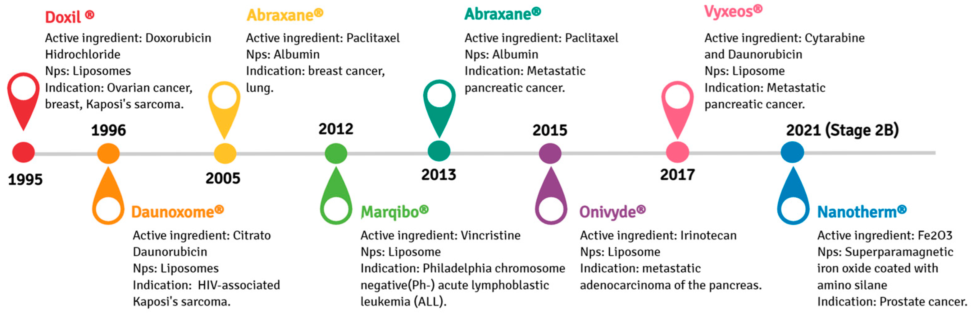
  52. Presant, C.A.; Scolaro, M.; Kennedy, P.; Blayney, D.; Flanagan, B.; Lisak, J.; Presant, J. Liposomal daunorubicin treatment of HIV-associated Kaposi’s sarcoma. Lancet 1993, 341, 1242–1243.
  53. Northfelt, D.W.; Martin, F.J.; Working, P.; Volberding, P.A.; Russell, J.; Newman, M.; Amantea, M.A.; Kaplan, L.D. Doxorubicin encapsulated in liposomes containing surface-bound polyethylene glycol: Pharmacokinetics, tumor localization, and safety in patients with AIDS-related Kaposi’s sarcoma. J. Clin. Pharmacol. 1996, 36, 55–63.
  54. Khawar, I.A.; Kim, J.H.; Kuh, H.-J. Improving drug delivery to solid tumors: Priming the tumor microenvironment. J. Control. Release 2015, 201, 78–89.
  55. Gordon, A.N.; Granai, C.O.; Rose, P.G.; Hainsworth, J.; Lopez, A.; Weissman, C.; Rosales, R.; Sharpington, T. Phase II Study of Liposomal Doxorubicin in Platinum- and Paclitaxel-Refractory Epithelial Ovarian Cancer. J. Clin. Oncol. 2000, 18, 3093–3100.
  56. Pillai, G. Nanomedicines for cancer therapy: An update of FDA approved and those under various stages of development. SOJ Pharm. Pharm. Sci. 2014, 1, 13.
  57. Guarneri, V.; Dieci, M.V.; Conte, P. Enhancing intracellular taxane delivery: Current role and perspectives of nanoparticle albumin-bound paclitaxel in the treatment of advanced breast cancer. Expert Opin. Pharmacother. 2012, 13, 395–406.
  58. Desai, N.; Trieu, V.; Yao, Z.; Louie, L.; Ci, S.; Yang, A.; Tao, C.; De, T.; Beals, B.; Dykes, D. Increased antitumor activity, intratumor paclitaxel concentrations, and endothelial cell transport of cremophor-free, albumin-bound paclitaxel, ABI-007, compared with cremophor-based paclitaxel. Clin. Cancer Res. Off. J. Am. Assoc. Cancer Res. 2006, 12, 1317–1324.
  59. Kapoor, M.; Lee, S.L.; Tyner, K.M. Liposomal drug product development and quality: Current US experience and perspective. AAPS J. 2017, 19, 632–641.
  60. Euliss, L.E.; DuPont, J.A.; Gratton, S.; DeSimone, J. Imparting size, shape, and composition control of materials for nanomedicine. Chem. Soc. Rev. 2006, 35, 1095–1104.
  61. Sebaaly, C.; Greige-Gerges, H.; Stainmesse, S.; Fessi, H.; Charcosset, C. Effect of composition, hydrogenation of phospholipids and lyophilization on the characteristics of eugenol-loaded liposomes prepared by ethanol injection method. Food Biosci. 2016, 15, 1–10.
  62. Tsuji, T.; Morita, S.-y.; Ikeda, Y.; Terada, T. Enzymatic fluorometric assays for quantifying all major phospholipid classes in cells and intracellular organelles. Sci. Rep. 2019, 9, 1–13.
  63. Yadav, D.; Sandeep, K.; Pandey, D.; Dutta, R.K. Liposomes for drug delivery. J. Biotechnol. Biomater 2017, 7, 1–8.
  64. Lombardo, D.; Calandra, P.; Barreca, D.; Magazù, S.; Kiselev, M.A. Soft interaction in liposome nanocarriers for therapeutic drug delivery. Nanomaterials 2016, 6, 125.
  65. Liu, W.; Ye, A.; Han, F.; Han, J. Advances and challenges in liposome digestion: Surface interaction, biological fate, and GIT modeling. Adv. Colloid Interface Sci. 2019, 263, 52–67.
  66. Rawicz, W.; Olbrich, K.C.; McIntosh, T.; Needham, D.; Evans, E. Effect of chain length and unsaturation on elasticity of lipid bilayers. Biophys. J. 2000, 79, 328–339.
  67. Monteiro, N.; Martins, A.; Reis, R.L.; Neves, N.M. Liposomes in tissue engineering and regenerative medicine. J. R. Soc. Interface 2014, 11, 20140459.
  68. Frezard, F. Liposomes: From biophysics to the design of peptide vaccines. Braz. J. Med. Biol. Res. 1999, 32, 181–189.
  69. Taylor, T.M.; Weiss, J.; Davidson, P.M.; Bruce, B.D. Liposomal nanocapsules in food science and agriculture. Crit. Rev. Food Sci. Nutr. 2005, 45, 587–605.
  70. Laouini, A.; Jaafar-Maalej, C.; Limayem-Blouza, I.; Sfar, S.; Charcosset, C.; Fessi, H. Preparation, characterization and applications of liposomes: State of the art. J. Colloid Sci. Biotechnol. 2012, 1, 147–168.
  71. Zawada, Z.H. Vesicles with a double bilayer. Cell. Mol. Biol. Lett. 2004, 9, 589–602.
  72. Inglut, C.T.; Sorrin, A.J.; Kuruppu, T.; Vig, S.; Cicalo, J.; Ahmad, H.; Huang, H.-C. Immunological and toxicological considerations for the design of liposomes. Nanomaterials 2020, 10, 190.
  73. Ferreira, P.G.; Ferreira, V.F.; da Silva, F.D.C.; Freitas, C.S.; Pereira, P.R.; Paschoalin, V.M.F. Chitosans and Nanochitosans: Recent Advances in Skin Protection, Regeneration, and Repair. Pharmaceutics 2022, 14, 1307.
  74. Bozzuto, G.; Molinari, A. Liposomes as nanomedical devices. Int. J. Nanomed. 2015, 10, 975.
  75. Lee, S.-C.; Lee, K.-E.; Kim, J.-J.; Lim, S.-H. The effect of cholesterol in the liposome bilayer on the stabilization of incorporated retinol. J. Liposome Res. 2005, 15, 157–166.
  76. Sharifi, F.; Zhou, R.; Lim, C.; Jash, A.; Abbaspourrad, A.; Rizvi, S.S. Generation of liposomes using a supercritical carbon dioxide eductor vacuum system: Optimization of process variables. J. CO2 Util. 2019, 29, 163–171.
  77. Beltrán-Gracia, E.; López-Camacho, A.; Higuera-Ciapara, I.; Velázquez-Fernández, J.B.; Vallejo-Cardona, A.A. Nanomedicine review: Clinical developments in liposomal applications. Cancer Nanotechnol. 2019, 10, 1–40.
  78. Lu, R.-M.; Chen, M.-S.; Chang, D.-K.; Chiu, C.-Y.; Lin, W.-C.; Yan, S.-L.; Wang, Y.-P.; Kuo, Y.-S.; Yeh, C.-Y.; Lo, A. Targeted drug delivery systems mediated by a novel Peptide in breast cancer therapy and imaging. PLoS ONE 2013, 8, e66128.
  79. Maranhão, R.C.; Vital, C.G.; Tavoni, T.M.; Graziani, S.R. Clinical experience with drug delivery systems as tools to decrease the toxicity of anticancer chemotherapeutic agents. Expert Opin. Drug Deliv. 2017, 14, 1217–1226.
  80. Yingchoncharoen, P.; Kalinowski, D.S.; Richardson, D.R. Lipid-based drug delivery systems in cancer therapy: What is available and what is yet to come. Pharmacol. Rev. 2016, 68, 701–787.
  81. Caddeo, C.; Pons, R.; Carbone, C.; Fernàndez-Busquets, X.; Cardia, M.C.; Maccioni, A.M.; Fadda, A.M.; Manconi, M. Physico-chemical characterization of succinyl chitosan-stabilized liposomes for the oral co-delivery of quercetin and resveratrol. Carbohydr. Polym. 2017, 157, 1853–1861.
  82. Elmoslemany, R.M.; Abdallah, O.Y.; El-Khordagui, L.K.; Khalafallah, N.M. Propylene glycol liposomes as a topical delivery system for miconazole nitrate: Comparison with conventional liposomes. AAPS PharmSciTech 2012, 13, 723–731.
  83. Manconi, M.; Mura, S.; Sinico, C.; Fadda, A.M.; Vila, A.; Molina, F. Development and characterization of liposomes containing glycols as carriers for diclofenac. Colloids Surf. Physicochem. Eng. Asp. 2009, 342, 53–58.
  84. Lee, Y.; Thompson, D. Stimuli-responsive liposomes for drug delivery. Wiley Interdiscip. Rev. Nanomed. Nanobiotechnol. 2017, 9, e1450.
  85. Immordino, M.L.; Dosio, F.; Cattel, L. Stealth liposomes: Review of the basic science, rationale, and clinical applications, existing and potential. Int. J. Nanomed. 2006, 1, 297.
  86. Allen, T.M.; Cullis, P.R. Liposomal drug delivery systems: From concept to clinical applications. Adv. Drug Deliv. Rev. 2013, 65, 36–48.
  87. Perry, J.L.; Reuter, K.G.; Kai, M.P.; Herlihy, K.P.; Jones, S.W.; Luft, J.C.; Napier, M.; Bear, J.E.; DeSimone, J.M. PEGylated PRINT nanoparticles: The impact of PEG density on protein binding, macrophage association, biodistribution, and pharmacokinetics. Nano Lett. 2012, 12, 5304–5310.
  88. Nogueira, E.; Loureiro, A.; Nogueira, P.; Freitas, J.; Almeida, C.R.; Härmark, J.; Hebert, H.; Moreira, A.; Carmo, A.M.; Preto, A. Liposome and protein based stealth nanoparticles. Faraday Discuss. 2013, 166, 417–429.
  89. Dams, E.T.; Laverman, P.; Oyen, W.J.; Storm, G.; Scherphof, G.L.; Van der Meer, J.W.; Corstens, F.H.; Boerman, O.C. Accelerated blood clearance and altered biodistribution of repeated injections of sterically stabilized liposomes. J. Pharmacol. Exp. Ther. 2000, 292, 1071–1079.
  90. Ishida, T.; Kiwada, H. Accelerated blood clearance (ABC) phenomenon upon repeated injection of PEGylated liposomes. Int. J. Pharm. 2008, 354, 56–62.
  91. Ishida, T.; Ichihara, M.; Wang, X.; Yamamoto, K.; Kimura, J.; Majima, E.; Kiwada, H. Injection of PEGylated liposomes in rats elicits PEG-specific IgM, which is responsible for rapid elimination of a second dose of PEGylated liposomes. J. Control. Release 2006, 112, 15–25.
  92. Lila, A.S.A.; Kiwada, H.; Ishida, T. The accelerated blood clearance (ABC) phenomenon: Clinical challenge and approaches to manage. J. Control. Release 2013, 172, 38–47.
  93. Ambegia, E.; Ansell, S.; Cullis, P.; Heyes, J.; Palmer, L.; MacLachlan, I. Stabilized plasmid–lipid particles containing PEG-diacylglycerols exhibit extended circulation lifetimes and tumor selective gene expression. Biochim. Et Biophys. Acta (BBA)-Biomembr. 2005, 1669, 155–163.
  94. Webb, M.S.; Saxon, D.; Wong, F.M.; Lim, H.J.; Wang, Z.; Bally, M.B.; Choi, L.S.; Cullis, P.R.; Mayer, L.D. Comparison of different hydrophobic anchors conjugated to poly (ethylene glycol): Effects on the pharmacokinetics of liposomal vincristine. Biochim. Et Biophys. Acta (BBA)-Biomembr. 1998, 1372, 272–282.
More
Upload a video for this entry
Information
Contributors MDPI registered users' name will be linked to their SciProfiles pages. To register with us, please refer to https://encyclopedia.pub/register : , Patricia Ribeiro Pereira , Cyntia Silva Freitas , Vania Margaret Flosi Paschoalin
View Times: 1.5K
Revisions: 2 times (View History)
Update Date: 23 Dec 2022
1000/1000
Hot Most Recent
Notice
You are not a member of the advisory board for this topic. If you want to update advisory board member profile, please contact office@encyclopedia.pub.
OK
Confirm
Only members of the Encyclopedia advisory board for this topic are allowed to note entries. Would you like to become an advisory board member of the Encyclopedia?
Yes
No
Academic Video Service