Your browser does not fully support modern features. Please upgrade for a smoother experience.
Submitted Successfully!
 +1 credit
Thank you for your contribution! You can also upload a video entry or images related to this topic.
For video creation, please contact our Academic Video Service.
| Version | Summary | Created by | Modification | Content Size | Created at | Operation |
|---|---|---|---|---|---|---|
| 1 | Yuliya S. Krasikova | -- | 2840 | 2022-12-15 08:31:26 | | | |
| 2 | Dean Liu | Meta information modification | 2840 | 2022-12-16 03:48:49 | | | | |
| 3 | Dean Liu | -4 word(s) | 2836 | 2022-12-19 09:18:48 | | | | |
| 4 | Dean Liu | Meta information modification | 2836 | 2022-12-30 06:07:04 | | |
Video Upload Options
We provide professional Academic Video Service to translate complex research into visually appealing presentations. Would you like to try it?
Cite
If you have any further questions, please contact Encyclopedia Editorial Office.
Krasikova, Y.S.; Lavrik, O.I.; Rechkunova, N.I. The Xeroderma Pigmentosum Group A Protein. Encyclopedia. Available online: https://encyclopedia.pub/entry/38803 (accessed on 26 July 2026).
Krasikova YS, Lavrik OI, Rechkunova NI. The Xeroderma Pigmentosum Group A Protein. Encyclopedia. Available at: https://encyclopedia.pub/entry/38803. Accessed July 26, 2026.
Krasikova, Yuliya S., Olga I. Lavrik, Nadejda I. Rechkunova. "The Xeroderma Pigmentosum Group A Protein" Encyclopedia, https://encyclopedia.pub/entry/38803 (accessed July 26, 2026).
Krasikova, Y.S., Lavrik, O.I., & Rechkunova, N.I. (2022, December 15). The Xeroderma Pigmentosum Group A Protein. In Encyclopedia. https://encyclopedia.pub/entry/38803
Krasikova, Yuliya S., et al. "The Xeroderma Pigmentosum Group A Protein." Encyclopedia. Web. 15 December, 2022.
Copy Citation
Nucleotide excision repair (NER) is a central DNA repair pathway responsible for removing a wide variety of DNA-distorting lesions from the genome. The highly choreographed cascade of core NER reactions requires more than 30 polypeptides. The xeroderma pigmentosum group A (XPA) protein plays an essential role in the NER process. XPA interacts with almost all NER participants and organizes the correct NER repair complex. In the absence of XPA’s scaffolding function, no repair process occurs. Researchers briefly summarize the knowledge about the XPA protein structure and analyze the formation of contact with its protein partners during NER complex assembling.
XPA
nucleotide excision repair (NER)
DNA repair
1. Introduction
The nucleotide excision repair (NER) pathway is the most universal repair pathway for the removal of a wide range of structurally unrelated DNA lesions, including UV photolesions (e.g., cyclobutane pyrimidine dimers (CPDs) and pyrimidine-pyrimidone (6-4)-photoproducts (6-4PPs)), intrastrand crosslinks, reactive oxygen species-induced base alterations, and bulky adducts of DNA bases with reactive metabolites of some chemical carcinogens or chemotherapeutic agents [1][2][3]. Mutations in NER-related genes are associated with an autosomal recessive disease called xeroderma pigmentosum (XP) [4]. XP is characterized by extreme sensitivity of the skin to sunlight and a dramatically increased risk of skin cancer [5][6]. A subset of XP patients developed a profound neurodegenerative condition known as XP neurological disease [7]. XP patients can be classified into seven complementation groups, XP-A through XP-G, depending on the specific gene that is affected [1]. Patients with known mutations in the XPA gene have the most severe form of XP, indicating a critical role of the XPA protein in the NER process.
Initially, XPA was considered to be the sole damage recognition factor [8]; then, it was proposed that the XPA–RPA complex performs the first recognition [9][10]. Later, enough data was accumulated to suggest that lesions are primarily recognized by the XPC–RAD23B–CEN2 complex [3], and XPA takes part in the damage verification process (together with TFIIH) and overall plays the role of an organizing or scaffold component of NER [11][12][13].
Three-dimensional structures of XPA give researchers an opportunity to propose a spatial arrangement of XPA inside NER machinery. Today, several spatial structures of human XPA exist: two solution structures of the minimal DNA-binding domain (DBD) determined by NMR spectroscopy (Protein Data Bank (PDB) IDs 1XPA and 1D4U) [14][15], a crystal structure of a redefined XPA DBD (PDB ID: 6J44), and a crystal structure of the XPA–DNA complex (PDB ID: 6LAE) [16][17]. The series of crystal structures of a yeast homolog of XPA (Rad14) in a complex with different lesion-containing DNA substrates illustrates the DNA-binding capacity of the XPA DBD [18][19][20]. Recent advances in cryo-electron microscopy (cryo-EM) gave investigators a unique chance to look into the structure of a TFIIH–XPA–DNA complex (PDB: 6RO4) [21] and confirm the biochemical data about XPA localization inside the DNA repair bubble [22]. Furthermore, cryo-EM data have expanded researchers' knowledge about the modulation of TFIIH activity by XPA and XPG.
2. XPA’s Structure and DNA-Binding and Protein-Binding Abilities
XPA is one of the smallest proteins inside the NER machine: human XPA is 31 kDa and consists of only 273 amino acid residues (aa). XPA is composed of a central globular domain (aa 98–219) that is flanked by dynamically disordered N and C termini (Figure 1A). This kind of structural organization, when structured globular domains are combined with disordered regions, is very common among eukaryotic proteins [23]. Frequently, disordered proteins (entirely disordered or containing disordered sequences, as in XPA) interact with or function as hubs in protein interaction networks or play a central role in an ordered assembly of macromolecular machines [24][25]. Indeed, both low-sequence-complexity parts enable the XPA protein to interact with a variety of protein partners. Future experiments will shed light on disordered XPA part properties: do they do folding upon binding to protein partners (and should be called “intrinsically disordered”), or do these regions not adopt a specific three-dimensional structure during functioning. In addition, the N terminus contains a conserved nuclear localization signal (NLS) that lies in the 13-residue stretch from aa 30 to 42 [26][27] (Figure 1A). The NLS is a tag that ensures that the protein is sorted into the nucleus, but in the case of XPA, things are not so simple, and researchers discuss this matter below.
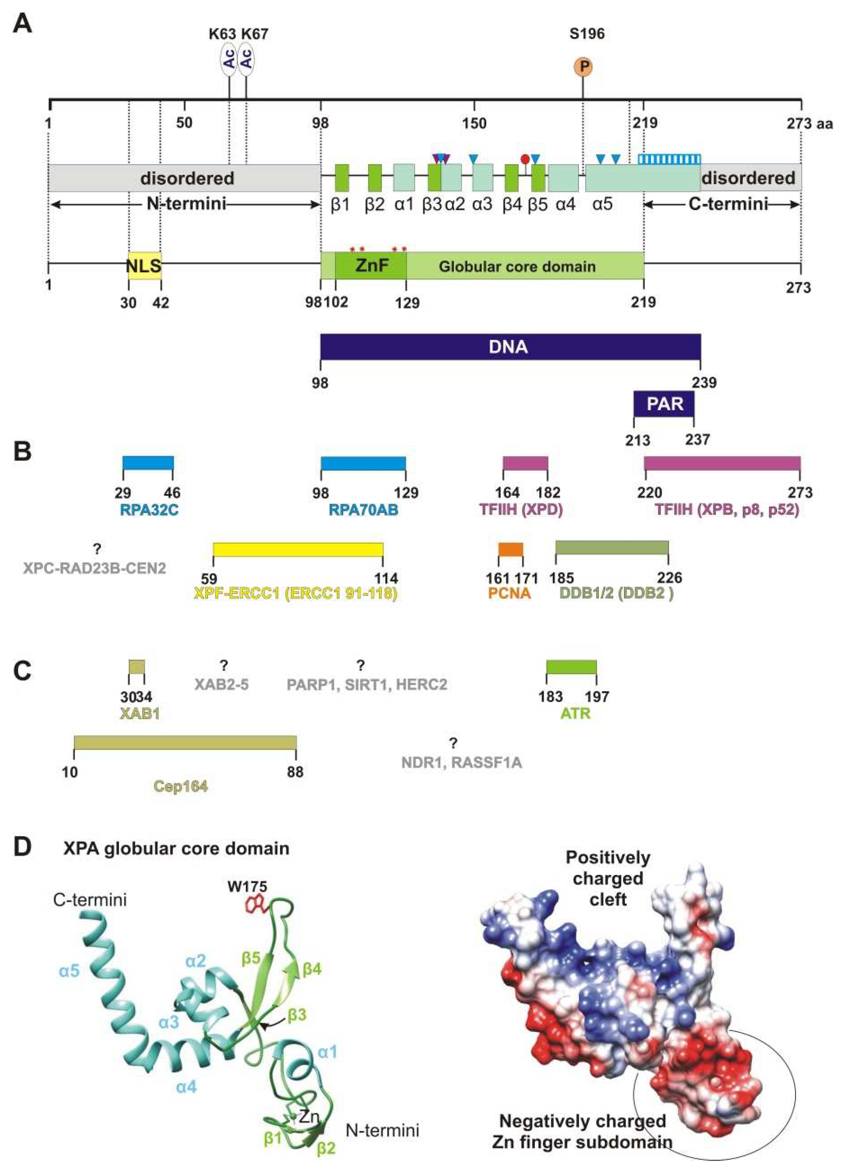
Figure 1. XPA’s structure and interaction partners. (A) The map of the XPA domain structure and known points of PTMs: phosphorylation at S196 and acetylation at K63 and K67. Secondary-structure elements are shown according to crystal structures PDB 6LAE and 6J44: β-strands are green (β1: aa 103–104, β2: aa 111–112, β3: aa 138–140, β4: aa 164–167, and β5: aa 178–172), and α-helices are light blue (α1: aa 116–121, α2: aa 141–148, α3: aa 151–157, α4: aa 183–194, and α5: aa 197–239). Positively charged residues K141, K151, K179, R207, and R211, which are directly involved in interactions with backbones of a DNA duplex, are shown as blue triangles. Two residues (Thr140 and Thr142, indicated as purple triangles) interact with the DNA backbone through a van der Waals contact and a hydrogen bond, respectively. Extended helix α5 contains several positively charged residues (Lys217/218/221/222/224/236 and Arg227/228/231/237) that are possibly involved in DNA binding, which are shown as a blue striped box. Conserved residue Trp175 intercalates into unpaired bases of single-stranded DNA (ssDNA) at the ss–dsDNA junction and is displayed as a red circle. Unstructured N- and C-terminal regions are gray. Zinc-coordinated conserved cysteine residues (C105, C108, C126, and C129) are presented as red asterisks and a Zn-finger motif (ZnF, aa 102–129) colored green. The N terminus accommodates a nuclear localization signal (NLS, aa 30–42), which is yellow. DNA-binding (aa 98–239) and poly(ADP-ribose) (PAR)-binding (aa 213–237) motifs are mapped to the overall XPA structure and are highlighted in dark blue. (B) Interaction sites for NER protein partners on XPA, which are aligned with the XPA residues involved in each interaction. Proteins whose interaction sites are unknown are gray. (C) XPA interaction partners outside NER. (D) A structural model of the XPA globular core domain (PDB ID: 6LAE). A ribbon diagram with color codes according to (A). The Trp175 residue is shown in red. A distribution of the electrostatic potential on the surface for the same structure: a positive charge is shown in blue, and a negative charge is red. The structures were generated using UCSF Chimera software (version 1.16).
The central domain contains a C4-type Zn-finger (ZnF) motif that has the sequence Cys105-X2-Cys108-X17-Cys126-X2-Cys129 [28] (Figure 1A). Side chains of cysteines Cys105/108 and Cys126/129 coordinate the zinc ion [14]. Although the ZnF of XPA is essential for DNA binding and for NER activity [29], the ZnF core itself is not directly in contact with DNA [16] but rather properly ensures the DBD folding [30]. Moreover, the zinc-containing subdomain is even negatively charged due to many glutamate and aspartate residues [14] (Figure 1D). Notably, the UvrA protein has two units of the same C4 type of the ZnF motif [28].
Originally, the minimal DNA-binding domain (DBD) of XPA was mapped to a central globular core between residues 98 and 219 [14][15][29][31][32]. This region contains a sheet-helix-hairpin motif (residues 138–182) and a helix-turn-helix motif (residues 183–230) that form a shallow clamp-like or right hand-like structure (the sheet-helix-hairpin motif as fingers and helix-turn-helix as a thumb) with a positively charged inner surface (Figure 1D). The internal curvature of the basic cleft fits well to the diameter of a standard B-form dsDNA [14]. Subsequent studies have found that some residues on the C-terminal side beyond the minimal DBD domain are also involved in binding to DNA substrates [33][34][35]. Later, the XPA DBD was redefined and extended by 20 additional C-terminal residues (Asp220–Thr239). The redefined XPA DBD (aa 98–239) can bind to DNA with an affinity nearly identical to that of the full-length XPA protein [34][35]. The crystal structure revealed that the C-terminal extension folds as a long α-helix (α5 in Figure 1A) with a basic residue cluster resulting in the formation of a consecutive positively charged surface [16][17]. Interestingly, structural superposition of the human XPA DBD on a yeast Rad14–DNA complex implies that the α5 extension (Asp217–Thr239) cannot directly come into contact with a DNA substrate.
XPA has an ability to recognize some bulky lesions [22][36] and especially prefers to bind to kinked and branched DNA structures [37][38][39]. All of these DNA structures contain an ss–dsDNA junction. XPA binds to the duplex part of the junction in a non-sequence-specific manner via electrostatic interactions between the positively charged cleft and negatively charged phosphate backbones of the DNA duplex [17] (Figure 1F and Figure 2). This intermediate is further stabilized by hydrogen bonding of the side chain hydroxyl group of Thr142, and a van der Waals contact is formed between the side chain Cβ atom of Thr140 and DNA’s phosphate moieties (Figure 1A and Figure 2). Thereafter, Trp175 from the top of the hairpin between β4 and β5 is stacked with bases of the unpaired ssDNA at the junction, thereby giving rise to a stable conformation of this β-hairpin [17][21]. According to the energy calculations, binding to ssDNA in the 3′→5′ direction is more favorable than that in the 5′→3′ direction (relative to α5), but it is not observed experimentally [29][38].
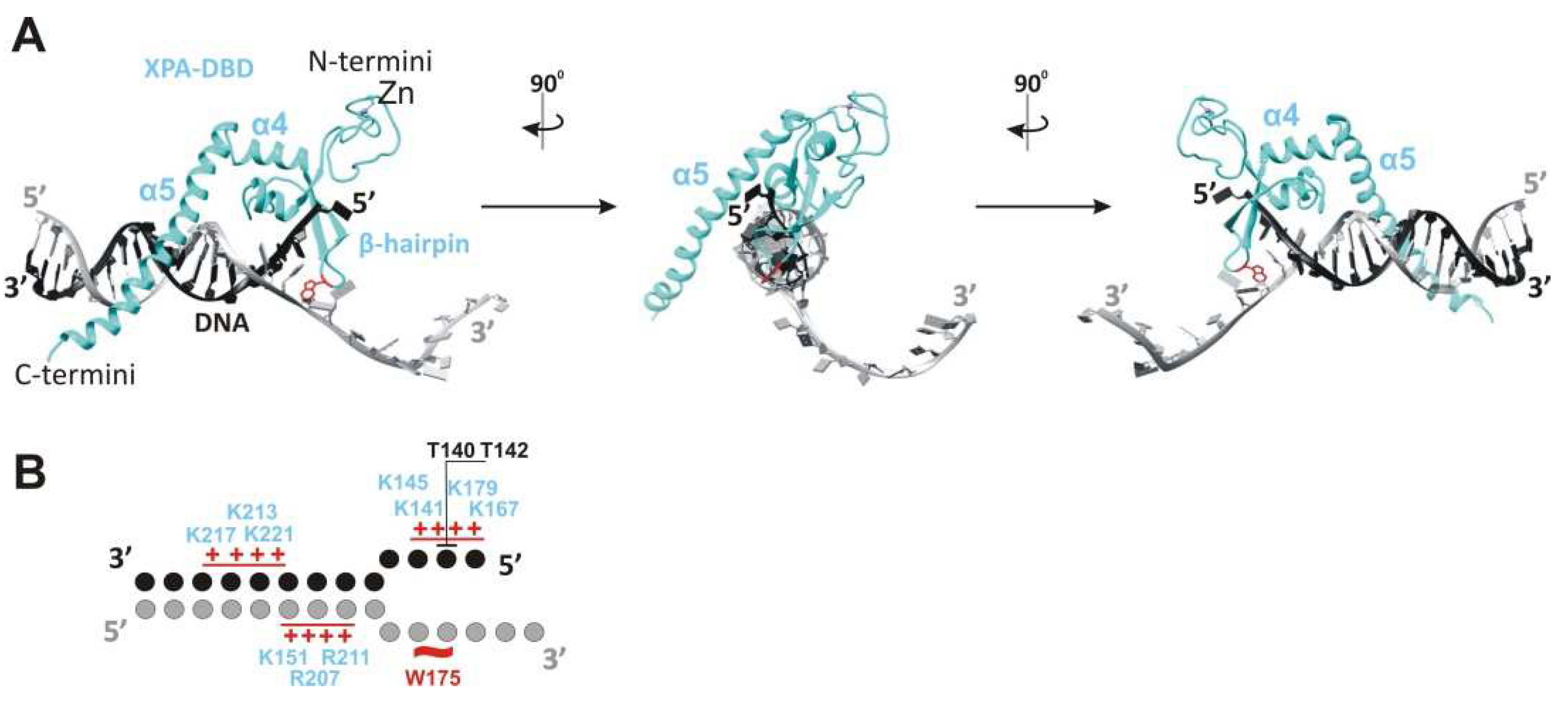
Figure 2. XPA interaction with the ss–dsDNA junction. (A) Cryo-EM structure PDB: 6RO4 provides details of the XPA–DBD interaction with the ss–dsDNA junction. XPA demarcates the 5′ edge of the DNA repair bubble. XPA inserts its intercalating β-hairpin between DNA single strands at the junction. Red colored Trp175 from the tip of the β-hairpin stacks against the base of the DNA 3′-extension at the junction. The structures were generated using UCSF Chimera software (version 1.16). (B) Schematic representation of the interactions between side chains of the XPA and DNA junction, according to cryo-EM structure PDB: 6RO4 [21] and crystal structure PDB: 6LAE. DNA nucleotides are indicated as circles. Patches of positively charged residues in proximity to the DNA backbone are indicated by red pluses. Hydrogen bonding of T142 and a van der Waals contact of T140 are indicated as black lines.
Recently, it was shown with atomic force microscopy, scanning force microscopy, and mathematical modeling that XPA undergoes episodic one-dimensional diffusion to search the DNA for damage [40]. The functional meaning of XPA’s damage recognition ability is not clear, and today, XPA is considered only as a protein scaffold element inside the NER complex. Anyway, XPA interacts with the proteins involved in every step of NER, from damage recognition to DNA synthesis. Yet, it is unknown how many contacts XPA could engage in concurrently, and it is possible that XPA interacts with these proteins not simultaneously but in the order of them joining the repair machinery. Figure 1B lists XPA’s protein interaction partners directly involved in NER according to the following process steps:
2.1. Initial Damage Recognition
XPA interacts physically with DDB2 through aa 185–226, and this interaction can be seen both in vitro and in vivo [41]. UV-damaged DNA-binding protein (DDB1/2) is a heterodimeric protein consisting of subunits DDB1 and DDB2/XPE and has an extraordinarily high binding affinity and specificity for CPD and 6-4PP [2][3]. The biological role of the XPA–DDB2 interaction is unclear, but because the DDB2 interaction site overlaps with the poly(ADP-ribose) [PAR]-binding motif (aa 213–237), researchers can speculate that the XPA–DDB2 complex is involved in PAR-dependent chromatin remodeling together with PARP1 and XPC [42]. XPC (which functions in the complex with proteins RAD23B and CEN2) is the protein sensor responsible for the detection of a wide variety of DNA lesions that are repaired through the global genome NER (GG-NER) pathway [43]. XPA interacts with XPC and stimulates its binding to the DNA [44][45]. The XPC interaction site in the XPA sequence and the biological meaning of this interaction are unknown.
2.2. Damage Verification
The TFIIH complex is the key protein for the damage verification step. Depending on the context, the TFIIH composition changes from a core of seven subunits, including translocase XPB and helicase XPD, to ten subunits, through the addition of three CAK (Cdk-activating kinase module) kinase subunits [21][46]. XPA’s whole C terminus is involved in an interaction with the TFIIH protein [47]. Recent cryo-EM data expanded the knowledge about the TFIIH–XPA interaction [21]. XPA (together with nuclease XPG) facilitates the CAK kinase module release from the core TFIIH and stabilizes an alternative conformation of TFIIH, where the XPD helicase assumes the open conformation for the functioning. In this complex, XPA forms a bridge between the XPB and the XPD, and moreover, XPA’s extended α5 helix and ATPase XPB form a positively charged tunnel that holds the DNA within. Thus, by trapping the DNA within a duplex tunnel, XPA may keep the NER machinery on the DNA during lesion scanning and processing [21].
2.3. Pre-Incision Complex Formation
Immediately after TFIIH opens the DNA repair bubble, the undamaged ssDNA that is being formed is bound by replication protein A (RPA) [48]. RPA is a heterotrimer consisting of subunits RPA70, RPA32, and RPA14 [49][50]. XPA interacts with two of them: RPA70 (which contains OB-fold domains A, B, and C) and RPA32 (which contains the D OB-fold domain) [51]. It was shown recently that the interaction between XPA (aa 29–46) and RPA32C is important for the initial association of XPA with NER complexes, whereas the interaction between XPA (aa 98–126) and RPA70AB is needed for structural shaping of the complex to enable the dual incision reaction [52]. Pre-incision complex formation is completed by the engagement of nuclease XPF–ERCC1, which is recruited by the XPA through the interaction with ERCC1 [53][54][55][56][57]. XPA aa 59–114 are responsible for this interaction. In particular, the Gly72–Phe75 region (also known as the G motif) and the Glu78–Glu84 region (i.e., the E motif) are the residues necessary for the binding of XPF–ERCC1. Biochemical data have shown that the ZnF motif (aa 102–129) is partially involved in this contact, but the NMR and molecular dynamics simulation revealed that only the 14-amino acid sequence (aa 67–80) mediates this interaction.
2.4. Dual Incision, Resynthesis, and Ligation
After the first incision, proliferating cell nuclear antigen (PCNA) joins the NER complex. PCNA is the processivity factor for DNA polymerases. XPA has been found to interact directly with PCNA via the APIM sequence (the AlkB homolog 2 PCNA-interacting motif), and it has been shown that XPA and PCNA colocalize to the damaged DNA foci in a cell culture [58][59][60]. XPA−/− cells complemented with XPA containing a mutated APIM sequence have high UV sensitivity and a deficient repair of CPDs and 6-4PPs and are consequently more arrested in the S phase as compared to XPA−/− cells complemented with wild-type XPA. Notably, XPA colocalizes with PCNA in the replication foci and is loaded onto a newly synthesized DNA in undamaged cells; thus, it is possible that this interaction is required for DNA-processing pathways other than NER.
2.5. XPA Dimerization
It is well-known that isolated XPA easily forms a homodimer. Moreover, in vitro, it can form the XPA2–RPA complex [61]. Instead, XPA has been widely assumed to be a monomer participating in the mechanism of NER (the first statement about the monomeric functional form was published by [62]). Accordingly, the physiological meaning of the XPA dimerization and the structural mechanism of this process are still unclear. Recently obtained molecular dynamics simulation data indicate that some residues make a contribution to the intermolecular interactions in XPA homodimers, but this needs to be validated by another approach [63]. Notably, XPA is not the sole NER protein that demonstrates easily dimerization characters, but as in the case of XPA, there is no functional explanation found for this ability [64].
2.6. PTM Proteins
Today, it is known that XPA is precisely tuned by several PTMs [13][65][66]. Obviously, XPA interacts with proteins that provide these modifications and removes them, but researchers know the interaction site only for the ATR kinase: the α4 helix [67] (Figure 1C). Interaction sites with proteins facilitating these modifications—RASSF1A and NDR1—have not been determined either. The only interaction site that has been identified is the one for the Cep164 protein, which functions in ATR-mediated checkpoint activation; it interacts with XPA through aa 10–88 of XPA [68].
2.7. XABs
Using the yeast two-hybrid system, researchers identified a novel set of XPA-interacting proteins that was designated as XABs [69]. XAB1 is a cytoplasmic protein with GTPase activity and binds to the N-terminal region (residues 1–52) of XPA, and the region “aa 30–34” is directly involved in this interaction (Figure 1C). The XAB1-interacting site overlaps with the NLS and raises a question about XAB1′s role in the cytoplasmic sequestering of XPA. Among the five found XABs, only XAB2 has a nuclear function and is intensively investigated. It has been reported that XAB2 interacts with the proteins involved in transcription-coupled NER (TC-NER), for example, CSA, CSB, and RNA polymerase II [70]. XAB2 contains 15 TPR (tetratricopeptide repeat) motifs and appears to have a role in transcription and pre-mRNA splicing [71]. Generally, XAB2 serves as a guardian of POLR2A (the largest catalytic subunit of RNA polymerase II) expression to ensure global gene expression and to antagonize cell senescence [72]. Furthermore, XAB2 promotes homologous recombination and facilitates histone acetylation events linked to homologous recombination [73]. Immunoprecipitation studies have revealed that XAB2 interacts with endonucleases ERCC1–XPF and XPG outside NER under the conditions that favor the formation of R-loops [74]. Altogether, XAB2 is involved in R-loop removal and pre-mRNA splicing; both processes are linked to transcription [75]. Future experiments will shed light on the possible involvement of XPA in processes where XAB2 is active.
References
- Gillet, L.C.; Schärer, O.D. Molecular mechanisms of mammalian global genome nucleotide excision repair. Chem Rev. 2006, 106, 253–276.
- Mu, H.; Geacintov, N.E.; Broyde, S.; Yeo, J.E.; Schärer, O.D. Molecular basis for damage recognition and verification by XPC-RAD23B and TFIIH in nucleotide excision repair. DNA Repair 2018, 71, 33–42.
- Kusakabe, M.; Onishi, Y.; Tada, H.; Kurihara, F.; Kusao, K.; Furukawa, M.; Iwai, S.; Yokoi, M.; Sakai, W.; Sugasawa, K. Mechanism and regulation of DNA damage recognition in nucleotide excision repair. Genes Environ. 2019, 41, 2.
- Rapin, I.; Lindenbaum, Y.; Dickson, D.W.; Kraemer, K.H.; Robbins, J.H. Cockayne syndrome and xeroderma pigmentosum. Neurology 2000, 55, 1442–1449.
- DiGiovanna, J.J.; Kraemer, K.H. Shining a light on xeroderma pigmentosum. J. Invest. Dermatol. 2012, 132, 785–796.
- Fassihi, H.; Sethi, M.; Fawcett, H.; Wing, J.; Chandler, N.; Mohammed, S.; Craythorne, E.; Morley, A.M.; Lim, R.; Turner, S.; et al. Deep phenotyping of 89 xeroderma pigmentosum patients reveals unexpected heterogeneity dependent on the precise molecular defect. Proc. Natl. Acad. Sci. USA 2016, 113, 1236–1245.
- Krasikova, Y.; Rechkunova, N.; Lavrik, O. Nucleotide Excision Repair: From Molecular Defects to Neurological Abnormalities. Int. J. Mol. Sci. 2021, 22, 6220.
- Cleaver, J.E.; States, J.C. The DNA damage-recognition problem in human and other eukaryotic cells: The XPA damage binding protein. Biochem. J. 1997, 328, 1–12.
- Wakasugi, M.; Sancar, A. Order of assembly of human DNA repair excision nuclease. J. Biol. Chem. 1999, 274, 18759–18768.
- Thoma, B.S.; Vasquez, K.M. Critical DNA damage recognition functions of XPC-hHR23B and XPA-RPA in nucleotide excision repair. Mol. Carcinog. 2003, 38, 1–13.
- Li, C.L.; Golebiowski, F.M.; Onishi, Y.; Samara, N.L.; Sugasawa, K.; Yang, W. Tripartite DNA Lesion Recognition and Verification by XPC, TFIIH, and XPA in Nucleotide Excision Repair. Mol. Cell 2015, 59, 1025–1034.
- Sugitani, N.; Sivley, R.M.; Perry, K.E.; Capra, J.A.; Chazin, W.J. XPA: A key scaffold for human nucleotide excision repair. DNA Repair 2016, 44, 123–135.
- Borszéková Pulzová, L.; Ward, T.A.; Chovanec, M. XPA: DNA Repair Protein of Significant Clinical Importance. Int. J. Mol. Sci. 2020, 21, 2182.
- Ikegami, T.; Kuraoka, I.; Saijo, M.; Kodo, N.; Kyogoku, Y.; Morikawa, K.; Tanaka, K.; Shirakawa, M. Solution structure of the DNA- and RPA-binding domain of the human repair factor XPA. Nat. Struct. Biol. 1998, 5, 701–706.
- Buchko, G.W.; Daughdrill, G.W.; de Lorimier, R.; Rao, B.K.; Isern, N.G.; Lingbeck, J.M.; Taylor, J.S.; Wold, M.S.; Gochin, M.; Spicer, L.D.; et al. Interactions of human nucleotide excision repair protein XPA with DNA and RPA70 Delta C327: Chemical shift mapping and 15N NMR relaxation studies. Biochemistry 1999, 38, 15116–15128.
- Lian, F.M.; Yang, X.; Yang, W.; Jiang, Y.L.; Qian, C. Structural characterization of the redefined DNA-binding domain of human XPA. Biochem. Biophys. Res. Commun. 2019, 514, 985–990.
- Lian, F.M.; Yang, X.; Jiang, Y.L.; Yang, F.; Li, C.; Yang, W.; Qian, C. New structural insights into the recognition of undamaged splayed-arm DNA with a single pair of non-complementary nucleotides by human nucleotide excision repair protein XPA. Int. J. Biol. Macromol. 2020, 148, 466–474.
- Koch, S.C.; Kuper, J.; Gasteiger, K.L.; Simon, N.; Strasser, R.; Eisen, D.; Geiger, S.; Schneider, S.; Kisker, C.; Carell, T. Structural insights into the recognition of cisplatin and AAF-dG lesion by Rad14 (XPA). Proc. Natl. Acad. Sci. USA 2015, 112, 8272–8277.
- Simon, N.; Ebert, C.; Schneider, S. Structural basis for bulky-Adduct DNA-lesion recognition by the nucleotide excision repair protein Rad14. Chemistry 2016, 22, 10782–10785.
- Ebert, C.; Simon, N.; Schneider, S.; Carell, T. Structural insights into the recognition of N(2) -Aryl- and C8-Aryl DNA lesions by the repair protein XPA/ Rad14. Chembiochem 2017, 18, 1379–1382.
- Kokic, G.; Chernev, A.; Tegunov, D.; Dienemann, C.; Urlaub, H.; Cramer, P. Structural basis of TFIIH activation for nucleotide excision repair. Nat. Commun. 2019, 10, 2885.
- Krasikova, Y.S.; Rechkunova, N.I.; Maltseva, E.A.; Petruseva, I.O.; Lavrik, O.I. Localization of xeroderma pigmentosum group A protein and replication protein A on damaged DNA in nucleotide excision repair. Nucleic Acids Res. 2010, 38, 8083–8094.
- Wright, P.E.; Dyson, H.J. Intrinsically disordered proteins in cellular signalling and regulation. Nat. Rev. Mol. Cell Biol. 2015, 16, 18–29.
- Dunker, A.K.; Cortese, M.S.; Romero, P.; Iakoucheva, L.M.; Uversky, V.N. Flexible nets. The roles of intrinsic disorder in protein interaction networks. FEBS J. 2005, 272, 5129–5148.
- Kim, P.M.; Sboner, A.; Xia, Y.; Gerstein, M. The role of disorder in interaction networks: A structural analysis. Mol. Syst. Biol. 2008, 4, 179.
- Miyamoto, I.; Miura, N.; Niwa, H.; Miyazaki, J.; Tanaka, K. Mutational analysis of the structure and function of the xeroderma pigmentosum group A complementing protein. Identification of essential domains for nuclear localization and DNA excision repair. J. Biol. Chem. 1992, 267, 12182–12187.
- Barve, A.; Ghaskadbi, S.; Ghaskadbi, S. Structural and Sequence Similarities of Hydra Xeroderma Pigmentosum A Protein to Human Homolog Suggest Early Evolution and Conservation. BioMed Res. Int. 2013, 2013, 854745.
- Tanaka, K.; Miura, N.; Satokata, I.; Miyamoto, I.; Yoshida, M.C.; Satoh, Y.; Kondo, S.; Yasui, A.; Okayama, H.; Okada, Y. Analysis of a human DNA excision repair gene involved in group A xeroderma pigmentosum and containing a zinc-finger domain. Nature 1990, 348, 73–76.
- Buchko, G.W.; Tung, C.S.; McAteer, K.; Isern, N.G.; Spicer, L.D.; Kennedy, M.A. DNA-XPA interactions: A 31P NMP and molecular modeling study of dCCAATTAACC association with the minimal DNA-binding domain (M98-F219) of the Nucleotide Excision Repair protein XPA. Nucleic Acids Res. 2001, 29, 2635–2643.
- Smirnova, J.; Zhukova, L.; Witkiewicz-Kucharczyk, A.; Kopera, E.; Oledzki, J.; Wysłouch-Cieszyńska, A.; Palumaa, P.; Hartwig, A.; Bal, W. Quantitative electrospray ionization mass spectrometry of zinc finger oxidation: The reaction of XPA zinc finger with H(2)O(2). Anal Biochem 2007, 369, 226–231.
- Kuraoka, I.; Morita, E.H.; Saijo, M.; Matsuda, T.; Morikawa, K.; Shirakawa, M.; Tanaka, K. Identification of a damaged-DNA binding domain of the XPA protein. Mutat. Res. 1996, 362, 87–95.
- Buchko, G.W.; Ni, S.; Thrall, B.D.; Kennedy, M.A. Structural features of the minimal DNA binding domain (M98-F219) of human nucleotide excision repair protein XPA. Nucleic Acids Res. 1998, 26, 2779–2788.
- Hilton, B.; Shkriabai, N.; Musich, P.R.; Kvaratskhelia, M.; Shell, S.; Zou, Y. A new structural insight into XPA-DNA interactions. Biosci. Rep. 2014, 34, e00162.
- Sugitani, N.; Shell, S.M.; Soss, S.E.; Chazin, W.J. Redefining the DNA-binding domain of human XPA. J. Am. Chem. Soc. 2014, 136, 10830–10833.
- Sugitani, N.; Voehler, M.W.; Roh, M.S.; Topolska-Wos, A.M.; Chazin, W.J. Analysis of DNA binding by human factor xeroderma pigmentosum complementation group A (XPA) provides insight into its interactions with nucleotide excision repair substrates. J. Biol. Chem. 2017, 292, 16847–16857.
- Missura, M.; Buterin, T.; Hindges, R.; Hubscher, U.; Kasparkova, J.; Brabec, V.; Naegeli, H. Double-check probing of DNA bending and unwinding by XPA-RPA: An architectural function in DNA repair. EMBO J. 2001, 20, 3554–3564.
- Camenisch, U.; Dip, R.; Schumacher, S.B.; Schuler, B.; Naegeli, H. Recognition of helical kinks by xeroderma pigmentosum group A protein triggers DNA excision repair. Nat. Struct. Mol. Biol. 2006, 13, 278–284.
- Yang, Z.; Roginskaya, M.; Colis, L.C.; Basu, A.K.; Shell, S.M.; Liu, Y.; Musich, P.R.; Harris, C.M.; Harris, T.M.; Zou, Y. Specific and efficient binding of xeroderma pigmentosum complementation group A to double-strand/single-strand DNA junctions with 3'- and/or 5'-ssDNA branches. Biochemistry 2006, 45, 15921–15930.
- Krasikova, Y.S.; Rechkunova, N.I.; Maltseva, E.A.; Lavrik, O.I. RPA and XPA interaction with DNA structures mimicking intermediates of the late stages in nucleotide excision repair. PLoS ONE 2018, 13, e0190782.
- Beckwitt, E.C.; Jang, S.; Carnaval Detweiler, I.; Kuper, J.; Sauer, F.; Simon, N.; Bretzler, J.; Watkins, S.C.; Carell, T.; Kisker, C.; et al. Single molecule analysis reveals monomeric XPA bends DNA and undergoes episodic linear diffusion during damage search. Nat. Commun. 2020, 11, 1356.
- Wakasugi, M.; Kasashima, H.; Fukase, Y.; Imura, M.; Imai, R.; Yamada, S.; Cleaver, J.E.; Matsunaga, T. Physical and functional interaction between DDB and XPA in nucleotide excision repair. Nucleic Acids Res. 2009, 37, 516–525.
- Blessing, C.; Apelt, K.; van den Heuvel, D.; Gonzalez-Leal, C.; Rother, M.B.; van der Woude, M.; González-Prieto, R.; Yifrach, A.; Parnas, A.; Shah, R.G.; et al. XPC-PARP complexes engage the chromatin remodeler ALC1 to catalyze global genome DNA damage repair. Nat. Commun. 2022, 13, 4762.
- Schärer, O.D. Nucleotide excision repair in eukaryotes. Cold Spring Harb. Perspect. Biol. 2013, 5, a012609.
- Bunick, C.G.; Miller, M.R.; Fuller, B.E.; Fanning, E.; Chazin, W.J. Biochemical and structural domain analysis of xeroderma pigmentosum complementation group C protein. Biochemistry 2006, 45, 14965–14979.
- Krasikova, Y.S.; Rechkunova, N.I.; Maltseva, E.A.; Petruseva, I.O.; Silnikov, V.N.; Zatsepin, T.S.; Oretskaya, T.S.; Schärer, O.D.; Lavrik, O.I. Interaction of nucleotide excision repair factors XPC-HR23B, XPA, and RPA with damaged DNA. Biochemistry 2008, 73, 886–896.
- Tsutakawa, S.E.; Tsai, C.L.; Yan, C.; Bralić, A.; Chazin, W.J.; Hamdan, S.M.; Schärer, O.D.; Ivanov, I.; Tainer, J.A. Envisioning how the prototypic molecular machine TFIIH functions in transcription initiation and DNA repair. DNA Repair 2020, 96, 102972.
- Park, C.H.; Mu, D.; Reardon, J.T.; Sancar, A. The general transcription-repair factor TFIIH is recruited to the excision repair complex by the XPA protein independent of the TFIIE transcription factor. J. Biol. Chem. 1995, 270, 4896–4902.
- Chen, R.; Wold, M.S. Replication protein A: Single-stranded DNA's first responder: Dynamic DNA-interactions allow replication protein A to direct single-strand DNA intermediates into different pathways for synthesis or repair. Bioessays 2014, 36, 1156–1161.
- Wold, M.S. Replication protein A: A heterotrimeric, single-stranded DNA-binding protein required for eukaryotic DNA metabolism. Annu. Rev. Biochem. 1997, 66, 61–92.
- Krasikova, Y.S.; Rechkunova, N.I.; Lavrik, O.I. Replication protein A as a major eukaryotic single-stranded DNA-binding protein and its role in DNA repair. Mol. Biol. 2016, 50, 735–750.
- Topolska-Woś, A.M.; Sugitani, N.; Cordoba, J.J.; Le Meur, K.V.; Le Meur, R.A.; Kim, H.S.; Yeo, J.E.; Rosenberg, D.; Hammel, M.; Schärer, O.D.; et al. A key interaction with RPA orients XPA in NER complexes. Nucleic Acids Res. 2020, 48, 2173–2188.
- Kim, M.; Kim, H.S.; D'Souza, A.; Gallagher, K.; Jeong, E.; Topolska-Wós, A.; Ogorodnik Le Meur, K.; Tsai, C.L.; Tsai, M.S.; Kee, M.; et al. Two interaction surfaces between XPA and RPA organize the preincision complex in nucleotide excision repair. Proc. Natl. Acad. Sci. USA 2022, 119, e2207408119.
- Li, L.; Elledge, S.J.; Peterson, C.A.; Bales, E.S.; Legerski, R.J. Specific association between the human DNA repair proteins XPA and ERCC1. Proc. Natl. Acad. Sci. USA 1994, 91, 5012–5016.
- Park, C.H.; Sancar, A. Formation of a ternary complex by human XPA, ERCC1, and ERCC4(XPF) excision repair proteins. Proc. Natl. Acad. Sci. USA 1994, 91, 5017–5021.
- Tsodikov, O.V.; Ivanov, D.; Orelli, B.; Staresincic, L.; Shoshani, I.; Oberman, R.; Schärer, O.D.; Wagner, G.; Ellenberger, T. Structural basis for the recruitment of ERCC1-XPF to nucleotide excision repair complexes by XPA. EMBO J. 2007, 26, 4768–4776.
- Croteau, D.L.; Peng, Y.; Van Houten, B. DNA repair gets physical: Mapping an XPA-binding site on ERCC1. DNA Repair 2008, 7, 819–826.
- Fadda, E. Conformational determinants for the recruitment of ERCC1 by XPA in the nucleotide excision repair (NER) Pathway: Structure and dynamics of the XPA binding motif. Biophys J. 2013, 104, 2503–2511.
- Gilljam, K.M.; Feyzi, E.; Aas, P.A.; Sousa, M.M.; Muller, R.; Vagbo, C.B.; Catterall, T.C.; Liabakk, N.B.; Slupphaug, G.; Drablos, F.; et al. Identification of a novel, widespread, and functionally important PCNA-binding motif. J. Cell Biol. 2009, 186, 645–654.
- Gilljam, K.M.; Müller, R.; Liabakk, N.B.; Otterlei, M. Nucleotide excision repair is associated with the replisome and its efficiency depends on a direct interaction between XPA and PCNA. PLoS ONE 2012, 7, e49199.
- Hara, K.; Uchida, M.; Tagata, R.; Yokoyama, H.; Ishikawa, Y.; Hishiki, A.; Hashimoto, H. Structure of proliferating cell nuclear antigen (PCNA) bound to an APIM peptide reveals the universality of PCNA interaction. Acta Cryst. F Struct. Biol. Commun. 2018, 74, 214–221.
- Yang, Z.G.; Liu, Y.; Mao, L.Y.; Zhang, J.T.; Zou, Y. Dimerization of human XPA and formation of XPA(2)-RPA protein complex. Biochemistry 2002, 41, 13012–13020.
- Rademakers, S.; Volker, M.; Hoogstraten, D.; Nigg, A.L.; Moné, M.J.; Van Zeeland, A.A.; Hoeijmakers, J.H.; Houtsmuller, A.B.; Vermeulen, W. Xeroderma pigmentosum group A protein loads as a separate factor onto DNA lesions. Mol. Cell. Biol. 2003, 23, 5755–5767.
- Pradhan, S.; Sarma, H.; Mattaparthi, V.S.K. Investigation of the probable homo-dimer model of the Xeroderma pigmentosum complementation group A (XPA) protein to represent the DNA-binding core. J. Biomol. Struct. Dyn. 2019, 37, 3322–3336.
- Muniesa-Vargas, A.; Theil, A.F.; Ribeiro-Silva, C.; Vermeulen, W.; Lans, H. XPG: A multitasking genome caretaker. Cell Mol. Life Sci. 2022, 79, 166.
- Park, J.M.; Kang, T.H. Transcriptional and posttranslational regulation of nucleotide excision repair: The guardian of the genome against ultraviolet radiation. Int. J. Mol. Sci. 2016, 17, 1840.
- Rechkunova, N.I.; Maltseva, E.A.; Lavrik, O.I. Post-translational Modifications of Nucleotide Excision Repair Proteins and Their Role in the DNA Repair. Biochemistry 2019, 84, 1008–1020.
- Shell, S.M.; Li, Z.; Shkriabai, N.; Kvaratskhelia, M.; Brosey, C.; Serrano, M.A.; Chazin, W.J.; Musich, P.R.; Zou, Y. Checkpoint kinase ATR promotes nucleotide excision repair of UV-induced DNA damage via physical interaction with xeroderma pigmentosum group A. J. Biol. Chem. 2009, 284, 24213–24222.
- Pan, Y.R.; Lee, E.Y. UV-dependent interaction between Cep164 and XPA mediates localization of Cep164 at sites of DNA damage and UV sensitivity. Cell Cycle 2009, 8, 655–664.
- Nitta, M.; Saijo, M.; Kodo, N.; Matsuda, T.; Nakatsu, Y.; Tamai, H.; Tanaka, K. A novel cytoplasmic GTPase XAB1 interacts with DNA repair protein XPA. Nucleic Acids Res. 2000, 28, 4212–4218.
- Nakatsu, Y.; Asahina, H.; Citterio, E.; Rademakers, S.; Vermeulen, W.; Kamiuchi, S.; Yeo, J.P.; Khaw, M.C.; Saijo, M.; Kodo, N.; et al. XAB2, a novel tetratricopeptide repeat protein involved in transcription-coupled DNA repair and transcription. J. Biol. Chem. 2000, 275, 34931–34937.
- Kuraoka, I.; Ito, S.; Wada, T.; Hayashida, M.; Lee, L.; Saijo, M.; Nakatsu, Y.; Matsumoto, M.; Matsunaga, T.; Handa, H.; et al. Isolation of XAB2 complex involved in pre-mRNA splicing, transcription, and transcription-coupled repair. J. Biol. Chem. 2008, 283, 940–950.
- Hou, S.; Qu, D.; Li, Y.; Zhu, B.; Liang, D.; Wei, X.; Tang, W.; Zhang, Q.; Hao, J.; Guo, W.; et al. XAB2 depletion induces intron retention in POLR2A to impair global transcription and promote cellular senescence. Nucleic Acids Res. 2019, 47, 8239–8254.
- Onyango, D.O.; Howard, S.M.; Neherin, K.; Yanez, D.A.; Stark, J.M. Tetratricopeptide repeat factor XAB2 mediates the end resection step of homologous recombination. Nucleic Acids Res. 2016, 44, 5702–5716.
- Goulielmaki, E.; Tsekrekou, M.; Batsiotos, N.; Ascensão-Ferreira, M.; Ledaki, E.; Stratigi, K.; Chatzinikolaou, G.; Topalis, P.; Kosteas, T.; Altmüller, J.; et al. The splicing factor XAB2 interacts with ERCC1-XPF and XPG for R-loop processing. Nat. Commun. 2021, 12, 3153.
- Donnio, L.M.; Cerutti, E.; Magnani, C.; Neuillet, D.; Mari, P.O.; Giglia-Mari, G. XAB2 dynamics during DNA damage-dependent transcription inhibition. Elife 2022, 11, e77094.
More
Information
Subjects:
Biochemistry & Molecular Biology; Dermatology; Oncology
Contributors
MDPI registered users' name will be linked to their SciProfiles pages. To register with us, please refer to https://encyclopedia.pub/register
:
View Times:
1.1K
Revisions:
4 times
(View History)
Update Date:
30 Dec 2022
Table of Contents


Notice
You are not a member of the advisory board for this topic. If you want to update advisory board member profile, please contact office@encyclopedia.pub.
OK
Confirm
Only members of the Encyclopedia advisory board for this topic are allowed to note entries. Would you like to become an advisory board member of the Encyclopedia?
Yes
No
${ textCharacter }/${ maxCharacter }
Submit
Cancel
Back
Comments
${ item }
|
${ item.createdUser.fullName }
${ item.createdAt }
${ item.vote }
${ item.reply }
Delete
${ reply.createdUser.fullName }
${ reply.createdAt }
${ reply.vote }
Delete
There is no reply to this comment~
${ item.replyTextCharacter }/${ item.replyMaxCharacter }
Submit
Cancel
More
No more~
There is no comment~
${ textCharacter }/${ maxCharacter }
Submit
Cancel
${ selectedItem.replyTextCharacter }/${ selectedItem.replyMaxCharacter }
Submit
Cancel
Confirm
Are you sure to Delete?
Yes
No