Submitted Successfully!
To reward your contribution, here is a gift for you: A free trial for our video production service.
Thank you for your contribution! You can also upload a video entry or images related to this topic.
Version Summary Created by Modification Content Size Created at Operation
1 -- 1841 2022-12-13 10:19:36 |
2 format change Meta information modification 1841 2022-12-14 04:35:15 |

Video Upload Options

Do you have a full video?

Confirm

Are you sure to Delete?
Cite
If you have any further questions, please contact Encyclopedia Editorial Office.
Zhang, Y.;  Yu, W.;  Flynn, C.;  Chang, W.;  Zhang, L.;  Wang, M.;  Zheng, W.;  Li, P. Functions of NLRP3 Inflammasome in Intracerebral Hemorrhage. Encyclopedia. Available online: https://encyclopedia.pub/entry/38697 (accessed on 07 July 2024).
Zhang Y,  Yu W,  Flynn C,  Chang W,  Zhang L,  Wang M, et al. Functions of NLRP3 Inflammasome in Intracerebral Hemorrhage. Encyclopedia. Available at: https://encyclopedia.pub/entry/38697. Accessed July 07, 2024.
Zhang, Yuan, Wanpeng Yu, Christopher Flynn, Wenguang Chang, Lei Zhang, Man Wang, Wanhong Zheng, Peifeng Li. "Functions of NLRP3 Inflammasome in Intracerebral Hemorrhage" Encyclopedia, https://encyclopedia.pub/entry/38697 (accessed July 07, 2024).
Zhang, Y.,  Yu, W.,  Flynn, C.,  Chang, W.,  Zhang, L.,  Wang, M.,  Zheng, W., & Li, P. (2022, December 13). Functions of NLRP3 Inflammasome in Intracerebral Hemorrhage. In Encyclopedia. https://encyclopedia.pub/entry/38697
Zhang, Yuan, et al. "Functions of NLRP3 Inflammasome in Intracerebral Hemorrhage." Encyclopedia. Web. 13 December, 2022.
Functions of NLRP3 Inflammasome in Intracerebral Hemorrhage
Edit

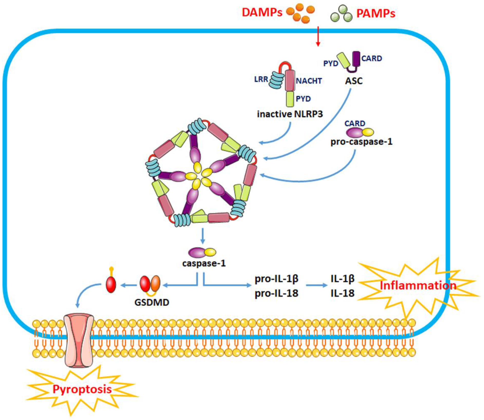
The pathophysiological process of intracerebral hemorrhage (ICH) is very complex, involving various mechanisms such as apoptosis, oxidative stress and inflammation. As one of the key factors, the inflammatory response is responsible for the pathological process of acute brain injury and is associated with the prognosis of patients. Abnormal or dysregulated inflammatory responses after ICH can aggravate cell damage in the injured brain tissue. The NOD-like receptor family pyrin domain-containing 3 (NLRP3) inflammasome is a multiprotein complex distributed in the cytosol, which can be triggered by multiple signals. The NLRP3 inflammasome is activated after ICH, thus promoting neuroinflammation and aggravating brain edema.

intracerebral hemorrhage secondary brain injury NLRP3 inflammasome

1. Introduction

Intracerebral hemorrhage (ICH) refers to the hemorrhage caused by a ruptured blood vessel in the non-traumatic brain parenchyma, which accounts for 20% to 30% of all strokes, with an acute mortality rate of 30% to 40%. The cause is mainly related to cerebrovascular lesions. The pathophysiological process of ICH is very complex, involving various mechanisms such as apoptosis, oxidative stress and inflammation. As one of the key factors, the inflammatory response is responsible for the pathological process of acute brain injury and is associated with the prognosis of patients [1]. Immunity and inflammation play important roles in the process of secondary brain injury (SBI) following ICH. Abnormal or dysregulated inflammatory responses after ICH can aggravate cell damage in the injured brain tissue [2]. It has been reported that the inflammatory responses following ICH begin with the secretion of activated microglia inflammatory factors. After ICH, white blood cells and other components of the peripheral blood enter the brain tissue, activate microglia and promote the production of local inflammatory factors [3]. These inflammatory factors, together with cleavage products resulting from cell death, accelerate the disruption of the blood–brain barrier, damage the brain parenchyma around the lesion and cause the expansion of edema around the hematoma, thereby causing serious damage to the brain tissue.
The NOD-like receptor family pyrin domain-containing 3 (NLRP3) inflammasome is a multiprotein complex distributed in the cytosol, which can be activated by a variety of signals, including bacterial, fungi and viral components, endogenous danger signals and particulates in the environment [4]. Previous studies have shown that the activation of the NLRP3 inflammasome is related to the occurrence and progression of different diseases, including atherosclerosis, metabolic disorders and inflammatory bowel disease [5][6]. As a critical component of innate immunity upon tissue injury, the NLRP3 inflammasome is activated after ICH, thus promoting neuroinflammation and aggravating brain edema. A growing body of evidence suggests that the NLRP3 inflammasome is activated and exhibits a detrimental effect on the brain following ICH [7][8][9].

2. Functions of the NLRP3 Inflammasome in ICH

2.1. Activation of the NLRP3 Inflammasome

The NLRP3 inflammasome consists of the effector protein pro-caspase-1, the adapter protein apoptosis-associated speck-like protein (ASC) and the sensor protein NLRP3 and orchestrates innate immune responses against cell stress and infection by regulating the caspase-1-dependent pathway and releasing proinflammatory cytokines such as interleukin-1β (IL-1β) and IL-18 [10].
The sensor protein NLRP3 consists of a carboxy-terminal leucine-rich repeat (LRR) domain, a NACHT domain and a pyrin domain (PYD). Upon binding to the corresponding ligands, thee LRR domain regulates the function of LRRs. The activation of the NLRP3 inflammasome involves two stages, i.e., the priming and activation stages (Figure 1). The priming stage is induced by the recognition of two molecular patterns, i.e., the damage-associated molecular patterns (DAMPs) and the pathogen-associated molecular patterns (PAMPs) [11]. This can activate the NF-κB signaling pathway and promote the expression of precursor proteins, including pro-IL-1β, pro-IL-18 and NLRP3. The activation stage is triggered by multiple stimuli that exist during metabolic imbalance, infection or tissue injury. When dangerous signals are recognized, the NLRP3 protein structure changes and exposes its PYD domain, which will bind to the PYD of ASC by forming a PYD–PYD interaction. Subsequently, ASC recruits the cysteine protease pro-caspase-1 to assemble the inflammasome complex via interacting with the caspase recruitment domain (CARD). Pro-caspase-1 is activated by self-cleavage to form active caspase-1. Then, caspase-1 dissociates gasdermin D (GSDMD) to release its N-terminal domain, which in turn binds to phosphatidylinositol phosphates and phosphatidylserine in the cytomembrane to generate pores, thereby inducing a lytic form of cell death, known as “pyroptosis”. In addition, caspase-1 can induce the transformation of IL-1β and IL-18 precursors into mature IL-1β and IL-18 and eventually aggravate the inflammatory responses and related complications.
Figure 1. The recognition of DAMPs and PAMPs induces a change in the NLRP3 protein structure and the exposure of its PYD domain to bind to the PYD of ASC, thus forming a PYD–PYD interaction. Then, ASC recruits the cysteine protease pro-caspase-1 to assemble the inflammasome complex via interacting with CARD. Pro-caspase-1 is activated by self-cleavage to form active caspase-1. Activated caspase-1 dissociates GSDMD and releases its N-terminal domain, which interacts with the cell membrane to produce pores and induce “pyroptosis”. In addition, caspase-1 can also induce the transformation of IL-1β and IL-18 precursors into mature IL-1β and IL-18 to induce inflammatory responses.

2.2. Modulation of NLRP3 Inflammasome Activity as a Therapeutic Strategy for SBI after ICH

The NLRP3 inflammasome is a multimolecular complex in the cytoplasm that mediates caspase-1 processing and proinflammatory cytokine maturation, including IL-1β and IL-18. Various risk factors such as Ca2+ mobilization, Na+ influx, K+ efflux, chloride efflux, mitochondrial dysfunction, oxidative stress and lysosomal damage are involved in the activation of the NLRP3 inflammasome to mediate neuroinflammatory responses after ICH [9][12]. Activating the NLRP3 inflammasome generates high levels of inflammatory cytokines, triggers an inflammatory response and recruits other immune cells to clear DAMPs after hemorrhage. However, overactivation of the NLRP3 inflammasome can result in persistent neuroinflammation and brain injury after ICH. Thus, assessing the role of the NLRP3 inflammasome in the processes associated with ICH may provide new strategies for ICH therapy.
As a crucial component of innate immunity upon tissue injury, the NLRP3 inflammasome is activated after ICH, thereby promoting neuroinflammation and aggravating brain edema [8]. It has been reported that the expression of NLRP3 is gradually upregulated in the perihematoma tissue within 1–5 days after ICH, and the NLRP3 inflammasome is responsible for the complement-induced neuroinflammation, which eventually leads to abnormal neurological functions [9]. Activating the NLRP3 inflammasome can promote neuroinflammation via caspase-1 processing and IL-1β generation following ICH. Nevertheless, ICH-induced NLRP3 inflammasome activation can promote neutrophil infiltration, trigger the inflammatory response, impair neurological functions and aggravate brain edema after ICH [9].
In recent years, numerous studies have been conducted around the functions of the NLRP3 inflammasome in ICH. Brain injury induced by inflammation after ICH can be alleviated by directly or indirectly inhibiting NLRP3 inflammasome activation. For example, histone deacetylase 10 (HDAC10) downregulates protein tyrosine phosphatase nonreceptor type 22 (PTPN22) expression by binding to and deacetylating the PTPN22 promoter, which inhibits NLRP3 inflammasome activation and alleviates inflammation after ICH in rats. PTPN22 is a protein tyrosine phosphatase involved in the cellular immune response and inflammation and is related to various autoimmune diseases. PTPN22 binds and dephosphorylates NLRP3 following proinflammatory injury, thus promoting NLRP3 activation and IL-1β secretion. Interfering with PTPN22 can reduce the expression of IL-1β and IL-18 by inhibiting the activation of the NLRP3 inflammasome to reduce inflammatory responses, thereby improving neurological dysfunction and reducing cerebral edema in ICH rats [13]. Mammalian sterile-20-like kinase 4 (MST4) is a member of the glucokinase (GCK) subfamily, which directly phosphorylates TRAF6 to suppress inflammation. MST4 phosphorylates and activates TRAF6, which further activates the NLRP3 inflammasome by regulating the TLR/IL-1R signaling pathway [7]. Overexpression of MST4 negatively regulates the NLRP3 inflammasome and reduces the expression of tumor necrosis factor-α (TNF-α) and IL-1β, indicating that NLRP3 and MST4 may be potential therapeutic targets for neuroinflammation after ICH [7].
Mitochondrial dysfunction plays an essential role in the activation of the NLRP3 inflammasome [14]. Mitophagy is involved in the maintenance of mitochondrial homeostasis via selective degradation of damaged mitochondria, which prevents inflammation by inhibiting the NLRP3 inflammasome pathway [15]. Therefore, modulating mitophagy to inhibit NLRP3 inflammasome activation can become a therapeutic approach for alleviating secondary brain injury (SBI) after ICH. Chen et al. revealed that the activation of the Nrf2/Optineurin (OPTN) pathway can mediate mitophagy to alleviate SBI by suppressing the NLRP3 inflammasome following ICH. OPTN is a multifunctional ubiquitin-binding autophagy receptor that interacts with Nrf2 to mediate mitophagy and eliminate damaged mitochondria following ICH. Suppressing Nrf2/OPTN could enhance NLRP3 inflammasome activation, downregulate mitophagy levels and increase BBB disruption and brain edema, with more severe neurological deficits after ICH [16]. FUN14 domain-containing 1 (FUNDC1) is a mitophagy receptor that is overexpressed after ICH. FUN14 can suppress the NLRP3 inflammasome via regulation of mitophagy, thus alleviating ICH-induced injury. However, silencing FUNDC1 promotes NLRP3-mediated inflammation, thereby suppressing mitophagy and exacerbating ICH injury [17]. Thus, it can be seen that modulating mitophagy to inhibit NLRP3 inflammasome activation is an important therapeutic strategy for alleviating ICH injury.
Many drugs can improve brain dysfunction and protect against SBI after ICH by acting NLRP3 inflammasome pathways (Table 1). For example, pioglitazone, edaravone and adiponectin significantly reduced brain edema and attenuated neurological deficits after ICH, as well as decreased the expression of IL-1β, IL-18, caspase-1 and NF-κB through suppressing the expression of NLRP3 [8][18][19]. Glibenclamide markedly reduced the neurological deficit and brain edema after ICH by decreasing the expression of ASC and caspase-1 and suppressing the activation of the NLRP3 inflammasome to maintain BBB integrity [20]. Memantine reduces ONOO- production by inhibiting neuronal nitric oxide synthase (nNOS) phosphorylation at ser1412, which further inhibits MMP-9 expression and NLRP3 inflammasome activation, protecting the blood–brain barrier integrity and alleviating neurological deficits in ICH rats [21]. Atorvastatin can protect the neurological function and reduce neuroinflammation and neuronal apoptosis by reducing the expression of TNF-α, IL-6 and IL-1β in ICH model mice. Furthermore, atorvastatin can decrease the expression of NLRP3 and cleaved caspase-1 and reverse the increase in toll-like receptor 4 (TLR4) and myeloid differentiation factor 88 (MyD88), indicating that atorvastatin suppresses NLRP3 inflammasome activation in glial cells of ICH model mice through inhibiting MyD88- and TLR4-associated pathways [22]. Some traditional Chinese medicine such as isoliquiritigenin, silymarin, baicalein and cordycepin can also exert antioxidant and anti-inflammatory roles by significantly inhibiting the activation of the Nrf2 dependent-NF-κB pathway and NLRP3 inflammasome, which in turn mitigates brain edema and improves neurological deficits after ICH [23][24][25][26][27]. This evidence indicates that the suppression of NLRP3 inflammasome activation might be a therapeutic target for ICH recovery.
Table 1. Potential NLRP3 inflammasome inhibitors for protecting against SBI after ICH.

Drugs

Models

Efficacy

References

Pioglitazone

blood-induced mouse ICH model

brain edema↓, lactate↑

[18]

Edaravone

autologous blood-induced rat ICH model

IL-1β↓, caspase-1↓, NF-κB↓, brain edema↓, neurological deficits↓

[19]

Adiponectin

autologous blood-induced rat ICH model

IL-1β↓, IL-18↓, brain edema↓, neurological deficits↓

[8]

Glibenclamide

autologous blood-induced mouse ICH model

IL-1β↓, IL-18↓, IL-6↓, TNF-α↓, brain edema↓, disrupted BBB↓, neurological deficits↓

[20]

Memantine

collagenase-induced rat ICH model

IL-1β↓, disrupted BBB↓, neurological deficits↓

[21]

Atorvastatin

collagenase-induced mouse ICH model

IL-1β↓, IL-6↓, TNF-α↓, brain edema↓, neurological deficits↓

[22]

Isoliquiritigenin

collagenase IV-induced rat ICH model

NF-κB↓, IL-1β↓, brain edema↓, disrupted BBB↓, neurological deficits↓

[26]

Silymarin

collagenase II-induced mouse ICH model

NF-κB↓, caspase-1↓, IL-1β↓

[24]

Baicalein

collagenase VII-induced rat ICH model

ROS↓, SOD↑, GSH-Px↑, ASC↓, caspase-1↓

[25]

Cordycepin

autologous blood-induced mouse ICH model

IL-1β↓, IL-18↓, brain edema↓, neurological deficits↓

[27]

References

  1. Askenase, M.H.; Sansing, L.H. Stages of the Inflammatory Response in Pathology and Tissue Repair after Intracerebral Hemorrhage. Semin. Neurol. 2016, 36, 288–297.
  2. Tschoe, C.; Bushnell, C.D.; Duncan, P.W.; Alexander-Miller, M.A.; Wolfe, S.Q. Neuroinflammation after Intracerebral Hemorrhage and Potential Therapeutic Targets. J. Stroke 2020, 22, 29–46.
  3. Seoane, P.I.; Lee, B.; Hoyle, C.; Yu, S.; Lopez-Castejon, G.; Lowe, M.; Brough, D. The NLRP3-inflammasome as a sensor of organelle dysfunction. J. Cell Biol. 2020, 219, e202006194.
  4. Liu, Y.; Li, C.; Yin, H.; Zhang, X.; Li, Y. NLRP3 Inflammasome: A Potential Alternative Therapy Target for Atherosclerosis. Evid.-Based Complement. Altern. Med. Ecam 2020, 2020, 1561342.
  5. Wani, K.; AlHarthi, H.; Alghamdi, A.; Sabico, S.; Al-Daghri, N.M. Role of NLRP3 Inflammasome Activation in Obesity-Mediated Metabolic Disorders. Int. J. Environ. Res. Public Health 2021, 18, 511.
  6. Wu, X.; Zhang, Y.; Zhang, Y.; Xia, L.; Yang, Y.; Wang, P.; Xu, Y.; Ren, Z.; Liu, H. MST4 attenuates NLRP3 inflammasome-mediated neuroinflammation and affects the prognosis after intracerebral hemorrhage in mice. Brain Res. Bull. 2021, 177, 31–38.
  7. Wang, S.; Yao, Q.; Wan, Y.; Wang, J.; Huang, C.; Li, D.; Yang, B. Adiponectin reduces brain injury after intracerebral hemorrhage by reducing NLRP3 inflammasome expression. Int. J. Neurosci. 2020, 130, 301–308.
  8. Ma, Q.; Chen, S.; Hu, Q.; Feng, H.; Zhang, J.H.; Tang, J. NLRP3 inflammasome contributes to inflammation after intracerebral hemorrhage. Ann. Neurol. 2014, 75, 209–219.
  9. Zhang, Y.; Zhang, S.; Li, B.; Luo, Y.; Gong, Y.; Jin, X.; Zhang, J.; Zhou, Y.; Zhuo, X.; Wang, Z.; et al. Gut microbiota dysbiosis promotes age-related atrial fibrillation by lipopolysaccharide and glucose-induced activation of NLRP3-inflammasome. Cardiovasc. Res. 2022, 118, 785–797.
  10. Chen, Z.; Chopp, M.; Zacharek, A.; Li, W.; Venkat, P.; Wang, F.; Landschoot-Ward, J.; Chen, J. Brain-Derived Microparticles (BDMPs) Contribute to Neuroinflammation and Lactadherin Reduces BDMP Induced Neuroinflammation and Improves Outcome After Stroke. Front. Immunol. 2019, 10, 2747.
  11. Li, C.; Chen, M.; He, X.; Ouyang, D. A mini-review on ion fluxes that regulate NLRP3 inflammasome activation. Acta Biochim. Biophys. Sin. 2021, 53, 131–139.
  12. Wang, L.; Zheng, S.; Zhang, L.; Xiao, H.; Gan, H.; Chen, H.; Zhai, X.; Liang, P.; Zhao, J.; Li, Y. Histone Deacetylation 10 Alleviates Inflammation After Intracerebral Hemorrhage via the PTPN22/NLRP3 Pathway in Rats. Neuroscience 2020, 432, 247–259.
  13. Zhou, R.; Yazdi, A.S.; Menu, P.; Tschopp, J. A role for mitochondria in NLRP3 inflammasome activation. Nature 2011, 469, 221–225.
  14. Lin, Q.; Li, S.; Jiang, N.; Jin, H.; Shao, X.; Zhu, X.; Wu, J.; Zhang, M.; Zhang, Z.; Shen, J.; et al. Inhibiting NLRP3 inflammasome attenuates apoptosis in contrast-induced acute kidney injury through the upregulation of HIF1A and BNIP3-mediated mitophagy. Autophagy 2021, 17, 2975–2990.
  15. Cheng, Y.; Liu, M.; Tang, H.; Chen, B.; Yang, G.; Zhao, W.; Cai, Y.; Shang, H. iTRAQ-Based Quantitative Proteomics Indicated Nrf2/OPTN-Mediated Mitophagy Inhibits NLRP3 Inflammasome Activation after Intracerebral Hemorrhage. Oxidative Med. Cell Longev. 2021, 2021, 6630281.
  16. Zheng, S.; Jian, D.; Gan, H.; Wang, L.; Zhao, J.; Zhai, X. FUNDC1 inhibits NLRP3-mediated inflammation after intracerebral hemorrhage by promoting mitophagy in mice. Neurosci. Lett. 2021, 756, 135967.
  17. Kim, H.; Lee, J.E.; Yoo, H.J.; Sung, J.H.; Yang, S.H. Effect of Pioglitazone on Perihematomal Edema in Intracerebral Hemorrhage Mouse Model by Regulating NLRP3 Expression and Energy Metabolism. J. Korean Neurosurg. Soc. 2020, 63, 689–697.
  18. Miao, H.; Jiang, Y.; Geng, J.; Zhang, B.; Zhu, G.; Tang, J. Edaravone Administration Confers Neuroprotection after Experimental Intracerebral Hemorrhage in Rats via NLRP3 Suppression. J. Stroke Cerebrovasc. Dis. Off. J. Natl. Stroke Assoc. 2020, 29, 104468.
  19. Xu, F.; Shen, G.; Su, Z.; He, Z.; Yuan, L. Glibenclamide ameliorates the disrupted blood-brain barrier in experimental intracerebral hemorrhage by inhibiting the activation of NLRP3 inflammasome. Brain Behav. 2019, 9, e01254.
  20. Chen, X.; Xiang, X.; Xie, T.; Chen, Z.; Mou, Y.; Gao, Z.; Xie, X.; Song, M.; Huang, H.; Gao, Z.; et al. Memantine protects blood-brain barrier integrity and attenuates neurological deficits through inhibiting nitric oxide synthase ser1412 phosphorylation in intracerebral hemorrhage rats: Involvement of peroxynitrite-related matrix metalloproteinase-9/NLRP3 inflammasome activation. Neuroreport 2021, 32, 228–237.
  21. Chen, D.; Sui, L.; Chen, C.; Liu, S.; Sun, X.; Guan, J. Atorvastatin suppresses NLRP3 inflammasome activation in intracerebral hemorrhage via TLR4- and MyD88-dependent pathways. Aging 2022, 14, 462–476.
  22. Ikeda, H. Effect of Ni2+ on Ca2+-stimulated adenosine triphosphatase in the microsomal fraction of the rat parotid gland in vitro. Comp. Biochem. Physiol. C Comp. Pharmacol. Toxicol. 1985, 81, 7–9.
  23. Yuan, R.; Fan, H.; Cheng, S.; Gao, W.; Xu, X.; Lv, S.; Ye, M.; Wu, M.; Zhu, X.; Zhang, Y. Silymarin prevents NLRP3 inflammasome activation and protects against intracerebral hemorrhage. Biomed. Pharmacother. Biomed. Pharmacother. 2017, 93, 308–315.
  24. Chen, X.; Zhou, Y.; Wang, S.; Wang, W. Mechanism of Baicalein in Brain Injury After Intracerebral Hemorrhage by Inhibiting the ROS/NLRP3 Inflammasome Pathway. Inflammation 2022, 45, 590–602.
  25. Zeng, J.; Chen, Y.; Ding, R.; Feng, L.; Fu, Z.; Yang, S.; Deng, X.; Xie, Z.; Zheng, S. Isoliquiritigenin alleviates early brain injury after experimental intracerebral hemorrhage via suppressing ROS- and/or NF-kappaB-mediated NLRP3 inflammasome activation by promoting Nrf2 antioxidant pathway. J. Neuroinflammation 2017, 14, 119.
  26. Cheng, Y.; Wei, Y.; Yang, W.; Song, Y.; Shang, H.; Cai, Y.; Wu, Z.; Zhao, W. Cordycepin confers neuroprotection in mice models of intracerebral hemorrhage via suppressing NLRP3 inflammasome activation. Metab. Brain Dis. 2017, 32, 1133–1145.
  27. Rutsch, A.; Kantsjo, J.B.; Ronchi, F. The Gut-Brain Axis: How Microbiota and Host Inflammasome Influence Brain Physiology and Pathology. Front. Immunol. 2020, 11, 604179.
More
Information
Subjects: Cell Biology
Contributors MDPI registered users' name will be linked to their SciProfiles pages. To register with us, please refer to https://encyclopedia.pub/register : , , , , , , ,
View Times: 500
Revisions: 2 times (View History)
Update Date: 14 Dec 2022
1000/1000
Video Production Service