Submitted Successfully!
To reward your contribution, here is a gift for you: A free trial for our video production service.
Thank you for your contribution! You can also upload a video entry or images related to this topic.
Version Summary Created by Modification Content Size Created at Operation
1 -- 3424 2022-12-07 09:31:22 |
2 format correct Meta information modification 3424 2022-12-08 03:07:47 | |
3 format correct -2 word(s) 3422 2022-12-09 09:57:51 |

Video Upload Options

Do you have a full video?

Confirm

Are you sure to Delete?
Cite
If you have any further questions, please contact Encyclopedia Editorial Office.
Awasthi, A.;  Corrie, L.;  Vishwas, S.;  Gulati, M.;  Kumar, B.;  Chellappan, D.K.;  Gupta, G.;  Eri, R.D.;  Dua, K.;  Singh, S.K. Gut Dysbiosis and Diabetic Foot Ulcer. Encyclopedia. Available online: https://encyclopedia.pub/entry/38186 (accessed on 27 July 2024).
Awasthi A,  Corrie L,  Vishwas S,  Gulati M,  Kumar B,  Chellappan DK, et al. Gut Dysbiosis and Diabetic Foot Ulcer. Encyclopedia. Available at: https://encyclopedia.pub/entry/38186. Accessed July 27, 2024.
Awasthi, Ankit, Leander Corrie, Sukriti Vishwas, Monica Gulati, Bimlesh Kumar, Dinesh Kumar Chellappan, Gaurav Gupta, Rajaraman D. Eri, Kamal Dua, Sachin Kumar Singh. "Gut Dysbiosis and Diabetic Foot Ulcer" Encyclopedia, https://encyclopedia.pub/entry/38186 (accessed July 27, 2024).
Awasthi, A.,  Corrie, L.,  Vishwas, S.,  Gulati, M.,  Kumar, B.,  Chellappan, D.K.,  Gupta, G.,  Eri, R.D.,  Dua, K., & Singh, S.K. (2022, December 07). Gut Dysbiosis and Diabetic Foot Ulcer. In Encyclopedia. https://encyclopedia.pub/entry/38186
Awasthi, Ankit, et al. "Gut Dysbiosis and Diabetic Foot Ulcer." Encyclopedia. Web. 07 December, 2022.
Gut Dysbiosis and Diabetic Foot Ulcer
Edit

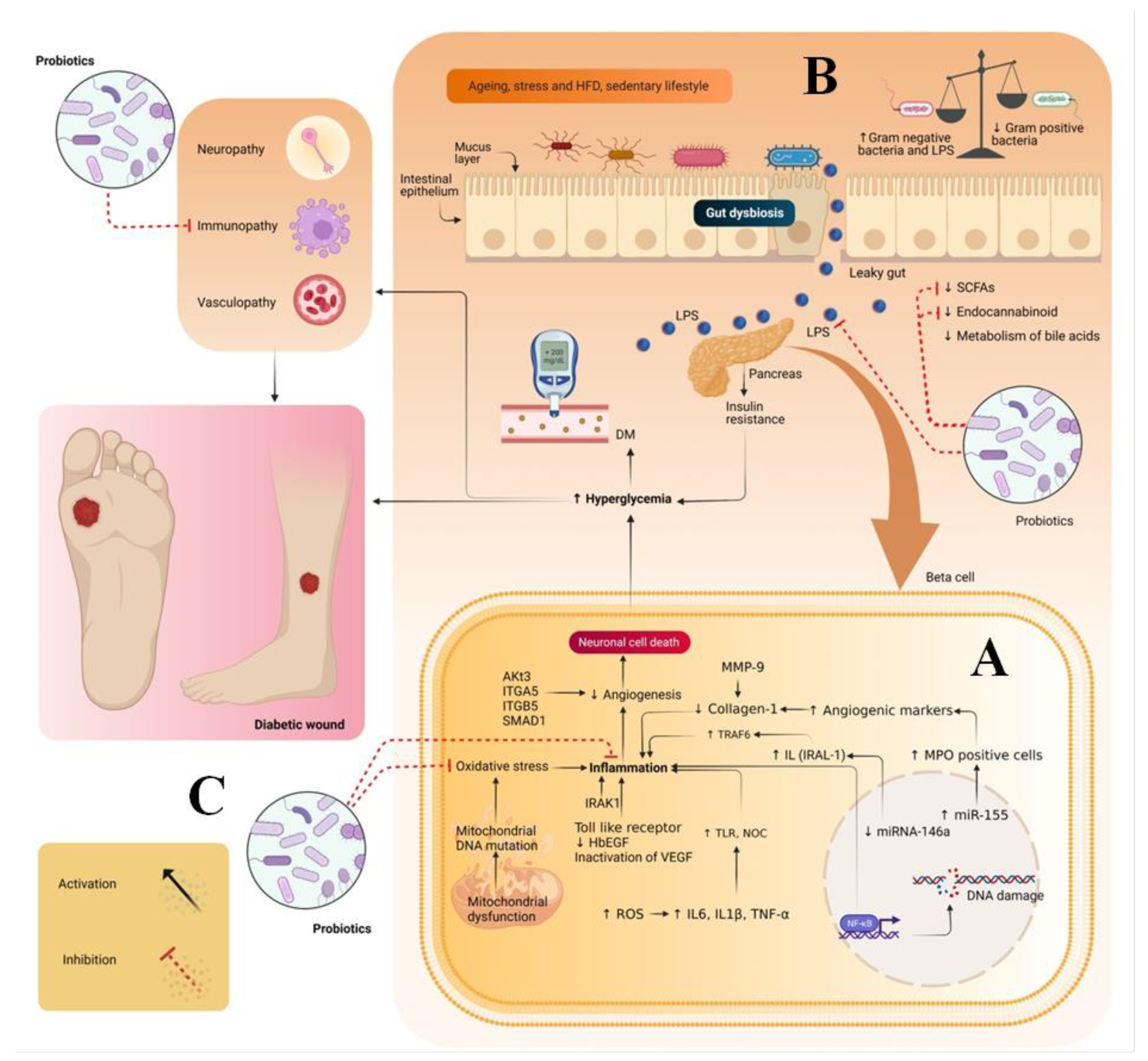
Diabetic foot ulcer (DFU) is a multifactorial disease and one of the complications of diabetes. The global burden of DFU in the health sector is increasing at a tremendous rate due to its cost management related to hospitalization, medical costs and foot amputation. Hence, to manage DFU/DWs, various attempts have been made, including treating wounds systematically/topically using synthetic drugs, herbal drugs, or tissue engineering based surgical dressings. However, less attention has been paid to the intrinsic factors that are also the leading cause of diabetes mellitus (DM) and its complications. One such factor is gut dysbiosis, which is one of the major causes of enhancing the counts of Gram-negative bacteria. These bacteria produce lipopolysaccharides, which are a major contributing factor toward insulin resistance and inflammation due to the generation of oxidative stress and immunopathy.

diabetic foot ulcer pathogenesis sources of probiotics therapeutic potential of probiotics on DFU market status of probiotics patents on probiotics

1. Introduction

Diabetic foot ulcer (DFU) is the one of the most common complications of diabetes. The global prevalence of DFU due to diabetes is 25%. It is an open sore wound that occurs in the foot. It generally occurs due to the hypoxia and oxidative stress caused by reactive oxygen species, a decrease in the level of growth factors (GFs), nucleic acids and the lack of glycemic control. DFU has reached the 10th position in terms of the annual economic burden of diabetics [1]. this situation has arisen because of a lack of existing treatment strategies to promote wound healing. In DFU, delayed wound healing occurs [2]. The common reason for this is the extended inflammatory response that leads to impairment in keratinocyte migration, collagen synthesis, vascularization, fibroblast migration, epithelialization, collagen proliferation, differentiation and migration. Overall, these contributing factors often result in amputation and even the death of the DFU patient. The global prevalence of amputation due to DFU in 2022 is reported to be 10–15% [3].
The treatment of DFU is challenging, as it involves multiple stages, etiologies and degrees of severity that vary among the diabetic mellitus (DM) patients. The existing formulations on the market provide adequate glycemic control. However, these are unable to treat the various stages of DFU in DM patients. Therefore, this increases the burden of medications on patients suffering to DFU, because the delay in wound healing may also be dependent on the severity of the wound, rather than only glycemic control. Hence, for wound healing, the administration of antibiotics or anti-inflammatory agents is also required. Other approaches that are used to manage DFU include plastic surgery, orthopedics, vascular surgery, offloading, antibiotics (ciprofloxacin, vancomycin, clindamycin and piperacillin/tazobactam), herbal drugs (curcumin, quercetin, aloe vera, achlefan and panchavalkla), synthetic drugs (mevastatin, simvastatin, naltrexone and azelnidipine), growth factors (GFs), nucleic acids gene based delivery, novel drug delivery systems (NDDSs) such as nanostructured lipid carriers, nanoemulsion, nanoparticles and dressings such as gauze, films, foams or, hydrocolloid-based dressings as well as polysaccharide- and polymer-based dressings etc. The limitation of surgery is that in DM patients, there is a slow progression of wound healing. Once the patient has undergone surgery, the wounds take a long time to heal, leaving the patient susceptible to infections. The limitation of synthetic and herbal drugs is their poor solubility and permeability, while the limitations of GFs and nucleic acid are their high cost and low stability. The limitation associated with the NDDS is their low retainability at the injured site, if used topically; additionally, to enhance their retention, they have to be further incorporated into nanomaterials, which increases the cost of therapy. Dressings which are currently available to manage DFU have some limitations, such as the inability to absorb the exudate and high cost. Antibiotics can decrease microbial load but not heal the wound [1][2][3]. These treatment strategies are expensive and underline the need for a multi-disciplinary, cost-effective approach to control hyperglycemia with the potential to target different stages of DFU. In recent years, probiotics have gained tremendous attention for the management of various metabolic diseases due to their anti-infective, antioxidant, anti-inflammatory, anti-diabetic and immunomodulatory activities. In the case of DFU, probiotics help to maintain the levels of short chain fatty acids, gut hormones and the endocannabinoid system that helps in maintaining glucose homeostasis, decreasing inflammation and providing immunity to the DFU patients. Probiotics are part of various food products that are consumed on a daily basis. They help to manage gut microbiota function and impart immunomodulation. They also have a commercial status in the form of probiotic drinks and foods [4]. Despite having such potential, they have been clinically less explored for their potential in the management of DFU.

2. Pathogenesis of Diabetic Wounds

During hyperglycemia, the levels of micro-ribulose nucleic acid (miR)-155, miR-191, miR-200b, miR-15b, miR-200, and miR-205–5p are increased while those of miRNA-146a and miR-132 are decreased. The overactivation of miR-155, miR-191 and miR-200b results an increase in the level of myeloperoxidase (MPO)-positive cells and C-reactive protein levels, which, in turn, leads to impairment in angiogenic markers such as collagen 1, transforming growth factor (GF) beta-1 and alpha-smooth muscle actin. In addition, they prolong the inflammatory phase of wound healing and impede the wound healing process. Besides these factors, the overactivation of miR-15b, miR-200 and miR-205–5p results in the impairment of the vasoendothelial GF pathways and impedes the wound healing process. The decrease in the levels of miRNA-146a and miR-132 activates the tumor necrosis factor receptor-associated factor 6 (TRAF6), interleukin-1 receptor associated kinase 1 (IRAK1) and toll-like receptors. The overactivation of these pathways results in an increase in the level of inflammatory markers that prolongs the inflammatory phase and delays the wound healing process [3]. In addition to this, in DFU, the level of matrix mettalo proteinase (MMP) also gets increased, which inhibits the migration of keratinocytes toward the wound site and impairs collagen synthesis. This delays the wound healing process [1].
High blood glucose levels also result in idiopathic complications, viz. neuropathy, immunopathy and vasculopathy. Neuropathy affects sensory, motor and autonomic nerves. In sensory neuropathy, there is a loss of pain leading to unnoticed trauma, which, in turn, may lead to ulcer formation. In motor neuropathy, weakness and wasting of intrinsic foot muscles occur, which results in abnormal gait and foot deformities that can lead to ulceration. In autonomic neuropathy, sweat glands get suppressed, which results in a decrease in the sweating rate at the foot site. This makes the skin dry and brittle and leads to secondary infections and, finally, ulceration. Vasculopathy is a general term used to describe any disease affecting blood vessels. It is generally of two types: microanginopathy and macroanginopathy. Microanginopathy occurs when there is deposition of glycoproteins and blood clots on the surface of the basement of the vessels. This deposition makes the walls of the vessels thicker and causes leakage from them, leading to ulceration. Macroanginopathy includes the deposition of fats and blood clots in the blood vessels. This decreases the blood flow in the vessels, which leads to necrosis and, finally, ulceration. In the case of immunopathy, there is a decrease in immunity due to the decrease in the level of polymorpholeukocytes, intracellular killing rate and GFs, coupled with an excess of metalloproteinases. This prolongs the inflammatory phase and delays the wound healing process (Figure 1) [2].
Figure 1. (A) Pathogenesis of DFU (B) Gut dysbiosis and its relation with pathogenesis of DFU and (C) the role of probiotics in the treatment of DFU. ↑ indicates upregulation and symbol ↓ indicates downregulation.

3. Therapeutic Potential of Probiotics in Treating DW

DW is associated with oxidative stress, inflammation and immunopathy. Hence, probiotics can play a major role in the therapy of DW. Probiotics have multiple therapeutic actions, such as antioxidant, anti-inflammatory, immunomodulatory and antidiabetic (Figure 1) [5]. Probiotics exert antioxidant effects by decreasing the oxidative stress generated by mitochondrial dysfunction and reactive oxygen species. It is known that SOD has a short half-life and low bioavailability. They enhance the antioxidant effect by releasing antioxidant enzymes such as SOD and catalase. In mitochondrial dysfunction, oxidative stress is produced by the generation of superoxide reactive oxygen species. When probiotics are consumed, SOD enzymes are produced that help in the breakdown of superoxide ions into hydrogen peroxide and water, thereby decreasing oxidative stress. Therefore, probiotics are suitable for the local delivery of SOD in bowel-related disease. In addition, probiotics also produce catalase enzymes that help in cellular antioxidant defense and promote the decomposition of hydrogen peroxide, which, in turn, inhibits the production of hydroxyl radicals by Fenton reaction. Probiotics also produce antioxidant metabolites such as glutathione butyrate and folate. These metabolites eliminate hydrogen peroxide, peroxynitrite and hydroxyl radicals with the help of selenium-dependent glutathione peroxidase enzyme and reduce oxidative stress [6].
Nuclear factor-kappa B (NF-ĸB) is a key signaling channel which is responsible for inflammation. It is present in the cytoplasm in an inactive form, bound to an inhibitory molecule, i.e., IĸB. During inflammation, IĸB molecule breaks down, which results in the release of NF-ĸB to activate the inflammatory cascades. A probiotics strain such as Lactobacillus rhamnosus GG or Lactobacillus casei DN-114 001 inhibits the breakdown of the inhibitory molecule- IĸB and reduces the expression of proinflammatory cytokines such as IL-8. In addition, probiotics trigger toll-like receptors, which initiate beta-defensins and exert anti-inflammatory actions [7].
Probiotics exert immunomodulatory actions by interacting with antigen presenting and release chemical mediator cytokines such as interleukins (ILs), tumor necrosis factor, interferons, transforming GF and chemokines from immune cells (lymphocytes, granulocytes, macrophages, mast cells, epithelial cells, and dendritic cells (DCs)), which further regulate the innate and adaptive immune system. In addition, probiotics help in enhancing the production of cytokines, activate the tight junctions of the intestinal barrier against intercellular bacterial invasion, encourage the secretion of immunoglobulin A and production of antibacterial substances and compete with new pathogenic microorganisms for enterocyte adherence. Through these processes, probiotics regulate intestinal epithelial health. An early, innate immune response is also induced by probiotics through phagocytosis, polymorphonuclear (PMN) cell recruitment and tumor necrotic factor-alpha production [8].
Probiotics have an anti-diabetic effect because they help in the production of SCFA, which enhances the release of incretin hormones that influence glucose levels. In addition, probiotics reduce the level of LPS, making them useful for the treatment of gut dysbiosis and type 2 diabetes mellitus. Probiotics also help to increase the levels of GLP-1 and insulinotropic hormones in enteroendocrine L-cells [9]. This optimizes glucose metabolism, reduces cell damage and improves insulin sensitivity. Among several animal models used for DM, it has been reported in 91 research papers that probiotics prevent DM onset by down-regulating certain inflammatory cytokines, such as interferons (IFN) and IL-2 or IL-1, or by increasing anti-inflammatory IL-10 production. It is also claimed that probiotics produce a defensive wall that prevents pathogenic bacterial species from colonizing the epithelium [10].
Studies related to the antioxidant, anti-inflammatory, immunomodulation and anti-diabetic property of probiotics are depicted in the Table 1.
Table 1. Probiotic compositions, indicating their pharmacological activity and their outcomes.
Probiotic Strain Assay Results References
Antioxidant effect      
Bacillus amyloliquefaciens,
Starmerella bombicola, and
Lactobacillus brevis
DPPH, ABTS
  • ABTS antioxidant activity tests of Bacillus amyloliquefaciens (400 µg/mL) showed 1.01-, 1.03- and 1.05-fold increases in antioxidant activity in comparison to Lactobacillus brevis, Starmerella bombicola and blueberry fruit extract without probiotic bacteria
  • A DPPH radical assay revealed that Bacillus amyloliquefaciens (1600 µg/mL) led to an increase in antioxidant activity by 1.01-, 1- and 1.23-fold as compared to Lactobacillus brevis, Starmerella bombicola, and blueberry fruit extract without probiotic bacteria
[11]
Bifidobacterium breve, Rhamnosus GG, Probionebacterium freudenreichii and Lactobacillus retueria, DPPH, ABTS
  • A DPPH antioxidant scavenging assay revealed that Probionebacterium freudenreichii (100 µg/mL) strain led to 1.01-, 1.12-, 1.06-, 1.05- and 1.04-fold increases in antioxidant activity in comparison to Lactobacillus retueria, Bifidobacterium breve and Lactobacillus rhamnosus, ascorbic acid, and butylated hydroxytoluene
  • ABTS antioxidant activity tests of Probionebacterium freudenreichii ( (100 µg/mL) strain revealed an increase in antioxidant activity by 1-, 1-, 1.06-, 1.01- and 1.01-fold as compared to Lactobacillus rhamnosus, Lactobacillus retueria, Bifidobacterium breve, ascorbic acid, and Butylated hydroxytoluene
[12]
BS1, BS2, BV TAOC, MDA, SOD
  • TAOC results revealed that BV led to 1.17-, 1.11- and 2.5-fold increase in antioxidant activity in comparison to BS2, BS1and saline-treated group (Control)
  • MDA study: BS2 treated groups showed 3.6-, 1.05- and 1.11-fold decreases in MDA level as compared to control, BS1 and BV1 treated groups
  • SOD study showed that BS2 treated groups exhibited an increase in antioxidant activity by 1.7-, 1.2- and 1.4-fold in comparison to control, BS1 and BV1 treated groups
[13]
Enterococcus faecium DPPH, Superoxide, Hydroxyl scavenging assay
  • DPPH assay showed that Enterococcus faecium (10 mg/mL) led to a 1.08-fold increase in antioxidant activity as compared to ascorbic acid
  • Superoxide scavenging assay revealed Enterococcus faecium (10 mg/mL) led to a 1.13-fold increase in antioxidant activity in comparison to ascorbic acid
  • Hydroxyl scavenging assay result revealed that Enterococcus faecium (10 mg/mL) led to a 1.42-fold in antioxidant activity as compared to ascorbic acid
[14]
Lactobacillus acidophilus DPPH
  • SY (0.2 mg/mL) led to a 1.16-, 1- and 1.04-fold increase in antioxidant activity in comparison to control, SWY and WY, respectively
[6]
Lactobacillus plantarum, Lactobacillus rhamnosus, Lactobacillus casei, DPPH
  • DPPH assay revealed that Lactobacillus rhamnosus (0.1 mg/mL) led to a 1.21-, 1.19- and 1.46-fold increase in antioxidant activity as compared to Lactobacillus casei, Lactobacillus plantarum and cashew milk-yoghurt without probiotic strain
[15]
Lactobacillus plantarum DM5 DPPH, Superoxide anion, Hydroxyl
  • Lactobacillus plantarum DM5 (1010 CFU/mL) has 20% and 30% higher hydroxyl radical activity than Lactobacillus acidophilus and Lactobacillus plantarum
  • Lactobacillus plantarum DM5 (1010 CFU/mL) showed 31% and 22% higher superoxide anion scavenging activity than Lactobacillus Plantarum and Lactobacillus acidophilus
  • Lactobacillus plantarum DM5 (1010 CFU/mL) exhibited an increase in DPPH scavenging activity by 43% and 33%, as compared to Lactobacillus plantarum and Lactobacillus acidophilus
[16]
Lactobacillus paracasei A-4, Lactobacillus plantarum A-7, Lactobacillus paracasei BL-12, Lactobacillus paracasei DU-8, Lactococcus lactis T-8 DPPH
  • Lactobacillus plantarum A-7 1 mg/mL) exhibited increase in antioxidant activity by 1.22-, 2.81-, 3.19-, 1.01-, 3.47- and 5.41-fold as compared to Lactobacillus paracasei A-4, Lactobacillus paracasei BL-12, Lactobacillus paracasei DU-8, Lactobacillus brevis O-9, Lactococcus lactis T-8 and Control milk respectively
[17]
Anti-inflammatory      
Probiotic strain Design/
participants
Results References
Bifidobacterium animalis ssp.
lactis 420 (900 billion CFU/day)
Randomized/50
  • Improved bacterial dysbiosis and immunity
  • Reconstructed the balance of intestinal flora
[18]
Lactobacillus acidophilus La-5
and Bifidobacterium BB-12 (106 CFU/g each)
Randomized double-blind/210
  • Decreased inflammation
  • Increased bacterial count in the intestine and colon
[19]
Lactobacillus acidophilus, Lactobacillus casei, Bifidobacterium bifidum, Lactobacillus fermentum (2 × 10⁹ CFU/g each) Randomized double-blind/48
  • Improved glucose homeostasis.
  • Decreased oxidative stress and inflammation
[20]
Lactobacillus acidophilus, Lactobacillus infantis, Bifidobacterium bifidum, Lactobacillus fermentum and Bifidobacterium longum (6 billion CFU each) Randomized double-blind/
52
  • Decreased proinflammatory mediators of inflammation
[21]
Lactobacillus plantarum OLL2712 (5 × 10⁹ CFU) Randomized/
130
  • Decreased chronic inflammation
  • Decreased HbA1c level
[22]
Immunomodulatory effect      
Probiotics strain Animal model/other Results References
Bifidobacterium longum KACC 91563(100 billion CFU/g) Male BALB/c mice
  • Improved systemic immunity
  • Regulated T and B-cell proliferation
  • Inhibited the Th1cytokine imbalance and immune cytokine production
[23]
Bifidobacterium longum CCUG 52486 (5 × 10⁸ CFU/day) Human
  • Increased NK cell activity
  • Increased the number of IgG+ memory B-cells
[24]
Lactobacillus casei Shirota (1.3 × 10¹⁰ CFU/day) Human
  • Increased innate immunity by increasing levels of natural killer cell activity
  • Increased inflammatory status by promoting IL-10/IL-12 ratio
[25]
Lactobacillus casei; CRL 431 (109 cells/day) Female BALB/c mice
  • Increased mucosal activity
  • Maintain homeostasis at the mucosal level
  • Increased phagocytosis
  • Increased IL-10 levels
[26]
Limosilactobacillus fermentum
(109 CFU/mL)
Female Balb/c mice
  • Modulated inflammatory cytokines
  • Stimulated response of the immune system
[27]
Antidiabetic effect      
Probiotic strain Animal model Results References
Lactobacillus casei (4.0 × 10⁹
CFU/rat/day)
Rat
  • ↓BGL
[28]
Lactobacillus casei and Bifidio bifidum (1 × 10⁷ cfu/mL) Wistar rat
  • ↓ BGL, ↓ HbA1c, ↓ TC, ↓ TGs
  • ↓ LDL, ↓ VLDL, ↑ HDL
[29]
Lactobacillus.casei (109 CFU/mL) Mice
  • ↓ BGL, ↓ insulin
  • ↓ insulin-like growth factor I, ↓ C-peptide
[30]
Lactobacillus casei CCFM419 (109 CFU) Mice
  • ↓ Fasting and postprandial blood glucose
  • ↓ glucose intolerance, ↓ IR, ↓ TNFα, ↓ IL-6, ↑ GLP-1
[31]
Lactobacillus. Gasseri (6 × 10⁷ cfu/g) Rat
  • ↓ BGL, ↓ IR, ↓ inflammation
  • ↑ SCFA, ↑ insulin secretion
[32]
Lactobacillus plantarum CCFM0236 (8 × 10⁹ cfu/mL) Mice
  • ↓ Food intake, ↓ BGL, ↓ HbA1c, ↓ leptin level, ↓ insulin level
  • ↓ TNFα, ↓ HOMA-IR index, ↑ activities of GPx
[33]
Lactobacillus.plantarum, strain Ln4 (5 × 10⁸ cfu/day) Male mice
  • ↓ Weight gain, ↓ epididymal fat mass, ↓ total plasma TG level
  • ↓ HOMA-IR, ↑ glucose tolerance, ↑ insulin response
[34]
Lactobacillus.plantarum MTCC5690 and Lactobacillus fermentum MTCC5689 (1.5 × 10⁹ colonies/day) C57BL/6J male mice
  • ↓ IR, ↓ glucose intolerance, ↓ glucose level, ↓ lipid level, ↓ TNFα ↓IL6
  • ↑ gene expression patterns of intestinal tight junction
[35]
Lactobacillus.rhamnoss, Lactobacillus.acidophilus, Bifidio bifidumi (6 × 10⁸ CFU each) Mice
  • ↓ Intestinal permeability, ↓ LPS translocation, ↓ low-grade systemic inflammation
  • ↓ glucose tolerance, ↓ hyperphagic behavior, ↓ hypothalamic insulin, and leptin resistance
[36]
ABTS 2,2′-azino-bis(3-ethylbenzothiazoline-6-sulfonic) acid, CFU/g; Colony forming units/gram, TAOC; Total antioxidant capacity, MDA; maleic dialdehyde, GSH-PX; Glutathione peroxidase, SOD; Superoxide dismutase, BS1; Bacillus subtilis1, BS2; Bacillus subtilis2, BV; Bacillus velezensisis, SY; Probiotic fat-free yogurt, SWY; Probiotic semi-fat yogurt, WY; Probiotic full fat yogurt; DPPH; 2,2-DiPhenyl-2-Picryl hydrazyl hydrate. Here sign ↓ indicates decrease in the level and ↑ indicates increase in the level.
With regard to the therapeutic potential of probiotics, various studies have been carried out in the field of DW healing, which are discussed below.
In one of these studies, Peral et al. (2010) investigated the effect of Lactobacillus plantarum against chronic infected leg ulcers in diabetic patients. In their trial, 14 diabetic and 20 non-diabetic patients having venous leg ulcers were considered. For the treatment, topically Lactobacillus plantarum was applied to both diabetic and non-diabetic patients with venous leg ulcers. After 30 days of topical treatment with Lactobacillus plantarum, it was observed that 43% of diabetics and 50% of non-diabetic patients showed complete wound healing. Therefore, it was concluded that Lactobacillus plantarum accelerated wound healing in diabetic and non-diabetic patients by exerting antibacterial and anti-inflammatory actions, reducing apoptotic, neutrophils, and necrotic cells and modifying IL-8 production [37].
In another study, Majid et al. (2016) examined the effect of Lactobacillus casei and its exopolysaccharide against DW in induced male Wistar diabetic rats. The results revealed that the topical application of Lactobacillus casei and its exopolysaccharide showed 1.4-fold and 1.1-fold increase in wound contraction within 14 days as compared to negative and control groups [38].
Similarly, Mohseni et al. (2018) investigated the effect of probiotic supplementation on metabolic status and wound healing in patients with DFU. They performed a double-blind, randomized and placebo-controlled trial. In their trial, 60 patients aged 40–85 years old and having grade 3 (deep ulcer with cellulitis) DFU were considered. These 60 patients were casually distributed into two groups (30 patients on each side) to receive either placebo or oral probiotic capsule (Lactobacillus fermentum, Lactobacillus casei, Lactobacillus acidophilus, and Bifidobacterium bifidum) every day for 12 weeks. The dose of the probiotic capsule was 2 × 10⁹ CFU/g each. After 12 weeks, it was observed that compared to the placebo group, the probiotics-treated groups showed a significant reduction in ulcer length (−1.3 ± 0.9 cm for probiotic vs. −0.8 ± 0.7 cm for placebo, p = 0.01), ulcer width (−1.1 ± 0.7 cm for probiotic vs. −0.7 ± 0.7 cm for placebo, p = 0.02) and ulcer depth (−0.5 ± 0.3 cm for probiotic vs. −0.3 ± 0.3 cm for placebo, p = 0.02). Moreover, it was also observed that probiotics not only reduced the ulcer length, size and depth, but also helped in the downregulation of blood glucose level, total serum cholesterol, high sensitivity C-reactive protein (hs-CRP), malondialdehyde (MDA) levels, augmented plasma nitric oxide (NO) and total antioxidant capacity (TAC), indicating the potential of probiotics in treating DFU [39].
In another study, Gonzalez et al. (2018) explored the effect of clindamycin/cefotaxime and Lactobacillus acidophilus against micro-organisms isolated from the foot of DFU patients. The turbidimetric method was used for the bioassay. Three types of bacteria were isolated from DFUs strain, i.e., strain 1 (Pseudomonas sp.), strain 2 (yeast-like cell) and strain 3 (Enterobacter sp.). Then, clindamycin/cefotaxime and Lactobacillus acidophilus were tested against micro-organisms isolated from the foot of DFU patients. Clindamycin was used against all the strains isolated from DFU patients at concentrations of 0.15 μg/mL, 0.25 μg/mL, and 50 μg/mL. It was observed that clindamycin was only effective against strain three; the percentages of inhibition were 18, 88, and 89, respectively. Meanwhile, cefotaxime at concentrations of 0.15 μg/mL, 0.25 μg/mL, and 50 μg/mL showed an effect against all the three strains. The percentages of inhibition of cefotaxime at a dose of 0.15 μg/mL against strains 1, 2 and 3 were 85, 70 and 55, respectively. At a dose of 0.25 μg/mL cefotaxime showed a good percentage of inhibition against strains 1, 2 and 3, i.e., 87, 68, and 60, respectively. At a dose, 50 μg/mL cefotaxime showed percentages of inhibition for strains 1, 2 and 3 of 88, 65 and 76, respectively. When Lactobacillus acidophilus was tested against all these at concentrations of 40 mg/mL, 400 mg/mL, and 800 mg/mL, it was observed that it was only effective against strains 1 and 3. For strains 1 and 3, Lactobacillus acidophilus showed percentages inhibition of 3% and 9%, respectively, at a dose of 40 mg/mL. At dose of 400 mg/mL, Lactobacillus acidophilus showed percentages of inhibition against strains 1 and 3 which of 34 and 18, respectively. Similarly, at a dose of 800 mg/mL, Lactobacillus acidophilus showed 40% inhibition for strain 1 and 26% inhibition for strain 3, indicating the antibacterial potential of probiotics against the micro-organisms that are responsible for DFU [40].

References

  1. Awasthi, A.; Gulati, M.; Kumar, B.; Kaur, J.; Vishwas, S.; Khursheed, R.; Porwal, O.; Alam, A.; Kr, A.; Corrie, L.; et al. Review Article Recent Progress in Development of Dressings Used for Diabetic Wounds with Special Emphasis on Scaffolds. BioMed Res. Int. 2022, 2022, 1659338.
  2. Awasthi, A.; Singh, S.K.; Kumar, B.; Gulati, M.; Kumar, R.; Wadhwa, S.; Khursheed, R.; Corrie, L.; KR, A.; Kumar, R.; et al. Treatment Strategies Against Diabetic Foot Ulcer: Success so Far and the Road Ahead. Curr. Diabetes Rev. 2020, 17, 421–436.
  3. Awasthi, A.; Vishwas, S.; Gulati, M.; Corrie, L.; Kaur, J.; Khursheed, R.; Alam, A.; Alkhayl, F.F.A.; Khan, F.R.; Nagarethinam, S.; et al. Expanding Arsenal against Diabetic Wounds Using Nanomedicines and Nanomaterials: Success so Far and Bottlenecks. J. Drug Deliv. Sci. Technol. 2022, 74, 103534.
  4. Yahfoufi, N.; Mallet, J.F.; Graham, E.; Matar, C. Role of Probiotics and Prebiotics in Immunomodulation. Curr. Opin. Food Sci. 2018, 20, 82–91.
  5. Plaza-Diaz, J.; Ruiz-Ojeda, F.J.; Gil-Campos, M.; Gil, A. Mechanisms of Action of Probiotics. Adv. Nutr. 2019, 10, S49–S66.
  6. Tavakoli, M.; Habibi Najafi, M.B.; Mohebbi, M. Effect of the Milk Fat Content and Starter Culture Selection on Proteolysis and Antioxidant Activity of Probiotic Yogurt. Heliyon 2019, 5, e01204.
  7. Sharma, S.; Singh, A.; Sharma, S.; Kant, A.; Sevda, S.; Taherzadeh, M.J.; Garlapati, V.K. Functional Foods as a Formulation Ingredients in Beverages: Technological Advancements and Constraints. Bioengineered 2021, 12, 11055–11075.
  8. Azad, M.A.K.; Sarker, M.; Wan, D. Immunomodulatory Effects of Probiotics on Cytokine Profiles. Biomed Res. Int. 2018, 2018, 8063647.
  9. Pegah, A.; Abbasi-Oshaghi, E.; Khodadadi, I.; Mirzaei, F.; Tayebinia, H. Probiotic and Resveratrol Normalize GLP-1 Levels and Oxidative Stress in the Intestine of Diabetic Rats. Metab. Open 2021, 10, 100093.
  10. Denkova, R.; Goranov, B.; Teneva, D.; Kostov, G. Antimicrobial Activity of Probiotic Microorganisms: Mechanisms of Interaction and Methods of Examination, 1st ed.; Z.-Mendez Villas: Polvdiv, Bulgaria, 2017; pp. 201–212.
  11. Oh, B.T.; Jeong, S.Y.; Velmurugan, P.; Park, J.H.; Jeong, D.Y. Probiotic-Mediated Blueberry (Vaccinium Corymbosum L.) Fruit Fermentation to Yield Functionalized Products for Augmented Antibacterial and Antioxidant Activity. J. Biosci. Bioeng. 2017, 124, 542–550.
  12. Helmy, S.A. Chemical Composition and Antimicrobial Activity of Some Essential Oils and Their Major Constituents. Int. J. Acad. Res. 2012, 4, 124–137.
  13. Li, A.; Wang, Y.; Li, Z.; Qamar, H.; Mehmood, K.; Zhang, L.; Liu, J.; Zhang, H.; Li, J. Probiotics Isolated from Yaks Improves the Growth Performance, Antioxidant Activity, and Cytokines Related to Immunity and Inflammation in Mice. Microb. Cell Fact. 2019, 18, 112.
  14. Abdhul, K.; Ganesh, M.; Shanmughapriya, S.; Kanagavel, M.; Anbarasu, K.; Natarajaseenivasan, K. Antioxidant activity of exopolysaccharide from probiotic strain Enterococcus faecium (BDU7) from Ngari. Int. J. Biol. Macromol. 2014, 70, 450–454.
  15. Shori, A.B.; Aljohani, G.S.; Al-zahrani, A.J.; Al-sulbi, O.S.; Baba, A.S. Viability of Probiotics and Antioxidant Activity of Cashew Milk-Based Yogurt Fermented with Selected Strains of Probiotic lactobacillus Spp. LWT 2022, 153, 112482.
  16. Das, D.; Goyal, A. Antioxidant Activity and γ-Aminobutyric Acid (GABA) Producing Ability of Probiotic Lactobacillus plantarum DM5 Isolated from Marcha of Sikkim. LWT—Food Sci. Technol. 2015, 61, 263–268.
  17. Uugantsetseg, E.; Batjargal, B. Antioxidant Activity of Probiotic Lactic Acid Bacteria Isolated from Mongolian Airag. Mong. J. Chem. 2014, 15, 73–78.
  18. Mäkelä, S.M.; Forssten, S.D.; Kailajärvi, M.; Langén, V.L.; Scheinin, M.; Tiihonen, K.; Ouwehand, A.C. Effects of Bifidobacterium animalis Ssp. Lactis 420 on Gastrointestinal Inflammation Induced by a Nonsteroidal Anti-Inflammatory Drug: A Randomized, Placebo-Controlled, Double-Blind Clinical Trial. Br. J. Clin. Pharmacol. 2021, 87, 4625–4635.
  19. Shadnoush, M.; Hosseini, R.S.; Khalilnezhad, A.; Navai, L.; Goudarzi, H.; Vaezjalali, M. Effects of Probiotics on Gut Microbiota in Patients with Inflammatory Bowel Disease: A Double-Blind, Placebo-Controlled Clinical Trial. Korean J. Gastroenterol. 2015, 65, 215–221.
  20. Babadi, M.; Khorshidi, A.; Aghadavood, E.; Samimi, M.; Kavossian, E.; Bahmani, F.; Mafi, A.; Shafabakhsh, R.; Satari, M.; Asemi, Z. The Effects of Probiotic Supplementation on Genetic and Metabolic Profiles in Patients with Gestational Diabetes Mellitus: A Randomized, Double-Blind, Placebo-Controlled Trial. Probiotics Antimicrob. Proteins 2019, 11, 1227–1235.
  21. Zaharuddin, L.; Mokhtar, N.M.; Muhammad Nawawi, K.N.; Raja Ali, R.A. A Randomized Double-Blind Placebo-Controlled Trial of Probiotics in Post-Surgical Colorectal Cancer. BMC Gastroenterol. 2019, 19, 131.
  22. Trial, A.A.R.P. Nutrients Effects of 12-Week Ingestion of Yogurt Containing Lactobacillus Plantarum OLL2712 on Glucose Metabolism and Chronic Inflammation in Prediabetic Adults: A Randomized Placebo-Controlled Trial. Nutrients. 2020, 12, 374.
  23. Choi, M.; Lee, Y.; Lee, N.K.; Bae, C.H.; Park, D.C.; Paik, H.D.; Park, E. Immunomodulatory Effects by Bifidobacterium longum KACC 91563 in Mouse Splenocytes and Macrophages. J. Microbiol. Biotechnol. 2019, 29, 1739–1744.
  24. Enani, S.M.; Childs, C.E.; Przemska, A.; Maidens, C.; Dong, H.; Rowland, I.; Tuohy, K.; Todd, S.; Gosney, M.; Yaqoob, P. Effects of a Novel Probiotic, Bifidobacterium longum Bv. Infantis CCUG 52486 with Prebiotic on the B-Cell Response to Influenza Vaccination. Proc. Nutr. Soc. 2014, 73, 52486.
  25. Dong, H.; Rowland, I.; Thomas, L.V.; Yaqoob, P. Immunomodulatory Effects of a Probiotic Drink Containing Lactobacillus casei Shirota in Healthy Older Volunteers. Eur. J. Nutr. 2013, 52, 1853–1863.
  26. Villena, J.; Salva, S.; Agüero, G.; Alvarez, S. Immunomodulatory and Protective Effect of Probiotic Lactobacillus casei against Candida Albicans Infection in Malnourished Mice. Microbiol. Immunol. 2011, 55, 434–445.
  27. D’ambrosio, S.; Ventrone, M.; Fusco, A.; Casillo, A.; Dabous, A.; Cammarota, M.; Corsaro, M.M.; Donnarumma, G.; Schiraldi, C.; Cimini, D. Limosilactobacillus fermentum from Buffalo Milk Is Suitable for Potential Biotechnological Process Development and Inhibits Helicobacter Pylori in a Gastric Epithelial Cell Model. Biotechnol. Rep. 2022, 34, e00732.
  28. Zhang, Y.; Guo, X.; Guo, J.; He, Q.; Li, H.; Song, Y.; Zhang, H. Lactobacillus casei Reduces Susceptibility to Type 2 Diabetes via Microbiota-Mediated Body Chloride Ion Influx. Sci. Rep. 2014, 4, 5654.
  29. Sharma, P.; Bhardwaj, P.; Singh, R. Administration of Lactobacillus casei and Bifidobacterium Bifidum Ameliorated Hyperglycemia, Dyslipidemia, and Oxidative Stress in Diabetic Rats. Int. J. Prev. Med. 2016, 7, 102.
  30. Asgharzadeh, F.; Tanomand, A.; Ashoori, M.R.; Asgharzadeh, A.; Zarghami, N. Investigating the Effects of Lactobacillus casei on Some Biochemical Parameters in Diabetic Mice. J. Endocrinol. Metab. Diabetes S. Afr. 2017, 22, 47–50.
  31. Wang, G.; Li, X.; Zhao, J.; Zhang, H.; Chen, W. Lactobacillus casei CCFM419 Attenuates Type 2 Diabetes via a Gut Microbiota Dependent Mechanism. Food Funct. 2017, 8, 3155–3164.
  32. Niibo, M.; Shirouchi, B.; Umegatani, M.; Morita, Y.; Ogawa, A.; Sakai, F.; Kadooka, Y.; Sato, M. Probiotic Lactobacillus gasseri SBT2055 Improves Insulin Secretion in a Diabetic Rat Model. J. Dairy Sci. 2019, 102, 997–1006.
  33. Li, X.; Wang, N.; Yin, B.; Fang, D.; Jiang, T.; Fang, S.; Zhao, J.; Zhang, H.; Wang, G.; Chen, W. Effects of Lactobacillus plantarum CCFM0236 on Hyperglycaemia and Insulin Resistance in High-Fat and Streptozotocin-Induced Type 2 Diabetic Mice. J. Appl. Microbiol. 2016, 121, 1727–1736.
  34. Lee, E.; Jung, S.R.; Lee, S.Y.; Lee, N.K.; Paik, H.D.; Lim, S. Il Lactobacillus plantarum Strain Ln4 Attenuates Diet-Induced Obesity, Insulin Resistance, and Changes in Hepatic MRNA Levels Associated with Glucose and Lipid Metabolism. Nutrients 2018, 10, 643.
  35. Balakumar, M.; Prabhu, D.; Sathishkumar, C.; Prabu, P.; Rokana, N.; Kumar, R.; Raghavan, S.; Soundarajan, A.; Grover, S.; Batish, V.K.; et al. Improvement in Glucose Tolerance and Insulin Sensitivity by Probiotic Strains of Indian Gut Origin in High-Fat Diet-Fed C57BL/6J Mice. Eur. J. Nutr. 2018, 57, 279–295.
  36. Bagarolli, R.A.; Tobar, N.; Oliveira, A.G.; Araújo, T.G.; Carvalho, B.M.; Rocha, G.Z.; Vecina, J.F.; Calisto, K.; Guadagnini, D.; Prada, P.O.; et al. Probiotics Modulate Gut Microbiota and Improve Insulin Sensitivity in DIO Mice. J. Nutr. Biochem. 2017, 50, 16–25.
  37. Peral, M.C.; Rachid, M.M.; Gobbato, N.M.; Huaman Martinez, M.A.; Valdez, J.C. Interleukin-8 Production by Polymorphonuclear Leukocytes from Patients with Chronic Infected Leg Ulcers Treated with Lactobacillus plantarum. Clin. Microbiol. Infect. 2010, 16, 281–286.
  38. Ahmadi Majd, S.; Khorasgani, M.R.; Talebi, A. Study of Diabetic Cutaneous Wound Healing in Rats Treated with Lactobacillus casei and Its Exopolysaccharide. Int. J. Adv. Biotechnol. Res. 2016, 7, 2083–2092.
  39. Mohseni, S.; Bayani, M.; Bahmani, F.; Tajabadi-Ebrahimi, M.; Bayani, M.A.; Jafari, P.; Asemi, Z. The Beneficial Effects of Probiotic Administration on Wound Healing and Metabolic Status in Patients with Diabetic Foot Ulcer: A Randomized, Double-Blind, Placebo-Controlled Trial. Diabetes. Metab. Res. Rev. 2018, 34, e2970.
  40. González, M.P.B.; Quiñones-Gutiérrez, Y. Antibiosis of Cefotaxime/Clindamycin and Lactobacillus acidophilus on Related Bacteria to Diabetic Foot Ulcer. Food Nutr. Sci. 2018, 9, 277–289.
More
Information
Contributors MDPI registered users' name will be linked to their SciProfiles pages. To register with us, please refer to https://encyclopedia.pub/register : , , , , , , , , ,
View Times: 444
Revisions: 3 times (View History)
Update Date: 09 Dec 2022
1000/1000
Video Production Service