Submitted Successfully!
To reward your contribution, here is a gift for you: A free trial for our video production service.
Thank you for your contribution! You can also upload a video entry or images related to this topic.
Version Summary Created by Modification Content Size Created at Operation
1 -- 1892 2022-11-22 10:04:30 |
2 Format correction -3 word(s) 1889 2022-11-23 02:35:01 | |
3 Format correction -1 word(s) 1888 2022-11-24 07:40:25 |

Video Upload Options

Do you have a full video?

Confirm

Are you sure to Delete?
Cite
If you have any further questions, please contact Encyclopedia Editorial Office.
Rykova, E.;  Ershov, N.;  Damarov, I.;  Merkulova, T. Approaches to Identify Functional SNPs in mRNA 3′UTRs. Encyclopedia. Available online: https://encyclopedia.pub/entry/35814 (accessed on 01 July 2024).
Rykova E,  Ershov N,  Damarov I,  Merkulova T. Approaches to Identify Functional SNPs in mRNA 3′UTRs. Encyclopedia. Available at: https://encyclopedia.pub/entry/35814. Accessed July 01, 2024.
Rykova, Elena, Nikita Ershov, Igor Damarov, Tatiana Merkulova. "Approaches to Identify Functional SNPs in mRNA 3′UTRs" Encyclopedia, https://encyclopedia.pub/entry/35814 (accessed July 01, 2024).
Rykova, E.,  Ershov, N.,  Damarov, I., & Merkulova, T. (2022, November 22). Approaches to Identify Functional SNPs in mRNA 3′UTRs. In Encyclopedia. https://encyclopedia.pub/entry/35814
Rykova, Elena, et al. "Approaches to Identify Functional SNPs in mRNA 3′UTRs." Encyclopedia. Web. 22 November, 2022.
Approaches to Identify Functional SNPs in mRNA 3′UTRs
Edit

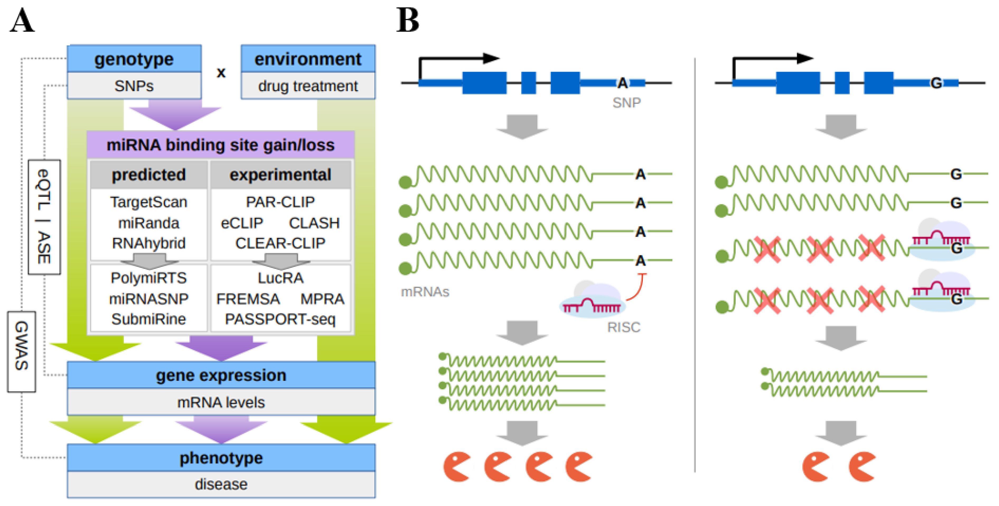
The complementary interaction of microRNAs (miRNAs) with their binding sites in the 3′untranslated regions (3′UTRs) of target gene mRNAs represses translation, playing a leading role in gene expression control. MiRNA recognition elements (MREs) in the 3′UTRs of genes often contain single nucleotide polymorphisms (SNPs), which can change the binding affinity for target miRNAs leading to dysregulated gene expression. Accumulated data suggest that these SNPs can be associated with various human pathologies (cancer, diabetes, neuropsychiatric disorders, and cardiovascular diseases) by disturbing the interaction of miRNAs with their MREs located in mRNA 3′UTRs. Numerous data show the role of SNPs in 3′UTR MREs in individual drug susceptibility and drug resistance mechanisms. This work brief the data on such SNPs focusing on the most rigorously proven cases. Some SNPs belong to conventional genes from the drug-metabolizing system (in particular, the genes coding for cytochromes P450 (CYP 450), phase II enzymes (SULT1A1 and UGT1A), and ABCB3 transporter and their expression regulators (PXR and GATA4)). Other examples of SNPs are related to the genes involved in DNA repair, RNA editing, and specific drug metabolisms. The gene-by-gene studies and genome-wide approaches utilized or potentially utilizable to detect the MRE SNPs associated with individual response to drugs discussed.

microRNA (miRNA) miRNA target sites single nucleotide polymorphisms (SNPs) 3′untranslated regions (3′UTRs)

1. Introduction

Recently, large scale sequencing and genome-wide association studies (GWASs) have identified tens of thousands of variants (mainly, single nucleotide polymorphisms, SNPs) associated with various human traits and diseases [1][2][3]. Of them, nearly 90% are located in noncoding regions [4][5] and can impact gene expression in several ways. A large group of the SNPs mainly residing in promoter and enhancer regions influences transcription initiation by creating or disrupting transcription factor binding sites or changing the affinity of transcription factors for their cognate sites [6][7][8][9]. Many SNPs affect RNA splicing [6][10]. In addition, 3′untranslated regions (3′UTRs) of genes often contain the variants which are able to change the binding affinity of targeting miRNAs, thereby interfering with gene expression at a posttranscriptional level [11][12].
According to the current data, miRNAs interact in a complementary manner with their sites in the 3′UTRs of the mRNAs of target genes causing template degradation and translational repression, thus playing a leading role in the posttranscriptional mechanisms of gene expression regulation [13]. MicroRNAs (miRNAs), an evolutionarily conserved class of endogenous 18–24-nucleotide noncoding RNAs, form a RISC complex together with one of the AGO family proteins and auxiliary proteins and are involved in translation repression or cleavage of target mRNAs [14][15]. So far, over 2600 miRNAs have been identified in humans [16]. One miRNA has the potential to target many genes (on the average, approximately 500 genes) and, according to the estimates, over 60% of the human mRNAs are potential targets of miRNAs [17].
Currently, a significant amount of data suggest that the coordinated action of miRNAs plays an important role in the regulation of biological processes, including the cell cycle, cell growth and differentiation, migration, apoptosis, and stress response [18]. Numerous studies have also shown that an altered expression of miRNAs contributes to the development of a wide range of human pathologies, such as cancer, diabetes, neuropsychiatric disorders, cardiovascular diseases, and infection diseases [19][20][21][22][23]. The SNPs interfering with the interaction of miRNAs with their target sites, located in both miRNA seed sequences and mRNA 3′UTRs, are frequently associated with the same diseases (for review, see [12]). In addition, some data suggest the involvement of SNPs in 3′UTR miRNA target sequences in individual drug susceptibility and drug resistance mechanisms [24][25][26]

2. Modern Approaches to Identify Functional SNPs in 3′UTR miRNA Target Sequences

An integrated set of in silico, in vitro, and in vivo approaches has been designed so far to detect the SNPs located in mRNA 3′UTRs and influencing (both deteriorating and enhancing) the interaction of miRNAs with their target sites (Figure 1).
Figure 1. (A) Principal scheme of discovery of functional SNPs that result in changes of microRNA binding to their response elements in the mRNA 3’UTR; (B) the hypothetical mechanism of the individual drug response due to the mirSNP. SNP(A>G) in 3′UTR of mRNA leads to the gain of the miRNA response element allowing miRNA-mRNA binding which hinders gene expression at a posttranscriptional level.
As a rule, in silico approaches rely on a certain algorithm predicting miRNA binding sites that make it possible to assess the functionality of these sites or correspondingly rank them based on the type of seed match, site accessibility, evolutionary conservation, nucleotide environment, thermodynamic characteristics of the duplex, and so on. In particular, the databases PolymiRTS 3.0 (http://www.targetscan.org/vert_71/) (accessed on 10 October 2022) and miRNASNP-v3 (http://bioinfo.bjmu.edu.cn/mirsnp/search/) (accessed on 10 October 2022) or the tool SubmiRine (https://research.nhgri.nih.gov/software/SubmiRine/) (accessed on 10 October 2022) use the popular de novo prediction algorithm TargetScan (http://www.targetscan.org/vert_71/) (accessed on 10 October 2022). This algorithm utilizes the so-called context+ score regression model to assess the site efficiency, which takes into account the contribution of the parameters, such as seed-pairing stability, target-site abundance, local AU content, site location within 3′UTR, and 3′-supplementary pairing (and a more recent context++ model, based on 14 parameters, including the degree of site conservation). Thus, the SNPs are selected that either differ in their context+ score or absent in one of the allelic variants. An alternative in silico approach used, in particular, in miRNASNP-v3, as well as popular and effective in silico method to study individual sites is the calculation of the minimum free energy (MFE) of hybridization between a miRNA seed and its cognate mRNA target (for example, using MicroInspector, RNAfold and RNAhybrid) [27][28][29].
A set of experimental approaches allowing for gene-by-gene or genome-wide studies is used to verify the obtained predictions. The main in vitro gene-by-gene approaches are luciferase reporter assay and fluorescent-based RNA electrophoretic mobility shift assay (FREMSA or simply RNA EMSA).
The luciferase reporter assay is used in almost all studies on assessment of the effect of a nucleotide substitution on the efficiency of miRNA–mRNA interaction. This method utilizes the plasmid constructs carrying a mutated or a reference allele of miRNA recognition site located in the mRNA 3′UTR inserted downstream of the reporter luciferase gene. Each plasmid construct is transfected alone [30] or cotransfected with the miRNA into the culture of selected eukaryotic cells to monitor the reporter expression [31]. Either siRNA overexpressing plasmids [32], miRNA mimics (individually [11][27][28][33] or a library of miRNA mimics [34] are used for cotransfection. Deletion and mutagenesis of the putative miRNA response element (MRE) in the plasmid construct are used to confirm the location of binding site for each miRNA [35]. Both transient [11][27][28][33] and stable transfection of cultivated cells with the plasmid reporter construct with concurrent cotransfection of mimic miRNA are used [36].
Although many papers assert that the luciferase reporter assay is able to determine the differences in the binding affinities between the wild-type and alternative alleles, this method in fact can record only the suppressive effect of a miRNA on the expression of its putative targets [31][32][37], which only allows the effect of the corresponding nucleotide substitution on the affinity of miRNA–mRNA interaction to be assumed. In addition, a considerable limitation of this method is the tissue-specificity of the studied effects, which requires that several cell lines are used [38].
FREMSA is an in vitro technique which is able to provide direct evidence of the interaction between miRNA and mRNA and to assess the effect of an SNP on the efficiency of this interaction [27][39][40]. This method utilizes the RNA oligonucleotides corresponding to the mature miRNA species and the cognate mRNA fragments corresponding to the miRNA targeting sequences. The fragments are 5′-labeled with different fluorescent dyes. The reaction mixture is separated by gel electrophoresis; the bands representing miRNAs, mRNAs, and miRNA–mRNA complexes are visualized according to different wavelengths of fluorescence dyes [27][39][40].
Correlation analysis of the expression of the miRNA and mRNA carrying different alleles in tissue samples is an informative in vivo method, widely used to assess the potential regulatory effects of miRNAs on their targets [32][34][41]. Another approach utilizes the transient transfection of primary human cells with miRNA mimics or the miRNA mimic negative controls in order to evaluate their influence on the target enzyme activity or mRNA concentrations [35]. The miRNA inhibitors, leading to a decrease in their levels in cells and the coupled decrease in the levels of mRNA targets, are also used [28].
To date, over 400,000 SNPs have been identified in putative miRNA binding sites (mirSNPs) by in silico methods, which poses the challenge of designing the genome-wide approaches to search for functional SNPs affecting miRNA binding [38]. The advent of state-of-the-art genome-wide technologies has brought about some such methods. In particular, the MPRA (massive parallel reporter analysis) [42] made it possible in 2018 to develop the PASSPORT-seq (parallel assessment of polymorphisms in miRNA target sites by sequencing) approach, which involves pooled synthesis, parallel cloning, and single-well transfection followed by next-generation sequencing (NGS) to functionally test hundreds of mirSNPs at once [38]. First, the utility of the assay was demonstrated by study of 100 randomly selected mirSNPs in HEK293, HepG2, and HeLa cells. Using traditional individual luciferase assay, a gold-standard platform for in vitro assessing gene expression, 17 of the tested 21 PASSPORT-seq results were confirmed. Second, the application of PASSPORT-seq technology to 111 mirSNPs in 3′UTR of 17 pharmacogenes showed a significant effect of 33 variants in at least one cell line (HeLa, HepG2, HEK293, or HepaRG). Several examples from this list are included below.
NGS was also used to design AGO crosslinking immunoprecipitation (CLIP) technologies that allow for the identification of endogenously relevant transcriptome-wide sites bound by AGO (eCLIP, PAR-CLIP, and HITS-CLIP). However, these technologies fail to reveal which particular miRNAs bind to each mRNA target [43][44]. A ligation step added to the CLIP technology makes it possible to sequence the chimeric miRNA–mRNA molecules, thereby showing the pairs that have hybridized (crosslinking, ligation, and sequencing of hybrids, CLASH) [45]. Powell et al. [44] used this approach to generate a liver-expressed pharmacogene-relevant map of miRNA–mRNA interactions and target sites. In their study, the chimeric-eCLIP was used to experimentally define the miRNA–mRNA interactome in primary human hepatocytes from a pool of 100 donors. Based on these results, an extensive map of miRNA binding of each gene was developed, including the pharmacogenes expressed in primary hepatocytes. Using the high-throughput PASSPORT-seq assay in HepaRG cells, they further tested the functional impact of 262 genetic variants in these miRNA binding sites. These 262 variants were selected among approximately 3600 PharmGKB variants as having the coordinates overlapping their chimeric-eCLIP–identified target sites. As a result, they discovered 24 mirSNPs considered to be functional in HepaRG cells. The share of the detected functional SNPs in their work is lower as compared with the earlier study of this group reported by Ipe et al. [38] although both studies were carried out using the same approach. The putative cause of this discrepancy is that in the later study Powell et al. used only one cell line versus four lines used in the earlier study by Ipe et al. [44]. Another possible cause is that in the later study [44] the SNPs were involved initially localized not only in 3′UTR, but also in the gene coding regions. In particular, the combination of chimeric-eCLIP with PASSPORT-seq successfully identified the functional mirSNPs at the end of the DPYD pharmacogene coding region that influenced the hsa-mir-27b interaction with the earlier validated binding site [46]. Note that these results suggest a potentially important role of the binding to target sequences not only in the 3′UTR but in the coding regions as well.

References

  1. Tam, V.; Patel, N.; Turcotte, M.; Bossé, Y.; Paré, G.; Meyre, D. Benefits and Limitations of Genome-Wide Association Studies. Nat. Rev. Genet. 2019, 20, 467–484.
  2. Buniello, A.; MacArthur, J.A.L.; Cerezo, M.; Harris, L.W.; Hayhurst, J.; Malangone, C.; McMahon, A.; Morales, J.; Mountjoy, E.; Sollis, E.; et al. The NHGRI-EBI GWAS Catalog of Published Genome-Wide Association Studies, Targeted Arrays and Summary Statistics 2019. Nucleic Acids Res. 2019, 47, D1005–D1012.
  3. Claussnitzer, M.; Cho, J.H.; Collins, R.; Cox, N.J.; Dermitzakis, E.T.; Hurles, M.E.; Kathiresan, S.; Kenny, E.E.; Lindgren, C.M.; MacArthur, D.G.; et al. A Brief History of Human Disease Genetics. Nature 2020, 577, 179–189.
  4. Hindorff, L.A.; Sethupathy, P.; Junkins, H.A.; Ramos, E.M.; Mehta, J.P.; Collins, F.S.; Manolio, T.A. Potential Etiologic and Functional Implications of Genome-Wide Association Loci for Human Diseases and Traits. Proc. Natl. Acad. Sci. USA 2009, 106, 9362–9367.
  5. Maurano, M.T.; Humbert, R.; Rynes, E.; Thurman, R.E.; Haugen, E.; Wang, H.; Reynolds, A.P.; Sandstrom, R.; Qu, H.; Brody, J.; et al. Systematic Localization of Common Disease-Associated Variation in Regulatory DNA. Science 2012, 337, 1190–1195.
  6. Huang, Q. Genetic Study of Complex Diseases in the Post-GWAS Era. J. Genet. Genom. 2015, 42, 87–98.
  7. Degtyareva, A.O.; Antontseva, E.V.; Merkulova, T.I. Regulatory SNPs: Altered Transcription Factor Binding Sites Implicated in Complex Traits and Diseases. Int. J. Mol. Sci. 2021, 22, 6454.
  8. Jin, Y.; Jiang, J.; Wang, R.; Qin, Z.S. Systematic Evaluation of DNA Sequence Variations on in Vivo Transcription Factor Binding Affinity. Front. Genet. 2021, 12, 667866.
  9. Tseng, C.-C.; Wong, M.-C.; Liao, W.-T.; Chen, C.-J.; Lee, S.-C.; Yen, J.-H.; Chang, S.-J. Genetic Variants in Transcription Factor Binding Sites in Humans: Triggered by Natural Selection and Triggers of Diseases. Int. J. Mol. Sci. 2021, 22, 4187.
  10. Dufner-Almeida, L.G.; do Carmo, R.T.; Masotti, C.; Haddad, L.A. Understanding Human DNA Variants Affecting Pre-MRNA Splicing in the NGS Era. Adv. Genet. 2019, 103, 39–90.
  11. Li, B.; Dong, J.; Yu, J.; Fan, Y.; Shang, L.; Zhou, X.; Bai, Y. Pinpointing MiRNA and Genes Enrichment over Trait-Relevant Tissue Network in Genome-Wide Association Studies. BMC Med. Genom. 2020, 13, 191.
  12. Chhichholiya, Y.; Suryan, A.K.; Suman, P.; Munshi, A.; Singh, S. SNPs in MiRNAs and Target Sequences: Role in Cancer and Diabetes. Front. Genet. 2021, 12, 793523.
  13. Moszyńska, A.; Gebert, M.; Collawn, J.F.; Bartoszewski, R. SNPs in MicroRNA Target Sites and Their Potential Role in Human Disease. Open Biol. 2017, 7, 170019.
  14. Fabian, M.R.; Sonenberg, N.; Filipowicz, W. Regulation of MRNA Translation and Stability by MicroRNAs. Annu. Rev. Biochem. 2010, 79, 351–379.
  15. Wilczynska, A.; Bushell, M. The Complexity of MiRNA-Mediated Repression. Cell Death. Differ. 2015, 22, 22–33.
  16. Kozomara, A.; Birgaoanu, M.; Griffiths-Jones, S. MiRBase: From MicroRNA Sequences to Function. Nucleic Acids Res. 2019, 47, D155–D162.
  17. Nakano, M.; Iwakami, C.; Fukami, T.; Nakajima, M. Identification of MiRNAs That Regulate Human CYP2B6 Expression. Drug Metab. Pharmacokinet. 2021, 38, 100388.
  18. Bartel, D.P. MicroRNAs: Target Recognition and Regulatory Functions. Cell 2009, 136, 215–233.
  19. Condrat, C.E.; Thompson, D.C.; Barbu, M.G.; Bugnar, O.L.; Boboc, A.; Cretoiu, D.; Suciu, N.; Cretoiu, S.M.; Voinea, S.C. MiRNAs as Biomarkers in Disease: Latest Findings Regarding Their Role in Diagnosis and Prognosis. Cells 2020, 9, 276.
  20. He, B.; Zhao, Z.; Cai, Q.; Zhang, Y.; Zhang, P.; Shi, S.; Xie, H.; Peng, X.; Yin, W.; Tao, Y.; et al. MiRNA-Based Biomarkers, Therapies, and Resistance in Cancer. Int. J. Biol. Sci. 2020, 16, 2628–2647.
  21. Chakraborty, C.; Sharma, A.R.; Sharma, G.; Lee, S.-S. Therapeutic Advances of MiRNAs: A Preclinical and Clinical Update. J. Adv. Res. 2021, 28, 127–138.
  22. Paul, S.; Reyes, P.R.; Garza, B.S.; Sharma, A. MicroRNAs and Child Neuropsychiatric Disorders: A Brief Review. Neurochem. Res. 2020, 45, 232–240.
  23. Paul, S.; Bravo Vázquez, L.A.; Uribe, S.P.; Manzanero Cárdenas, L.A.; Ruíz Aguilar, M.F.; Chakraborty, S.; Sharma, A. Roles of MicroRNAs in Carbohydrate and Lipid Metabolism Disorders and Their Therapeutic Potential. Biochimie 2021, 187, 83–93.
  24. Swart, M.; Dandara, C. Genetic Variation in the 3’-UTR of CYP1A2, CYP2B6, CYP2D6, CYP3A4, NR1I2, and UGT2B7: Potential Effects on Regulation by MicroRNA and Pharmacogenomics Relevance. Front. Genet. 2014, 5, 167.
  25. Li, M.-P.; Hu, Y.-D.; Hu, X.-L.; Zhang, Y.-J.; Yang, Y.-L.; Jiang, C.; Tang, J.; Chen, X.-P. MiRNAs and MiRNA Polymorphisms Modify Drug Response. Int. J. Environ. Res. Public Health 2016, 13, 1096.
  26. Zanger, U.M.; Klein, K.; Kugler, N.; Petrikat, T.; Ryu, C.S. Epigenetics and MicroRNAs in Pharmacogenetics. Adv. Pharmacol. 2018, 83, 33–64.
  27. Knox, B.; Wang, Y.; Rogers, L.J.; Xuan, J.; Yu, D.; Guan, H.; Chen, J.; Shi, T.; Ning, B.; Kadlubar, S.A.; et al. A Functional SNP in the 3′-UTR of TAP2 Gene Interacts with MicroRNA Hsa-miR-1270 to Suppress the Gene Expression. Environ. Mol. Mutagen. 2018, 59, 134–143.
  28. Yu, X.; Dhakal, I.B.; Beggs, M.; Edavana, V.K.; Williams, S.; Zhang, X.; Mercer, K.; Ning, B.; Lang, N.P.; Kadlubar, F.F.; et al. Functional Genetic Variants in the 3′-Untranslated Region of Sulfotransferase Isoform 1A1 (SULT1A1) and Their Effect on Enzymatic Activity. Toxicol. Sci. 2010, 118, 391–403.
  29. Omariba, G.; Xu, F.; Wang, M.; Li, K.; Zhou, Y.; Xiao, J. Genome-Wide Analysis of MicroRNA-Related Single Nucleotide Polymorphisms (SNPs) in Mouse Genome. Sci. Rep. 2020, 10, 5789.
  30. Zheng, Z.; Xue, F.; Wang, H.; He, Y.; Zhang, L.; Ma, W.; Zhang, C.; Guan, Y.; Ye, F.; Wen, Y.; et al. A Single Nucleotide Polymorphism-Based Formula to Predict the Risk of Propofol TCI Concentration Being over 4 µg ML−1 at the Time of Loss of Consciousness. Pharmacogenom. J. 2022, 22, 109–116.
  31. Clément, T.; Salone, V.; Rederstorff, M. Dual Luciferase Gene Reporter Assays to Study MiRNA Function. Methods Mol. Biol. 2015, 1296, 187–198.
  32. Li, S.; Xu, K.; Gu, D.; He, L.; Xie, L.; Chen, Z.; Fan, Z.; Zhu, L.; Du, M.; Chu, H.; et al. Genetic Variants in RPA1 Associated with the Response to Oxaliplatin-Based Chemotherapy in Colorectal Cancer. J. Gastroenterol. 2019, 54, 939–949.
  33. Hua, H.; Zeng, J.; Xing, H.; He, Y.; Han, L.; Zhang, N.; Yang, M. The RNA Editing Enzyme ADAR Modulated by the Rs1127317 Genetic Variant Diminishes EGFR-TKIs Efficiency in Advanced Lung Adenocarcinoma. Life Sci. 2022, 296, 120408.
  34. Papageorgiou, I.; Court, M.H. Identification and Validation of the MicroRNA Response Elements in the 3′-Untranslated Region of the UDP Glucuronosyltransferase (UGT) 2B7 and 2B15 Genes by a Functional Genomics Approach. Biochem. Pharmacol. 2017, 146, 199–213.
  35. Papageorgiou, I.; Court, M.H. Identification and Validation of MicroRNAs Directly Regulating the UDP-Glucuronosyltransferase 1A Subfamily Enzymes by a Functional Genomics Approach. Biochem. Pharmacol. 2017, 137, 93–106.
  36. Nakano, M.; Mohri, T.; Fukami, T.; Takamiya, M.; Aoki, Y.; McLeod, H.L.; Nakajima, M. Single-Nucleotide Polymorphisms in Cytochrome P450 2E1 (CYP2E1) 3′-Untranslated Region Affect the Regulation of CYP2E1 by MiR-570. Drug Metab. Dispos. 2015, 43, 1450–1457.
  37. Tomasello, L.; Cluts, L.; Croce, C.M. Experimental Validation of MicroRNA Targets: Mutagenesis of Binding Regions. Methods Mol. Biol. 2019, 1970, 331–339.
  38. Ipe, J.; Collins, K.S.; Hao, Y.; Gao, H.; Bhatia, P.; Gaedigk, A.; Liu, Y.; Skaar, T.C. PASSPORT-Seq: A Novel High-Throughput Bioassay to Functionally Test Polymorphisms in Micro-RNA Target Sites. Front Genet. 2018, 9, 219.
  39. Zeng, L.; Chen, Y.; Wang, Y.; Yu, L.-R.; Knox, B.; Chen, J.; Shi, T.; Chen, S.; Ren, Z.; Guo, L.; et al. MicroRNA Hsa-MiR-370-3p Suppresses the Expression and Induction of CYP2D6 by Facilitating MRNA Degradation. Biochem. Pharmacol. 2017, 140, 139–149.
  40. Yu, D.; Chen, S.; Li, D.; Knox, B.; Guo, L.; Ning, B. FREMSA: A Method That Provides Direct Evidence of the Interaction between MicroRNA and MRNA. Methods Mol. Biol. 2020, 2102, 557–566.
  41. Wei, R.; Yang, F.; Urban, T.J.; Li, L.; Chalasani, N.; Flockhart, D.A.; Liu, W. Impact of the Interaction between 3′-UTR SNPs and MicroRNA on the Expression of Human Xenobiotic Metabolism Enzyme and Transporter Genes. Front. Genet. 2012, 3, 248.
  42. Kheradpour, P.; Ernst, J.; Melnikov, A.; Rogov, P.; Wang, L.; Zhang, X.; Alston, J.; Mikkelsen, T.S.; Kellis, M. Systematic Dissection of Regulatory Motifs in 2000 Predicted Human Enhancers Using a Massively Parallel Reporter Assay. Genome Res. 2013, 23, 800–811.
  43. Li, D.; Chen, M.; Hong, H.; Tong, W.; Ning, B. Integrative Approaches for Studying the Role of Noncoding RNAs in Influencing Drug Efficacy and Toxicity. Expert. Opin. Drug Metab. Toxicol. 2022, 18, 151–163.
  44. Powell, N.R.; Zhao, H.; Ipe, J.; Liu, Y.; Skaar, T.C. Mapping the MiRNA-mRNA Interactome in Human Hepatocytes and Identification of Functional MirSNPs in Pharmacogenes. Clin. Pharmacol. Ther. 2021, 110, 1106–1118.
  45. Helwak, A.; Kudla, G.; Dudnakova, T.; Tollervey, D. Mapping the Human MiRNA Interactome by CLASH Reveals Frequent Noncanonical Binding. Cell 2013, 153, 654–665.
  46. Offer, S.M.; Butterfield, G.L.; Jerde, C.R.; Fossum, C.C.; Wegner, N.J.; Diasio, R.B. MicroRNAs MiR-27a and MiR-27b Directly Regulate Liver Dihydropyrimidine Dehydrogenase Expression through Two Conserved Binding Sites. Mol. Cancer Ther. 2014, 13, 742–751.
More
Information
Contributors MDPI registered users' name will be linked to their SciProfiles pages. To register with us, please refer to https://encyclopedia.pub/register : , , ,
View Times: 508
Revisions: 3 times (View History)
Update Date: 24 Nov 2022
1000/1000
Video Production Service