Submitted Successfully!
To reward your contribution, here is a gift for you: A free trial for our video production service.
Thank you for your contribution! You can also upload a video entry or images related to this topic.
Version Summary Created by Modification Content Size Created at Operation
1 -- 4142 2022-11-18 16:20:22 |
2 update references and layout + 10 word(s) 4152 2022-11-21 03:08:39 |

Video Upload Options

Do you have a full video?

Confirm

Are you sure to Delete?
Cite
If you have any further questions, please contact Encyclopedia Editorial Office.
Sonawala, K.;  Ramalingam, S.;  Sellamuthu, I. LncRNAs in Cancer Stem Cell Signaling Pathways. Encyclopedia. Available online: https://encyclopedia.pub/entry/35316 (accessed on 27 July 2024).
Sonawala K,  Ramalingam S,  Sellamuthu I. LncRNAs in Cancer Stem Cell Signaling Pathways. Encyclopedia. Available at: https://encyclopedia.pub/entry/35316. Accessed July 27, 2024.
Sonawala, Kevina, Satish Ramalingam, Iyappan Sellamuthu. "LncRNAs in Cancer Stem Cell Signaling Pathways" Encyclopedia, https://encyclopedia.pub/entry/35316 (accessed July 27, 2024).
Sonawala, K.,  Ramalingam, S., & Sellamuthu, I. (2022, November 18). LncRNAs in Cancer Stem Cell Signaling Pathways. In Encyclopedia. https://encyclopedia.pub/entry/35316
Sonawala, Kevina, et al. "LncRNAs in Cancer Stem Cell Signaling Pathways." Encyclopedia. Web. 18 November, 2022.
LncRNAs in Cancer Stem Cell Signaling Pathways
Edit

Initially entitled as junk matter, non-coding RNAs are an exceptional class of RNAs constituting a majority of the transcriptional output in living cells, which are not translated into functional proteins. They are not only responsible for regulating the expression of the gene at the transcriptional and post-transcriptional stages but also for mediating various cellular processes such as heterochromatin formation, epigenetic modifications, signal transduction and so on. It is quite evident from one research that the abnormal expression of LncRNAs plays a significant role in cancer stem cells (CSCs)’ metabolism. hey regulate gene expression by the following approaches: as a modulator of gene expression; as a decoy to lead the transcription factor elsewhere from a target site; as a competitor to hinder the attachment of other molecules to the target site; as a chaperone for molecules to attach to a certain segment and as a scaffold that enhances the association of different proteins into different complexes.

non-coding RNAs lncRNAs cancer stem cells signaling pathways

1. Focus on LncRNAs

One of the types of non-coding RNAs that span higher than 200 nucleotides in length, possess no mechanism to code for proteins, and are seen to have a little expression in certain tissues are considered as long non-coding RNAs. Some lncRNAs may show exceptions to these criteria. It is quite evident from the recent research that the abnormal expression of LncRNAs plays a significant role in cancer stem cells (CSCs)’ metabolism. They regulate gene expression by the following approaches: as a modulator of gene expression; as a decoy to lead the transcription factor elsewhere from a target site; as a competitor to hinder the attachment of other molecules to the target site; as a chaperone for molecules to attach to a certain segment and as a scaffold that enhances the association of different proteins into different complexes [1].
Different types of lncRNAs regulate the functions of CSCs by altering and modulating different transcription factors, enzymes, niches, and signaling pathways. LncRNA H19 is seen to maintain the stemness in glioma [2] and BCSCs [3]. Contrastingly, lncRNA ROR plays a repressive role in glioma SCs by negatively altering the expressions of stem cell factor KLF4 [4]. LncRNA CUDR leads to high H3K4 trimethylation by activating the associations between SET1A and pRB1in hepatocellular carcinoma (HCC) [5]. It works with Cyclin-D to stimulate the self-renewal and proliferation of hepatocellular carcinoma stem cells by increasing the expression of telomerase reverse transcriptase (TERT) and C-Myc [6]. In prostate cancer, the PCR2-LncRNA HOTAIR association represses the androgen receptor transcription by complexing to its 5′-flanking promoter region and enhancing prostate cancer stem/progenitor cells and invasion [7]. In breast cancer, LncRNA HOTAIR leads to the induction of epithelial–mesenchymal transition (EMT) by underregulating the STAT3 pathway [8]. LncRNA uc.283-plus [9], LncRNA CRNDE [10], and LncRNA XIST [11] are all known to be overregulated in glioma stem cells. Two lncRNAs, PRNCR1 and PCGEM1, are found to be overexpressed in different prostate cancers and cause the androgen receptor (AR)–associated transcriptional mechanisms to induce the growth of prostate cancer [12][13]. Maternally expressed gene 3 lncRNA (MEG3) was observed to stimulate p53 and promote p53 signaling, as well as increasing p53 associations with the promoters of its target genes [14]. Recurrent hypermethylation in the MEG3 promoter is commonly seen in human tumors, such as pituitary cancer [15], meningioma [16], and leukemia [17]. Higher expression of MEG3 works in a tumor suppressive manner by oppressing cell proliferation in meningioma and HCC cell lines [18].

2. The Interplay of LncRNAs in CSCs Signaling Pathways

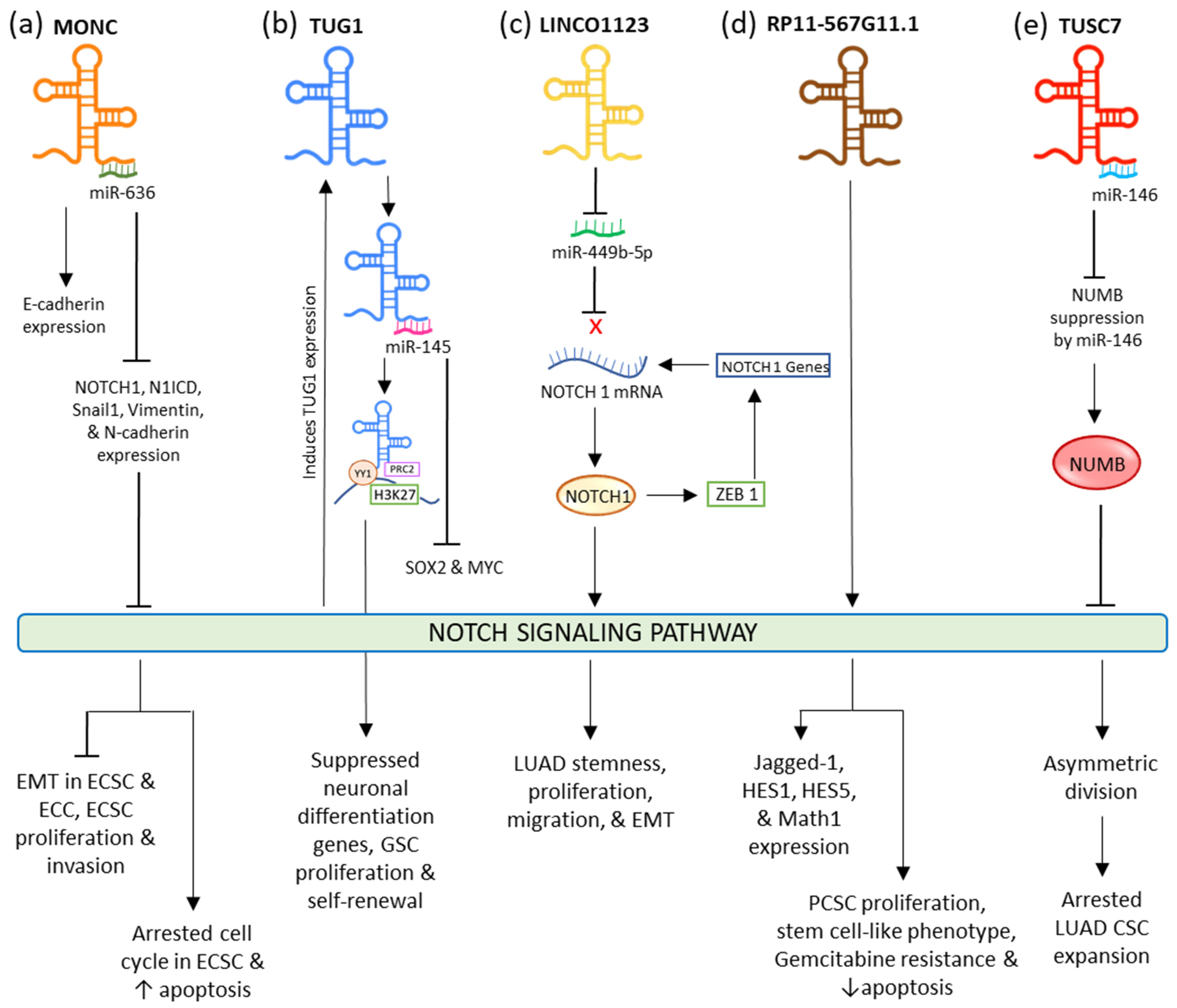
2.1. NOTCH Signaling Pathway

NOTCH signaling is a majorly conserved signaling pathway that carries out juxtracrine signaling between cells. NOTCH signaling takes place by the binding of two jagged ligands (Jagged1; Jagged2) and a transmembrane delta-like ligand either 1, 2, 3, or 4 (DLL1, DLL2, DLL3, DLL4) and one cell to a NOTCH receptor either 1, 2, 3, or 4 (NOTCH1, NOTCH2, NOTCH3, NOTCH4) on the neighboring cell. In the endoplasmic reticulum, NOTCH is synthesized and glycosylated by the soluble ER enzymes. In the Golgi, the S1 cleavage generates a heterodimer of NOTCH extracellular domain (NECD) and transmembrane NOTCH intracellular domain (TM-NICD). It then is transported to the plasma membrane to permit ligand binding. This binding is modulated by Deltex and obstructed by the activity of NUMB. After this, the S2 cleavage takes place, the NECD is cleaved from the TM-NICD domain by TNF-α ADAM metalloprotease converting enzyme (TACE) ADAM17 or a disintegrin and metalloproteinase domain-10 (ADAM10). In the signal-sending cell, the NECD stays confined to the ligand and experiences further recycling. Meanwhile, in the signal-sending cell, S3 cleavage is carried out by γ-Secretase, which releases the NICD segment. It is then translocated to the nucleus, where it associates with transcriptional regulators to instigate transcription of the NOTCH target genes.
NOTCH signaling plays a prominent role in the modulation of cell-fate specification, and progenitor cell differentiation, maintenance, and proliferation during organogenesis as well as during the early development of the hematopoietic cells and Central Nervous System [19][20]. In stem cell niches, it is crucial in the differential activities of some stem cells, stem cell proliferation, and their interaction with the microenvironment. The carcinogenicity of the pathway was first observed in human T-cell acute lymphoblastic leukemia (T-ALL), where, due to the release of N1ICD, NOTCH-1 was seen to be activated [21]. Since then, abnormal activation of the NOTCH family led to tumorigenesis of various cancers, such as pancreatic [22], breast [23], and cervical cancer [24]. This pathway is observed to operate as both oncogenic and tumor-suppressive, this is dependent on the tissue type, cancer cell type, as well as the stage involved. NOTCH-1 stimulates cancer development at an early stage of cervical cancer, while it represses cancer growth at a later stage. As an oncogene, NOTCH is seen to be overexpressed in T-ALL, pancreatic, breast, gastric, and colon cancer [22][23][25][26][27]. As an example, mutations leading to activated NOTCH1 can stimulate the development of T-ALL [28]. PCSCs were observed to have increased levels of NOTCH1, NOTCH3, Jag1, Jag2, as well as the NOTCH target gene Hes1; this was responsible for the maintenance of CSCs stemness [29]. Meanwhile, its expression is downregulated in myeloid malignancies, skin, non-small cell lung cancer, liver, prostate, and certain breast cancers [30][31][32][33][34] where it plays a tumor-suppressive role. It was observed that NOTCH-1 activation is responsible for inducing the MMP-2 and MMP-9 expression and stimulation while lowering the regulation of NOTCH-1 involved in lowering the MMP-2 and MMP-9 activation to hinder cell progression in breast cancer and pancreatic cancer cells [35].
In endometrial carcinoma (EC), tumor-suppressive MONC sponges oncogenic miR-636 to negatively regulate it. The upregulation of this lncRNA restricted the expression of NOTCH1, N1ICD, Vimentin, Snail1, and N-Cadherin, while the expression of E-Cadherin was fostered. This eventually inhibited the NOTCH signaling pathway and EMT resulting in suppressed proliferation and invasion, and induced apoptosis, and aided the arrest of the cell cycle at the G0/G1 phase in endometrial cancer cells and cancer stem cells (ECCs and ECSCs) (Figure 1a) [36]. Increased expression of lncRNA TUG1 (Taurine Upregulated Gene 1) is seen in different types of cancers such as bladder carcinoma, stomach cancer, and bone sarcoma [37][38][39], while its decreased expression is seen in non-small cell lung cancer (NSCLC) [40]. This lncRNA is credible for sustaining the stemness properties and proliferation of glioma stem cells (GSC) in glioblastoma via neutralizing miR-145 by sponging it in the cytoplasm and modulating transcription factors MYC and SOX2. In the nucleus, it forms a polycomb structure with YY1, PRC2, and H3K27, from which histone H3K27 undergoes locus-specific methylation via the activity of YY1 to limit the neuronal differentiation genes (Figure 1b). In lung adenocarcinoma (LUAD), ZEB1, an EMT transcriptional factor, activates LINC01123 and NOTCH1. LINC01123 functions by sponging miR-449b-5p and blocking its inhibitory effect on NOTCH1 mRNA; this results in the expression of NOTCH1, thus mediating the promotion of the NOTCH1-dependent NOTCH signaling pathway. This results in the lncRNA facilitating increased stemness, proliferation, migration, malignant transformation, and EMT in LUAD (Figure 2c) [41]. In pancreatic cancer, RP11-567G11.1 is observed to upregulate the NOTCH signaling pathway and the expression of its downstream components. This aberrant activation resulted in the progression of the cell cycle, PCSCs proliferation, tumorigenesis, and decreased apoptosis. RP11-567G11.1 also mediated the chemoresistance to antimetabolite Gemcitabine (Figure 2d) [42]. LncRNA TUSC-7 is responsible for inactivated NOTCH signaling in lung adenocarcinoma (LUAD) stem cells. This leads to the disruption in the symmetric division, causing the appointment of asymmetric division and ultimately arresting the CSC expansion.
Figure 1. Interaction of different lncRNAs and lincRNA with NOTCH signaling pathway.
It does so by inactivating the miR-146-mediated repression of NUMB, which plays a significant role in restraining the NOTCH signaling. It is responsible for restraining the ligand from binding by hindering the NOTCH receptor’s endosome transportation to the plasma membrane. When TUSC-7 is sponged to miR-146, its degradation toward NUMB is abolished, resulting in the inactivation of NOTCH signaling (Figure 1e) [43].

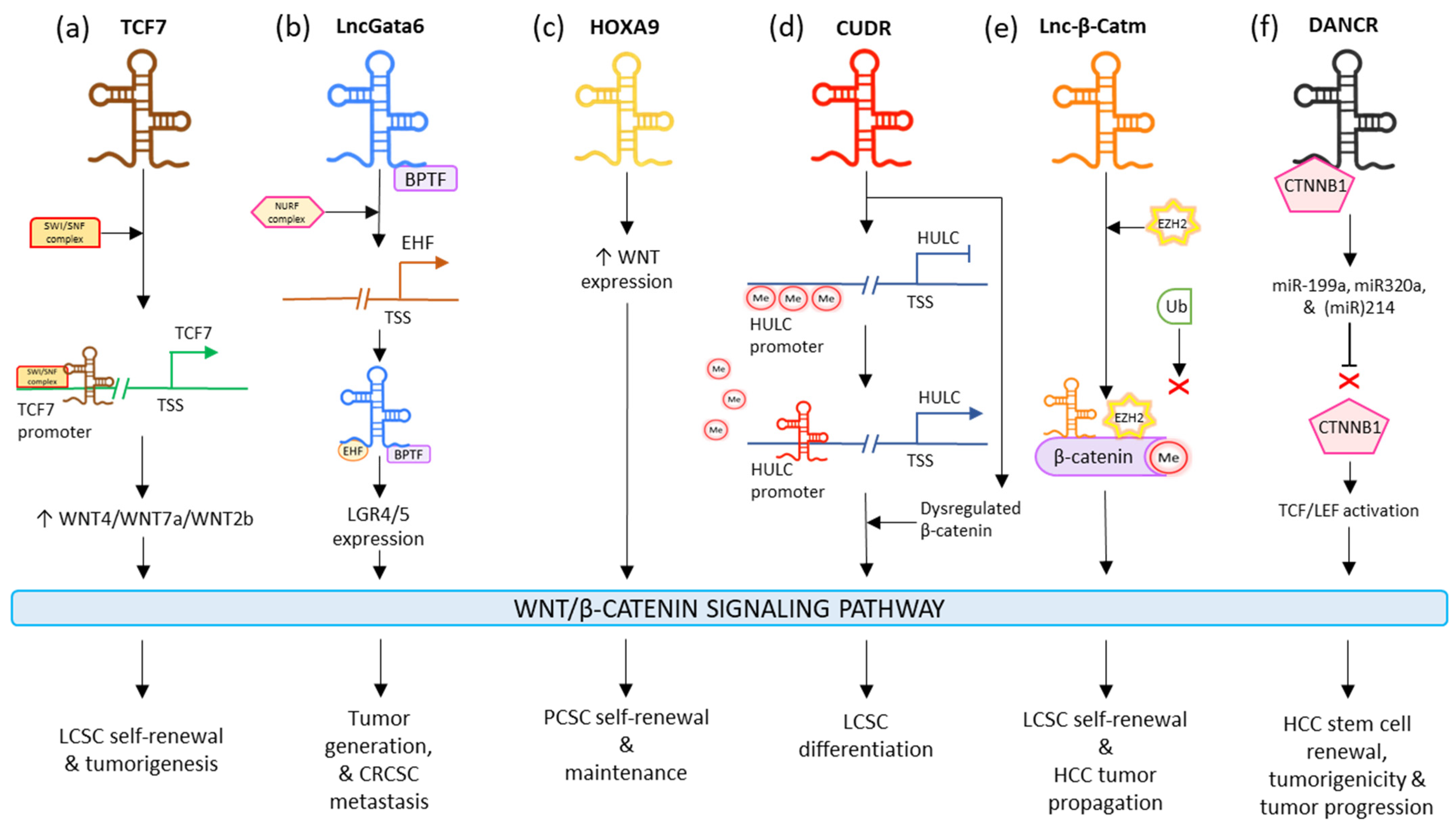
2.2. WNT Signaling Pathway

WNT signaling significantly participates in different biological activities, such as cell proliferation, differentiation, tissue regeneration, and organogenesis [44][45][46][47][48]. This pathway is split into β-Catenin-dependent and β-Catenin-independent signaling; the former is known as the canonical pathway which is the WNT/β-Catenin signaling pathway and the latter is known as the non-canonical pathways comprised of WNT/calcium signaling and WNT/planar cell polarity [PCP]-signaling pathways [49][50].
The canonical WNT signaling pathway is initiated when WNT-associated ligands link to Frizzled, a transmembrane receptor and the low-density lipoprotein receptor-related protein (LRP). Phosphorylation of LRP is then carried out, due to which Dishevelled proteins (DVL) become polymerized at the plasma membrane. This further hinders the destruction complex; comprising APC, AXIN, and GSK3β. This leads to the accumulation of β-Catenin in the cytoplasm, which then undergoes nuclear translocation. Once inside, β-Catenin is associated with the T-cell factor (TCF) and lymphoid enhancer factor (LEF); it functions by regulating multiple cellular mechanisms, thus behaving as a transcriptional switch [51].
In the non-canonical calcium-signaling pathway, WNT binds to DVL to stimulate phospholipase C (PLC) which releases calcium ions. These intracellular ions stimulate the downstream protein kinase C (PKC) and calcium/calmodulin-dependent protein kinase II (CaMKII) which in turn positively affect the nuclear factor (NFAT) [52]. In the non-canonical PCP-signaling pathway, the planar cell polarity is triggered by WNT–Frizzled receptor associations that activate DVL. Activated DVL then modulates RHOA, RAC, and CDC42 GTPases. It further triggers the JUN N-terminal kinase, which then activates nuclear factor-dependent transcription of activator protein-1 and stimulated T cells after nuclear transfer.
The canonical pathway is mostly known as regulating cell proliferation, while noncanonical pathways manage cells’ polarity and movement. However, both signaling pathways participate in tumorigenesis and can also become involved in different cellular processes [53]. In the same way, adenomatous polyposis coli (APC) and β-Catenin not only regulate cell proliferation but are also involved in cell-to-cell adhesion [54]. In colon cancer, R-SPONDIN/LGR5/RNF43 mutations are linked with the deregulation of WNT leading to tumor development [51]. Approximately 92% of sporadic colorectal cancers are observed to have at least an alteration in the regulators of the WNT pathway [55]. In a wide range of breast cancers, plummeted levels of nuclear β-Catenin are reported, as well as seeing regular plummeted expression of the receptors and ligands. However, unlike colorectal cancer, the development of breast cancer is not due to the genetic regulation of the signaling components of the WNT pathway, but is due to other factors such as epigenetics [56]. In different types of leukemias, the deregulated WNT pathway leads to increased WNT activity, eventually triggering leukemogenesis. In liver-cancer stem cells (LCSC), activation of canonical WNT signaling is linked with elevated self-renewal capacity of cells [57]. WNT signaling was seen to elevate the expression of a colon cancer SC marker CD44v6, which resulted in an aggravated metastatic capacity of the CSCs [58]. In the breast cancer niche, the cross talk of Periostin with WNT1 and WNT3a stimulates WNT signaling, resulting in the sustained phenotype of cancer stem cells [59].
LncTCF7 appoints the SWI/SNF to the TCF7 promoter and improves its expression to surge the levels of WNT7a/WNT4/WNT2b, which activates the WNT signaling pathway. The lncTCF7-associated WNT stimulation results in the maintenance of the self-renewal and tumorigenic ability of LCSCs (Figure 2a) [57]. LncGata6 enhances tumor generation and metastasis in colorectal CSCs. LncGata6 associates with Bromodomain PHD Finger Transcription Factor (BPTF) and adopts the Nucleosome Remodeling Factor (NURF) complex to trigger ETS homologous factor (EHF) transcription, which makes up the lncGata6–BPTF–EHF axis to generate LGR4/5 expression and eventually activate the WNT signaling pathway (Figure 2b) [60]. LncRNA HOTTIP is observed to drive the stimulation of the WNT/β-Catenin signaling pathway; this induces the cell generation and chemoresistance in osteosarcoma. It is also responsible for elevating the stemness of PCSCs by regulating the WNT pathway. HOXA9 regulates the stimulation of the WNT pathway in PCSCs by promoting WNT. HOXA9 targeted stem cell transcription factors (SOX2, Nanog, OCT4, and LIN28) and markers (ALDH1, CD44, and CD133) in order to sustain PCSC properties such as inducing CSC expansion and self-renewal (Figure 2c) [61]. Interestingly, LncRNA CUDR stimulates lncRNA HULC by obstructing the methylation of the HULC promoter; additionally, it dysregulates β-Catenin thus promoting malignant differentiation in human-liver stem cells (Figure 2d) [62]. LincRNA-p21 is seen to reduce the activity of β-Catenin signaling, ultimately mediating the negative regulation of self-renewal, viability, and glycolysis of colorectal-cancer stem cells (CRCSCs) [63]. The levels of lncRNA H19 are observed to plummet in bladder cancer tissues. H19 is complexed with the enhancer of zeste homolog 2 (EZH2), such that this association results in the activation of WNT/β-Catenin. This activation reduces the expression of E-cad and increases tumor metastasis. Upregulated H19 is also responsible for promoting bladder-cancer cell migration [64]. Lnc-β-Catm catalyzes the methylation process of β-Catenin, by binding to it along with methyltransferase EZH2. This limits the ubiquitination of β-Catenin and thereby stabilizes it, which eventually drives the activation of the WNT/β-Catenin signaling pathway. This activation promotes self-renewal and maintenance of LCSCs and tumor progression of HCC (Figure 2e) [65]. In HCC, microRNAs miR214, miR320a, and miR-199a are seen to have a suppressive effect on CTNNB1; DANCR blocks this suppression by associating with tethering to CTNNB1. This induces the activation of TCF/LEF by β-Catenin, which guides the positive regulation of HCC stem cells’ self-renewal, tumorigenicity, and cancer progression (Figure 2f) [66]. In gastric cancer, HOTAIR was seen to be majorly regulated in cisplatin-resistant gastric cancer cells and tissues. It was observed that the higher expression of HOTAIR promoted gastric-cancer cell proliferation, enhanced the transition of cell cycle G1/S, as well as decreased cancer cell apoptosis by activating WNT/β-Catenin signaling. Moreover, the lncRNA was also responsible for binding and inhibiting miR-126 expression; this resulted in the promotion of VEGFA and PIK3R2 expression and activation of the PI3K/AKT/MRP1 pathway [67].
Figure 2. Influence of different lncRNAs on WNT/β-Catenin CSC signaling pathway.

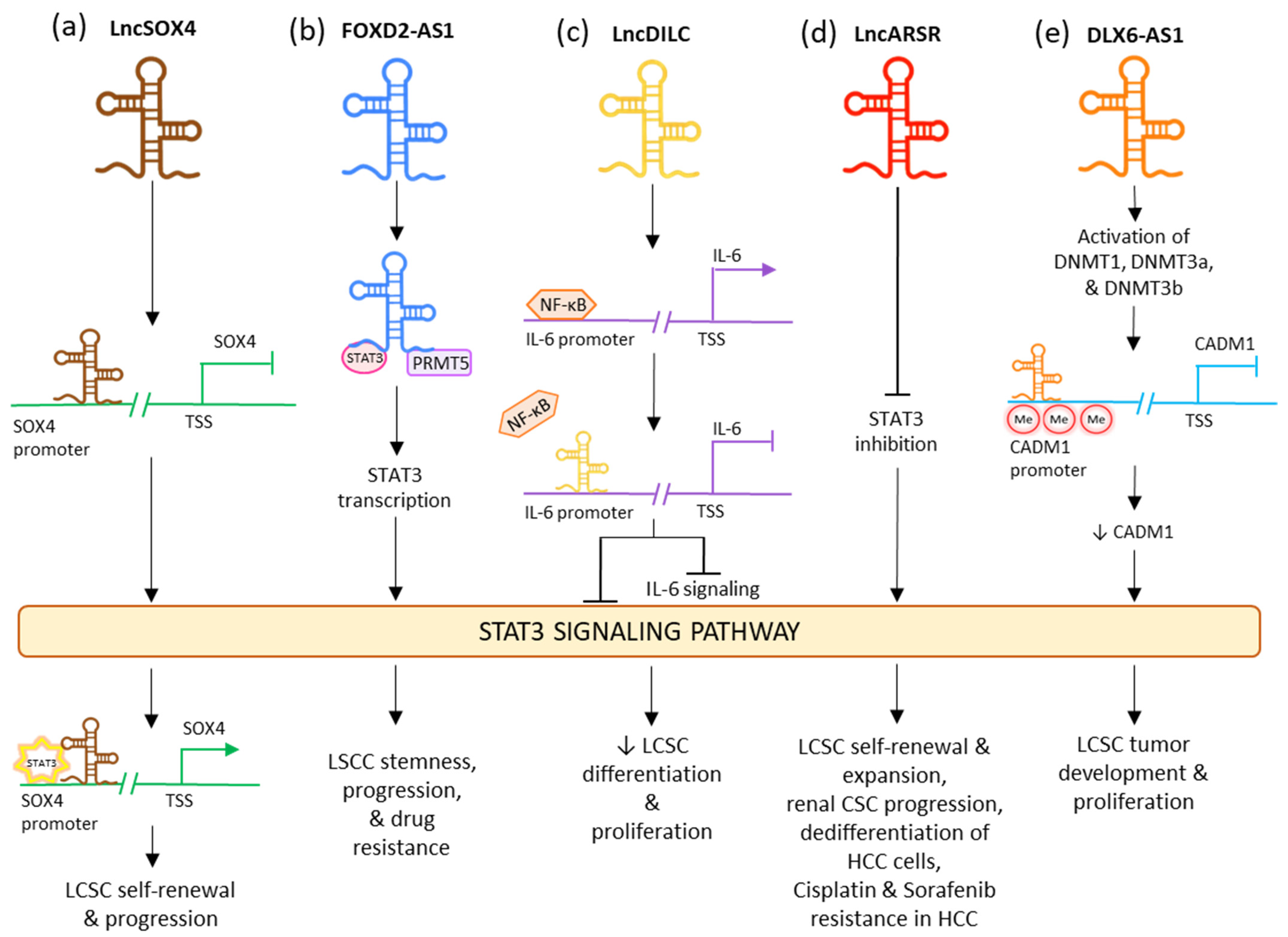
2.3. STAT Signaling Pathway

In humans, the STAT family incorporates STAT-1, 2, 3, 4, 5A, 5B, and 6 [68]. Among these seven proteins, STAT-3 and STAT-5 demonstrate the highest association with tumor progression. Their activation regulates a broad range of functions, such as cell cycle progression, proliferation, apoptosis, angiogenesis, as well as immune evasion [69][70][71]. STAT-3 is primarily responsible for tumor growth, proliferation, and sustenance due to its activity in stromal cells, immune cells, and the tumor microenvironment [72][73][74][75][76][77]. The negative modulation of STAT-3 activation is carried out by protein repressors of stimulated STATs (PIAS), tyrosine phosphatases, and oppressors of cytokine signaling (SOCS) [78].
Abnormal activation of the pathway is seen in CSCs from different cancers such as breast, blood, glia, and prostate. In prostate cancer, stem-like cells were seen to overexpress various genes linked to JAK/STAT signaling pathway, such as IFNK, IFNGR, IL6, CSF2, and STAT1 [79], and stimulate STAT3 to prominently regulate JAK/STAT signaling molecules in mammalian CSC-like cells [80]. In glioblastoma, obstruction of STAT3 in CSCs shuts down proliferation, as well as lowering the expression of OLIG2 and NESTIN (neural stem cell gene), and enhancing the expression of βIII-Tubulin, which acts as a neuronal differentiator [81]. Thus, in glioblastoma, JAK/STAT signaling is significant for CSC proliferation and is responsible for positively regulating glioblastoma stemness. In acute myeloid leukemia (AML), activated JAK/STAT signaling led to the growth and sustenance of CSCs [82].
Abnormal STAT3 activation, because of tyrosine 705 phosphorylation signaling, led to the oncogenesis and induction of malignancy [83][84][85][86]. Activation of STAT3 in the tumor causes transfer of indicators from the different growth factors and cytokines [87] which in turn induces certain target genes such as Cyclin-D, c-Myc, and CDC25A. These factors ultimately activate the proliferation of cells, suppress the apoptotic genes [88], and upregulate anti-apoptotic genes such as Beta2-Macroglobulin, B-Cell CLL/Lymphoma-2 (BCL2), and BCLXL [89]. The induction of STAT3 by IL-6 [90] or stress factors [91] leads to progress in the self-renewal of prostate CSCs and tumor-propagation [92]. VEGF binds to VEGFR-2/JAK2/STAT3, leading to the activation of STAT3 and upregulation of Myc and SOX2. This eventually results in enhanced self-renewal of breast and lung CSC. Thus, apart from angiogenesis, VEGF is also responsible for CSCs’ self-renewal and tumor-initiating via the VEGFR-2/STAT3 signaling [93].
LncRNA downregulated in liver-cancer stem cell (lnc-DILC) acts as a tumor suppressor by declining the proliferation rates of CSC and differentiation of the LCSCs [94]. It also has an intense hindering effect on the autocrine pathway of IL-6/JAK2/STAT3 signaling [95]. This lncRNA lowers the expression of STAT3 as well as IL-6 and also limits the translocation of STAT3 to the nucleus. It also acts by inhibiting the NF-κB-mediated induction of IL-6. In various tumor stem cells, IL-6 is highly expressed by the NF-κB signaling pathway which is triggered by growth factors such as TNF-α and IL-1β (Figure 3c) [96][97]. LncRNA DILC and lncSOX4 are well known for controlling CSC properties by the STAT3 pathway in liver cancer. Lnc-DILC can also regulate the JAK2/STAT3 signaling. Overexpression of lnc-DILC lowers the elevated levels of phospho-STAT3 protein, reduces the translocation of STAT3 to the nucleus, and oppresses the transcriptional activity of STAT3-responsive segments. In CSC spheroids, the decreased regulation of lnc-DILC activates TNF-α and IL-1β-triggered IL-6 expression. Thus, this lncRNA can synchronize the crosstalk between the inflammatory signaling and the autocrine IL-6/STAT3 pathway to affect the expansion of LCSC [98]. On the other hand, lncSOX4 increased regulation has been associated with critical liver cancer. It is positively regulated in the CD133+ liver cancer cell population and CSC spheroids and is crucial for the self-renewing activities and tumorigenic capability of the liver CSCs. LncSOX4 crosstalks with STAT3 and inducts it to the promoter region of SOX4, stimulating the H3K4me3 and H3K27ac alterations to increase the SOX4 promoter stimulation and thus SOX4 expression. The lncSOX4/STAT3-dependent SOX4 expression is significant for sustaining liver CSC proliferation (Figure 3a) [99]. Upregulated FOXD2-AS1 was identified in maintaining the stemness in laryngeal squamous cell carcinoma (LSCC) and decreasing the reactions to chemotherapy drugs, while its under-regulation had opposing consequences. FOXD2-AS1 is complexed as a scaffold with STAT3 and PRMT5, stimulating the transcription STAT3, which is important for maintaining tumor stemness and enabling chemotherapeutic resistance. Thus, FOXD2-AS1 acts as an upstream activator of STAT3 and aids cancer progression (Figure 3b) [100]. LncARSR overexpression is seen in CD24+/CD133+ LCSCs as well as in hepatoma spheres augmented with CSC. Its upregulation causes blockage of STAT3 inhibition, thus activating the STAT3 signaling. STAT3 also plays a downstream role in regulation by lncARSR in HCC cells. LncARSR is responsible for LCSC expansion by rising the dedifferentiation of hepatoma cells and increasing the self-renewal, progression, and tumorigenesis capacity of LCSCs. It also aids drug resistance and tumor recurrence (Figure 3d) [101]. In early LSCC, the levels of lncRNA PTCSC3 in plasma are low. The increased expression of PTCSC3 causes repression of LSCC cell proliferation; thus, the lncRNA acts as a tumor suppressor. In UM-SCC-17A cells, PTCSC3 hindered STAT3 and HOTAIR’s expression, while STAT3 was responsible for increasing the expression of HOTAIR. Thus, it can be stipulated that the oppressive effect of PTCSC3 on tumor progression can be connected to lncRNA HOTAIR [100]. The downregulation of LncRNA DLX6-AS1 may cause an inhibitory effect on the methylation of the CADM1 promoter. This may lead to the inactivation of the STAT3 signaling pathway. Upregulated CADM1 results in the suppression of the stem cell characteristics of liver CSCs [102]. In HCC, overactivated lncRNA DLX6-AS1 confers the methylation of CADM1 promoter via activation of methyltransferases DNMTs and expression of SOX4 in LCSCs and thus upregulating the STAT3 signaling pathway. This led to decreased expressions of SOX2, OCT-4, CD13, and CD133 in LCSCs and induced tumor formation and proliferation (Figure 3e) [102].
Figure 3. Influence of different lncRNAs on STAT3 CSC signaling pathway.

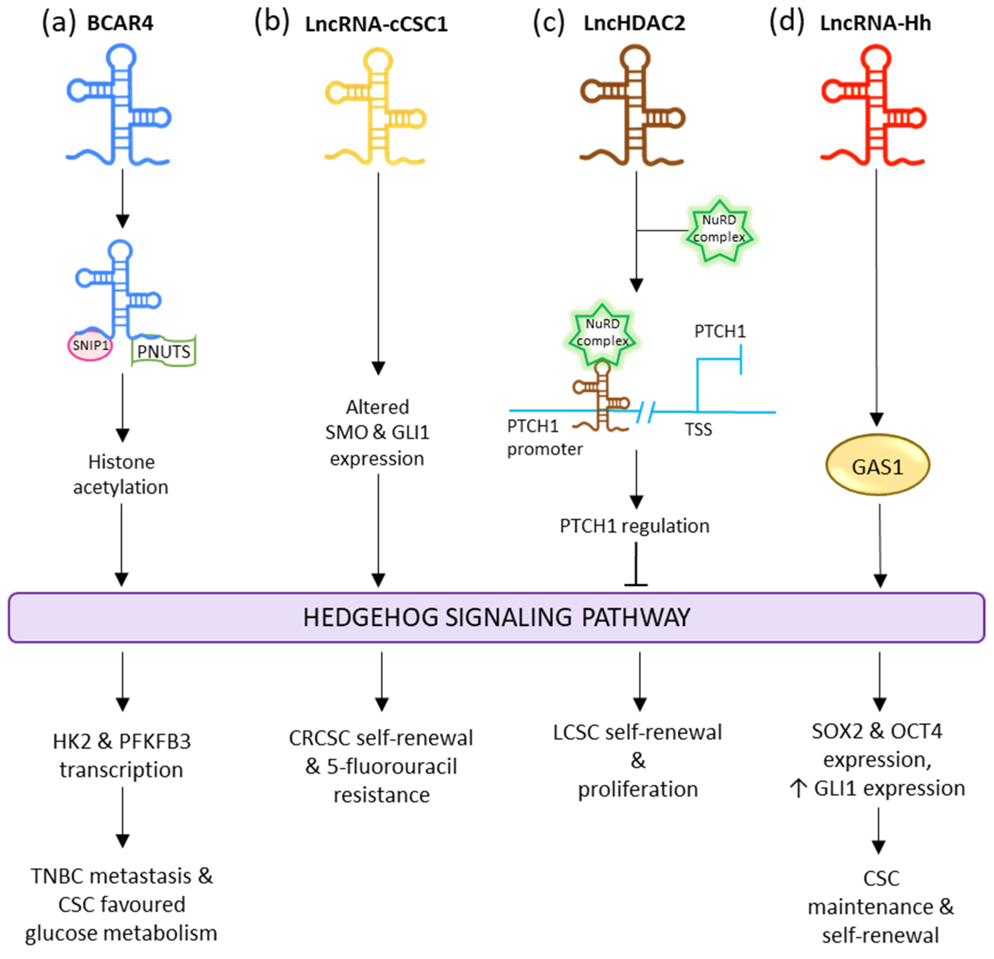
2.4. Hedgehog Signaling Pathway

Abnormal induction of Hedgehog is linked with the proliferation of cancer cells, neoplastic transformations, malignancy, metastasis, and multi-drug resistance of a wide range of tumors. When Hedgehog is not present, PTCH1 localizes in the cilia and avoids the membrane localization and stimulation of SMO. SMO after internalization undergoes degradation. The full-length GLI (GLIFL) is altered by protein kinase A (PKA), glycogen synthase kinase-3 (GSK3), casein kinase 1 (CK1), and the E3 ubiquitin ligase β-TrCP. It then undergoes cleavage to form transcriptional oppressor GLIR. The stimulated form of GLI (GLIA) is repressed by SUFU. Then, GLIR undergoes nuclear translocation and hinders the expression of its target genes. When the Hedgehog binds to PTCH1, it internalizes and carries out the translocation and induction of SMO. GLIFL avoids phosphorylation by PKA, GSK3, and CK1, which causes activation of GLI (GLIA). GLIA then undergoes nuclear translocation where it activates the expression of Hedgehog target genes [103].
The activated Hedgehog pathway is involved in the growth, survival, migration, and proliferation [104] of a wide range of cancers such as multiple myeloma, pancreatic adenocarcinoma [105], breast cancer [106], and chronic myelogenous leukemia (CML) [107]. The first evidence of deregulated Hedgehog signaling and cancer was seen in Gorlin syndrome, which is caused due to the inducing mutation in the Patched gene [108]. Patients with this syndrome display different cancer types such as basal cell carcinomas (BCCs) [109], medulloblastoma [110][111], and rhabdomyosarcoma [112][113]. The paracrine stimulation of the Hedgehog pathway is seen in different tumor types such as prostate [114], pancreatic [115], colorectal, and esophageal cancers [116]. In chronic myelogenous leukemia (CML), the loss of SMO impaired the self-renewal capacity of the hematopoietic stem cells and hindered the initiation of CML by the oncoprotein: BCR-ABL1 [117]. Sonic Hedgehog, SMO, and GLI1 are highly aggravated in CML suggesting that activation of the Hedgehog pathway is a significant activator of CML progression [118]. Hedgehog inactivation in MCF-7-derived CSCs caused a reduction in the cells via the downregulation of OCT4, NESTIN, and Nanog, suggesting that Hedgehog signaling in breast CSCs upregulates stem cell markers to retain a self-renewing phenotype [119]
This signaling plays a crucial role in the sustenance of colon CD133+ CSCs, which show the increased gene expression levels of GLI1, PTCH1, GLI2, SHH, and HHIP concerning all CD133- cells in colon carcinoma [120].
Yes-associated protein 1 (YAP) is responsible for upregulating the transcription of lncRNA BCAR4, which interacts with SNIP1 and PNUTS to regulate the p300-dependent histone acetylation carried by H3K27ac of the non-canonical Hedgehog and GLI2 transcriptional cascade. This stimulates the transcription of enzymes HK2 and PFKFB3; thus, allowing BCAR4 to promote metastasis and invasion in triple-negative breast cancer as well as reprogram glucose metabolism in favor of the CSCs (Figure 4a) [121]. LncRNA-cCSC1 is seen to be overexpressed in colorectal cancer cells (CRC) and CRCSCs. The exhaustion of lncRNA-cCSC1 prominently hindered the self-renewal activity of the CRCSCs and decreased their chemo-drug resistance to 5-fluorouracil. Meanwhile increased expression of lncRNA-cCSC1 led to triggered self-renewal of CSCs as well as assisting in 5-fluorouracil drug resistance. Abnormal expression of this lncRNA resulted in the transfiguration of SMO and GLI family zinc finger 1 (GLI1) expression in the Hedgehog signaling pathway (Figure 4b) [122]. LncRNA lncHDAC2 is overexpressed in LCSCs. In the LCSC niche, it is associated with histone deacetylase-2 (HDAC2). LncHDAC2 regulated PTCH1 downregulation to stimulate Hedgehog signaling, thus increasing the self-renewal activity of LCSCs (Figure 4c) [123]. Overexpression of ASAP1-IT1 is seen in cholangiocarcinoma (CCA) tissues and cells. This lncRNA is responsible for activating the Hedgehog pathway via the increased regulation of SMO and GLI1, thus inducing cell proliferation, cell migration, and the progression of EMT [124].
Cells 11 03492 g005
Figure 4. Influence of different lncRNAs on Hedgehog CSC signaling pathway.
LncRNA EGOT acts as a significant modulator in breast cancer. In gastric cancer, with elevated expression, it acts as an oncogene. EGOT-regulated Cyclin D1 expression in GC is modulated by the Hedgehog pathway. Its upregulation is related to gastric cancer cell growth, cell cycle progression, and lymphatic metastasis [125]. LncRNA HCG18 participates as an oncogenic lncRNA in nasopharyngeal cancer (NPC) progression. It does so by modulating WNT/β-Catenin signaling and the Hedgehog pathway. Upregulation of HCG18 is associated with tumor progressive effects via the stimulation of cell proliferation and metastasis [126]. LncRNA-Hh is known to target GAS1 to induce the activation of Hedgehog signaling. The activated Hedgehog signaling raises the expression of GLI1 and stimulates the expression of SOX2 and OCT4 to regulate CSC maintenance and self-renewal (Figure 4d) [127].

References

  1. Wang, K.C.; Chang, H.Y. Molecular Mechanisms of Long Noncoding RNAs. Mol. Cell 2011, 43, 904–914.
  2. Li, W.; Jiang, P.; Sun, X.; Xu, S.; Ma, X.; Zhan, R. Suppressing H19 Modulates Tumorigenicity and Stemness in U251 and U87MG Glioma Cells. Cell. Mol. Neurobiol. 2016, 36, 1219–1227.
  3. Peng, F.; Li, T.T.; Wang, K.L.; Xiao, G.Q.; Wang, J.H.; Zhao, H.D.; Kang, Z.J.; Fan, W.J.; Zhu, L.L.; Li, M.; et al. H19/Let-7/LIN28 Reciprocal Negative Regulatory Circuit Promotes Breast Cancer Stem Cell Maintenance. Cell Death Dis. 2017, 8, e2569.
  4. Feng, S.; Yao, J.; Chen, Y.; Geng, P.; Zhang, H.; Ma, X.; Zhao, J.; Yu, X. Expression and Functional Role of Reprogramming-Related Long Noncoding RNA (LincRNA-ROR) in Glioma. J. Mol. Neurosci. 2015, 56, 623–630.
  5. Wu, M.; An, J.; Zheng, Q.; Xin, X.; Lin, Z.; Li, X.; Li, H.; Lu, D. Double Mutant P53 (N340Q/L344R) Promotes Hepatocarcinogenesis through Upregulation of Pim1 Mediated by PKM2 and LncRNA CUDR. Oncotarget 2016, 7, 66525–66539.
  6. Pu, H.; Zheng, Q.; Li, H.; Wu, M.; An, J.; Gui, X.; Li, T.; Lu, D. CUDR Promotes Liver Cancer Stem Cell Growth through Upregulating TERT and C-Myc. Oncotarget 2015, 6, 40775–40798.
  7. Li, L.; Dang, Q.; Xie, H.; Yang, Z.; He, D.; Liang, L.; Song, W.; Yeh, S.; Chang, C. Correction: Infiltrating Mast Cells Enhance Prostate Cancer Invasion via Altering LncRNA-HOTAIR/PRC2-Androgen Receptor (AR)-MMP9 Signals and Increased Stem/Progenitor Cell Population. Oncotarget 2016, 7, 83828.
  8. Alves, C.P.; Fonseca, A.S.; Muys, B.R.; Bueno, R.D.B.E.L.; Burger, M.C.; De Souza, J.E.S.; Valente, V.; Zago, M.A.; Silva, W.A. Brief Report: The LincRNA Hotair Is Required for Epithelial-to-Mesenchymal Transition and Stemness Maintenance of Cancer Cell Lines. Stem Cells 2013, 31, 2827–2832.
  9. Galasso, M.; Dama, P.; Previati, M.; Sandhu, S.; Palatini, J.; Coppola, V.; Warner, S.; Sana, M.E.; Zanella, R.; Abujarour, R.; et al. A Large Scale Expression Study Associates Uc.283-plus LncRNA with Pluripotent Stem Cells and Human Glioma. Genome Med. 2014, 6, 76.
  10. Wang, Y.; Wang, Y.; Li, J.; Zhang, Y.; Yin, H.; Han, B. CRNDE, a Long-Noncoding RNA, Promotes Glioma Cell Growth and Invasion through MTOR Signaling. Cancer Lett. 2015, 367, 122–128.
  11. Ghafouri-Fard, S.; Dashti, S.; Farsi, M.; Taheri, M.; Mousavinejad, S.A. X-Inactive-Specific Transcript: Review of Its Functions in the Carcinogenesis. Front. Cell Dev. Biol. 2021, 9, 690522.
  12. Yang, L.; Lin, C.; Jin, C.; Yang, J.C.; Tanasa, B.; Li, W.; Merkurjev, D.; Ohgi, K.A.; Meng, D.; Zhang, J.; et al. LncRNA-Dependent Mechanisms of Androgen-Receptor-Regulated Gene Activation Programs. Nature 2013, 500, 598–602.
  13. Popadiuk, C.M.; Xiong, J.; Wells, M.G.; Andrews, P.G.; Dankwa, K.; Hirasawa, K.; Lake, B.B.; Kao, K.R. Antisense Suppression of Pygopus2 Results in Growth Arrest of Epithelial Ovarian Cancer. Clin. Cancer Res. 2006, 12, 2216–2223.
  14. Zhou, Y.; Zhong, Y.; Wang, Y.; Zhang, X.; Batista, D.L.; Gejman, R.; Ansell, P.J.; Zhao, J.; Weng, C.; Klibanski, A. Activation of P53 by MEG3 Non-Coding RNA. J. Biol. Chem. 2007, 282, 24731–24742.
  15. Gibb, E.A.; Brown, C.J.; Lam, W.L. The Functional Role of Long Non-Coding RNA in Human Carcinomas. Mol. Cancer 2011, 10, 38.
  16. Zhang, X.; Gejman, R.; Mahta, A.; Zhong, Y.; Rice, K.A.; Zhou, Y.; Cheunsuchon, P.; Louis, D.N.; Klibanski, A. Maternally Expressed Gene 3, an Imprinted Noncoding RNA Gene, Is Associated with Meningioma Pathogenesis and Progression. Cancer Res. 2010, 70, 2350–2358.
  17. Benetatos, L.; Hatzimichael, E.; Dasoula, A.; Dranitsaris, G.; Tsiara, S.; Syrrou, M.; Georgiou, I.; Bourantas, K.L. CpG Methylation Analysis of the MEG3 and SNRPN Imprinted Genes in Acute Myeloid Leukemia and Myelodysplastic Syndromes. Leuk. Res. 2010, 34, 148–153.
  18. Braconi, C.; Kogure, T.; Valeri, N.; Huang, N.; Nuovo, G.; Costinean, S.; Negrini, M.; Miotto, E.; Croce, C.M.; Patel, T. MicroRNA-29 Can Regulate Expression of the Long Non-Coding RNA Gene MEG3 in Hepatocellular Cancer. Oncogene 2011, 30, 4750–4756.
  19. Chiba, S. Concise Review: Notch Signaling in Stem Cell Systems. Stem Cells 2006, 24, 2437–2447.
  20. Karamboulas, C.; Ailles, L. Developmental Signaling Pathways in Cancer Stem Cells of Solid Tumors. Biochim. Biophys. Acta (BBA)-Gen. Subj. 2013, 1830, 2481–2495.
  21. Ellisen, L.W.; Bird, J.; West, D.C.; Soreng, A.L.; Reynolds, T.C.; Smith, S.D.; Sklar, J. TAN-1, the Human Homolog of the Drosophila Notch Gene, Is Broken by Chromosomal Translocations in T Lymphoblastic Neoplasms. Cell 1991, 66, 649–661.
  22. Ma, Y.-C.; Shi, C.; Zhang, Y.-N.; Wang, L.-G.; Liu, H.; Jia, H.-T.; Zhang, Y.-X.; Sarkar, F.H.; Wang, Z.-S. The Tyrosine Kinase C-Src Directly Mediates Growth Factor-Induced Notch-1 and Furin Interaction and Notch-1 Activation in Pancreatic Cancer Cells. PLoS ONE 2012, 7, e33414.
  23. Wu, F.; Stutzman, A.; Mo, Y.Y. Notch Signaling and Its Role in Breast Cancer. Front. Biosci. 2007, 12, 4370–7383.
  24. Song, L.L.; Peng, Y.; Yun, J.; Rizzo, P.; Chaturvedi, V.; Weijzen, S.; Kast, W.M.; Stone, P.J.B.; Santos, L.; Loredo, A.; et al. Notch-1 Associates with IKKα and Regulates IKK Activity in Cervical Cancer Cells. Oncogene 2008, 27, 5833–5844.
  25. Qiao, L.; Wong, B.C.Y. Role of Notch Signaling in Colorectal Cancer. Carcinogenesis 2009, 30, 1979–1986.
  26. Zhou, W.; Fu, X.-Q.; Zhang, L.-L.; Zhang, J.; Huang, X.; Lu, X.-H.; Shen, L.; Liu, B.-N.; Liu, J.; Luo, H.-S.; et al. The AKT1/NF-KappaB/Notch1/PTEN Axis Has an Important Role in Chemoresistance of Gastric Cancer Cells. Cell Death Dis. 2013, 4, e847.
  27. Zhang, Y.; Li, B.; Ji, Z.Z.; Zheng, P.S. Notch1 Regulates the Growth of Human Colon Cancers. Cancer 2010, 116, 5207–5218.
  28. Ranganathan, P.; Weaver, K.L.; Capobianco, A.J. Notch Signalling in Solid Tumours: A Little Bit of Everything but Not All the Time. Nat. Rev. Cancer 2011, 11, 338–351.
  29. Abel, E.V.; Kim, E.J.; Wu, J.; Hynes, M.; Bednar, F.; Proctor, E.; Wang, L.; Dziubinski, M.L.; Simeone, D.M. The Notch Pathway Is Important in Maintaining the Cancer Stem Cell Population in Pancreatic Cancer. PLoS ONE 2014, 9, e91983.
  30. Kannan, S.; Sutphin, R.M.; Hall, M.G.; Golfman, L.S.; Fang, W.; Nolo, R.M.; Akers, L.J.; Hammitt, R.A.; McMurray, J.S.; Kornblau, S.M.; et al. Notch Activation Inhibits AML Growth and Survival: A Potential Therapeutic Approach. J. Exp. Med. 2013, 210, 321–337.
  31. Lefort, K.; Mandinova, A.; Ostano, P.; Kolev, V.; Calpini, V.; Kolfschoten, I.; Devgan, V.; Lieb, J.; Raffoul, W.; Hohl, D.; et al. Notch1 Is a P53 Target Gene Involved in Human Keratinocyte Tumor Suppression through Negative Regulation of ROCK1/2 and MRCKα Kinases. Genes Dev. 2007, 21, 562–577.
  32. Konishi, J.; Yi, F.; Chen, X.; Vo, H.; Carbone, D.P.; Dang, T.P. Notch3 Cooperates with the EGFR Pathway to Modulate Apoptosis through the Induction of Bim. Oncogene 2009, 29, 589–596.
  33. Viatour, P.; Ehmer, U.; Saddic, L.A.; Dorrell, C.; Andersen, J.B.; Lin, C.; Zmoos, A.F.; Mazur, P.K.; Schaffer, B.E.; Ostermeier, A.; et al. Notch Signaling Inhibits Hepatocellular Carcinoma Following Inactivation of the RB Pathway. J. Exp. Med. 2011, 208, 1963–1976.
  34. Gupta, A.; Wang, Y.; Browne, C.; Kim, S.; Case, T.; Paul, M.; Wills, M.L.; Matusik, R.J. Neuroendocrine Differentiation in the 12T-10 Transgenic Prostate Mouse Model Mimics Endocrine Differentiation of Pancreatic Beta Cells. Prostate 2008, 68, 50–60.
  35. Parr, C.; Watkins, G.; Jiang, W.G. The Possible Correlation of Notch-1 and Notch-2 with Clinical Outcome and Tumour Clinicopathological Parameters in Human Breast Cancer. Int. J. Mol. Med. 2004, 14, 779–786.
  36. Li, Y.; Huo, J.; He, J.; Ma, X. LncRNA MONC Suppresses the Malignant Phenotype of Endometrial Cancer Stem Cells and Endometrial Carcinoma Cells by Regulating the MiR-636/GLCE Axis. Cancer Cell Int. 2021, 21, 331.
  37. Tan, J.; Qiu, K.; Li, M.; Liang, Y. Double-Negative Feedback Loop between Long Non-Coding RNA TUG1 and MiR-145 Promotes Epithelial to Mesenchymal Transition and Radioresistance in Human Bladder Cancer Cells. FEBS Lett. 2015, 589, 3175–3181.
  38. Cao, W.J.; Wu, H.L.; He, B.S.; Zhang, Y.S.; Zhang, Z.Y. Analysis of Long Non-Coding RNA Expression Profiles in Gastric Cancer. World J. Gastroenterol. 2013, 19, 3658–3664.
  39. Zhang, Q.; Geng, P.L.; Yin, P.; Wang, X.L.; Jia, J.P.; Yao, J. Down-Regulation of Long Non-Coding RNA TUG1 Inhibits Osteosarcoma Cell Proliferation and Promotes Apoptosis. Asian Pac. J. Cancer Prev. 2013, 14, 2311–2315.
  40. Zhang, E.-B.; Yin, D.-D.; Sun, M.; Kong, R.; Liu, X.-H.; You, L.-H.; Han, L.; Xia, R.; Wang, K.-M.; Yang, J.-S.; et al. P53-Regulated Long Non-Coding RNA TUG1 Affects Cell Proliferation in Human Non-Small Cell Lung Cancer, Partly through Epigenetically Regulating HOXB7 Expression. Cell Death Dis. 2014, 5, e1243.
  41. Zhang, M.; Han, Y.; Zheng, Y.; Zhang, Y.; Zhao, X.; Gao, Z.; Liu, X. ZEB1-Activated LINC01123 Accelerates the Malignancy in Lung Adenocarcinoma through NOTCH Signaling Pathway. Cell Death Dis. 2020, 11, 981.
  42. Huang, R.; Nie, W.; Yao, K.; Chou, J. Depletion of the LncRNA RP11-567G11.1 Inhibits Pancreatic Cancer Progression. Biomed. Pharmacother. 2019, 112, 108685.
  43. Huang, G.; Wang, M.; Li, X.; Wu, J.; Chen, S.; Du, N.; Li, K.; Wang, J.; Xu, C.; Ren, H.; et al. TUSC7 Suppression of Notch Activation through Sponging MiR-146 Recapitulated the Asymmetric Cell Division in Lung Adenocarcinoma Stem Cells. Life Sci. 2019, 232, 116630.
  44. Acebron, S.P.; Karaulanov, E.; Berger, B.S.; Huang, Y.L.; Niehrs, C. Mitotic Wnt Signaling Promotes Protein Stabilization and Regulates Cell Size. Mol. Cell 2014, 54, 663–674.
  45. Atlasi, Y.; Noori, R.; Gaspar, C.; Franken, P.; Sacchetti, A.; Rafati, H.; Mahmoudi, T.; Decraene, C.; Calin, G.A.; Merrill, B.J.; et al. Wnt Signaling Regulates the Lineage Differentiation Potential of Mouse Embryonic Stem Cells through Tcf3 Down-Regulation. PLoS Genet. 2013, 9, e1003424.
  46. Clevers, H.; Loh, K.M.; Nusse, R. An Integral Program for Tissue Renewal and Regeneration: Wnt Signaling and Stem Cell Control. Science 2014, 3, 346.
  47. Green, J.L.; Inoue, T.; Sternberg, P.W. Opposing Wnt Pathways Orient Cell Polarity during Organogenesis. Cell 2008, 134, 646–656.
  48. Zhan, T.; Rindtorff, N.; Boutros, M. Wnt Signaling in Cancer. Oncogene 2017, 36, 1461–1473.
  49. Grumolato, L.; Liu, G.; Mong, P.; Mudbhary, R.; Biswas, R.; Arroyave, R.; Vijayakumar, S.; Economides, A.N.; Aaronson, S.A. Canonical and Noncanonical Wnts Use a Common Mechanism to Activate Completely Unrelated Coreceptors. Genes Dev. 2010, 24, 2517–2530.
  50. Katoh, M. Canonical and Non-Canonical WNT Signaling in Cancer Stem Cells and Their Niches: Cellular Heterogeneity, Omics Reprogramming, Targeted Therapy and Tumor Plasticity (Review). Int. J. Oncol. 2017, 51, 1357–1369.
  51. Dieter, S.M.; Glimm, H.; Ball, C.R. Colorectal Cancer-initiating Cells Caught in the Act. EMBO Mol. Med. 2017, 9, 856–858.
  52. Kahn, M. Can We Safely Target the WNT Pathway? Nat. Rev. Drug Discov. 2014, 13, 513–532.
  53. Mirabelli, C.K.; Nusse, R.; Tuveson, D.A.; Williams, B.O. Perspectives on the Role of Wnt Biology in Cancer. Sci. Signal. 2019, 12, eaay4494.
  54. Amin, N.; Cavallaro, U. The Wnt Signaling Pathways and Cell Adhesion. Front. Biosci. 2012, 17, 784–804.
  55. Schatoff, E.M.; Leach, B.I.; Dow, L.E. WNT Signaling and Colorectal Cancer. Curr. Color. Cancer Rep. 2017, 13, 101–110.
  56. Klarmann, G.J.; Decker, A.; Farrar, W.L. Epigenetic Gene Silencing in the Wnt Pathway in Breast Cancer. Epigenetics 2008, 3, 59–63.
  57. Wang, Y.; He, L.; Du, Y.; Zhu, P.; Huang, G.; Luo, J.; Yan, X.; Ye, B.; Li, C.; Xia, P.; et al. The Long Noncoding RNA LncTCF7 Promotes Self-Renewal of Human Liver Cancer Stem Cells through Activation of Wnt Signaling. Cell Stem Cell 2015, 16, 413–425.
  58. Todaro, M.; Gaggianesi, M.; Catalano, V.; Benfante, A.; Iovino, F.; Biffoni, M.; Apuzzo, T.; Sperduti, I.; Volpe, S.; Cocorullo, G.; et al. CD44v6 Is a Marker of Constitutive and Reprogrammed Cancer Stem Cells Driving Colon Cancer Metastasis. Cell Stem Cell 2014, 14, 342–356.
  59. Malanchi, I.; Santamaria-Martínez, A.; Susanto, E.; Peng, H.; Lehr, H.A.; Delaloye, J.F.; Huelsken, J. Interactions between Cancer Stem Cells and Their Niche Govern Metastatic Colonization. Nature 2012, 481, 85–91.
  60. Zhu, P.; Wu, J.; Wang, Y.; Zhu, X.; Lu, T.; Liu, B.; He, L.; Ye, B.; Wang, S.; Meng, S.; et al. LncGata6 Maintains Stemness of Intestinal Stem Cells and Promotes Intestinal Tumorigenesis. Nat. Cell Biol. 2018, 20, 1134–1144.
  61. Li, Z.; Zhao, L.; Wang, Q. Overexpression of Long Non-Coding RNA HOTTIP Increases Chemoresistance of Osteosarcoma Cell by Activating the Wnt/β-Catenin Pathway. Am. J. Transl. Res. 2016, 8, 2385.
  62. Gui, X.; Li, H.; Li, T.; Pu, H.; Lu, D. Long Noncoding RNA CUDR Regulates HULC and β-Catenin to Govern Human Liver Stem Cell Malignant Differentiation. Mol. Ther. 2015, 23, 1843–1853.
  63. Wang, J.; Lei, Z.J.; Guo, Y.; Wang, T.; Qin, Z.Y.; Xiao, H.L.; Fan, L.L.; Chen, D.F.; Bian, X.W.; Liu, J.; et al. MiRNA-Regulated Delivery of LincRNA-P21 Suppresses β-Catenin Signaling and Tumorigenicity of Colorectal Cancer Stem Cells. Oncotarget 2015, 6, 37852–37870.
  64. Luo, M.; Li, Z.; Wang, W.; Zeng, Y.; Liu, Z.; Qiu, J. Long Non-Coding RNA H19 Increases Bladder Cancer Metastasis by Associating with EZH2 and Inhibiting E-Cadherin Expression. Cancer Lett. 2013, 333, 213–221.
  65. Zhu, P.; Wang, Y.; Huang, G.; Ye, B.; Liu, B.; Wu, J.; Du, Y.; He, L.; Fan, Z. Lnc-β-Catm elicits EZH2-dependent β-catenin stabilization and sustains liver CSC self-renewal. Nat. Struct. Mol. Biol. 2016, 23, 631–639.
  66. Yuan, S.-X.; Wang, J.; Yang, F.; Tao, Q.-F.; Zhang, J.; Wang, L.-L.; Yang, Y.; Liu, H.; Wang, Z.-G.; Xu, Q.-G.; et al. Long Noncoding RNA DANCR Increases Stemness Features of Hepatocellular Carcinoma by Derepression of CTNNB1. Hepatology 2016, 63, 499–511.
  67. Yan, J.; Dang, Y.; Liu, S.; Zhang, Y.; Zhang, G. LncRNA HOTAIR Promotes Cisplatin Resistance in Gastric Cancer by Targeting MiR-126 to Activate the PI3K/AKT/MRP1 Genes. Tumor Biol. 2016, 37, 16345–16355.
  68. Gatta, L.B.; Melocchi, L.; Bugatti, M.; Missale, F.; Lonardi, S.; Zanetti, B.; Cristinelli, L.; Belotti, S.; Simeone, C.; Ronca, R.; et al. Hyper-Activation of STAT3 Sustains Progression of Non-Papillary Basal-Type Bladder Cancer via FOSL1 Regulome. Cancers 2019, 11, 1219.
  69. Kim, J.H.; Choi, H.S.; Kim, S.L.; Lee, D.S. The PAK1-Stat3 Signaling Pathway Activates IL-6 Gene Transcription and Human Breast Cancer Stem Cell Formation. Cancers 2019, 11, 1527.
  70. White, C.L.; Jayasekara, W.S.N.; Picard, D.; Chen, J.; Watkins, D.N.; Cain, J.E.; Remke, M.; Gough, D.J. A Sexually Dimorphic Role for STAT3 in Sonic Hedgehog Medulloblastoma. Cancers 2019, 11, 1702.
  71. Yun, J.W.; Lee, S.; Kim, H.M.; Chun, S.; Engleman, E.G.; Kim, H.C.; Kang, E.S. A Novel Type of Blood Biomarker: Distinct Changes of Cytokine-Induced Stat Phosphorylation in Blood t Cells between Colorectal Cancer Patients and Healthy Individuals. Cancers 2019, 11, 1157.
  72. Severin, F.; Frezzato, F.; Visentin, A.; Martini, V.; Trimarco, V.; Carraro, S.; Tibaldi, E.; Maria Brunati, A.; Piazza, F.; Semenzato, G.; et al. In Chronic Lymphocytic Leukemia the JAK2/STAT3 Pathway Is Constitutively Activated and Its Inhibition Leads to CLL Cell Death Unaffected by the Protective Bone Marrow Microenvironment. Cancers 2019, 11, 1939.
  73. Morgan, E.L.; Macdonald, A. JAK2 Inhibition Impairs Proliferation and Sensitises Cervical Cancer Cells to Cisplatin-Induced Cell Death. Cancers 2019, 11, 1934.
  74. Basu, R.; Kulkarni, P.; Qian, Y.; Walsh, C.; Arora, P.; Davis, E.; Duran-Ortiz, S.; Funk, K.; Ibarra, D.; Kruse, C.; et al. Growth Hormone Upregulates Melanocyte-Inducing Transcription Factor Expression and Activity via JAK2-STAT5 and SRC Signaling in GH Receptor-Positive Human Melanoma. Cancers 2019, 11, 1352.
  75. Maurer, B.; Kollmann, S.; Pickem, J.; Hoelbl-Kovacic, A.; Sexl, V. STAT5A and STAT5B—Twins with Different Personalities in Hematopoiesis and Leukemia. Cancers 2019, 11, 1726.
  76. Moll, H.P.; Mohrherr, J.; Blaas, L.; Musteanu, M.; Stiedl, P.; Grabner, B.; Zboray, K.; König, M.; Stoiber, D.; Rülicke, T.; et al. A Mouse Model to Assess STAT3 and STAT5A/B Combined Inhibition in Health and Disease Conditions. Cancers 2019, 11, 1226.
  77. Valle-Mendiola, A.; Soto-Cruz, I. Energy Metabolism in Cancer: The Roles of STAT3 and STAT5 in the Regulation of Metabolism-Related Genes. Cancers 2020, 12, 124.
  78. Seif, F.; Khoshmirsafa, M.; Aazami, H.; Mohsenzadegan, M.; Sedighi, G.; Bahar, M. The Role of JAK-STAT Signaling Pathway and Its Regulators in the Fate of T Helper Cells. Cell Commun. Signal. 2017, 15, 23.
  79. Birnie, R.; Bryce, S.D.; Roome, C.; Dussupt, V.; Droop, A.; Lang, S.H.; Berry, P.A.; Hyde, C.F.; Lewis, J.L.; Stower, M.J.; et al. Gene Expression Profiling of Human Prostate Cancer Stem Cells Reveals a Pro-Inflammatory Phenotype and the Importance of Extracellular Matrix Interactions. Genome Biol. 2008, 9, R83.
  80. Zhou, J.; Wulfkuhle, J.; Zhang, H.; Gu, P.; Yang, Y.; Deng, J.; Margolick, J.B.; Liotta, L.A.; Petricoin, E.; Zhang, Y. Activation of the PTEN/MTOR/STAT3 Pathway in Breast Cancer Stem-like Cells Is Required for Viability and Maintenance. Proc. Natl. Acad. Sci. USA 2007, 104, 16158–16163.
  81. Sherry, M.M.; Reeves, A.; Wu, J.K.; Cochran, B.H. STAT3 Is Required for Proliferation and Maintenance of Multipotency in Glioblastoma Stem Cells. Stem Cells 2009, 27, 2383–2392.
  82. Cook, A.M.; Li, L.; Ho, Y.; Lin, A.; Li, L.; Stein, A.; Forman, S.; Perrotti, D.; Jove, R.; Bhatia, R. Role of Altered Growth Factor Receptor-Mediated JAK2 Signaling in Growth and Maintenance of Human Acute Myeloid Leukemia Stem Cells. Blood 2014, 123, 2826–2837.
  83. Zhang, H.F.; Lai, R. STAT3 in Cancer-Friend or Foe? Cancers 2014, 6, 1408–1440.
  84. Vultur, A.; Cao, J.; Arulanandam, R.; Turkson, J.; Jove, R.; Greer, P.; Craig, A.; Elliott, B.; Raptis, L. Cell-to-Cell Adhesion Modulates Stat3 Activity in Normal and Breast Carcinoma Cells. Oncogene 2004, 23, 2600–2616.
  85. Steinman, R.A.; Wentzel, A.; Lu, Y.; Stehle, C.; Grandis, J.R. Activation of Stat3 by Cell Confluence Reveals Negative Regulation of Stat3 by Cdk2. Oncogene 2003, 22, 3608–3615.
  86. Gkouveris, I.; Nikitakis, N.; Karanikou, M.; Rassidakis, G.; Sklavounou, A. Erk1/2 Activation and Modulation of STAT3 Signaling in Oral Cancer. Oncol. Rep. 2014, 32, 2175–2182.
  87. Yan, S.; Li, Z.; Thiele, C.J.; Yan, S.; Li, Z.; Thiele, C.J. Inhibition of STAT3 with Orally Active JAK Inhibitor, AZD1480, Decreases Tumor Growth in Neuroblastoma and Pediatric Sarcomas In Vitro and In Vivo. Oncotarget 2013, 4, 433–445.
  88. Ivanov, V.N.; Bhoumik, A.; Krasilnikov, M.; Raz, R.; Owen-Schaub, L.B.; Levy, D.; Horvath, C.M.; Ronai, Z. Cooperation between STAT3 and C-Jun Suppresses Fas Transcription. Mol. Cell 2001, 7, 517–528.
  89. Barré, B.; Avril, S.; Coqueret, O. Opposite Regulation of Myc and P21 Waf1 Transcription by STAT3 Proteins. J. Biol. Chem. 2003, 278, 2990–2996.
  90. Kroon, P.; Berry, P.A.; Stower, M.J.; Rodrigues, G.; Mann, V.M.; Simms, M.; Bhasin, D.; Chettiar, S.; Li, C.; Li, P.K.; et al. JAK-STAT Blockade Inhibits Tumor Initiation and Clonogenic Recovery of Prostate Cancer Stem-like Cells. Cancer Res. 2013, 73, 5288–5298.
  91. Qu, Y.; Oyan, A.M.; Liu, R.; Hua, Y.; Zhang, J.; Hovland, R.; Popa, M.; Liu, X.; Brokstad, K.A.; Simon, R.; et al. Generation of Prostate Tumor–Initiating Cells Is Associated with Elevation of Reactive Oxygen Species and IL-6/STAT3 Signaling. Cancer Res. 2013, 73, 7090–7100.
  92. Rybak, A.P.; Bristow, R.G.; Kapoor, A. Prostate Cancer Stem Cells: Deciphering the Origins and Pathways Involved in Prostate Tumorigenesis and Aggression. Oncotarget 2015, 6, 1900–1919.
  93. Zhao, D.; Pan, C.; Sun, J.; Gilbert, C.; Drews-Elger, K.; Azzam, D.J.; Picon-Ruiz, M.; Kim, M.; Ullmer, W.; El-Ashry, D.; et al. VEGF Drives Cancer-Initiating Stem Cells through VEGFR-2/Stat3 Signaling to Upregulate Myc and SOX2. Oncogene 2014, 34, 3107–3119.
  94. Gu, L.Q.; Xing, X.L.; Cai, H.; Si, A.F.; Hu, X.R.; Ma, Q.Y.; Zheng, M.L.; Wang, R.Y.; Li, H.Y.; Zhang, X.P. Long Non-Coding RNA DILC Suppresses Cell Proliferation and Metastasis in Colorectal Cancer. Gene 2018, 666, 18–26.
  95. Wang, X.; Sun, W.; Shen, W.; Xia, M.; Chen, C.; Xiang, D.; Ning, B.; Cui, X.; Li, H.; Li, X.; et al. Long Non-Coding RNA DILC Regulates Liver Cancer Stem Cells via IL-6/STAT3 Axis. J. Hepatol. 2016, 64, 1283–1294.
  96. Iliopoulos, D.; Hirsch, H.A.; Struhl, K. An Epigenetic Switch Involving NF-ΚB, Lin28, Let-7 MicroRNA, and IL6 Links Inflammation to Cell Transformation. Cell 2009, 139, 693–706.
  97. Kagoya, Y.; Yoshimi, A.; Kataoka, K.; Nakagawa, M.; Kumano, K.; Arai, S.; Kobayashi, H.; Saito, T.; Iwakura, Y.; Kurokawa, M. Positive Feedback between NF-ΚB and TNF-α Promotes Leukemia-Initiating Cell Capacity. J. Clin. Investig. 2014, 124, 528–542.
  98. Magagula, L.; Gagliardi, M.; Naidoo, J.; Mhlanga, M. Lnc-Ing Inflammation to Disease. Biochem. Soc. Trans. 2017, 45, 953–962.
  99. Chen, Z.; Huang, L.; Wu, Y.; Zhai, W.; Zhu, P.; Gao, Y. LncSOX4 Promotes the Self-Renewal of Liver Tumour-Initiating Cells through Stat3-Mediated SOX4 Expression. Nat. Commun. 2016, 7, 12598.
  100. Li, W.; Chen, Y.; Nie, X. Regulatory Mechanisms of LncRNAs and Their Target Gene Signaling Pathways in Laryngeal Squamous Cell Carcinoma. Front. Pharmacol. 2020, 11, 1140.
  101. Yang, C.; Cai, W.C.; Dong, Z.T.; Guo, J.W.; Zhao, Y.J.; Sui, C.J.; Yang, J. mei LncARSR Promotes Liver Cancer Stem Cells Expansion via STAT3 Pathway. Gene 2019, 687, 73–81.
  102. Wu, D.M.; Zheng, Z.H.; Zhang, Y.B.; Fan, S.H.; Zhang, Z.F.; Wang, Y.J.; Zheng, Y.L.; Lu, J. Down-Regulated LncRNA DLX6-AS1 Inhibits Tumorigenesis through STAT3 Signaling Pathway by Suppressing CADM1 Promoter Methylation in Liver Cancer Stem Cells. J. Exp. Clin. Cancer Res. 2019, 38, 237.
  103. Yang, L.; Xie, G.; Fan, Q.; Xie, J. Activation of the Hedgehog-Signaling Pathway in Human Cancer and the Clinical Implications. Oncogene 2010, 29, 469–481.
  104. Peacock, C.D.; Wang, Q.; Gesell, G.S.; Corcoran-Schwartz, I.M.; Jones, E.; Kim, J.; Devereux, W.L.; Rhodes, J.T.; Huff, C.A.; Beachy, P.A.; et al. Hedgehog Signaling Maintains a Tumor Stem Cell Compartment in Multiple Myeloma. Proc. Natl. Acad. Sci. USA 2007, 104, 4048–4053.
  105. Dembinski, J.L.; Krauss, S. Characterization and Functional Analysis of a Slow Cycling Stem Cell-like Subpopulation in Pancreas Adenocarcinoma. Clin. Exp. Metastasis 2009, 26, 611–623.
  106. Liu, S.; Dontu, G.; Mantle, I.D.; Patel, S.; Ahn, N.S.; Jackson, K.W.; Suri, P.; Wicha, M.S. Hedgehog Signaling and Bmi-1 Regulate Self-Renewal of Normal and Malignant Human Mammary Stem Cells. Cancer Res. 2006, 66, 6063–6071.
  107. Long, B.; Zhu, H.; Zhu, C.; Liu, T.; Meng, W. Activation of the Hedgehog Pathway in Chronic Myelogeneous Leukemia Patients. J. Exp. Clin. Cancer Res. 2011, 30, 8.
  108. Johnson, R.L.; Rothman, A.L.; Xie, J.; Goodrich, L.V.; Bare, J.W.; Bonifas, J.M.; Quinn, A.G.; Myers, R.M.; Cox, D.R.; Epstein, E.H.; et al. Human Homolog of Patched, a Candidate Gene for the Basal Cell Nevus Syndrome. Science 1996, 272, 1668–1671.
  109. Muzio, L.L. Nevoid Basal Cell Carcinoma Syndrome (Gorlin Syndrome). Orphanet J. Rare Dis. 2008, 3, 32.
  110. Dahmane, N.; Lee, J.; Robins, P.; Heller, P.; Ruiz I Altaba, A. Activation of the Transcription Factor Gli1 and the Sonic Hedgehog Signalling Pathway in Skin Tumours. Nature 1997, 389, 876–881.
  111. Goodrich, L.V.; Milenković, L.; Higgins, K.M.; Scott, M.P. Altered Neural Cell Fates and Medulloblastoma in Mouse Patched Mutants. Science 1997, 277, 1109–1113.
  112. Vořechovský, I.; Tingby, O.; Hartman, M.; Strömberg, B.; Nister, M.; Collins, V.P.; Toftgård, R. Somatic Mutations in the Human Homologue of Drosophila Patched in Primitive Neuroectodermal Tumours. Oncogene 1997, 15, 361–366.
  113. Tostar, U.; Malm, C.J.; Meis-Kindblom, J.M.; Kindblom, L.G.; Toftgård, R.; Undén, A.B. Deregulation of the Hedgehog Signalling Pathway: A Possible Role for the PTCH and SUFU Genes in Human Rhabdomyoma and Rhabdomyosarcoma Development. J. Pathol. 2006, 208, 17–25.
  114. Fan, L.; Pepicelli, C.V.; Dibble, C.C.; Catbagan, W.; Zarycki, J.L.; Laciak, R.; Gipp, J.; Shaw, A.; Lamm, M.L.G.; Munoz, A.; et al. Hedgehog Signaling Promotes Prostate Xenograft Tumor Growth. Endocrinology 2004, 145, 3961–3970.
  115. Tian, H.; Callahan, C.A.; Dupree, K.J.; Darbonne, W.C.; Ahn, C.P.; Scales, S.J.; De Sauvage, F.J. Hedgehog Signaling Is Restricted to the Stromal Compartment during Pancreatic Carcinogenesis. Proc. Natl. Acad. Sci. USA 2009, 106, 4254–4259.
  116. Ma, X.; Sheng, T.; Zhang, Y.; Zhang, X.; He, J.; Huang, S.; Chen, K.; Sultz, J.; Adegboyega, P.A.; Zhang, H.; et al. Hedgehog Signaling Is Activated in Subsets of Esophageal Cancers. Int. J. Cancer 2006, 118, 139–148.
  117. Zhao, C.; Chen, A.; Jamieson, C.H.; Fereshteh, M.; Abrahamsson, A.; Blum, J.; Kwon, H.Y.; Kim, J.; Chute, J.P.; Rizzieri, D.; et al. Hedgehog Signalling Is Essential for Maintenance of Cancer Stem Cells in Myeloid Leukaemia. Nature 2009, 458, 776–779.
  118. Turner, K.A. Assessment of a Potential Therapeutic Target in the Hedgehog Pathway for the Eradication of Primitive Chronic Myeloid Leukemia Cells. Ph.D. Thesis, University of British Columbia, Vancouver, BC, Canada, 2017.
  119. Wang, X.; Zhang, N.; Huo, Q.; Sun, M.; Dong, L.; Zhang, Y.; Xu, G.; Yang, Q. Huaier Aqueous Extract Inhibits Stem-like Characteristics of MCF7 Breast Cancer Cells via Inactivation of Hedgehog Pathway. Tumor Biol. 2014, 35, 10805–10813.
  120. Varnat, F.; Duquet, A.; Malerba, M.; Zbinden, M.; Mas, C.; Gervaz, P.; Ruiz I Altaba, A. Human Colon Cancer Epithelial Cells Harbour Active HEDGEHOG-GLI Signalling That Is Essential for Tumour Growth, Recurrence, Metastasis and Stem Cell Survival and Expansion. EMBO Mol. Med. 2009, 1, 338–351.
  121. Fu, P.; Zheng, X.; Fan, X.; Lin, A. Role of Cytoplasmic LncRNAs in Regulating Cancer Signaling Pathways. J. Zhejiang Univ. Sci. B 2019, 20, 1.
  122. Zhou, H.; Xiong, Y.; Peng, L.; Wang, R.; Zhang, H.; Fu, Z. LncRNA-CCSC1 Modulates Cancer Stem Cell Properties in Colorectal Cancer via Activation of the Hedgehog Signaling Pathway. J. Cell. Biochem. 2020, 121, 2510–2524.
  123. Wu, J.; Zhu, P.; Lu, T.; Du, Y.; Wang, Y.; He, L.; Ye, B.; Liu, B.; Yang, L.; Wang, J.; et al. The Long Non-Coding RNA LncHDAC2 Drives the Self-Renewal of Liver Cancer Stem Cells via Activation of Hedgehog Signaling. J. Hepatol. 2019, 70, 918–929.
  124. Guo, L.; Zhou, Y.; Chen, Y.; Sun, H.; Wang, Y.; Qu, Y. LncRNA ASAP1-IT1 Positively Modulates the Development of Cholangiocarcinoma via Hedgehog Signaling Pathway. Biomed. Pharmacother. 2018, 103, 167–173.
  125. Peng, W.; Wu, J.; Fan, H.; Lu, J.; Feng, J. LncRNA EGOT Promotes Tumorigenesis Via Hedgehog Pathway in Gastric Cancer. Pathol. Oncol. Res. 2017, 25, 883–887.
  126. Li, L.; Ma, T.T.; Ma, Y.H.; Jiang, Y.F. LncRNA HCG18 Contributes to Nasopharyngeal Carcinoma Development by Modulating MiR-140/CCND1 and Hedgehog Signaling Pathway. Eur. Rev. Med. Pharmacol. Sci. 2019, 23, 10387–10399.
  127. Zhou, M.; Hou, Y.; Yang, G.; Zhang, H.; Tu, G.; Du, Y.E.; Wen, S.; Xu, L.; Tang, X.; Tang, S.; et al. LncRNA-Hh Strengthen Cancer Stem Cells Generation in Twist-Positive Breast Cancer via Activation of Hedgehog Signaling Pathway. Stem Cells 2016, 34, 55–66.
More
Information
Contributors MDPI registered users' name will be linked to their SciProfiles pages. To register with us, please refer to https://encyclopedia.pub/register : , ,
View Times: 373
Revisions: 2 times (View History)
Update Date: 21 Nov 2022
1000/1000
Video Production Service