Submitted Successfully!
To reward your contribution, here is a gift for you: A free trial for our video production service.
Thank you for your contribution! You can also upload a video entry or images related to this topic.
Version Summary Created by Modification Content Size Created at Operation
1 -- 1551 2022-10-20 13:02:14 |
2 format correct Meta information modification 1551 2022-10-21 03:32:38 |

Video Upload Options

Do you have a full video?

Confirm

Are you sure to Delete?
Cite
If you have any further questions, please contact Encyclopedia Editorial Office.
Deaconu, S.;  Deaconu, A.;  Marascu, G.;  Petre, I.;  Vatasescu, R. Vital Echocardiographic Data Prior to Ventricular Arrhythmia Ablation. Encyclopedia. Available online: https://encyclopedia.pub/entry/30514 (accessed on 28 April 2024).
Deaconu S,  Deaconu A,  Marascu G,  Petre I,  Vatasescu R. Vital Echocardiographic Data Prior to Ventricular Arrhythmia Ablation. Encyclopedia. Available at: https://encyclopedia.pub/entry/30514. Accessed April 28, 2024.
Deaconu, Silvia, Alexandru Deaconu, Gabriela Marascu, Ioana Petre, Radu Vatasescu. "Vital Echocardiographic Data Prior to Ventricular Arrhythmia Ablation" Encyclopedia, https://encyclopedia.pub/entry/30514 (accessed April 28, 2024).
Deaconu, S.,  Deaconu, A.,  Marascu, G.,  Petre, I., & Vatasescu, R. (2022, October 20). Vital Echocardiographic Data Prior to Ventricular Arrhythmia Ablation. In Encyclopedia. https://encyclopedia.pub/entry/30514
Deaconu, Silvia, et al. "Vital Echocardiographic Data Prior to Ventricular Arrhythmia Ablation." Encyclopedia. Web. 20 October, 2022.
Vital Echocardiographic Data Prior to Ventricular Arrhythmia Ablation
Edit

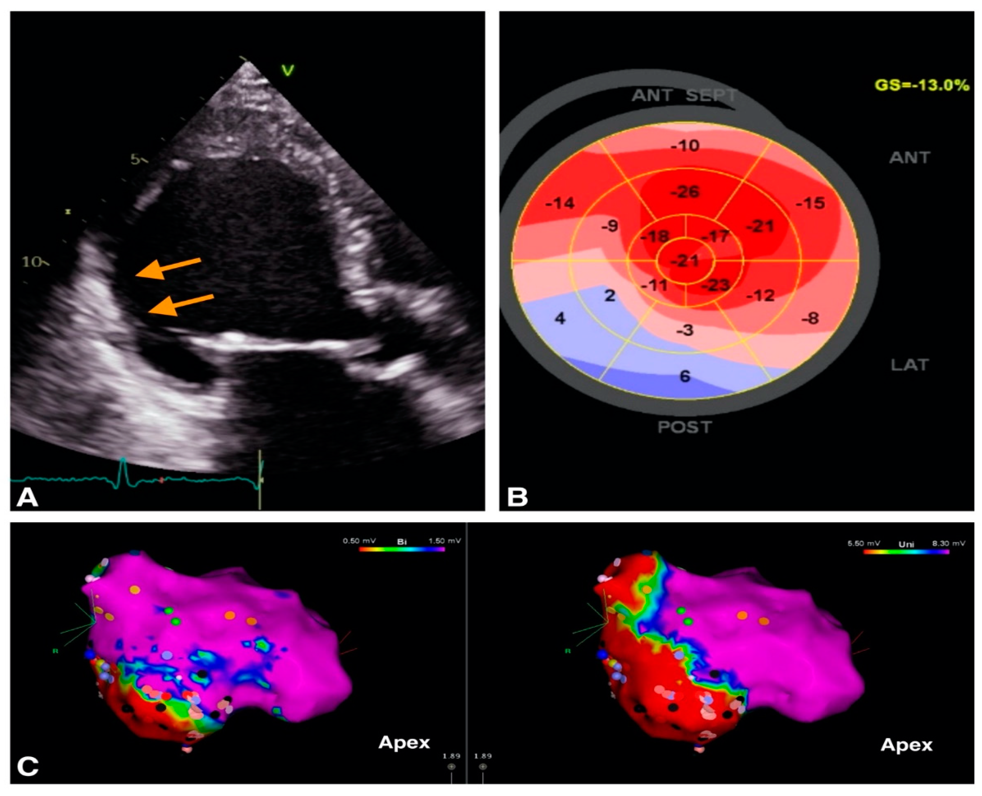
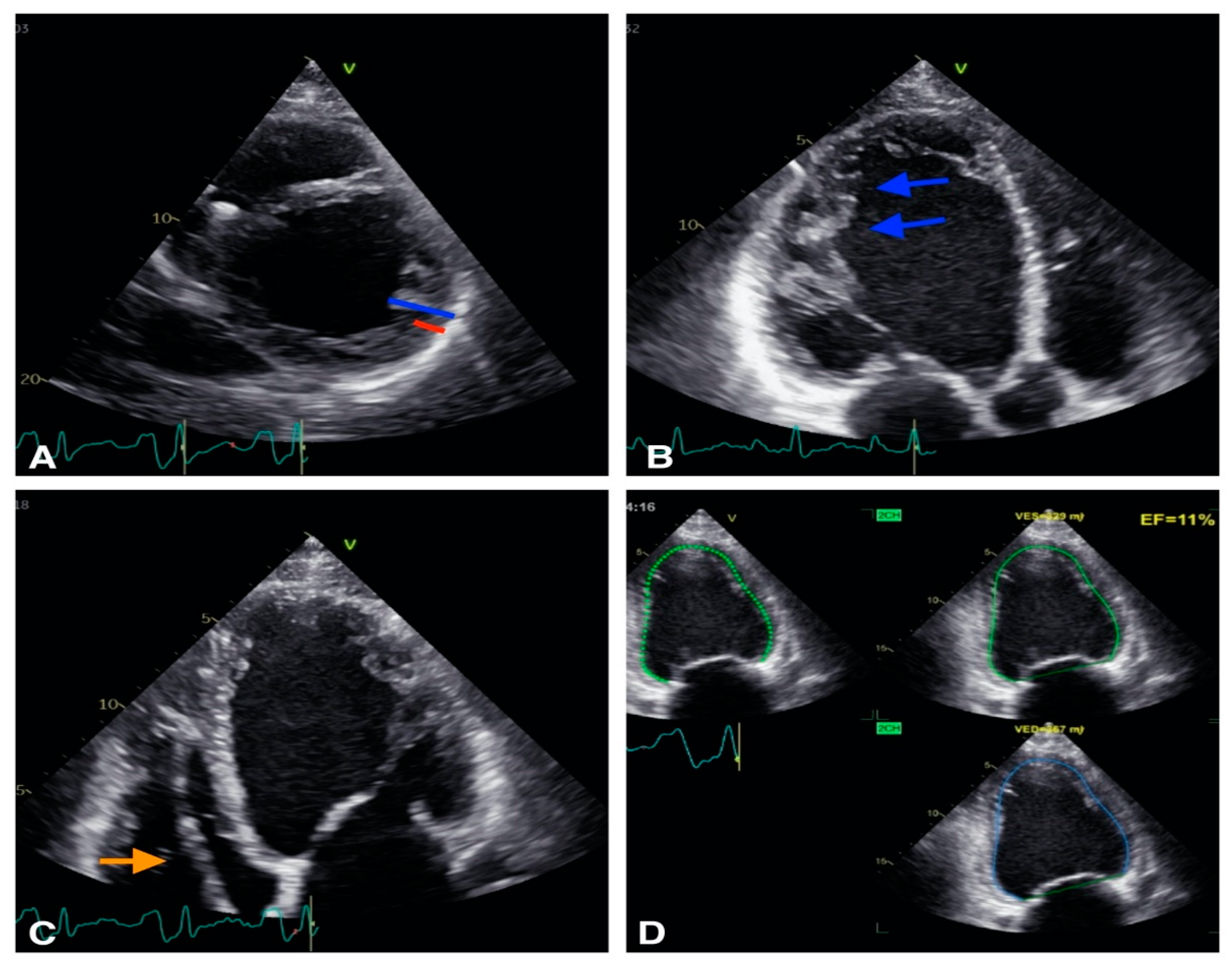
Ventricular arrhythmias (VA) are a major cause of sudden cardiac death (SCD). Echocardiography is the first widely available imaging tool which guides VA management strategies. Along with other invasive and noninvasive imaging techniques, it provides essential information for identification of VA substrate such as differentiation between ischemic and non-ischemic etiology and identification of structural heart disease. Both classic as well as novel echocardiographic techniques such as left ventricular strain measurement and mechanical dispersion assessment provide prognostic information and assist in risk stratification. Furthermore, intracardiac echocardiography may have an adjunctive role for the VA ablation by providing real-time visualization of cardiac structures, continuous monitoring of catheter location and early recognition of procedural complications.

echocardiography cardiac imaging ventricular arrhythmia intracardiac echocardiography

1. Introduction

Ventricular arrhythmias (VA) consist of a large spectrum of ventricular rhythm disturbances ranging from premature ventricular complexes to ventricular tachycardia and fibrillation. VA are a major cause of sudden cardiac death (SCD) and a significant burden on the patient’s quality of life. In the majority of cases, VA are associated with structural heart disease.
Cardiac imaging plays an essential role in revealing the underlying etiology of VA and for SCD risk stratification. Echocardiography is the first readily available, inexpensive and accurate imaging tool used to identify the structural changes associated with VA. It is usually followed by multimodality imaging techniques such as cardiac magnetic resonance (CMR), cardiac computed tomography (CT) and nuclear imaging which bring additional information about the VA substrate. Basic echocardiography measurements such as left ventricle ejection fraction (LVEF) have long been used to assess the risk of SCD and current guidelines still rely on it for the indication of a implantable cardiac defibrillator (ICD) [1].
Substrate ablation is an efficient interventional treatment for VA. For a safe and successful ablation there is a need to understand anatomical VA substrate along with the electric substrate defined by the electroanatomic mapping (EAM). This entry will focus on the necessary information provided by the echocardiography prior to the VA ablation procedure.

2. Role of Echocardiography in Identification of VA Substrate

2.1. VA in Structural Heart Disease

Echocardiography is useful in the diagnosis of structural heart disease [2]. Transthoracic echocardiography (TTE) is a widely available imaging technique which offers valuable information about VA substrate. The main objectives of TTE evaluation are assessment of left ventricle (LV) systolic function with estimation of LVEF, differential diagnosis between ischemic versus nonischemic etiology, assessment of valve function [3], identification of congenital heart disease [3] and different cardiomyopathies [3].
For the evaluation of LV systolic function, LVEF is the main parameter used in VA management. The ESC guidelines recommend performing echocardiography in all patients with suspected or known VA for assessment of LV function and identification of structural substrate [1]. In patients with structural heart disease, a LVEF < 35% estimated by echocardiography is associated with increased risk of VA and SCD [1]. Furthermore, indications for ICDs in primary prevention of SCD rely on LVEF [1]. The LVEF may be estimated using 2D or 3D LV volumes [1]. Furthermore, TTE can identify segmental LV contraction abnormalities and describe myocardial scars which may represent VA substrate (Figure 1).
Figure 1. Example of image integration from a patient with VT and old myocardial infarction; (A). 2D apical 3 chambers view showing the thin, hyperechogenic inferolateral wall (orange arrows). (B). Bull’s eye representation of peak longitudinal strain showing positive values in inferior wall providing a description of the myocardial scar (C) Electroanatomic bipolar (left) and unipolar (right) voltage maps in septal view, with an inferior area of low voltage corresponding to the scar identified by echocardiography.
Transthoracic and transesophageal echocardiography are the routine tools for the diagnosis and severity assessment of underlying valvular heart disease which may contribute to VA occurrence.
Echocardiography plays a significant role in the diagnosis and in management of patients with congenital heart disease and VA. Several studies have shown that right ventricle (RV) and LV dysfunction are risk factors for VA in congenital heart disease patients [2]. It is mandatory to establish the current status of the congenital heart disease and the need of correcting residual complications in order to limit the risk of VA recurrence. A frequent clinical scenario is the operated patient with tetralogy of Fallot with severe residual pulmonary regurgitation and severe dilatation of RV who develops ventricular tachycardia (VT). The treatment of pulmonary stenosis/regurgitation is discussed to promote RV reverse remodeling and maybe decrease the risk of malignant polymorphic VA [4].
A multicenter study performed by Koyak et al. showed that moderate to severe systemic or subpulmonary ventricular dysfunction were associated with a high risk of SCD, especially in adults with transposition of great arteries, Eisenmenger physiology and surgically repaired tetralogy of Fallot [5].
Finally, echocardiography may diagnose cardiomyopathy as the substrate for VA (Figure 2).
Figure 2. Example of a patient with non-compaction cardiomiopathy. (A): 2D short axis view, the non compaction myocardium (blue) is twice thicker than the compacted myocardium (red). (B): 2D apical 3 chambers view, trabeculations are visible (blue arrows). (C): 2D apical 4 chambers view: defibrillator lead is visibile in right ventricle (orange arrow). (D): Biplane EF is calculated at 11% showing severe LV systolic dysfunction.
The established echocardiographic criteria for diagnosis of the different cardiomyopathies are summarized in Table 1. However, CMR provides more precise information of VA substrate, as it is able to distinguish between ischemic and non-ischemic etiology and detect subtle structural abnormalities such as myocarditis, sarcoid or amyloid cardiomyopathies [6]. CMR also brings incremental information for the diagnosis of HCM and ARVD [6].
Table 1. Echocardiographic criteria for the diagnosis of cardiomyopathy.
Cardiomyopathy Echocardiographic Parameters
HCM [7][8][9]
-
LV maximal wall thickness > 15 mm *
-
Ratio of septal and posterior wall thickness ratio > 1.3 **
ARVC [10]
-
Major criteria:
Regional RV akinesia, dyskinesia, or aneurysm and one of the following:
  • PLAX RVOT ≥ 32 mm (corrected for body size [PLAX/BSA] ≥ 19 mm/m2)
  • PSAX RVOT ≥ 36 mm (corrected for body size [PSAX/BSA] ≥ 21 mm/m2)
  • FAC ≤ 33%
-
Minor criteria:
Regional RV akinesia or dyskinesia and one of the following:
  • PLAX RVOT ≥ 29 to <32 mm (corrected for body size [PLAX/BSA] ≥16 to <19 mm/m2)
  • PSAX RVOT ≥ 32 to <36 mm (corrected for body size [PSAX/BSA] ≥18 to <21 mm/m2)
  • FAC > 33% to ≤40%
NCCM [11]
-
Presence of numerous trabeculations and recesses: most proeminent at LV apex and least proeminent at the level of mitral valve
-
Ratio of thickness of NC and C myocardial layers at the site of maximal WT averages around 3.5 (2–3.5)
-
>3 trabeculations protrunding from the LV apex to the papillary muscles
-
Maximum linear length of NC-C myocardium
-
Planimetry of NC area on A4C
DCM [12]
-
LVIDd >112% (2 s.d.) corrected for age and BSA ****
-
LVEF < 45%
-
LVFS < 25%
-
Mitral regurgitation
-
SI < 1.5
-
Septal flash and apical rocking
-
Presence of dysynchrony indices
HCM—hypertrophic cardiomyopathy, ARVC—arrhythmogenic right ventricular cardiomyopathy, NCCM—noncompaction cardiomyopathy, DCM—dilated cardiomyopathy, SAM—systolic anterior motion, LVOT—left ventricular outflow tract, LV—left ventricle, RV—right ventricle, PLAX—parasternal long-axis view; RVOT—right ventricular outflow tract; BSA—body surface area; PSAX—parasternal short-axis FAC—fractional area change; NC—noncompacted, C—compacted, A4C—apical four chambers; LVIDd—Left ventricular internal dimension in diastole, LVEF—left ventricular ejection fraction, LVFS—left ventricular fractional shortening, SI—sphericity index; * > 13 mm in HCM relatives, ** > 1.5 in hypertensive patients, **** > 117% (2 s.d. plus 5%) increases the specificity and may be useful in familial screening.

2.2. VA in Structurally Normal Hearts

Almost 50% of SCD events occur in patients without structural heart disease [1]Table 2 summarizes the possible etiology of VA in patients without any obvious structural changes. There is data that “normal” structural hearts associated with VA and channelopathies actually show structural changes. A group by Scheirlynck et al. studied 175 patients with Brugada Syndrome (BrS) compared to 82 controls and found that BrS patients had lower longitudinal strain and more heterogeneous contractions than healthy controls [13]. Furthermore, BrS patients with a history of life-threatening VA had more heterogeneous LV contractions [13]. Therefore, LV mechanical dispersion may be a risk marker in BrS and its evaluation in prospective studies is needed [9]. Studies on RV function also seem to be relevant in BrS patients, in whom subtle contractile RV mechanical abnormalities were demonstrated, including impaired RV longitudinal strain and a greater contraction delay between the lateral and the septal aspect of the RV [14].
Table 2. Etiology of VA.
VA in Structural Heart Disease VA in Structurally Normal Heart
IHD
pARVC
HCM
DCM
NCCM
Valvular heart disease (i.e., aortic stenosis, mitral valve prolapse)
Congenital heart disease
Outflow tract VT
Fascicular VT
Interfascicular reentrant VT (Belhassen Tachycardia)
Papillary muscle VA
Channelopathies:
-
Long QT syndrome
-
Short QT syndrome
-
Brugada syndrome
-
Catecholaminergic polymorphic VT
-
Early repolarization syndrome
IHD—ischemic heart disease, ARVC—arrhythmogenic right ventricular cardiomyopathy, HCM—hypertrophic cardiomyopathy, DCM—dilated cardiomyopathy, NCCM—non compaction cardiomyopathy, VA—ventricular arrythmia, VT—ventricular tachycardia.
Other studies show an overlap between different VA etiology as for BrS and arrhythmogenic cardiomyopathy [15]. The presence of arrhythmogenic cardiomyopathy diagnostic criteria in BrS patients was associated with a trend towards higher arrhythmic risk [15].
Prior to VA ablation, echocardiographic data can be completed with cardiac magnetic resonance (CMR). CMR may be later combined with EAM information. Several studies have confirmed that low endocardial voltage corresponds to the presence of scar tissue as defined by the late gadolinium enhanced CMR [16]. Pre-procedural CMRs have been extremely helpful in localizing scar tissue in the ventricles and have helped to guide and target the arrhythmogenic tissue with catheter ablation [17].

References

  1. Priori, S.G.; Blomström-Lundqvist, C.; Mazzanti, A.; Blom, N.; Borggrefe, M.; Camm, J.; Elliott, P.M.; Fitzsimons, D.; Hatala, R.; Hindricks, G.; et al. 2015 ESC Guidelines for the management of patients with ventricular arrhythmias and the prevention of sudden cardiac deathThe Task Force for the Management of Patients with Ventricular Arrhythmias and the Prevention of Sudden Cardiac Death of the European Society of Cardiology (ESC)Endorsed by: Association for European Paediatric and Congenital Cardiology (AEPC). Eur. Heart J. 2015, 36, 2793–2867.
  2. Papadopoulos, C.H.; Oikonomidis, D.; Lazaris, E.; Nihoyannopoulos, P. Echocardiography and cardiac arrhythmias. Hell. J. Cardiol. 2018, 59, 140–149.
  3. Saric, M.; Armour, A.C.; Arnaout, M.S.; Chaudhry, F.A.; Grimm, R.A.; Kronzon, I.; Landeck, B.F.; Maganti, K.; Michelena, H.I.; Tolstrup, K. Guidelines for the Use of Echocardiography in the Evaluation of a Cardiac Source of Embolism. J. Am. Soc. Echocardiogr. 2016, 29, 1–42.
  4. Therrien, J.; Siu, S.C.; Harris, L.; Dore, A.; Niwa, K.; Janousek, J.; Williams, W.G.; Webb, G.; Gatzoulis, M.A. Impact of Pulmonary Valve Replacement on Arrhythmia Propensity Late After Repair of Tetralogy of Fallot. Circulation 2001, 103, 2489–2494.
  5. Koyak, Z.; Harris, L.; de Groot, J.R.; Silversides, C.K.; Oechslin, E.N.; Bouma, B.J.; Budts, W.; Zwinderman, A.H.; Van Gelder, I.C.; Mulder, B.J.M. Sudden Cardiac Death in Adult Congenital Heart Disease. Circulation 2012, 126, 1944–1954.
  6. Mahida, S.; Sacher, F.; Dubois, R.; Sermesant, M.; Bogun, F.; Haïssaguerre, M.; Jaïs, P.; Cochet, H. Cardiac Imaging in Patients With Ventricular Tachycardia. Circulation 2017, 136, 2491–2507.
  7. Williams, L.K.; Frenneaux, M.P.; Steeds, R.P. Echocardiography in hypertrophic cardiomyopathy diagnosis, prognosis, and role in management. Eur. J. Echocardiogr. 2009, 10, iii9–iii14.
  8. Hagège, A.A.; Dubourg, O.; Desnos, M.; Mirochnik, R.; Isnard, G.; Bonne, G.; Carrier, L.; Guicheney, P.; Bouhour, J.-B.; Schwartz, K.; et al. Familial hypertrophic cardiomyopathy. Cardiac ultrasonic abnormalities in genetically affected subjects without echocardiographic evidence of left ventricular hypertrophy. Eur. Heart J. 1998, 19, 490–499.
  9. Elliott, P.M.; Anastasakis, A.; Borger, M.A.; Borggrefe, M.; Cecchi, F.; Charron, P.; Hagege, A.A.; Lafont, A.; Limongelli, G.; Mahrholdt, H.; et al. 2014 ESC Guidelines on diagnosis and management of hypertrophic cardiomyopathy: The Task Force for the Diagnosis and Management of Hypertrophic Cardiomyopathy of the European Society of Cardiology (ESC). Eur. Heart J. 2014, 35, 2733–2779.
  10. Marcus, F.I.; McKenna, W.J.; Sherrill, D.; Basso, C.; Bauce, B.; Bluemke, D.A.; Calkins, H.; Corrado, D.; Cox, M.G.P.J.; Daubert, J.P.; et al. Diagnosis of arrhythmogenic right ventricular cardiomyopathy/dysplasia: Proposed Modification of the Task Force Criteria. Eur. Heart J. 2010, 31, 806–814.
  11. Chebrolu, L.H.; Mehta, A.M.; Nanda, N.C. Noncompaction cardiomyopathy: The role of advanced multimodality imaging techniques in diagnosis and assessment. Echocardiography 2017, 34, 279–289.
  12. Mathew, T.; Williams, L.; Navaratnam, G.; Rana, B.; Wheeler, R.; Collins, K.; Harkness, A.; Jones, R.; Knight, D.; O’Gallagher, K.; et al. Diagnosis and assessment of dilated cardiomyopathy: A guideline protocol from the British Society of Echocardiography. Echo Res. Pract. 2017, 4, G1–G13.
  13. Scheirlynck, E.; Malderen, S.V.; Motoc, A.; Lie, Ø.H.; de Asmundis, C.; Sieira, J.; Chierchia, G.-B.; Brugada, P.; Cosyns, B.; Droogmans, S. Contraction alterations in Brugada syndrome; association with life-threatening ventricular arrhythmias. Int. J. Cardiol. 2020, 299, 147–152.
  14. Mitroi, C.; García-Izquierdo, E.; García-Lunar, I.; Castro-Urda, V.; Toquero-Ramos, J.; Moñivas-Palomero, V.; Mingo-Santos, S.; Cavero, M.A.; Brugada, J.; Fernández-Lozano, I. Right ventricular function and dyssynchrony in Brugada syndrome: Highlighting the importance of the mechanical substrate in the right ventricular outflow tract. Int. J. Cardiol. 2021, 333, 233–238.
  15. Scheirlynck, E.; Chivulescu, M.; Lie, Ø.H.; Motoc, A.; Koulalis, J.; de Asmundis, C.; Sieira, J.; Chierchia, G.-B.; Brugada, P.; Cosyns, B.; et al. Worse Prognosis in Brugada Syndrome Patients With Arrhythmogenic Cardiomyopathy Features. JACC Clin. Electrophysiol. 2020, 6, 1353–1363.
  16. Perin, E.C.; Silva, G.V.; Sarmento-Leite, R.; Sousa, A.L.S.; Howell, M.; Muthupillai, R.; Lambert, B.; Vaughn, W.K.; Flamm, S.D. Assessing Myocardial Viability and Infarct Transmurality With Left Ventricular Electromechanical Mapping in Patients With Stable Coronary Artery Disease. Circulation 2002, 106, 957–961.
  17. Njeim, M.; Desjardins, B.; Bogun, F. Multimodality Imaging for Guiding EP Ablation Procedures. JACC Cardiovasc. Imaging 2016, 9, 873–886.
More
Information
Contributors MDPI registered users' name will be linked to their SciProfiles pages. To register with us, please refer to https://encyclopedia.pub/register : , , , ,
View Times: 270
Revisions: 2 times (View History)
Update Date: 21 Oct 2022
1000/1000