Submitted Successfully!
To reward your contribution, here is a gift for you: A free trial for our video production service.
Thank you for your contribution! You can also upload a video entry or images related to this topic.
Version Summary Created by Modification Content Size Created at Operation
1 -- 2136 2022-09-14 06:20:28 |
2 format correct + 66 word(s) 2202 2022-09-15 03:44:34 |

Video Upload Options

Do you have a full video?

Confirm

Are you sure to Delete?
Cite
If you have any further questions, please contact Encyclopedia Editorial Office.
Kalo, E.;  Read, S.;  Ahlenstiel, G. Reprogramming of α-Cells into Insulin Producing Cells. Encyclopedia. Available online: https://encyclopedia.pub/entry/27151 (accessed on 19 April 2024).
Kalo E,  Read S,  Ahlenstiel G. Reprogramming of α-Cells into Insulin Producing Cells. Encyclopedia. Available at: https://encyclopedia.pub/entry/27151. Accessed April 19, 2024.
Kalo, Eric, Scott Read, Golo Ahlenstiel. "Reprogramming of α-Cells into Insulin Producing Cells" Encyclopedia, https://encyclopedia.pub/entry/27151 (accessed April 19, 2024).
Kalo, E.,  Read, S., & Ahlenstiel, G. (2022, September 14). Reprogramming of α-Cells into Insulin Producing Cells. In Encyclopedia. https://encyclopedia.pub/entry/27151
Kalo, Eric, et al. "Reprogramming of α-Cells into Insulin Producing Cells." Encyclopedia. Web. 14 September, 2022.
Reprogramming of α-Cells into Insulin Producing Cells
Edit

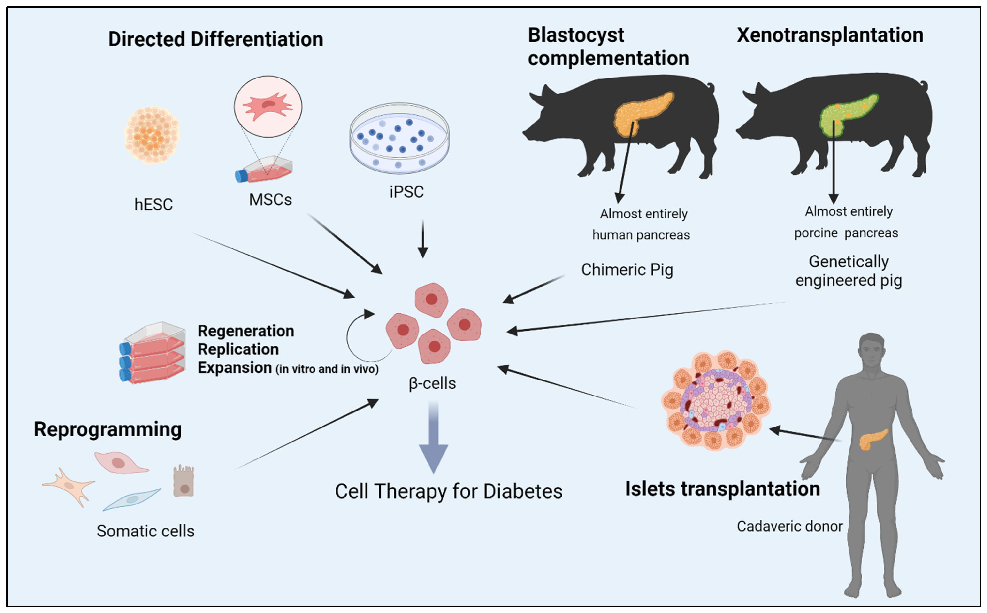
Numerous cell sources are being explored to replenish functional β-cell mass since the proof-of -concept for cell therapy of diabetes was laid down by transplantation of islets. Various strategies that aim to generate bone fide insulin producing cells are explored.  In particular on reprogramming and especially on α-cells conversion into insulin producing cells are focused here. A logical place to begin with for generating β-cells is to utilise the plasticity of closely related endoderm derived cell types like pancreatic non-β-cells and coaxing them to adopt a β-cell phenotype. Given the close ontogenetic relationship, functional similarity and dependency among these cells, the potential for interconversion is unequivocal. Phenotypic plasticity between pancreatic α-cells and β-cells is notably pronounced.

β-cell α-cell diabetes mellitus cell sources reprogramming insulin transcription factors small molecules

1. Introduction

Over the past years, the global incidence of diabetes mellitus has dramatically increased alongside the obesity epidemic. With more than 537 million people affected by the disease globally, it is projected to increase to more than half a billion by 2030 [1]. It is becoming an ever-growing burden threatening to overwhelm many health care systems and economies throughout the world [2].
Diabetes is described as a group of heterogeneous metabolic diseases characterized by common elements of hyperglycemia and glucose intolerance due to defects in insulin section, impaired effectiveness of insulin or both. In the progression of both type 1 diabetes mellitus (T1DM) and type 2 DM (T2DM), there comes a point where a threshold percentage of β-cells become dysfunctional, leading to a reliance on exogenous insulin administration for the treatment of patients with DM.
Although the isolation of insulin in 1921 marked a panacea that has drastically transformed diabetes from a terminal to a treatable illness, the precise temporal glucose control supplied by endogenous insulin-producing β-cells is not matched by the current methods of insulin delivery. Exogenous insulin therapy, as a result, can pose a risk of hypoglycemia that can lead to life threatening coma and premature death [3]. Consequently, intensive insulin therapy targeting near-euglycemia can prevent the risk of long-term microvascular and macrovascular complications. Data from large population studies have consistently supported the notion that tight glycemic control can effectively reduce the development of diabetes complication and conferred mortality and morbidity benefits [4][5][6].
Continuous glucose monitoring and exogenous insulin administration are considered the mainstay of treatment for diabetes. While it significantly leads to improvement of patient outcomes, it falls short in achieving optimal long-standing blood glycaemia control, especially for T1DM patients [7][8][9]. In a recent randomized, multicenter trial, results showed that the percentage of time that blood glucose remained within the target glycemic range (notwithstanding automated insulin delivery systems) was persistently suboptimal in patients with T1DM [10]. Consequently, therapies that result in perpetual reconstitution of a physiological blood glucose setpoint are an extremely sought approach in the long term.
Rather than modestly alleviating both type 1 and type 2 diabetes, β-cell replacement has the potential to reverse these conditions. A successful pancreas transplant can provide a closed-loop system that restores euglycemia without the risk of severe hypoglycemia, thus halting the development or progression of complications. Nonetheless, major surgery associated risks, chronic shortages of donor pancreata, graft rejection and the burden of life-long immunosuppression regimens set the utility of such an approach exclusively to a narrow group of uremic patients with brittle diabetes.
To overcome the need for major surgery associated with pancreas transplantation, research has focused on the development of protocols to isolate pancreatic islets. Despite the outstanding success of islet of Langerhans transplantation in lowering insulin dependence for years in patients with T1DM, the widespread application of this approach has been hampered by the scarcity of donors, as well as immunological challenges that are further aggravated by toxic effects of long-term immunosuppressive drugs [11]. Notwithstanding the pitfalls, the success of this approach has fueled efforts for a generation of bone fide functional β-cell mass that maintains the physiological oscillation of insulin secretion and responsiveness to dysregulated glycemia. Moreover, recent studies using autologous cell therapies may halt the requirement of lifelong immunosuppressive therapy.

2. Reprogramming Cells to Make Insulin

For the last 20 years, significant efforts have focused on the generation of human embryonic stem cells, hESC-derived β-cells, that exhibit sustainable insulin secretion features, which mainly mirror the dynamicity of native human islets. By replicating the signaling events and physiologically relevant cues that dictate fate transition through definitive endoderm and pancreatic endoderm to hormone-expressing cells during human pancreas development, β-like cells can be produced [12][13][14]. The particular interest in hESCs is supported by the fact that these cells have the capacity of extensive self-renewal and virtually can be differentiated into derivatives of all three germ layers.
Numerous studies have outlined differentiation protocols of insulin-producing cell types from hESCs [15][16][17][18][19][20][21]. However, reconstructing functional equivalent, mono-hormonal cells producing insulin under cell culture conditions has been intangible. This is likely because the in vitro systems utilized lack critical signals present in vivo, including the close interaction between exocrine, ductal and endocrine cells [15][20][22][23]. Among the critical concerns with hESC-derived therapeutic products is the occurrence of unwanted cell populations during in vitro differentiation that might interfere with the activity of the desired cell populations. Another concern is the risk of tumorigenicity. Moreover, the wider use of ESCs for research reasons is still largely hampered by numerous governmental bans concerning the use of human embryos in many countries, as well as by ethical and religious sensitivities.
Compared to ESC and induced pluripotent stem cells (iPSCs), adult stem cell-like, mesenchymal stem cells (MSC) are considered ideal candidates to generate functional beta cells for personalized medicine owing to their superior anti-inflammatory, immunomodulatory and angiogenic properties. Numerous studies have shown that MSCs isolated from ample tissues and organs, such as bone marrow, adipose tissue, Wharton’s jelly, umbilical cords matrix blood, placenta and dental pulps, possess a developmental plasticity to differentiate into functional insulin producing cells with similar cytoarchitecture and functionality to β-cells. Consequently, utilizing MSC transplantation for the treatment of diabetes has been the focus of randomized controlled trails (RCTs) for the last few years. Although several clinical trials have shown that MSCs can reduce hyperglycemia by increasing insulin secretion in humans, the lack of control arms in some small sample sizes, inconsistent methods of isolation and delivery of MSCs, adverse effects and the failure to sustain therapeutic effect longitudinally from MSC therapy were common limitations in almost all RCTs. Finally, genetically modified animals designed for xenotransplantation or interspecies chimera-derived human organs, by using the blastocyst complementation method, could potentially offer unlimited sources of β-cells. Figure 1 summarizes several strategies aimed at generation of bone fide β-cell for replacement in diabetes.
Figure 1. Strategies aimed at generation of bone fide β-cell for replacement therapy in diabetes. Abbreviations: human embryonic stem cells (hESC); mesenchymal stem cells (MSCs); induced pluripotent stem cells (iPSC). Figure was created with BioRender http://www.biorender.com.

3. Alpha to β-cell Reprogramming

A logical place to begin with for generating β-cells is to utilise the plasticity of closely related endoderm derived cell types like pancreatic non-β-cells and coaxing them to adopt a β-cell phenotype. Given the close ontogenetic relationship, functional similarity and dependency among these cells, the potential for interconversion is unequivocal [24]. Phenotypic plasticity between pancreatic α-cells and β-cells is notably pronounced. A prominent study several years ago demonstrated that inter-endocrine plasticity and proliferation of β-cells can be elicited upon increased metabolic demand or after substantial diphtherial toxin induced β-cell loss [25]. While this entry was performed in mice, there is no evidence to suggest that a similar intrinsic cell type interconversion event can occur in other settings of β-cell damage in humans. Of note, a similar phenomenon of β-cell loss may naturally occur in mice transitioning into adulthood from puberty.
Forced overexpression of key transcriptional regulators, the paired/homeodomain transcription factor (PAX4) [26], Insulin promoter factor 1 (PDX1) [27], or PDX1 and MAF BZIP Transcription Factor A (MAFA) [28], inhibition of DNA (cytosine-5)-methyltransferase 1 (DNMT1) and Aristaless related homeobox (ARX) [29], have also been shown to successfully drive α- to β-cell conversion. Yang et al. illustrated that PDX1 alone is not sufficient to allow the full reprogramming of glucagon-expressing cells [27]. This points to a chronotypic effect, where a subset of endocrine progenitors is primed for fate switch at the peri/postnatal stage, highlighting the role of the epigenome in reprogramming events. Importantly, the applicability of these findings to human cells needs to be determined, given that most of these studies were conducted in rodents. A subsequent study sought to better study human islet cell plasticity by lineage tracing and reprogramming α-cells with Mafa and Pdx1. Remarkably, insulin-producing α-cells retained α-cell markers, as evidenced by transcriptomic and proteomic characterization, while retaining insulin production and reversing diabetes for 6 months [30]. Several studies highlighted the type of pancreatic gene delivery systems for efficient intrinsic alpha cell transduction. Using adeno-associated virus (AAV) rather than adenoviral or lentiviral vectors has been shown to exhibit long-term gene expression induced reprogramming of pancreatic α-cells into functional β-cells [31].
The development of mature pancreatic endocrine cell subtypes represents the culmination of complex fate determining transcriptional programs that orchestrate the transition from one progenitor state to another. As transcription factors are sufficient to induce α- to β-cell reprogramming, it will be imperative to identify molecules that govern such a conversion as they would permit better control of the process [32]. For example, α-cells treated with glucagon-like peptide receptor agonists, exendin-4, can enhance pancreatic α-cells proliferation and their trans-differentiation into β-cells [33]. Similarly, sustained γ-Aminobutyric acid (GABA) exposure can induce concomitant α-cell to β-like cell neogenesis in vivo from human islet α-cells transplanted in mice by mobilization of duct-lining precursor cells that adopt an α cell identity [34]. GABA is a critical paracrine signal released by β-cells that inhibits glucagon-release from α-cells [35]. Artemisinin, a class of antimalarial agents, has been shown to impair α-cell identity in immortalized rodent cell lines by enhancing GABA receptor signaling, and by functionally inhibiting the ARX transcription factor [36]. In contrast, other studies have found that inhibition of ARX after long-term treatment by artemether, a derivative of artemisinin, did not furnish any α- to β-cell trans-differentiation in primary mouse islets, suggesting that these regulators may not represent a viable route to a novel diabetes therapy [37][38]. Nonetheless, in support of glucagon inhibition as a mechanism of α- to β- cell transition, the monoclonal antibody antagonist of the glucagon receptor (Ab-4) has been shown to enhance the formation of functional β-cell mass and their conversion from α-cell precursors in a rodent model of T1DM [39].

4. Conclusions and Future Perspectives

Despite the vast potential of recent advances in cellular reprogramming and trans-differentiation of insulin producing cells, many issues still need refining in the coming decades. These include the efficiency of the cell trans-differentiation protocols, their cost-effectiveness for large-scale differentiation and whether reprogrammed cells can sustain their new differentiation state, or whether they potentially return to their early fate. Furthermore, whether transdifferentiated β-cells can respond adequately to the multitude of physiological stimuli required to maintain metabolic equilibrium and upon increased metabolic demand. Lastly, are the reprogrammed cells able to evade the recurrent auto-immune attacks, particularly in the context of allogenic cell transplants, and might cell transplants possess increased oncogenic potential resulting from incomplete epigenetic conversion? Several technologies have been proposed to attend to the immune responses to and blood supply for transplants, including the use of physical shielding by encapsulating the reprogrammed cells in a device that allows for insulin and nutrients to efficiently cross through the membrane while blocking cells from trespassing. Other biological interventions include either modifying host immune system, using antibodies to block the immune reactions or gene editing of cells used for transplant.
Furthermore, the safe implementation of viral reprogramming technologies to the human setting need to be further assessed. The small molecules approach for reprogramming are more intriguing than virus-based methods, but they are very complex to develop for the regulation of a number of transcription factors. Moreover, impeding to clinical translation of the reprogramming approach still, is the relatively limited conversion rate into functional bone fide β-cells and presence of unwanted cells in the final product
A paradigm shift has been made toward understanding fundamental β-cell biology, development and reprogramming over the last 20 years. Although a plethora of cell sources have been suggested as a starting material en route to successful reprogramming, some cells may fail. Thus, it is difficult to speculate which cells will reach the finish line and lead to a replacement therapy for diabetes.
In conclusion, researchers have witnessed a remarkable two decades of significant progress in the methods of creating cellular transplant for the cure of diabetes. Current clinical trials for T1DM are still using hESC-derived pancreatic progenitors as a surrogate for cadaveric material, but the debate over whether to transplant a more mature cell population more similar to human islets is still ongoing. Hepatic, gastrointestinal and pancreatic exocrine cells, which are derived from common endodermal progenitor cells, have the potential to take the lead as cell sources for the development of a therapeutic product. Identical developmental transcription mechanisms and regulatory networks, analogous chromatin landscapes, physical proximity to the injured pancreas and a minimal need for epigenomic rearrangement are all tempting to conclude that these functionally related cells may yield more robust reprogramming outcomes, and offers a unique promise for cell replacement therapies for diabetes.

References

  1. International Diabetes Federation. Diabetes Facts & Figures; International Diabetes Federation: Brussels, Belgium, 2021; Available online: https://idf.org/aboutdiabetes/what-is-diabetes/facts-figures.html (accessed on 1 September 2022).
  2. Lin, X.; Xu, Y.; Pan, X.; Xu, J.; Ding, Y.; Sun, X.; Song, X.; Ren, Y.; Shan, P.F. Global, regional, and national burden and trend of diabetes in 195 countries and territories: An analysis from 1990 to 2025. Sci. Rep. 2020, 10, 14790.
  3. Chen, H.; Chen, G.; Zheng, X.; Guo, Y. Contribution of specific diseases and injuries to changes in health adjusted life expectancy in 187 countries from 1990 to 2013: Retrospective observational study. BMJ 2019, 364, l969.
  4. Holman, R.R.; Paul, S.K.; Bethel, M.A.; Matthews, D.R.; Neil, H.A. 10-year follow-up of intensive glucose control in type 2 diabetes. N. Engl. J. Med. 2008, 359, 1577–1589.
  5. Diabetes Control and Complications Trial (DCCT); Epidemiology of Diabetes Interventions and Complications (EDIC) Research Group. Effect of intensive diabetes therapy on the progression of diabetic retinopathy in patients with type 1 diabetes: 18 years of follow-up in the DCCT/EDIC. Diabetes 2015, 64, 631–642.
  6. Zinman, B.; Genuth, S.; Nathan, D.M. The Diabetes Control and Complications Trial/Epidemiology of Diabetes Interventions and Complications Study: 30th Anniversary Presentations. Diabetes Care 2014, 37, 8.
  7. Polonsky, K.S. The Past 200 Years in Diabetes. N. Engl. J. Med. 2012, 367, 1332–1340.
  8. NICE-Sugar Study Investigators. Hypoglycemia and risk of death in critically ill patients. N. Engl. J. Med. 2012, 367, 1108–1118.
  9. Bluestone, J.A.; Herold, K.; Eisenbarth, G. Genetics, pathogenesis and clinical interventions in type 1 diabetes. Nature 2010, 464, 1293–1300.
  10. Brown, S.A.; Kovatchev, B.P.; Raghinaru, D.; Lum, J.W.; Buckingham, B.A.; Kudva, Y.C.; Laffel, L.M.; Levy, C.J.; Pinsker, J.E.; Wadwa, R.P.; et al. Six-Month Randomized, Multicenter Trial of Closed-Loop Control in Type 1 Diabetes. N. Engl. J. Med. 2019, 381, 1707–1717.
  11. Shapiro, A.M.J.; Ricordi, C.; Hering, B.J.; Auchincloss, H.; Lindblad, R.; Robertson, R.P.; Secchi, A.; Brendel, M.D.; Berney, T.; Brennan, D.C.; et al. International Trial of the Edmonton Protocol for Islet Transplantation. N. Engl. J. Med. 2006, 355, 1318–1330.
  12. Oliver-Krasinski, J.M.; Stoffers, D.A. On the origin of the beta cell. Genes Dev. 2008, 22, 1998–2021.
  13. Assady, S.; Maor, G.; Amit, M.; Itskovitz-Eldor, J.; Skorecki, K.L.; Tzukerman, M. Insulin Production by Human Embryonic Stem Cells. Diabetes 2001, 50, 1691–1697.
  14. D’Amour, K.A.; Agulnick, A.D.; Eliazer, S.; Kelly, O.G.; Kroon, E.; Baetge, E.E. Efficient differentiation of human embryonic stem cells to definitive endoderm. Nat. Biotechnol. 2005, 23, 1534–1541.
  15. D’Amour, K.A.; Bang, A.G.; Eliazer, S.; Kelly, O.G.; Agulnick, A.D.; Smart, N.G.; Moorman, M.A.; Kroon, E.; Carpenter, M.K.; Baetge, E.E. Production of pancreatic hormone–expressing endocrine cells from human embryonic stem cells. Nat. Biotechnol. 2006, 24, 1392–1401.
  16. Kroon, E.; Martinson, L.A.; Kadoya, K.; Bang, A.G.; Kelly, O.G.; Eliazer, S.; Young, H.; Richardson, M.; Smart, N.G.; Cunningham, J.; et al. Pancreatic endoderm derived from human embryonic stem cells generates glucose-responsive insulin-secreting cells in vivo. Nat. Biotechnol. 2008, 26, 443–452.
  17. Nair, G.G.; Liu, J.S.; Russ, H.A.; Tran, S.; Saxton, M.S.; Chen, R.; Juang, C.; Li, M.-I.; Nguyen, V.Q.; Giacometti, S.; et al. Recapitulating endocrine cell clustering in culture promotes maturation of human stem-cell-derived beta cells. Nat. Cell Biol. 2019, 21, 263–274.
  18. Hogrebe, N.J.; Augsornworawat, P.; Maxwell, K.G.; Velazco-Cruz, L.; Millman, J.R. Targeting the cytoskeleton to direct pancreatic differentiation of human pluripotent stem cells. Nat. Biotechnol. 2020, 38, 460–470.
  19. Rezania, A.; Bruin, J.E.; Arora, P.; Rubin, A.; Batushansky, I.; Asadi, A.; O’Dwyer, S.; Quiskamp, N.; Mojibian, M.; Albrecht, T.; et al. Reversal of diabetes with insulin-producing cells derived in vitro from human pluripotent stem cells. Nat. Biotechnol. 2014, 32, 1121–1133.
  20. Pagliuca, F.W.; Millman, J.R.; Gürtler, M.; Segel, M.; Van Dervort, A.; Ryu, J.H.; Peterson, Q.P.; Greiner, D.; Melton, D.A. Generation of functional human pancreatic beta cells in vitro. Cell 2014, 159, 428–439.
  21. Russ, H.A.; Parent, A.V.; Ringler, J.J.; Hennings, T.G.; Nair, G.G.; Shveygert, M.; Guo, T.; Puri, S.; Haataja, L.; Cirulli, V.; et al. Controlled induction of human pancreatic progenitors produces functional beta-like cells in vitro. EMBO J. 2015, 34, 1759–1772.
  22. Naujok, O.; Burns, C.; Jones, P.M.; Lenzen, S. Insulin-producing surrogate beta-cells from embryonic stem cells: Are we there yet? Mol. Ther. 2011, 19, 1759–1768.
  23. Pagliuca, F.W.; Melton, D.A. How to make a functional beta-cell. Development 2013, 140, 2472–2483.
  24. Hamaguchi, K.; Utsunomiya, N.; Takaki, R.; Yoshimatsu, H.; Sakata, T. Cellular interaction between mouse pancreatic alpha-cell and beta-cell lines: Possible contact-dependent inhibition of insulin secretion. Exp. Biol. Med. 2003, 228, 1227–1233.
  25. Thorel, F.; Népote, V.; Avril, I.; Kohno, K.; Desgraz, R.; Chera, S.; Herrera, P.L. Conversion of adult pancreatic alpha-cells to beta-cells after extreme beta-cell loss. Nature 2010, 464, 1149–1154.
  26. Collombat, P.; Xu, X.; Ravassard, P.; Sosa-Pineda, B.; Dussaud, S.; Billestrup, N.; Madsen, O.D.; Serup, P.; Heimberg, H.; Mansouri, A. The ectopic expression of Pax4 in the mouse pancreas converts progenitor cells into alpha and subsequently beta cells. Cell 2009, 138, 449–462.
  27. Yang, Y.P.; Thorel, F.; Boyer, D.F.; Herrera, P.L.; Wright, C.V. Context-specific alpha- to-beta-cell reprogramming by forced Pdx1 expression. Genes Dev. 2011, 25, 1680–1685.
  28. Matsuoka, T.A.; Kawashima, S.; Miyatsuka, T.; Sasaki, S.; Shimo, N.; Katakami, N.; Kawamori, D.; Takebe, S.; Herrera, P.L.; Kaneto, H.; et al. Mafa Enables Pdx1 to Effectively Convert Pancreatic Islet Progenitors and Committed Islet alpha-Cells Into beta-Cells In Vivo. Diabetes 2017, 66, 1293–1300.
  29. Chakravarthy, H.; Gu, X.; Enge, M.; Dai, X.; Wang, Y.; Damond, N.; Downie, C.; Liu, K.; Wang, J.; Xing, Y.; et al. Converting Adult Pancreatic Islet alpha Cells into beta Cells by Targeting Both Dnmt1 and Arx. Cell Metab. 2017, 25, 622–634.
  30. Furuyama, K.; Chera, S.; Van Gurp, L.; Oropeza, D.; Ghila, L.; Damond, N.; Vethe, H.; Paulo, J.A.; Joosten, A.M.; Berney, T.; et al. Diabetes relief in mice by glucose-sensing insulin-secreting human alpha-cells. Nature 2019, 567, 43–48.
  31. Xiao, X.; Guo, P.; Shiota, C.; Zhang, T.; Coudriet, G.M.; Fischbach, S.; Prasadan, K.; Fusco, J.; Ramachandran, S.; Witkowski, P.; et al. Endogenous Reprogramming of Alpha Cells into Beta Cells, Induced by Viral Gene Therapy, Reverses Autoimmune Diabetes. Cell Stem Cell 2018, 22, 78–90.e4.
  32. Fomina-Yadlin, D.; Kubicek, S.; Walpita, D.; Dančik, V.; Hecksher-Sørensen, J.; Bittker, J.A.; Sharifnia, T.; Shamji, A.; Clemons, P.A.; Wagner, B.K.; et al. Small-molecule inducers of insulin expression in pancreatic α-cells. Proc. Natl. Acad. Sci. USA 2010, 107, 15099–15104.
  33. Lee, Y.S.; Lee, C.; Choung, J.S.; Jung, H.S.; Jun, H.S. Glucagon-Like Peptide 1 Increases beta-Cell Regeneration by Promoting alpha- to beta-Cell Transdifferentiation. Diabetes 2018, 67, 2601–2614.
  34. Ben-Othman, N.; Vieira, A.; Courtney, M.; Record, F.; Gjernes, E.; Avolio, F.; Hadzic, B.; Druelle, N.; Napolitano, T.; Navarro-Sanz, S.; et al. Long-Term GABA Administration Induces Alpha Cell-Mediated Beta-like Cell Neogenesis. Cell 2017, 168, 73–85.e11.
  35. Menegaz, D.; Hagan, D.W.; Almaça, J.; Cianciaruso, C.; Rodriguez-Diaz, R.; Molina, J.; Dolan, R.M.; Becker, M.W.; Schwalie, P.C.; Nano, R.; et al. Mechanism and effects of pulsatile GABA secretion from cytosolic pools in the human beta cell. Nat. Metab. 2019, 1, 1110–1126.
  36. Li, J.; Casteels, T.; Frogne, T.; Ingvorsen, C.; Honoré, C.; Courtney, M.; Huber, K.V.; Schmitner, N.; Kimmel, R.A.; Romanov, R.A.; et al. Artemisinins Target GABAA Receptor Signaling and Impair α Cell Identity. Cell 2016, 168, 86–100.e15.
  37. van der Meulen, T.L.S.; Noordeloos, E.; Donaldson, C.J.; Adams, M.W.; Noguchi, G.M.; Mawla, A.M.; Huising, M.O. Artemether Does Not Turn α Cells into β Cells. Cell Metab. 2018, 27, 218–225.e4.
  38. Ackermann, A.M.; Moss, N.G.; Kaestner, K.H. GABA and Artesunate Do Not Induce Pancreatic alpha-to-beta Cell Transdifferentiation In Vivo. Cell Metab. 2018, 28, 787–792.e3.
  39. Wang, M.Y.; Dean, E.D.; Quittner-Strom, E.; Zhu, Y.; Chowdhury, K.H.; Zhang, Z.; Zhao, S.; Li, N.; Ye, R.; Lee, Y.; et al. Glucagon blockade restores functional beta-cell mass in type 1 diabetic mice and enhances function of human islets. Proc. Natl. Acad. Sci. USA 2021, 118, e2022142118.
More
Information
Contributors MDPI registered users' name will be linked to their SciProfiles pages. To register with us, please refer to https://encyclopedia.pub/register : , ,
View Times: 556
Entry Collection: Biopharmaceuticals Technology
Revisions: 2 times (View History)
Update Date: 16 Sep 2022
1000/1000