Submitted Successfully!
To reward your contribution, here is a gift for you: A free trial for our video production service.
Thank you for your contribution! You can also upload a video entry or images related to this topic.
Version Summary Created by Modification Content Size Created at Operation
1 -- 2473 2022-09-06 10:48:55 |
2 format corrected. + 6 word(s) 2479 2022-09-07 04:39:10 |

Video Upload Options

Do you have a full video?

Confirm

Are you sure to Delete?
Cite
If you have any further questions, please contact Encyclopedia Editorial Office.
Proaño, B.;  Casani-Cubel, J.;  Benlloch, M.;  Rodriguez-Mateos, A.;  Navarro-Illana, E.;  Lajara-Romance, J.M.;  Ortí, J.E.D.L.R. Therapeutic Effect of Dutasteride in Amyotrophic Lateral Sclerosis. Encyclopedia. Available online: https://encyclopedia.pub/entry/26910 (accessed on 24 April 2024).
Proaño B,  Casani-Cubel J,  Benlloch M,  Rodriguez-Mateos A,  Navarro-Illana E,  Lajara-Romance JM, et al. Therapeutic Effect of Dutasteride in Amyotrophic Lateral Sclerosis. Encyclopedia. Available at: https://encyclopedia.pub/entry/26910. Accessed April 24, 2024.
Proaño, Belén, Julia Casani-Cubel, María Benlloch, Ana Rodriguez-Mateos, Esther Navarro-Illana, Jose María Lajara-Romance, Jose Enrique De La Rubia Ortí. "Therapeutic Effect of Dutasteride in Amyotrophic Lateral Sclerosis" Encyclopedia, https://encyclopedia.pub/entry/26910 (accessed April 24, 2024).
Proaño, B.,  Casani-Cubel, J.,  Benlloch, M.,  Rodriguez-Mateos, A.,  Navarro-Illana, E.,  Lajara-Romance, J.M., & Ortí, J.E.D.L.R. (2022, September 06). Therapeutic Effect of Dutasteride in Amyotrophic Lateral Sclerosis. In Encyclopedia. https://encyclopedia.pub/entry/26910
Proaño, Belén, et al. "Therapeutic Effect of Dutasteride in Amyotrophic Lateral Sclerosis." Encyclopedia. Web. 06 September, 2022.
Therapeutic Effect of Dutasteride in Amyotrophic Lateral Sclerosis
Edit

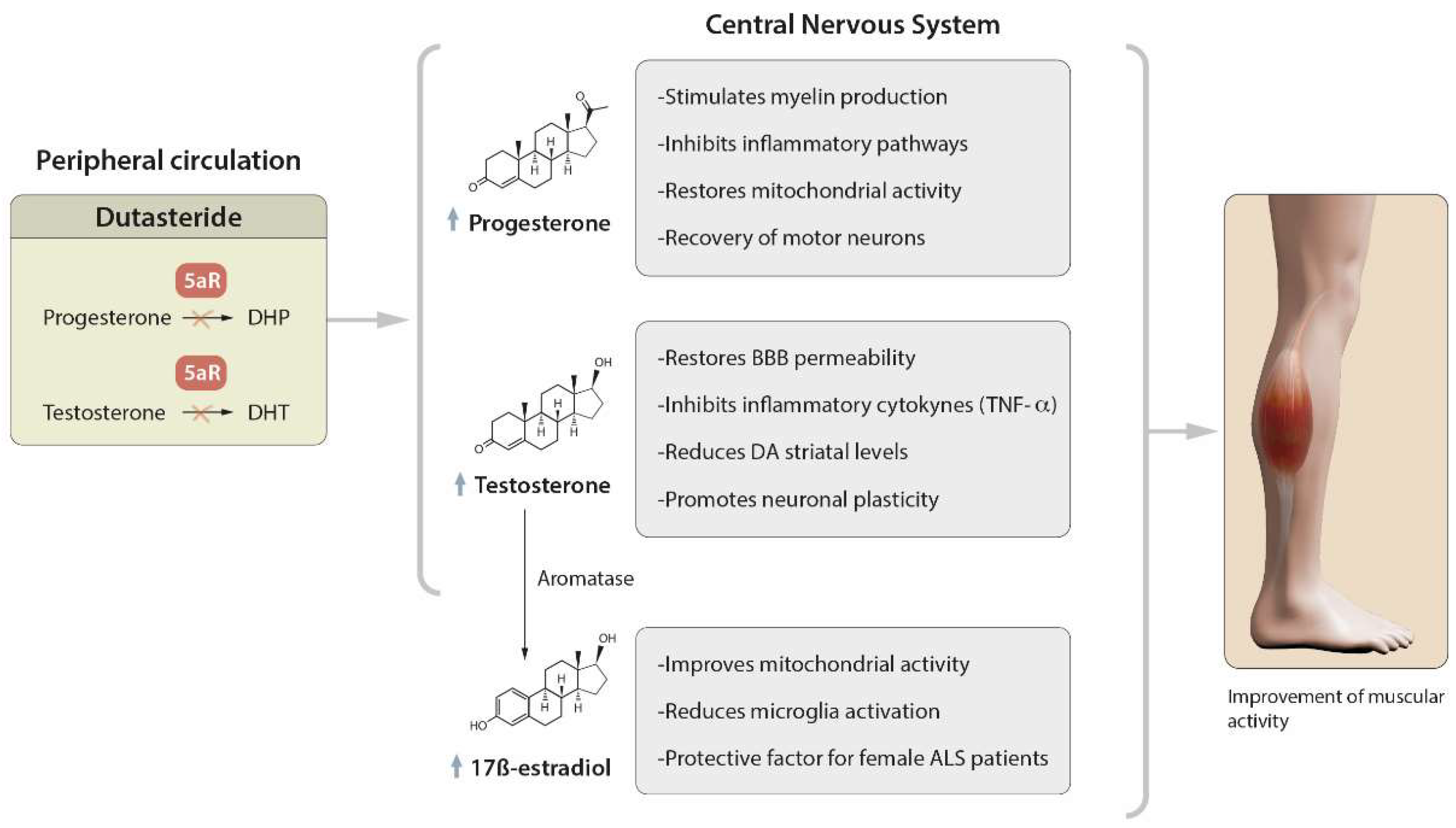
Amyotrophic lateral sclerosis (ALS) is a neurodegenerative disease that is characterized by the loss of upper and lower motor neurons (MNs) in the cerebral cortex, brainstem and spinal cord, with consequent weakness, atrophy and the progressive paralysis of all muscles. There is currently no medical cure, and riluzole and edaravone are the only two known approved drugs for treating this condition. However, they have limited efficacy, and hence there is a need to find new molecules. Dutasteride, a dual inhibitor of type 1 and type 2 5α-reductase (5AR) enzymes, the therapeutic purposes of which, to date, are the treatment of benign prostatic hyperplasia and androgenic alopecia, shows great anti-ALS properties by the molecular-topology methodology.

amyotrophic lateral sclerosis dutasteride neuroprotection oxidative stress inflammation TDP43 neurosteroids

1. Role of Dutasteride in the Activity of Steroid Hormones

Steroid hormones have been linked to neuroprotection because they participate in anti-inflammatory processes, are antioxidant, stabilize the BBB and reduce the accumulation of β-amyloid protein (AB) and excitotoxicity caused by glutamate excess [1]. Dutasteride administration in patients with benign prostatic hyperplasia diminishes the prostate size due to the DHT decrease and T increase [2].
In Amyotrophic lateral sclerosis (ALS), this increase in the serum T levels could help maintain the plasma FT levels, which can readily be aromatized in the CNS after crossing the BBB [3]. Similarly, the inhibition of the PROG transformation into DHP could result in a free PROG increase so that its neuroprotective effects can be seen.
PROG in the CNS stimulates myelin production mainly in animal models of neurological disorders [4]. Clinically, it is also considered to be a molecule with therapeutic potential because, due to its low molecular weight and fat-soluble nature, it diffuses easily into nervous tissue, in addition to interacting with multiple targets [5]. For example, patients with traumatic acute brain injury who took PROG had lower 30-day mortality and a better prognosis than the placebo group in the ProTEC trial [6]. Low levels of PROG (in men and women) have also been linked to bulbar ALS, a shorter time from symptom onset to diagnosis and a shorter survival time from diagnosis [4]. In a case–control study in menopausal women with ALS, longer reproductive periods (from menarche to menopause) were associated with a lower risk of developing the disease and longer survival, suggesting endogenous-estrogen mediation [7].
It has been suggested that both PROG and 17BE have a neuroprotective effect, mainly due to the lower proportion of women suffering from the disease. In addition, diagnosis is more frequent after menopause, and very few cases of pregnant women with ALS or pregnancies after the onset of the disease have been reported. On the rare occasion that the latter has occurred, pregnancy seems to have attenuated the symptoms of the pathology [8]. 17BE and T can also inhibit the activation of microglia cells, playing an anti-inflammatory role [9][10]. Furthermore, this anti-inflammatory effect seems to be enhanced with the combination of PROG administration, probably through the inhibition of the signaling pathways that lead to the activation of proinflammatory genes [11], with neuromotor benefits observed using a well-validated mouse model of lipopolysaccharide (LPS)-induced intrauterine inflammation (IUI) [12].
With regard to T, it restored the BBB selective permeability and tight-junction integrity and almost completely suppressed proinflammatory cytokine production, such as TNFα [13]. Furthermore, as already pointed out, DA has been linked to the pathogenesis of ALS and D2 dopamine receptor agonism, which modulates neuronal excitability and may increase MN survival [14]. In this sense, the administration of dutasteride increases the T in the brain in 1-methyl-4-phenyl-1,2,3,6-tetrahydropyridine (MPTP)-lesioned mice, which is related to a clearly beneficial effect in the DA activity due to the reversion of the striatal DA levels [15].
Moreover, insofar as the increase in FT has been related to the loss of the respiratory capacity in ALS, it should be remembered that dutasteride not only increases the T levels, but also the PROG levels. More precisely, Littim (2017) showed that the peripheral and brain levels of both hormones remain high after administering dutasteride [16].
All these mechanisms, summarized in Figure 1, could have a direct clinical impact in ALS patients, and specifically, regarding the possible benefits of T, it should be noted that androgenic therapies have been especially effective in the treatment of neurodegenerative diseases [17] to improve muscle mass and restore the function of the neuromuscular junction [18]. Clinically, muscle atrophy and a loss of strength are responsible for the physical disability in ALS. Androgen therapy stimulates MN recovery, leading to improved neuromuscular function [19] and increased lean mass [20]. These data seem to be supported by the recent work of Yu Jin Kim and colleagues, which showed that long-term hormone therapy (HT) is associated with a greater reduction in the risk of neurodegenerative diseases, and which highlighted the need for precision hormone therapy, as there are multiple factors that can influence its efficacy and safety (such as hormonal fluctuations during peri- and postmenopause associated with changes in the neuroimmune and genetic systems) [21].
Figure 1. Therapeutic action of dutasteride through steroid hormones. In peripheral circulation, the inhibitory effect of 5α-reductase (5AR) by dutasteride will reduce the dihydroprogesterone (DHP) and dihydrotestosterone (DHT) levels, with the consequent possible increase (in the progesterone (PROG) and testosterone (T) levels. Both PROG and T will be more available to cross the blood–brain barrier and exert their therapeutic actions in the central nervous system. Moreover, T is substrate for the synthesis of 17 β-estradiol (17BE), which also possesses a neuroprotective role. In sum, these properties should restore the muscular activity in ALS patients. ↑: increase of PROG, T or 17BE blood levels.

2. Regulatory Mechanisms of Protein Aggregation

The molecular-topology method that identified the therapeutic potential of dutasteride in ALS was based on a discriminant analysis using three computational models that considered the general activity and specific activity in ALS clinical trials and TDP-43 molecular binding due to its strong relationship with the pathogenesis of the disease.
Dutasteride showed binding affinity by hydrogen bond for the analyzed domains 4IUF and 4BS2 [22]. Both domains belong to the RRM family (RNA recognition motif), which consists of RNA-binding proteins involved in the recognition of proteins and post-transcriptional RNA processes [23][24], which can be found in TDP-43 and FUS [25]. Although the interaction sites with RRM reported for dutasteride by Gálvez et al. are a lysine (Lys145) and an aspartic acid (Asp174), the interaction potential is important because RRM1 oxidation has been shown to decrease the TDP-43 solubility and promote aggregate formation [26]. In this context, Liu et al. have proposed a new mechanism of TDP-43 aggregation, which could characterize the formation of large aggregation models with repeated helical and rope-like structures, helping to understand the amyloid-like aggregation phenomena of the TDP-43 protein in ALS [27].
Regarding mutated proteins, mice with SOD1 mutations present specific sex differences in the age of onset and the progression of the disease, which have been related to deficiencies in the expression of androgen receptors (ARs) in the spinal cord [28]. In fact, the same group found that AR depletion was associated with 5AR type 2 depletion [29].
Similarly, VCP mutants are known to bind to different polyglutamine (PolyQ) disease proteins, including AR variants. These abnormal aggregates disrupt the interaction of the double-strand break repair proteins, which, in turn, causes further damage to the DNA double helix. Fujita et al. performed an immunoprecipitation assay to test the interaction between VCP and polyQ disease proteins, finding that VCP binding to AR is dependent on the T concentrations, which suggests that the AR binding of ligands decreases the association with VCP, and thus, the formation of protein aggregates. Therefore, a T increase could also counteract the formation of aggregates dependent on AR binding, as is the case of not only VCP, but also TDP-43 [30]. This, in turn, would counteract C9ORF72 mutations, which are especially involved in the pathophysiology of sALS [31]. Conversely, it would be necessary to consider the possible relationship of these altered proteins with the increased steroid hormones after the administration of dutasteride. However, there are not many studies on this matter, although, interestingly, it has only been described that an increase in 17 β-estradiol enhanced autophagy and suppressed apoptosis to limit MN death in an NSC34 cell-like model that stably expresses the 25 kDa C-terminal fragment of TDP-43 [32]. 17 β-estradiol showed neuroprotector effects in a zebrafish model of C9ORF72-amyotrophic lateral sclerosis [33], which possibly confirms the 17 β-estradiol neuroprotective activity that is also in other protein alterations, such as C9ORF72.

3. Neuroprotective and Antioxidant Effects of Dutasteride

Neuroinflammation and OS are closely linked in the pathogeneses of neurodegenerative diseases [34]. Infiltrated and glial immune cells are one of the main producers of reactive oxygen species (ROS) and reactive nitrogen species (RNS) in CNS pathologies [35]. Although ROS are not believed to be the cause of ALS, they contribute to the progression of the disease [36].
Dutasteride has shown neuroprotective effects against glutamate toxicity in animal models. In a cell-viability-detection assay on mouse cortical neurons, the aim was to assess which molecules were more effective at counteracting the glutamate toxicity directly related to the pathogenesis of the disease. After testing 146 natural products and 424 FDA-approved drugs to determine their ability to protect neurons against NMDA (N-methyl-D-Aspartate)-induced cell death, dutasteride stood out, together with enalapril and finasteride. By using the in vivo imaging of primary cortical neurons labeled with tetramethylrhodamine ethyl ester, dutasteride was shown to attenuate the NMDA-induced breakdown of the mitochondrial membrane potential [37]. This finding would confirm the previously detected neuroprotective effect of dutasteride; it has been observed that it protects against chemical ischemia and the mitochondrial permeability transition in cultured neurons, which could be due to its role in the modulation of voltage-gated potassium channels [38].
No previous studies have shown the mechanisms and direct effect of dutasteride on the OS reduction in ALS. However, it is possible that the neuroprotective effects of dutasteride are again due to its hormonal products. FT shows great neuroprotective properties, as it has been observed that it improves human neurons and astrocyte survival, acting directly on the mitochondrial membrane, inhibiting the generation of ROS [39][40] and RNS [41] and increasing the sirtuin-1 expression [42]. In cell cultures and male animal models, T stimulates neuronal differentiation, maintaining neuronal plasticity [43], promoting synaptic density [44], increasing the connectivity of hypothalamic neurons [45] and stimulating neurite outgrowth [46]. This neuroprotective effect has been seen in female mice with induced spinal-cord injury, where T produced dendrite-length reduction, weight loss and muscle-fiber atrophy in portions of the quadriceps [47].
However, the neuroprotective effect of PROG should also be added to this T neuroprotective effect [48], which can be particularly seen in the Wobbler mouse model of ALS, and shows the selective loss of MN, astrocytosis and microgliosis in the spinal cord. In this model, PROG decreased the MN vacuolization with the preservation of the mitochondrial respiratory complex I activity, decreased the mitochondrial expression and nitric oxide synthase activity, increased manganese-dependent SOD (MnSOD), stimulated brain-derived neurotrophic factor, increased MN cholinergic phenotype and improved survival, with a concomitant decrease in the cell-death pathways. PROG also showed differential effects on glial cells, including an increased oligodendrocyte density and the downregulation of astrogliosis and microgliosis. These changes are associated with reduced anti-inflammatory markers and increased survival and muscle strength [49].
Finally, it has been seen that motor neurons (MNs) express high levels of 5α-reductase enzymes in spinal and bulbar muscular atrophy, which is a disease of a similar nature to ALS. This could imply that alterations in the T conversion are linked to neurodegenerative processes related to MN damage and neuroinflammation [50].

4. Efficacy of Dutasteride against Neuroinflammation

There is no direct evidence of the effects of dutasteride on inflammation in ALS patients, but it has been shown to decrease the gene-expression levels of the proinflammatory cytokines IL-1b and IL-18 in rats with prostate cancer [51]. Therefore, dutasteride might also have important anti-inflammatory activity. In fact, in vitro, dutasteride significantly reduces the secretion of both IL-6 and TNFα in lipopolysaccharide (LPS)-stimulated BV2 cells and decreases the microglia activation in the brain, hippocampus and plasma in an LPS-induced neuroinflammatory mouse model. This seems to demonstrate that dutasteride effectively suppresses the inflammatory response stimulated by LPS in the peripheral and central nervous systems, and that it could counteract the levels of inflammation markers and oxidation produced by abnormal protein aggregates [52]. In particular, there could be an anti-inflammatory effect with respect to FUS aggregates, the increase of which is proportional to the proinflammatory activity of microglia, mediated mainly by the TNFα increase.
Moreover, dutasteride can inhibit the Keap1–Nrf2 interaction, which seems to represent an important pathway in the development of ALS, by promoting inflammation mediated by an increase in OS, as already described [53].
Dutasteride activity related to the regulatory mechanisms of protein aggregation, neuroprotective and antioxidant effects, and neuroinflammation, is summarized Figure 2.
Figure 2. Dutasteride main effects to improve clinical outcome in ALS patients. Dutasteride decreases oxidative stress, including reactive oxygen species (ROS) and reactive nitrogen species (RNS), as well as glutamate toxicity, and increases progesterone (PROG) and testosterone (T) in systemic circulation, which promotes neuroprotection. It also has anti-inflammatory activity that prevents microglia activation and reduces the pro-inflammatory cytokines IL-6 and TNF-α. Moreover, it interacts with Keap1–Nrf2, which, in turn, contributes to the anti-inflammatory effect and oxidative stress. Finally, because of the interaction with the RRM domain, it could reduce the aggregation of TDP-43 and FUS, and because of the interaction with the androgen receptor, it may reduce the aggregation of mutant C9ORF2. ↓: decrease of certain activity (oxidative stress, glutamate toxicity, microglia activation, interaction Keap1-Nrf2, or aggregation of TDP-43, FUS or mutant C90RF72), or PROG, T, IL-6 or TNF-α blood levels, or ROS/RNS decrease. ↑: increase of PROG or T blood levels.

5. Possible Side Effects of Dutasteride

In general, dutasteride appears to be a fairly safe drug, and so its use is recommended. In this respect, a literature review by Hirshburg et al., in 2016 analyzed studies related to adverse events of 5-alpha reductase inhibitors in relation to prostate cancer, psychological effects, their use in women and sexual health.
In large and representative populations, an increase in the incidence of prostate cancer, or an increase in high-grade prostate cancers upon detection, or a variation in the survival rate were not associated with dutasteride. A direct link between the use of 5-alpha reductase inhibitors and depression was not established either. The same revision indicated that there were not many studies on the use of 5-alpha reductase inhibitors in women, but the current known risks to women include birth defects in male fetuses if taken during pregnancy, a decreased libido, headache, gastrointestinal problems, isolated cases of menstrual changes, acne and dizziness. Finally, it was reported that erectile dysfunction, a decreased libido and ejaculation disorders occurred in a fairly low percentage of men [54].
In addition, the long-term effect of dutasteride on sexual alterations is interesting as it was associated with a significant increase in impotence, a decreased libido and ejaculation disorders during the first year when compared with a healthy control, but there were no significant differences between the two groups in the second year [55].

References

  1. Carroll, J.C.; Rosario, E.R. The Potential Use of Hormone-Based Therapeutics for the Treatment of Alzheimer’s Disease. Curr. Alzheimer Res. 2012, 9, 18–34.
  2. Clark, R.V.; Hermann, D.J.; Cunningham, G.R.; Wilson, T.H.; Morrill, B.B.; Hobbs, S. Marked Suppression of Dihydrotestosterone in Men with Benign Prostatic Hyperplasia by Dutasteride, a Dual 5α-Reductase Inhibitor. J. Clin. Endocrinol. Metab. 2004, 89, 2179–2184.
  3. Jin, Y.; Penning, T.M. Steroid 5alpha-Reductases and 3alpha-Hydroxysteroid Dehydrogenases: Key Enzymes in Androgen Metabolism. Best Pract. Res. Clin. Endocrinol. Metab. 2001, 15, 79–94.
  4. Monachelli, G.G.; Meyer, M.; Rodríguez, G.E.; Garay, L.I.; Sica, R.E.P.; De Nicola, A.F.; Deniselle, M.C.G. Endogenous Progesterone Is Associated to Amyotrophic Lateral Sclerosis Prognostic Factors. Acta Neurol. Scand. 2011, 123, 60–67.
  5. Schumacher, M.; Denier, C.; Oudinet, J.P.; Adams, D.; Guennoun, R. Progesterone Neuroprotection: The Background of Clinical Trial Failure. J. Steroid Biochem. Mol. Biol. 2016, 160, 53–56.
  6. Wright, D.W.; Kellermann, A.L.; Hertzberg, V.S.; Clark, P.L.; Frankel, M.; Goldstein, F.C.; Salomone, J.P.; Dent, L.L.; Harris, O.A.; Ander, D.S.; et al. ProTECT: A Randomized Clinical Trial of Progesterone for Acute Traumatic Brain Injury. Ann. Emerg. Med. 2007, 49, 391–402.
  7. De Jong, S.; Huisman, M.; Sutedja, N.; Van Der Kooi, A.; De Visser, M.; Schelhaas, J.; Van Der Schouw, Y.; Veldink, J.; Van Den Berg, L.; De Jong, S.; et al. Endogenous Female Reproductive Hormones and the Risk of Amyotrophic Lateral Sclerosis. J. Neurol. 2013, 260, 507–512.
  8. Hancevic, M.; Bilic, H.; Sitas, B.; Pavlisa, G.; Borovecki, F.; Munitic, I.; Bilic, E. Attenuation of ALS Progression during Pregnancy—Lessons to Be Learned or Just a Coincidence? Neurol. Sci. 2019, 40, 1275–1278.
  9. Mhaouty-Kodja, S. Role of the Androgen Receptor in the Central Nervous System. Mol. Cell. Endocrinol. 2018, 465, 103–112.
  10. Subhramanyam, C.S.; Wang, C.; Hu, Q.; Dheen, S.T. Microglia-Mediated Neuroinflammation in Neurodegenerative Diseases. Semin. Cell Dev. Biol. 2019, 94, 112–120.
  11. Mouihate, A. TLR4-Mediated Brain Inflammation Halts Neurogenesis: Impact of Hormonal Replacement Therapy. Front. Cell. Neurosci. 2014, 8, 146.
  12. Novak, C.M.; Ozen, M.; McLane, M.; Alqutub, S.; Lee, J.Y.; Lei, J.; Burd, I. Progesterone Improves Perinatal Neuromotor Outcomes in a Mouse Model of Intrauterine Inflammation via Immunomodulation of the Placenta. Am. J. Reprod. Immunol. 2018, 79, e12842.
  13. Atallah, A.; Mhaouty-Kodja, S.; Grange-Messent, V. Chronic Depletion of Gonadal Testosterone Leads to Blood-Brain Barrier Dysfunction and Inflammation in Male Mice. J. Cereb. Blood Flow Metab. 2017, 37, 3161–3175.
  14. Huang, X.; Roet, K.C.D.; Zhang, L.; Brault, A.; Berg, A.P.; Jefferson, A.B.; Klug-McLeod, J.; Leach, K.L.; Vincent, F.; Yang, H.; et al. Human Amyotrophic Lateral Sclerosis Excitability Phenotype Screen: Target Discovery and Validation. Cell Rep. 2021, 35, 109224.
  15. Litim, N.; Bourque, M.; Al Sweidi, S.; Morissette, M.; Di Paolo, T. The 5α-Reductase Inhibitor Dutasteride but Not Finasteride Protects Dopamine Neurons in the MPTP Mouse Model of Parkinson’s Disease. Neuropharmacology 2015, 97, 86–94.
  16. Litim, N.; Morissette, M.; Caruso, D.; Melcangi, R.C.; Di Paolo, T. Effect of the 5α-Reductase Enzyme Inhibitor Dutasteride in the Brain of Intact and Parkinsonian Mice. J. Steroid Biochem. Mol. Biol. 2017, 174, 242–256.
  17. Vest, R.S.; Pike, C.J. Gender, Sex Steroid Hormones, and Alzheimer’s Disease. Horm. Behav. 2013, 63, 301–307.
  18. Bianchi, V.E.; Rizzi, L.; Bresciani, E.; Omeljaniuk, R.J.; Torsello, A. Androgen Therapy in Neurodegenerative Diseases. J. Endocr. Soc. 2020, 4, bvaa120.
  19. Fargo, K.N.; Foecking, E.M.; Jones, K.J.; Sengelaub, D.R.; Fargo, K.N.; Galbiati, M.; Foecking, E.M.; Poletti, A.; Jones, K.J. Neuroprotective Actions of Androgens on Motoneurons. Front. Neuroendocr. 2009, 30, 130–141.
  20. Wilborn, C.; Taylor, L.; Poole, C.; Foster, C.; Willoughby, D.; Kreider, R. Effects of a Purported Aromatase and 5α-Reductase Inhibitor on Hormone Profiles in College-Age Men. Int. J. Sport Nutr. Exerc. Metab. 2010, 20, 457–465.
  21. Kim, Y.J.; Soto, M.; Branigan, G.L.; Rodgers, K.; Brinton, R.D. Association between Menopausal Hormone Therapy and Risk of Neurodegenerative Diseases: Implications for Precision Hormone Therapy. Alzheimer’s Dement. Transl. Res. Clin. Interv. 2021, 7, e12174.
  22. Galvez-Llompart, M.; Zanni, R.; Garcia-Domenech, R.; Galvez, J. How Molecular Topology Can Help in Amyotrophic Lateral Sclerosis (ALS) Drug Development: A Revolutionary Paradigm for a Merciless Disease. Pharmaceuticals 2022, 15, 94.
  23. Lukavsky, P.J.; Daujotyte, D.; Tollervey, J.R.; Ule, J.; Stuani, C.; Buratti, E.; Baralle, F.E.; Damberger, F.F.; Allain, F.H.T. Molecular Basis of UG-Rich RNA Recognition by the Human Splicing Factor TDP-43. Nat. Struct. Mol. Biol. 2013, 20, 1443–1449.
  24. Kuo, P.H.; Chiang, C.H.; Wang, Y.T.; Doudeva, L.G.; Yuan, H.S. The Crystal Structure of TDP-43 RRM1-DNA Complex Reveals the Specific Recognition for UG- and TG-Rich Nucleic Acids. Nucleic Acids Res. 2014, 42, 4712–4722.
  25. Mistry, J.; Chuguransky, S.; Williams, L.; Qureshi, M.; Salazar, G.A.; Sonnhammer, E.L.L.; Tosatto, S.C.E.; Paladin, L.; Raj, S.; Richardson, L.J.; et al. Pfam: The Protein Families Database in 2021. Nucleic Acids Res. 2021, 49, D412–D419.
  26. Chang, C.K.; Chiang, M.H.; Toh, E.K.W.; Chang, C.F.; Huang, T.H. Molecular Mechanism of Oxidation-Induced TDP-43 RRM1 Aggregation and Loss of Function. FEBS Lett. 2013, 587, 575–582.
  27. Liu, W.; Li, C.; Shan, J.; Wang, Y.; Chen, G. Insights into the Aggregation Mechanism of RNA Recognition Motif Domains in TDP-43: A Theoretical Exploration. R. Soc. Open Sci. 2021, 8, 210160.
  28. McLeod, V.M.; Chiam, M.D.F.; Perera, N.D.; Lau, C.L.; Boon, W.C.; Turner, B.J. Mapping Motor Neuron Vulnerability in the Neuraxis of Male SOD1G93A Mice Reveals Widespread Loss of Androgen Receptor Occurring Early in Spinal Motor Neurons. Front. Endocrinol. 2022, 13, 122.
  29. McLeod, V.M.; Chiam, M.D.F.; Lau, C.L.; Rupasinghe, T.W.; Boon, W.C.; Turner, B.J. Dysregulation of Steroid Hormone Receptors in Motor Neurons and Glia Associates with Disease Progression in ALS Mice. Endocrinology 2020, 161, bqaa113.
  30. Fujita, K.; Nakamura, Y.; Oka, T.; Ito, H.; Tamura, T.; Tagawa, K.; Sasabe, T.; Katsuta, A.; Motoki, K.; Shiwaku, H.; et al. A Functional Deficiency of TERA/VCP/P97 Contributes to Impaired DNA Repair in Multiple Polyglutamine Diseases. Nat. Commun. 2013, 4, 1816.
  31. Cook, C.N.; Wu, Y.; Odeh, H.M.; Gendron, T.F.; Jansen-West, K.; del Rosso, G.; Yue, M.; Jiang, P.; Gomes, E.; Tong, J.; et al. C9orf72 Poly(GR) Aggregation Induces TDP-43 Proteinopathy. Sci. Transl. Med. 2020, 12, eabb3774.
  32. Zhou, F.; Dong, H.; Liu, Y.; Yan, L.; Sun, C.; Hao, P.; Liu, Y.; Zhai, J.; Liu, Y. Raloxifene, a Promising Estrogen Replacement, Limits TDP-25 Cell Death by Enhancing Autophagy and Suppressing Apoptosis. Brain Res. Bull. 2018, 140, 281–290.
  33. Boddy, S.; Islam, M.; Moll, T.; Kurz, J.; Burrows, D.; McGown, A.; Bhargava, A.; Julian, T.H.; Harvey, C.; Marshall, J.N.; et al. Unbiased Metabolome Screen Leads to Personalized Medicine Strategy for Amyotrophic Lateral Sclerosis. Brain Commun. 2022, 4, fcac069.
  34. Sies, H. Oxidative Stress: A Concept in Redox Biology and Medicine. Redox Biol. 2015, 4, 180.
  35. D’Ambrosi, N.; Cozzolino, M.; Carrì, M.T. Neuroinflammation in Amyotrophic Lateral Sclerosis: Role of Redox (Dys)Regulation. Antioxid. Redox Signal. 2018, 29, 15–36.
  36. Liu, J.; Wang, F. Role of Neuroinflammation in Amyotrophic Lateral Sclerosis: Cellular Mechanisms and Therapeutic Implications. Front. Immunol. 2017, 8, 1005.
  37. Merz, S.F.; Bengtson, C.P.; Tepohl, C.; Hagenston, A.M.; Bading, H.; Bas-Orth, C. A Microscopy-Based Small Molecule Screen in Primary Neurons Reveals Neuroprotective Properties of the FDA-Approved Anti-Viral Drug Elvitegravir. Mol. Brain 2020, 13, 124.
  38. Soskic, V.; Schrattenholz, A. WO2006058781A2-Finasteride, Dutasteride and Related Compounds for Preventing/Treating Neurologically-Associated Disorders-Google Patents. Available online: https://patents.google.com/patent/WO2006058781A2/en (accessed on 17 August 2022).
  39. Meydan, S.; Kus, I.; Tas, U. Effects of Testosterone on Orchiectomy-Induced Oxidative Damage in the Rat Hippocampus. J. Chem. Neuroanat. 2010, 40, 281–285.
  40. Son, S.W.; Lee, J.S.; Kim, H.G.; Kim, D.W.; Ahn, Y.C.; Son, C.G. Testosterone Depletion Increases the Susceptibility of Brain Tissue to Oxidative Damage in a Restraint Stress Mouse Model. J. Neurochem. 2016, 136, 106–117.
  41. Toro-Urrego, N.; Garcia-Segura, L.M.; Echeverria, V.; Barreto, G.E. Testosterone Protects Mitochondrial Function and Regulates Neuroglobin Expression in Astrocytic Cells Exposed to Glucose Deprivation. Front. Aging Neurosci. 2016, 8, 152.
  42. Ota, H.; Akishita, M.; Akiyoshi, T. Testosterone Deficiency Accelerates Neuronal and Vascular Aging of SAMP8 Mice: Protective Role of ENOS and SIRT1. PLoS ONE 2012, 7, e29598.
  43. Matsumoto, A. Hormonally Induced Neuronal Plasticity in the Adult Motoneurons. Brain Res. Bull. 1997, 44, 539–547.
  44. Leranth, C.; Petnehazy, O.; MacLusky, N.J. Gonadal Hormones Affect Spine Synaptic Density in the CA1 Hippocampal Subfield of Male Rats. J. Neurosci. 2003, 23, 1588–1592.
  45. Beyer, C.; Hutchison, J.B. Androgens Stimulate the Morphological Maturation of Embryonic Hypothalamic Aromatase-Immunoreactive Neurons in the Mouse. Brain Res. Dev. Brain Res. 1997, 98, 74–81.
  46. Marron, T.U.; Guerini, V.; Rusmini, P. Androgen-Induced Neurite Outgrowth Is Mediated by Neuritin in Motor Neurones. J. Neurochem. 2005, 92, 10–20.
  47. Byers, J.S.; Huguenard, A.L.; Kuruppu, D.; Liu, N.K.; Xu, X.M.; Sengelaub, D.R. Neuroprotective Effects of Testosterone on Motoneuron and Muscle Morphology Following Spinal Cord Injury. J. Comp. Neurol. 2012, 520, 2683–2696.
  48. Hong, Y.; Liu, Y.; Yu, D.; Wang, M.; Hou, Y. The Neuroprotection of Progesterone against Aβ-Induced NLRP3-Caspase-1 Inflammasome Activation via Enhancing Autophagy in Astrocytes. Int. Immunopharmacol. 2019, 74, 105669.
  49. De Nicola, A.F.; Meyer, M.; Garay, L.; Kruse, M.S.; Schumacher, M.; Guennoun, R.; Gonzalez Deniselle, M.C. Progesterone and Allopregnanolone Neuroprotective Effects in the Wobbler Mouse Model of Amyotrophic Lateral Sclerosis. Cell. Mol. Neurobiol. 2022, 42, 23–40.
  50. Fernández-Rhodes, L.E.; Kokkinis, A.D.; White, M.J.; Watts, C.A.; Auh, S.; Jeffries, N.O.; Shrader, J.A.; Lehky, T.J.; Li, L.; Ryder, J.E.; et al. Efficacy and Safety of Dutasteride in Patients with Spinal and Bulbar Muscular Atrophy: A Randomised Placebo-Controlled Trial. Lancet. Neurol. 2011, 10, 140–147.
  51. Mizoguchi, S.; Mori, K.; Shin, T.; Wang, Z.; DeFranco, D.B.; Yoshimura, N.; Mimata, H. Effects of Dutasteride in a Rat Model of Chemically Induced Prostatic Inflammation—Potential Role of Estrogen Receptor β. Prostate 2020, 80, 1413.
  52. Luo, D.; Han, L.; Gao, S.; Xiao, Z.; Zhou, Q.; Cheng, X.; Zhang, Y.; Zhou, W. Lincs Dataset-Based Repositioning of Dutasteride as an Anti-Neuroinflammation Agent. Brain Sci. 2021, 11, 1411.
  53. Wang, Y.; Xiao, C.Y.; Lin, H.Q.; Hu, J.S.; Ip, T.M.; Chi-Cheong Wan, D. Development of an Enzyme-Linked Immunosorbent Assay for Keap1-Nrf2 Interaction Inhibitors Identification. Redox Biol. 2020, 34, 101573.
  54. Hirshburg, J.M.; Kelsey, P.A.; Therrien, C.A.; Gavino, A.C.; Reichenberg, J.S. Adverse Effects and Safety of 5-Alpha Reductase Inhibitors (Finasteride, Dutasteride): A Systematic Review-PubMed. J. Clin. Aesthet Dermatol 2016, 9, 56–62.
  55. Roehrborn, C.G.; Boyle, P.; Nickel, J.C.; Hoefner, K.; Andriole, G. Efficacy and Safety of a Dual Inhibitor of 5-Alpha-Reductase Types 1 and 2 (Dutasteride) in Men with Benign Prostatic Hyperplasia. Urology 2002, 60, 434–441.
More
Information
Subjects: Clinical Neurology
Contributors MDPI registered users' name will be linked to their SciProfiles pages. To register with us, please refer to https://encyclopedia.pub/register : , , , , , ,
View Times: 848
Entry Collection: Neurodegeneration
Revisions: 2 times (View History)
Update Date: 07 Sep 2022
1000/1000