Your browser does not fully support modern features. Please upgrade for a smoother experience.
Submitted Successfully!
 +1 credit
Thank you for your contribution! You can also upload a video entry or images related to this topic.
For video creation, please contact our Academic Video Service.
| Version | Summary | Created by | Modification | Content Size | Created at | Operation |
|---|---|---|---|---|---|---|
| 1 | Nguyen Van Long | -- | 1454 | 2022-09-05 08:08:59 | | | |
| 2 | Conner Chen | Meta information modification | 1454 | 2022-09-06 09:45:02 | | |
Video Upload Options
We provide professional Academic Video Service to translate complex research into visually appealing presentations. Would you like to try it?
Cite
If you have any further questions, please contact Encyclopedia Editorial Office.
Trinh, X.; Long, N.; Anh, L.T.V.; Nga, P.T.; Giang, N.N.; Chien, P.N.; Nam, S.; Heo, C. The Process of Wound Healing. Encyclopedia. Available online: https://encyclopedia.pub/entry/26862 (accessed on 11 July 2026).
Trinh X, Long N, Anh LTV, Nga PT, Giang NN, Chien PN, et al. The Process of Wound Healing. Encyclopedia. Available at: https://encyclopedia.pub/entry/26862. Accessed July 11, 2026.
Trinh, Xuan-Tung, Nguyen-Van Long, Le Thi Van Anh, Pham Thi Nga, Nguyen Ngan Giang, Pham Ngoc Chien, Sun-Young Nam, Chan-Yeong Heo. "The Process of Wound Healing" Encyclopedia, https://encyclopedia.pub/entry/26862 (accessed July 11, 2026).
Trinh, X., Long, N., Anh, L.T.V., Nga, P.T., Giang, N.N., Chien, P.N., Nam, S., & Heo, C. (2022, September 05). The Process of Wound Healing. In Encyclopedia. https://encyclopedia.pub/entry/26862
Trinh, Xuan-Tung, et al. "The Process of Wound Healing." Encyclopedia. Web. 05 September, 2022.
Copy Citation
Wound healing is a recovering process of damaged tissues by replacing dysfunctional injured cellular structures. Wounds occur as a result of accidental or surgical trauma and from a variety of medical conditions. This wound often causes pain, inflammation, and loss of function, which affects a patient’s life and financial costs.
wound healing
natural compounds
bioactivity
anti-inflammation
1. Introduction
Wounds occur as a result of accidental or surgical trauma and from a variety of medical conditions. This wound often causes pain, inflammation, and loss of function, which affects a patient’s life and financial costs [1]. Wounds are classified as acute wounds or chronic wounds. Wound healing is a complex process of replacing damaged and dysfunctional cellular structures and tissue layers [2]. Acute wounds go through stages of healing, and signs of healing are well-defined within four weeks. Chronic wounds do not undergo normal progression through the healing phases, and healing is not apparent within four weeks. It can be said that the wound healing process depends on factors at the wound site, systemic mediators, type of injury, or any underlying disease [3]. Wound treatment is mainly performed by strategies such as physical closure of the wound margin, sutures, and dressings. When the wound is inaccessible, leave the wound open and let the damaged area clear itself and fill with connective tissue, and the healing process occurs sequentially through phases.
Natural compounds have been used for thousands of years to treat wounds. Natural compounds are found in many plants and animals, which are an abundantly available source for wound treatment. They have proven effective in healing through Chinese and Indian traditional medicines. Due to a vast number of natural compounds, reviews of those compounds would benefit readers and researchers in systematically finding interesting compounds and developing new products for wound healing treatment. Previously, many review papers discussed natural compounds for wound healing treatment [1][4][5][6][7][8][9][10][11][12]. For example, Ryall and colleagues discussed current advancements in skin delivery of natural bioactive compounds for wound management (e.g., turmeric, green tea, honey, garlic, aloe vera, etc.) [4]. Vitale et al. focused on medicinal plants’ phytochemistry and biological activity in wound healing [5]. Ataide and colleagues discussed the activities of pro-wound healing compounds and their mode of action [7]. Dumitru et al. discussed bee products for wound healing treatment [13]. Fana et al. reviewed natural wound healing compounds in traditional Iranian medicine [11]. Those reviews provided many natural compounds for wound healing treatment. However, they only gave tables or lists of natural compounds regarding categories, bioactivities, and mode of action. Those reviews lack discussion on which phase of wound healing natural compounds are affected. Readers might find difficulty when they want to search for information on interesting compounds (wound healing phase, category, chemical formula, mechanism, etc.).
2. The Process of Wound Healing
Wound healing is a process consisting of four phases: hemostasis, inflammation, proliferation, and remodeling. Illustration of the wound healing process is shown in Figure 1.
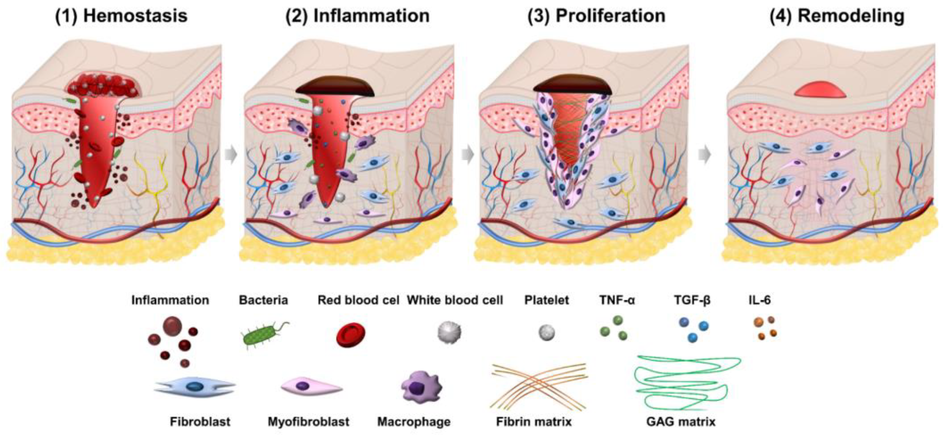
Figure 1. Illustration of four phases in the wound healing process.
2.1. Hemostasis Phase
Wound healing first begins with hemostasis. The lymphatic vessels are injured in this phase, and blood flows out to remove microorganisms and antigens [14]. The body will activate different clotting cascades and thrombocytes to agglomerate by exposed collagen. At the same time, platelets activate vasoconstriction to reduce blood loss and fill tissue gaps in injured vessels with blood clots containing cytokines and growth factors [15]. The clot contains the molecules fibrin, fibronectin, vitronectin, and thrombospondin, which form a temporary matrix as a scaffolding structure for the migration of leukocytes, keratinocytes, fibroblasts, and endothelial cells, and it is a reservoir of growth factors that stabilize blood clots and avoid bleeding.
2.2. Inflammation Phase
The second phase of wound healing is inflammation which focuses on cleaning the wound and preparing for new tissue formation in the wound. This stage has the appearance of neutrophils and lasts about 2–5 days from when the wound becomes infected. Neutrophils can phagocytize and secrete proteases (elastase, cathepsin G, proteinase 3) that help destroy bacteria in the wound and deco remove debris. Neutrophils also release mediators (TNF-α, IL-1 and IL-6) to amplify the inflammatory response, stimulating VEGF and IL-8 to respond to repair during wound healing [16]. The macrophage process then supports the ongoing process by phagocytosis of the debris and secretion of growth factors, chemokines, and cytokines [17]. Macrophages promote and address inflammation, eliminate apoptotic, and support cell proliferation and tissue recovery after injury [18]. In the inflammatory phase, there are often symptoms of edema, erythema and pain.
2.3. Proliferation Phase
The proliferation phase is the most important phase of the wound healing process and lasts from 6 to 21 days. During the proliferation phase of wound healing, the wound is healed with fresh collagen and extracellular matrix tissue. After that, the wound shrinks as new tissues develop. A new network of blood vessels must be created for granulation tissues to remain healthy and receive an adequate supply of nutrients and oxygen. The modulation of fibroblasts toward myofibroblasts promotes the formation of granulation tissue. The myofibroblasts are characterized by the capacity to produce force and synthesize extracellular matrix components that allow the contraction of granulation tissue [19]. By gripping the wound boundaries and pulling them together, myofibroblasts use a technique akin to that of smooth muscle cells to close the wound. In the initial stages of wound healing, granulation tissue appears pink or red and has an uneven texture. Furthermore, healthy granulation tissue is clot-resistant [20][21]. Dark granulation tissue may be brought on by an infection, ischemia, or insufficient perfusion. Near the conclusion of the proliferation phase, epithelial cells resurface the wound. Keeping wounds moist accelerates epithelialization. Epithelialization occurs when occlusive or semi-occlusive dressings are applied within 48 h after the injury. This is because adequate tissue humidity is maintained. One accomplishment of the proliferation phase is replacing the temporary fibrin matrix with a new matrix made of collagen fibers, proteoglycans, and fibronectin to restore the structure and function of tissues. Another crucial stage of healing is angiogenesis, or the ingrowth of new capillaries to replace previously damaged vessels and restore circulation. The creation of granulation tissue and epithelialization are other important phenomena in this healing period. In the proliferation phase of healing, fibroblasts are the most important cells [22][23]. For fibroblasts to migrate in the extracellular matrix, they must first recognize and interact with particular matrix components. Fibroblasts in the normal dermis are usually dormant and sparsely scattered, but they are active and plentiful in the provisional matrix wound site and granulation tissue [24][25]. Their migration and aggregation in the wound site necessitate morphological changes and the production and secretion of proteases to clear a passage from the ECM into the wound site. The chemotactic growth factors, cytokines, and chemokines concentration gradient, as well as the alignment of the fibrils in the ECM and provisional matrix, control the direction of fibroblast migration. Rather than crossing these fibrils, fibroblasts prefer to move along them [26][27]. To help them move through the matrix, fibroblasts produce proteolytic enzymes on a local level. Collagenase (MMP-1), gelatinases (MMP-2 and MMP-9) that destroy gelatin substrates, and stromelysin (MMP-3), which has various protein substrates in the ECM, are three kinds of MMPs released by fibroblasts [28][29]. After migrating into the matrix, fibroblasts change shape, settle down, and begin to proliferate and generate granulation tissue components such as collagen, elastin, and proteoglycans. Fibroblasts connect to the provisional fibrin matrix cables and begin producing collagen [19][30]. Type III collagen, like other extracellular matrix proteins and proteoglycans, is generated in high amounts at first [31]. Collagen mRNA is connected to polyribosomes on the endoplasmic reticulum, where new collagen chains are formed after transcription and processing. A crucial stage in this process involves proline and lysine residue hydroxylation.
2.4. Remodeling Phase
Closure of acute and chronic wounds is regarded as the wound healing endpoint in most clinical settings, yet wounds can continue to undergo remodeling or tissue maturation for months or even years [32][33]. This final stage of wound healing decides whether scarring will occur and whether the wound will reoccur. Regression of the neo vasculature, a periodic deposition to the ECM, and subsequent reconstruction of granulation tissue to scar tissue are all part of the remodeling phase [26]. Collagen III makes up the majority of granulation tissue, which is gradually replaced by the stronger collagen I as the wound heals. This occurs due to simultaneous collagen I production and collagen III lysis, followed by ECM remodeling [34]. In the remodeling phase, scar tissues are created, and it might take several months or years to complete, depending on the severity and location of the wound, and used therapeutic procedures. During this time, the new tissue gradually gets stronger and more flexible. Elasticity and tensile strength of the skin are both getting stronger because of collagen synthesis. After re-epithelialization, macrophages regain their phagocytic phenotype. Excessed cells and matrix no longer required for wound healing are phagocytosed by Mreg or M2c-like macrophages [24].
References
- Agyare, C.; Akindele, A.J.; Steenkamp, V. Natural Products and/or Isolated Compounds on Wound Healing. Evid.-Based Complement. Altern. Med. 2019, 2019, 4594965.
- Sorg, H.; Tilkorn, D.J.; Hager, S.; Hauser, J.; Mirastschijski, U. Skin Wound Healing: An Update on the Current Knowledge and Concepts. Eur. Surg. Res. 2017, 58, 81–94.
- Schreml, S.; Szeimies, R.-M.; Prantl, L.; Landthaler, M.; Babilas, P. Wound Healing in the 21st Century. J. Am. Acad. Dermatol. 2010, 63, 866–881.
- Ryall, C.; Duarah, S.; Chen, S.; Yu, H.; Wen, J. Advancements in Skin Delivery of Natural Bioactive Products for Wound Management: A Brief Review of Two Decades. Pharmaceutics 2022, 14, 1072.
- Vitale, S.; Colanero, S.; Placidi, M.; Di Emidio, G.; Tatone, C.; Amicarelli, F.; D’Alessandro, A.M. Phytochemistry and Biological Activity of Medicinal Plants in Wound Healing: An Overview of Current Research. Molecules 2022, 27, 3566.
- Ibrahim, N.; Wong, S.; Mohamed, I.; Mohamed, N.; Chin, K.-Y.; Ima-Nirwana, S.; Shuid, A. Wound Healing Properties of Selected Natural Products. Int. J. Environ. Res. Public Health 2018, 15, 2360.
- Artem Ataide, J.; Caramori Cefali, L.; Machado Croisfelt, F.; Arruda Martins Shimojo, A.; Oliveira-Nascimento, L.; Gava Mazzola, P. Natural Actives for Wound Healing: A Review. Phyther. Res. 2018, 32, 1664–1674.
- Pasupuleti, V.R.; Sammugam, L.; Ramesh, N.; Gan, S.H. Honey, Propolis, and Royal Jelly: A Comprehensive Review of Their Biological Actions and Health Benefits. Oxid. Med. Cell. Longev. 2017, 2017, 1259510.
- Viuda-Martos, M.; Ruiz-Navajas, Y.; Fernández-López, J.; Pérez-Álvarez, J.A.A. Functional Properties of Honey, Propolis, and Royal Jelly. J. Food Sci. 2008, 73, 117–124.
- Radha, M.H.; Laxmipriya, N.P. Evaluation of Biological Properties and Clinical Effectiveness of Aloe Vera: A Systematic Review. J. Tradit. Complement. Med. 2015, 5, 21–26.
- Fana, S.E.; Ahmadpour, F.; Rasouli, H.R.; Tehrani, S.S.; Maniati, M. The Effects of Natural Compounds on Wound Healing in Iranian Traditional Medicine: A Comprehensive Review. Complement. Ther. Clin. Pract. 2021, 42, 101275.
- Hajialyani, M.; Tewari, D.; Sobarzo-Sánchez, E.; Nabavi, S.M.; Farzaei, M.H.; Abdollahi, M. Natural Product-Based Nanomedicines for Wound Healing Purposes: Therapeutic Targets and Drug Delivery Systems. Int. J. Nanomed. 2018, 13, 5023–5043.
- Dumitru, C.D.; Neacsu, I.A.; Grumezescu, A.M.; Andronescu, E. Bee-Derived Products: Chemical Composition and Applications in Skin Tissue Engineering. Pharmaceutics 2022, 14, 750.
- Strodtbeck, F. Physiology of Wound Healing. Newborn Infant Nurs. Rev. 2001, 1, 43–52.
- Martin, P. Wound Healing--Aiming for Perfect Skin Regeneration. Science 1997, 276, 75–81.
- Eming, S.A.; Krieg, T.; Davidson, J.M. Inflammation in Wound Repair: Molecular and Cellular Mechanisms. J. Investig. Dermatol. 2007, 127, 514–525.
- Tziotzios, C.; Profyris, C.; Sterling, J. Cutaneous Scarring: Pathophysiology, Molecular Mechanisms, and Scar Reduction Therapeutics. J. Am. Acad. Dermatol. 2012, 66, 13–24.
- Koh, T.J.; DiPietro, L.A. Inflammation and Wound Healing: The Role of the Macrophage. Expert Rev. Mol. Med. 2011, 13, e23.
- Desmouliere, A.; Darby, I.A.; Laverdet, B.; Bonté, F. Fibroblasts and Myofibroblasts in Wound Healing. Clin. Cosmet. Investig. Dermatol. 2014, 7, 301.
- Grey, J.E.; Enoch, S.; Harding, K.G. Wound Assessment. BMJ 2006, 332, 285–288.
- Krzyszczyk, P.; Schloss, R.; Palmer, A.; Berthiaume, F. The Role of Macrophages in Acute and Chronic Wound Healing and Interventions to Promote Pro-Wound Healing Phenotypes. Front. Physiol. 2018, 9, 419.
- Johnson, K.E.; Wilgus, T.A. Vascular Endothelial Growth Factor and Angiogenesis in the Regulation of Cutaneous Wound Repair. Adv. Wound Care 2014, 3, 647–661.
- Miricescu, D.; Badoiu, S.C.; Stanescu-Spinu, I.-I.; Totan, A.R.; Stefani, C.; Greabu, M. Growth Factors, Reactive Oxygen Species, and Metformin—Promoters of the Wound Healing Process in Burns? Int. J. Mol. Sci. 2021, 22, 9512.
- Rodrigues, M.; Kosaric, N.; Bonham, C.A.; Gurtner, G.C. Wound Healing: A Cellular Perspective. Physiol. Rev. 2019, 99, 665–706.
- Moretti, L.; Stalfort, J.; Barker, T.H.; Abebayehu, D. The Interplay of Fibroblasts, the Extracellular Matrix, and Inflammation in Scar Formation. J. Biol. Chem. 2022, 298, 101530.
- Xue, M.; Jackson, C.J. Extracellular Matrix Reorganization During Wound Healing and Its Impact on Abnormal Scarring. Adv. Wound Care 2015, 4, 119–136.
- Chen, H.; Li, G.; Liu, Y.; Ji, S.; Li, Y.; Xiang, J.; Zhou, L.; Gao, H.; Zhang, W.; Sun, X.; et al. Pleiotropic Roles of CXCR4 in Wound Repair and Regeneration. Front. Immunol. 2021, 12, 668758.
- Pittayapruek, P.; Meephansan, J.; Prapapan, O.; Komine, M.; Ohtsuki, M. Role of Matrix Metalloproteinases in Photoaging and Photocarcinogenesis. Int. J. Mol. Sci. 2016, 17, 868.
- Levin, M.; Udi, Y.; Solomonov, I.; Sagi, I. Next Generation Matrix Metalloproteinase Inhibitors—Novel Strategies Bring New Prospects. Biochim. Biophys. Acta Mol. Cell Res. 2017, 1864, 1927–1939.
- Rajkumar, V.S.; Shiwen, X.; Bostrom, M.; Leoni, P.; Muddle, J.; Ivarsson, M.; Gerdin, B.; Denton, C.P.; Bou-Gharios, G.; Black, C.M.; et al. Platelet-Derived Growth Factor-β Receptor Activation Is Essential for Fibroblast and Pericyte Recruitment during Cutaneous Wound Healing. Am. J. Pathol. 2006, 169, 2254–2265.
- Wang, C.; Brisson, B.K.; Terajima, M.; Li, Q.; Hoxha, K.; Han, B.; Goldberg, A.M.; Sherry Liu, X.; Marcolongo, M.S.; Enomoto-Iwamoto, M.; et al. Type III Collagen Is a Key Regulator of the Collagen Fibrillar Structure and Biomechanics of Articular Cartilage and Meniscus. Matrix Biol. 2020, 85–86, 47–67.
- Zou, M.-L.; Teng, Y.-Y.; Wu, J.-J.; Liu, S.-Y.; Tang, X.-Y.; Jia, Y.; Chen, Z.-H.; Zhang, K.-W.; Sun, Z.-L.; Li, X.; et al. Fibroblasts: Heterogeneous Cells With Potential in Regenerative Therapy for Scarless Wound Healing. Front. Cell Dev. Biol. 2021, 9, 713605.
- Eriksson, E.; Liu, P.Y.; Schultz, G.S.; Martins-Green, M.M.; Tanaka, R.; Weir, D.; Gould, L.J.; Armstrong, D.G.; Gibbons, G.W.; Wolcott, R.; et al. Chronic Wounds: Treatment Consensus. Wound Repair Regen. 2022, 30, 156–171.
- Czubryt, M.P. Common Threads in Cardiac Fibrosis, Infarct Scar Formation, and Wound Healing. Fibrogenesis Tissue Repair 2012, 5, 19.
More
Information
Subjects:
Biochemistry & Molecular Biology
Contributors
MDPI registered users' name will be linked to their SciProfiles pages. To register with us, please refer to https://encyclopedia.pub/register
:
View Times:
11.9K
Revisions:
2 times
(View History)
Update Date:
06 Sep 2022
Table of Contents


Notice
You are not a member of the advisory board for this topic. If you want to update advisory board member profile, please contact office@encyclopedia.pub.
OK
Confirm
Only members of the Encyclopedia advisory board for this topic are allowed to note entries. Would you like to become an advisory board member of the Encyclopedia?
Yes
No
${ textCharacter }/${ maxCharacter }
Submit
Cancel
Back
Comments
${ item }
|
${ item.createdUser.fullName }
${ item.createdAt }
${ item.vote }
${ item.reply }
Delete
${ reply.createdUser.fullName }
${ reply.createdAt }
${ reply.vote }
Delete
There is no reply to this comment~
${ item.replyTextCharacter }/${ item.replyMaxCharacter }
Submit
Cancel
More
No more~
There is no comment~
${ textCharacter }/${ maxCharacter }
Submit
Cancel
${ selectedItem.replyTextCharacter }/${ selectedItem.replyMaxCharacter }
Submit
Cancel
Confirm
Are you sure to Delete?
Yes
No