Submitted Successfully!
To reward your contribution, here is a gift for you: A free trial for our video production service.
Thank you for your contribution! You can also upload a video entry or images related to this topic.
Version Summary Created by Modification Content Size Created at Operation
1 -- 2266 2022-08-23 17:53:20 |
2 layout Meta information modification 2266 2022-08-24 07:33:29 | |
3 layout Meta information modification 2266 2022-08-24 07:40:18 |

Video Upload Options

Do you have a full video?

Confirm

Are you sure to Delete?
Cite
If you have any further questions, please contact Encyclopedia Editorial Office.
Gött, H.;  Uhl, E. FGFR3-TACCs3 Fusions in Human Glioblastoma. Encyclopedia. Available online: https://encyclopedia.pub/entry/26407 (accessed on 27 June 2024).
Gött H,  Uhl E. FGFR3-TACCs3 Fusions in Human Glioblastoma. Encyclopedia. Available at: https://encyclopedia.pub/entry/26407. Accessed June 27, 2024.
Gött, Hanna, Eberhard Uhl. "FGFR3-TACCs3 Fusions in Human Glioblastoma" Encyclopedia, https://encyclopedia.pub/entry/26407 (accessed June 27, 2024).
Gött, H., & Uhl, E. (2022, August 23). FGFR3-TACCs3 Fusions in Human Glioblastoma. In Encyclopedia. https://encyclopedia.pub/entry/26407
Gött, Hanna and Eberhard Uhl. "FGFR3-TACCs3 Fusions in Human Glioblastoma." Encyclopedia. Web. 23 August, 2022.
FGFR3-TACCs3 Fusions in Human Glioblastoma
Edit

Glioma are the most frequent malignant primary CNS tumors in adults, with an incidence of 5–6 per 100,000 per year, with glioblastoma (with 3.2 per 100,000 per year) being the largest subgroup. The current therapy for glioblastoma is resection followed by radiochemotherapy and their prognosis is always fatal.Oncogenic fusion genes emerged as successful targets in several malignancies, such as chronic myeloic leukemia or lung cancer. Fusion of the fibroblast growth receptor 3 and the transforming acidic coiled coil containing protein – FGFR3-TACC3-fusion is prevalent in 3-4% of human glioblastoma. The fusion protein leads to constitutively activated kinase signaling of FGFR3 and thereby promotes cell proliferation and tumour progression. An overview on clinical and histomolecular features of FGFR3-TACC3-fusion positive glioblastoma is described and the cellular fuction of the fusion protein in glioblastoma cells is highlighted.

FGFR3-TACC3 glioblastoma tumor cells

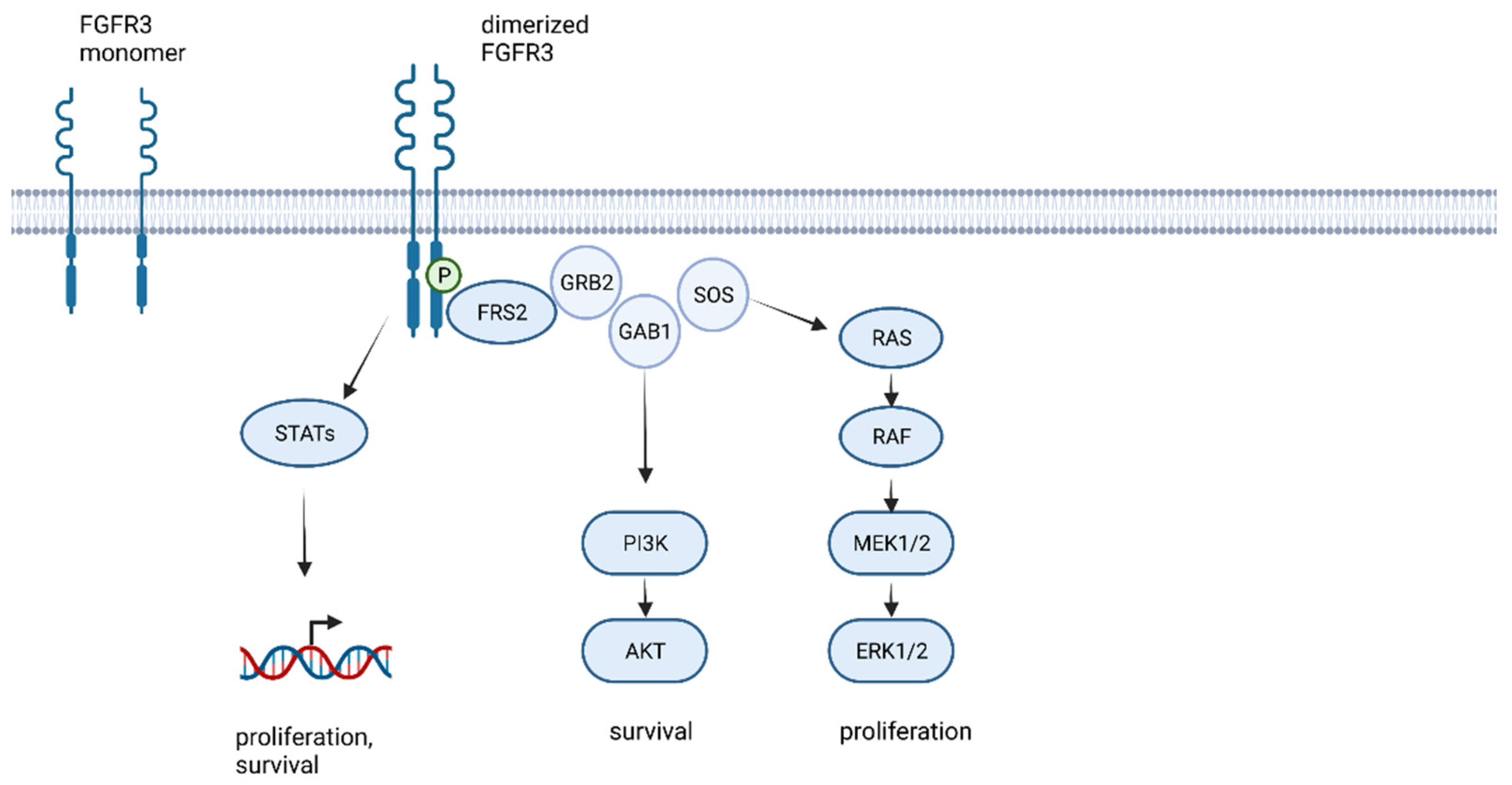
1. The FGFR Family and FGFR Signaling

The family of fibroblast growth factor receptors (FGFRs) consists of four membrane-bound tyrosine kinase receptors (RTK) FGFR1–4 and one kinase-lacking coreceptor FGFR5 or FGFR-like1 (FGFRL1). The structure among the FGFR1–4 family members is highly conserved: every FGFR consists of three immunoglobulin-like looped domains (Ig-domains), an acidic region (the acid box), a single transmembrane domain, a long juxtamembrane domain, and an intracellular domain with two tyrosine kinase domains [1]. While FGFR(1–3)IIIb is usually expressed in epithelial tissue, FGFR(1–3)IIIc is more prevalent in a mesenchymal environment [2]. Although splicing regulatory mechanisms are not yet fully understood, the switching of the expression between the isoforms during oncogenic or developmental processes, such as epithelial to mesenchymal transition, is possible [3].
Via the extracellular membrane domain, FGFRs can bind 22 known fibroblast growth factor ligands (FGF), as well as the FGFR-coreceptors heparan sulfate proteoglycans and klotho. In addition to these canonical binding partners, FGFRs are also able to bind not only extracellular matrix associated proteins and cell adhesion molecules, but also a broad spectrum of non-canonical signaling partners such as N-cadherins or galectins [4][5]. The numerous possible ligand-receptor combinations point out that FGFR-signaling is highly context-specific, and the role of the same receptor in two distinct tissues or pathologies can be completely different.
Ligand binding at the extracellular IgII and IgIII binding domain induces the homodimerization of two FGFR molecules, and interaction between their tyrosine kinase domain leads to the transautophosphorylation and initiation of downstream signaling pathways (Figure 1) [6][7][8][9]. The most relevant FGFR-downstream pathways in cancer are activated by the FGFR substrate 2 (FRS2) mediated binding of growth factor receptor bound protein 2 (GRB2). Binding of the RAS guanine exchange factor, son of sevenless homologue 1 (SOS) and the cofactor GRB2-associated-binding protein 1 (GAB1), forms a complex that activates RAS/MEK/MAPK and RAC/JNK and PI3K/AKT signaling. All these pathways result in activation of cell proliferation, cell survival, and STAT1 and 3 activation, resulting in the induction of gene translation [10][11].
Figure 1. FGFR kinase signaling and activation of MAPK pathways leading to cell proliferation and cell survival (created with biorender.com, accessed on 21 May 2022).
In addition to other mechanisms, FGFR-signaling is negatively regulated by miRNA-mediated degradation of the receptor [10][12] and SPROUTY (SPRY) proteins, which function as tumor suppressor genes by negative regulation of RAS/MAPK-activity [7][10][13][14].
The central function of FGF-FGFR in pathways related to cell proliferation and survival in combination with the variable and tissue specific complex regulatory loops implicate the consequences of disturbance of these mechanisms in cancer.
A whole genome analysis of more than 2662 adult human glioblastoma samples revealed the number of FGFR-aberrations and amplifications as generally sparse, with prevalence of FGFR1 aberrations of 51 in 3068, FGFR2 in 12 of 2662, FGFR3 in 13 of 2887, and FGFR4 in 9 out of 2456 investigated samples [15]. While little is known about the role of FGFR2 and FGFR4 in glioblastoma, FGFR1 is expressed in human glioma, and its expression level increases with the grade of malignancy [16]. The expression of stem cell transcription factors such as ZEB1, SOX2, and OLIG3 is regulated by FGFR1 [17]. FGFR3 expression is increased in the classical and neural subtype of glioblastoma, and Wang et al. saw improved survival and an expressome associated with differentiated cellular function in correlation with higher FGFR3 expression [18].

2. Histomolecular and Clinical Characteristics of FGFR3-TACC3 Fused Glioblastoma

There are several recurrent molecular and histological characteristics that were observed in an increasing number of investigations around FGFR3-TACC3 fusion-positive glioma: even in studies that were not limited to IDH-wildtype or former primary glioblastomas WHO grade 4, but included all forms of malignant gliomas, all gliomas that were positive for a FGFR3-TACC fusion presented the wildtype version of the IDH1 and IDH2 gene [6][12][19][20][21][22][23]. No FGFR3-TACC3 fusions were reported in oligodendroglioma, suggesting a restriction of the fusion to the astrocytic lineage of human glioma [20].
FGFR3-TACC3 fusions were mutually exclusive with EGFR-amplifications, a mutation that is present in about 80% of IDH-wildtype glioblastoma [20][21][22][24]. Mata et al. was the only group that found an EGFR-amplification in a FGFR3-TACC3 fusion-positive glioma [21]. Di Stefano et al. reported a trend towards the absence of the EGFRvIII variant that goes hand in hand with higher signaling activity of the EGFR-tyrosine kinase in FGFR3-TACC3 fusion-positive gliomas [22]. Furthermore, other authors reported an exclusivity of FGFR3-TACC3 fusions with amplifications of other tyrosine kinase receptors such as PDGFR, KIT, and MET [21][23][25].
FGFR3-TACC3 fusions were mutually exclusive with ATRX-loss and H3F3A mutations [21][26]. They occurred with less probability of harboring a co-occurring oncogenic TP53 mutation [21][26]. Regarding the frequency of CDKN2A inactivation, TERT-mutation, and cell cycle associated pathways, no difference in FGFR3-fusion-positive gliomas was described [21]. They were associated with a higher expression of stemness markers such as OLIG2 and GFAP [23].
While MDM4 alterations were absent in FGFR3-TACC3 fusion-positive gliomas, there is an association with a higher prevalence of MDM2 alterations and CDK4 amplifications, which can be found in 19% and 10%, respectively, of fusion-positive glioblastoma [23]. Even though CDK4 and MDM2 amplification itself is a favorable prognostic factor for the survival of glioblastoma patients, a subgroup of FGFR3-fusion-positive patients with CDK4 and MDM2 amplifications had an even better overall survival rate than those with CDK4 and MDM2 amplification that were negative for FGFR3 fusions [22].
Mata et al. performed methylation epic arrays on FGFR3-TACC3 positive glioblastoma, and their methylation profile most likely corresponded to the RTKII or mesenchymal subclass phenotype. In addition, FGFR3-fusion-positive glioblastomas harbored a generally lower overall mutational burden [21].
Remarkably, FGFR3-TACC3 positive gliomas were reported to possess specific morphological features that might reflect an initial step of tumorigenesis, as they can not only be found in glioblastoma, but also in FGFR3-fusion-positive lower grade glioma [23]. The specific recurrent morphological features of these tumors include monomorphous ovoid nuclei with nuclear palisading and attachment of the tumor cells towards blood vessels by parallel thin cytoplasmic processes, forming vague pseudorosettes [23]. Isolated tumor cells present with ovoid nuclei and ovoid cytoplasm and infiltrate the neuropil. A network of small capillary like vessels, arranged in an endocrinoid network, and spindled neoplastic cells embedded in a loose, myxoid background, with a tissue culture like appearance, is described as a “chickenwire pattern” by Broggi et al. [27]. In glioblastoma, these specific features were associated with different areas of higher cellular density, with anisocytosis, microvascular proliferation, and necrosis [23]. Furthermore, the tumors presented lower mitotic activity and signs of desmoplasia, such as CD34 labelling and microcalcifications, making differential diagnosis between astroblastoma, ependymoma, and angiocentric glioma, which share these morphological features, challenging [23]. These specific morphological features are reported to be present, at least focally, in 73% of FGFR3-fusion-positive tumors; however, as gliomas are known to be very heterogeneous, not all tumor areas reflect these features [23]. Gliani et al. investigated six FGFR3-TACC3 fusion-positive glioblastomas and five of them shared the described morphological characteristics [28]. Based on this observation, the group conducted prospective testing for FGFR3-TACC3 fusions in gliomas presenting with typical histomorphology. Two of the investigated tumors turned out to be negative for an FGFR3 fusion (the number of investigated cases is not published), concluding that the described recurrent morphological features are often shared by FGFR3-fusion-positive glioma, but the specificity for the molecular subtype is limited [28].
The epidemiology of FGFR3-TACC3 fusion-positive glioblastoma is mostly unspecific. However, Granberg et al. reported a female predominance, and Bielle et al. reported a sex ratio of 1:1 for this specific type of glioblastoma [23][25], which is interesting, as glioblastoma are usually more frequent in men [29]. The presence of a FGFR3 fusion in glioblastoma IDH wildtype is reported as a favorable prognostic factor, associated with a better overall survival compared to glioblastoma IDH-wildtype without FGFR3 fusion [30]. However, the survival rate is not better than the survival rate for IDH-mutated gliomas [22]. The analysis of imaging data of a large set of FGFR3 fusion-positive glioblastoma revealed an association with the occurrence in the cortical-subcortical region, insular, and temporal lobe location, which might be due to the specific role of FGFRs in the development of these brain areas [22]. FGFR3 fusion-positive tumors presented with recurrent radiogenomic features, including a less frequent eloquent location, poorly defined contrast-enhancing and non-enhancing tumor margins, as well as increased edema in glioblastoma and poorly defined tumor borders in lower grade glioma [22][31] Radiomic analysis for the classification of the FGFR3-TACC3 status of glioblastoma led to an area under the curve (AUC) of 0.87 in the first dataset and 0.754 in a second validation set, allowing the conclusion that FGFR3-TACC3 positive glioblastoma has a distinct radiomic signature [22].

3. The Functional Role of FGFR3-TACC3 in Glioblastoma Cells

The FGFR3-TACC3 fusion protein is involved in several cellular processes and signaling cascades, leading to FGFR3-TACC3 overexpression, increased kinase activity, and corresponding downstream signaling, morphological changes, increased cell growth, altered cellular metabolism, stress response, and even dysregulated mitotic progression, resulting in aneuploidy.
In the very first study on FGFR3-TACC3 fusion, Singh et al. transfected fibroblasts and astrocytes with the FGFR3-TACC3 fusion gene and observed not only anchorage independent growth of these cells in soft agar, but also a gain of proliferative capacity and the formation of glioma lesions, expressing the glioma stem cell markers OLIG2, phosphohistone H3, nestin, and GFAP, while EGFRvIII transfected astrocytes did not show these markers [19]. Stimulation with FGF did not affect the downstream signaling of FGFR3-fusion-positive cells, but instead a constitutive phosphorylation of the tyrosine kinase domain of FGFR3 and FRS2 was observed [19]. All of these effects were abolished by the tyrosine kinase inhibitor PD173074.
The kinase activity of the FGFR3-TACC3 fusion protein plays a crucial role in the proliferation and survival of glioblastoma cells. The ability to dimerize TACC3 leads to the presence of a constitutively dimerized, and thereby activated, FGFR3 tyrosine kinase domain in fusion-positive tumor cells and hyperactivated downstream signaling, resulting in an overexpression of phosphorylated FRS2, the initial intracellular binding partner of FGFR3, to activate ERK1/2 and AKT signaling [2][6][8][32][33]. Parker et al. detected an increased activation of pERK, but not of STAT3 and pAKT, in fusion-positive cells while other authors saw enhanced STAT3 and STAT1 activation [12][32]. Nelson et al. showed that only plasma membrane localized FGFR3-TACC3 fusion protein leads to formation of oncogenic foci in fusion transfected NIH3T3 cells. This goes along with increased MAPK signaling activation, while cytoplasmatic localized FGFR3-TACC3 does not induce oncogenic transformation, supporting the hypothesis that the oncogenic force of the fusion protein is dependent on its kinase activity [33]. The phosphorylation site Y746 is of major importance for activation of ERK, STAT and PI3K signaling. Y746 is hyperphosphorylated in FGFR3-TACC3 fusion-positive cells and MAPK activation is increased in fusion-positive cells [34]. Treatment with the kinase inhibitors BGJ398 and trametinib resulted in reduction in MAPK signaling and had an antitumor effect in FGFR3-TACCex11 and FGFR3-TACC3ex8 transfected cells, but the signaling and treatment response differed between the two isoforms [33].
TACC3 in its wildtype from is phosphorylated by Aurora-A and forms a complex with clathrin and ch-TOG, which is localized to the mitotic spindle apparatus and provides its stability [33]. When fused to the FGFR3-protein, TACC3 lacks a phosphorylation site for Aurora-A [33]. Confocal imaging of FGFR3-TACC3 showed an arc-shaped structure of the protein, bending over and enchasing the metaphase spindle poles, but not relocating to the mid body, leading to erratic mitotic segregation [19]. FGFR3-TACC3-positive cells exhibit a 3 to 5-fold higher number of errors in chromosomal segregation and resulting aneuploidy, an effect, that could be reduced to 80% by treatment with the kinase inhibitor PD173074 [19]. This, however, implies that the activity of FGFR3-TACC3 in aneuploidy induction is dependent on its kinase activity.
Gene ontology mapping of the dataset from TCGA showed enriched expression of genes that are related to oxidative phosphorylation, high mitochondrial activity, and biogenesis [35]. Additionally, increased mitochondrial DNA, mitochondrial mass, and higher levels of ATP were detected in FGFR3-TACC3 transfected astrocytes compared to control cells, and the fusion-positive cells showed an elevated basal and maximal oxygen consumption rate, as well as a mild increase in the extracellular acidification rate [35]. Gene expression levels of the respiratory complex proteins SDHB, UQCRC1, ATP5A1, and the mitochondrial membrane transporter VDAC1 were elevated in fusion-positive cells [35]. Anti-pY immunoprecipitation showed that only FGFR3-TACC3 fusion-positive cells contained phosphorylated PIN4, an activator of mitochondrial metabolism and anabolic response, leading to accumulation of reactive oxygen species and thereby, elevated expression of the transcription regulators PGC1α and ERRγ, which increase mitochondrial metabolism [35]. Acordingly, the FGFR3-TACC3-positive cells were sensitive towards treatment with mitochondrial inhibitors such as menadione, metformin, and tigecycline in vitro [35].
Besides this, FGFR3-TACC3 fusion leads to morphological changes characterized by the rounding up of the cells in HEK293T cells, the activation of cell signaling pathways related to chaperone activation, the stress response and regulation of tp53 expression, and the degradation and resistance to EGFR inhibitors in HNSCC and urothelial carcinoma cells [32][36][37].

References

  1. Harmer, N.J.; Ilag, L.L.; Mulloy, B.; Pellegrini, L.; Robinson, C.V.; Blundell, T.L. Towards a resolution of the stoichiometry of the fibroblast growth factor (FGF)-FGF receptor-heparin complex. J. Mol. Biol. 2004, 339, 821–834.
  2. Williams, S.V.; Hurst, C.D.; Knowles, M.A. Oncogenic FGFR3 gene fusions in bladder cancer. Hum. Mol. Genet. 2013, 22, 795–803.
  3. Warzecha, C.C.; Sato, T.K.; Nabet, B.; Hogenesch, J.B.; Carstens, R.P. ESRP1 and ESRP2 are epithelial cell-type-specific regulators of FGFR2 splicing. Mol. Cell 2009, 33, 591–601.
  4. Latko, M.; Czyrek, A.; Porębska, N.; Kucińska, M.; Otlewski, J.; Zakrzewska, M.; Opaliński, Ł. Cross-Talk between Fibroblast Growth Factor Receptors and Other Cell Surface Proteins. Cells 2019, 8, 455.
  5. Kucińska, M.; Porębska, N.; Lampart, A.; Latko, M.; Knapik, A.; Zakrzewska, M.; Otlewski, J.; Opaliński, Ł. Differential regulation of fibroblast growth factor receptor 1 trafficking and function by extracellular galectins. Cell Commun. Signal. 2019, 17, 65.
  6. Parker, B.C.; Engels, M.; Annala, M.; Zhang, W. Emergence of FGFR family gene fusions as therapeutic targets in a wide spectrum of solid tumours. J. Pathol. 2014, 232, 4–15.
  7. Jimenez-Pascual, A.; Siebzehnrubl, F.A. Fibroblast Growth Factor Receptor Functions in Glioblastoma. Cells 2019, 8, 715.
  8. Costa, R.; Carneiro, B.A.; Taxter, T.; Tavora, F.A.; Kalyan, A.; Pai, S.A.; Chae, Y.K.; Giles, F.J. FGFR3-TACC3 fusion in solid tumors: Mini review. Oncotarget 2016, 7, 55924–55938.
  9. Bae, J.H.; Boggon, T.J.; Tomé, F.; Mandiyan, V.; Lax, I.; Schlessinger, J. Asymmetric receptor contact is required for tyrosine autophosphorylation of fibroblast growth factor receptor in living cells. Proc. Natl. Acad. Sci. USA 2010, 107, 2866–2871.
  10. Ferguson, H.R.; Smith, M.P.; Francavilla, C. Fibroblast Growth Factor Receptors (FGFRs) and Noncanonical Partners in Cancer Signaling. Cells 2021, 10, 1201.
  11. Liang, G.; Liu, Z.; Wu, J.; Cai, Y.; Li, X. Anticancer molecules targeting fibroblast growth factor receptors. Trends Pharmacol. Sci. 2012, 33, 531–541.
  12. Parker, B.C.; Annala, M.J.; Cogdell, D.E.; Granberg, K.J.; Sun, Y.; Ji, P.; Li, X.; Gumin, J.; Zheng, H.; Hu, L.; et al. The tumorigenic FGFR3-TACC3 gene fusion escapes miR-99a regulation in glioblastoma. J. Clin. Invest. 2013, 123, 855–865.
  13. Lo, T.L.; Yusoff, P.; Fong, C.W.; Guo, K.; McCaw, B.J.; Phillips, W.A.; Yang, H.; Wong, E.S.M.; Leong, H.F.; Zeng, Q.; et al. The ras/mitogen-activated protein kinase pathway inhibitor and likely tumor suppressor proteins, sprouty 1 and sprouty 2 are deregulated in breast cancer. Cancer Res. 2004, 64, 6127–6136.
  14. Yusoff, P.; Lao, D.-H.; Ong, S.H.; Wong, E.S.M.; Lim, J.; Lo, T.L.; Leong, H.F.; Fong, C.W.; Guy, G.R. Sprouty2 inhibits the Ras/MAP kinase pathway by inhibiting the activation of Raf. J. Biol. Chem. 2002, 277, 3195–3201.
  15. Forbes, S.A.; Beare, D.; Boutselakis, H.; Bamford, S.; Bindal, N.; Tate, J.; Cole, C.G.; Ward, S.; Dawson, E.; Ponting, L.; et al. COSMIC: Somatic cancer genetics at high-resolution. Nucleic. Acids Res. 2017, 45, D777–D783.
  16. Gouazé-Andersson, V.; Delmas, C.; Taurand, M.; Martinez-Gala, J.; Evrard, S.; Mazoyer, S.; Toulas, C.; Cohen-Jonathan-Moyal, E. FGFR1 Induces Glioblastoma Radioresistance through the PLCγ/Hif1α Pathway. Cancer Res. 2016, 76, 3036–3044.
  17. Jimenez-Pascual, A.; Hale, J.S.; Kordowski, A.; Pugh, J.; Silver, D.J.; Bayik, D.; Roversi, G.; Alban, T.J.; Rao, S.; Chen, R.; et al. ADAMDEC1 Maintains a Growth Factor Signaling Loop in Cancer Stem Cells. Cancer Discov. 2019, 9, 1574–1589.
  18. Wang, Z.; Zhang, C.; Sun, L.; Liang, J.; Liu, X.; Li, G.; Yao, K.; Zhang, W.; Jiang, T. FGFR3, as a receptor tyrosine kinase, is associated with differentiated biological functions and improved survival of glioma patients. Oncotarget 2016, 7, 84587–84593.
  19. Singh, D.; Chan, J.M.; Zoppoli, P.; Niola, F.; Sullivan, R.; Castano, A.; Liu, E.M.; Reichel, J.; Porrati, P.; Pellegatta, S.; et al. Transforming fusions of FGFR and TACC genes in human glioblastoma. Science 2012, 337, 1231–1235.
  20. Ferguson, S.D.; Zhou, S.; Huse, J.T.; de Groot, J.F.; Xiu, J.; Subramaniam, D.S.; Mehta, S.; Gatalica, Z.; Swensen, J.; Sanai, N.; et al. Targetable Gene Fusions Associate with the IDH Wild-Type Astrocytic Lineage in Adult Gliomas. J. Neuropathol. Exp. Neurol. 2018, 77, 437–442.
  21. Mata, D.A.; Benhamida, J.K.; Lin, A.L.; Vanderbilt, C.M.; Yang, S.-R.; Villafania, L.B.; Ferguson, D.C.; Jonsson, P.; Miller, A.M.; Tabar, V.; et al. Genetic and epigenetic landscape of IDH-wildtype glioblastomas with FGFR3-TACC3 fusions. Acta Neuropathol. Commun. 2020, 8, 186.
  22. Di Stefano, A.L.; Picca, A.; Saragoussi, E.; Bielle, F.; Ducray, F.; Villa, C.; Eoli, M.; Paterra, R.; Bellu, L.; Mathon, B.; et al. Clinical, molecular, and radiomic profile of gliomas with FGFR3-TACC3 fusions. Neuro-oncology 2020, 22, 1614–1624.
  23. Bielle, F.; Di Stefano, A.-L.; Meyronet, D.; Picca, A.; Villa, C.; Bernier, M.; Schmitt, Y.; Giry, M.; Rousseau, A.; Figarella-Branger, D.; et al. Diffuse gliomas with FGFR3-TACC3 fusion have characteristic histopathological and molecular features. Brain Pathol. 2018, 28, 674–683.
  24. Di Stefano, A.L.; Fucci, A.; Frattini, V.; Labussiere, M.; Mokhtari, K.; Zoppoli, P.; Marie, Y.; Bruno, A.; Boisselier, B.; Giry, M.; et al. Detection, Characterization, and Inhibition of FGFR-TACC Fusions in IDH Wild-type Glioma. Clin. Cancer Res. 2015, 21, 3307–3317.
  25. Granberg, K.J.; Annala, M.; Lehtinen, B.; Kesseli, J.; Haapasalo, J.; Ruusuvuori, P.; Yli-Harja, O.; Visakorpi, T.; Haapasalo, H.; Nykter, M.; et al. Strong FGFR3 staining is a marker for FGFR3 fusions in diffuse gliomas. Neuro-oncology 2017, 19, 1206–1216.
  26. Schittenhelm, J.; Ziegler, L.; Sperveslage, J.; Mittelbronn, M.; Capper, D.; Burghardt, I.; Poso, A.; Biskup, S.; Skardelly, M.; Tabatabai, G. FGFR3 overexpression is a useful detection tool for FGFR3 fusions and sequence variations in glioma. Neurooncol. Pract. 2021, 8, 209–221.
  27. Broggi, G.; Piombino, E.; Altieri, R.; Romano, C.; Certo, F.; Barbagallo, G.M.V.; Vigneri, P.; Condorelli, D.; Colarossi, L.; Colarossi, C.; et al. Glioblastoma, IDH-Wild Type with FGFR3-TACC3 Fusion: When Morphology May Reliably Predict the Molecular Profile of a Tumor. A Case Report and Literature Review. Front. Neurol. 2022, 13, 823015.
  28. Gilani, A.; Davies, K.D.; Kleinschmidt-DeMasters, B.K. Can adult IDH-wildtype glioblastomas with FGFR3:TACC3 fusions be reliably predicted by histological features? Clin. Neuropathol. 2021, 40, 165–167.
  29. Ostrom, Q.T.; Patil, N.; Cioffi, G.; Waite, K.; Kruchko, C.; Barnholtz-Sloan, J.S. CBTRUS Statistical Report: Primary Brain and Other Central Nervous System Tumors Diagnosed in the United States in 2013–2017. Neuro-oncology 2020, 22, iv1–iv96.
  30. Wu, Z.; Lopes Abath Neto, O.; Bale, T.A.; Benhamida, J.; Mata, D.; Turakulov, R.; Abdullaev, Z.; Marker, D.; Ketchum, C.; Chung, H.-J.; et al. DNA methylation analysis of glioblastomas harboring FGFR3-TACC3 fusions identifies a methylation subclass with better patient survival. Acta Neuropathol. 2022, 144, 155–157.
  31. Vagvala, S.; Guenette, J.P.; Jaimes, C.; Huang, R.Y. Imaging diagnosis and treatment selection for brain tumors in the era of molecular therapeutics. Cancer Imaging 2022, 22, 19.
  32. Wu, Y.-M.; Su, F.; Kalyana-Sundaram, S.; Khazanov, N.; Ateeq, B.; Cao, X.; Lonigro, R.J.; Vats, P.; Wang, R.; Lin, S.-F.; et al. Identification of targetable FGFR gene fusions in diverse cancers. Cancer Discov. 2013, 3, 636–647.
  33. Nelson, K.N.; Meyer, A.N.; Wang, C.G.; Donoghue, D.J. Oncogenic driver FGFR3-TACC3 is dependent on membrane trafficking and ERK signaling. Oncotarget 2018, 9, 34306–34319.
  34. Nelson, K.N.; Meyer, A.N.; Siari, A.; Campos, A.R.; Motamedchaboki, K.; Donoghue, D.J. Oncogenic Gene Fusion FGFR3-TACC3 Is Regulated by Tyrosine Phosphorylation. Mol. Cancer Res. 2016, 14, 458–469.
  35. Frattini, V.; Pagnotta, S.M.; Tala; Fan, J.J.; Russo, M.V.; Lee, S.B.; Garofano, L.; Zhang, J.; Shi, P.; Lewis, G.; et al. A metabolic function of FGFR3-TACC3 gene fusions in cancer. Nature 2018, 553, 222–227.
  36. Daly, C.; Castanaro, C.; Zhang, W.; Zhang, Q.; Wei, Y.; Ni, M.; Young, T.M.; Zhang, L.; Burova, E.; Thurston, G. FGFR3-TACC3 fusion proteins act as naturally occurring drivers of tumor resistance by functionally substituting for EGFR/ERK signaling. Oncogene 2017, 36, 471–481.
  37. Lombardi, B.; Ashford, P.; Moya-Garcia, A.A.; Rust, A.; Crawford, M.; Williams, S.V.; Knowles, M.A.; Katan, M.; Orengo, C.; Godovac-Zimmermann, J. Unique signalling connectivity of FGFR3-TACC3 oncoprotein revealed by quantitative phosphoproteomics and differential network analysis. Oncotarget 2017, 8, 102898–102911.
More
Information
Subjects: Oncology
Contributors MDPI registered users' name will be linked to their SciProfiles pages. To register with us, please refer to https://encyclopedia.pub/register : ,
View Times: 701
Revisions: 3 times (View History)
Update Date: 24 Aug 2022
1000/1000
Video Production Service