Submitted Successfully!
To reward your contribution, here is a gift for you: A free trial for our video production service.
Thank you for your contribution! You can also upload a video entry or images related to this topic.
Version Summary Created by Modification Content Size Created at Operation
1 -- 3378 2022-08-19 10:39:33 |
2 format corrected. + 16 word(s) 3394 2022-08-19 11:24:01 | |
3 format corrected. -1 word(s) 3393 2022-08-25 10:09:34 |

Video Upload Options

Do you have a full video?

Confirm

Are you sure to Delete?
Cite
If you have any further questions, please contact Encyclopedia Editorial Office.
Yue, Q.;  Song, Y.;  Liu, Z.;  Zhang, L.;  Yang, L.;  Li, J. Receptor for Advanced Glycation End Products (RAGE). Encyclopedia. Available online: https://encyclopedia.pub/entry/26315 (accessed on 29 March 2024).
Yue Q,  Song Y,  Liu Z,  Zhang L,  Yang L,  Li J. Receptor for Advanced Glycation End Products (RAGE). Encyclopedia. Available at: https://encyclopedia.pub/entry/26315. Accessed March 29, 2024.
Yue, Qing, Yu Song, Zi Liu, Lin Zhang, Ling Yang, Jinlong Li. "Receptor for Advanced Glycation End Products (RAGE)" Encyclopedia, https://encyclopedia.pub/entry/26315 (accessed March 29, 2024).
Yue, Q.,  Song, Y.,  Liu, Z.,  Zhang, L.,  Yang, L., & Li, J. (2022, August 19). Receptor for Advanced Glycation End Products (RAGE). In Encyclopedia. https://encyclopedia.pub/entry/26315
Yue, Qing, et al. "Receptor for Advanced Glycation End Products (RAGE)." Encyclopedia. Web. 19 August, 2022.
Receptor for Advanced Glycation End Products (RAGE)
Edit

As a critical molecule in the onset and sustainment of inflammatory response, the receptor for advanced glycation end products (RAGE) has a variety of ligands, such as advanced glycation end products (AGEs), S100/calcium granule protein, and high-mobility group protein 1 (HMGB1). An increasing number studies have shown that RAGE ligand binding can initiate the intracellular signal cascade, affect intracellular signal transduction, stimulate the release of cytokines, and play a vital role in the occurrence and development of immune-related diseases, such as systemic lupus erythematosus, rheumatoid arthritis, and Alzheimer’s disease. In addition, other RAGE signaling pathways can play crucial roles in life activities, such as inflammation, apoptosis, autophagy, and endoplasmic reticulum stress. Therefore, the strategy of targeted intervention in the RAGE signaling pathway may have significant therapeutic potential, attracting increasing attention.

advanced glycation end-product receptor immune high-mobility group protein 1 nuclear factor kappa-B

1. RAGE Structure

The receptor for advanced glycation end products (RAGE) consists of extracellular, hydrophobic, transmembrane, and intracellular segments. The extracellular V-shaped region provides RAGE–ligand binding sites. Intracellular fragments can bind to various intracellular signal molecules and mediate signal transduction to cause cascade reactions, including full-length type, truncated C-terminal type, and truncated N-terminal type. The truncated C-terminal type is endogenous secretory soluble RAGE (esRAGE), which can be secreted by cells and contains only the extracellular segment. In contrast, the truncated N-terminal type consists of the transmembrane region and an intracellular component. Membrane-associated proteases can remove the transmembrane component of RAGE by hydrolysis, and the released extracellular segment can form soluble sRAGE with esRAGE. sRAGE can competitively bind to RAGE ligands, but binding to ligands terminates intracellular signal transduction due to the loss of transmembrane and intracellular fragments [1] (Figure 1).
Figure 1. RAGE structural organization. The extracellular domain comprises three domains: V, C1, and C2. One transmembrane receptor passes through the plasma membrane bilayer, followed by an intracellular cytoplasmic tail. The extracellular region without a transmembrane receptor and a cytoplasmic tail is called soluble RAGE (sRAGE).

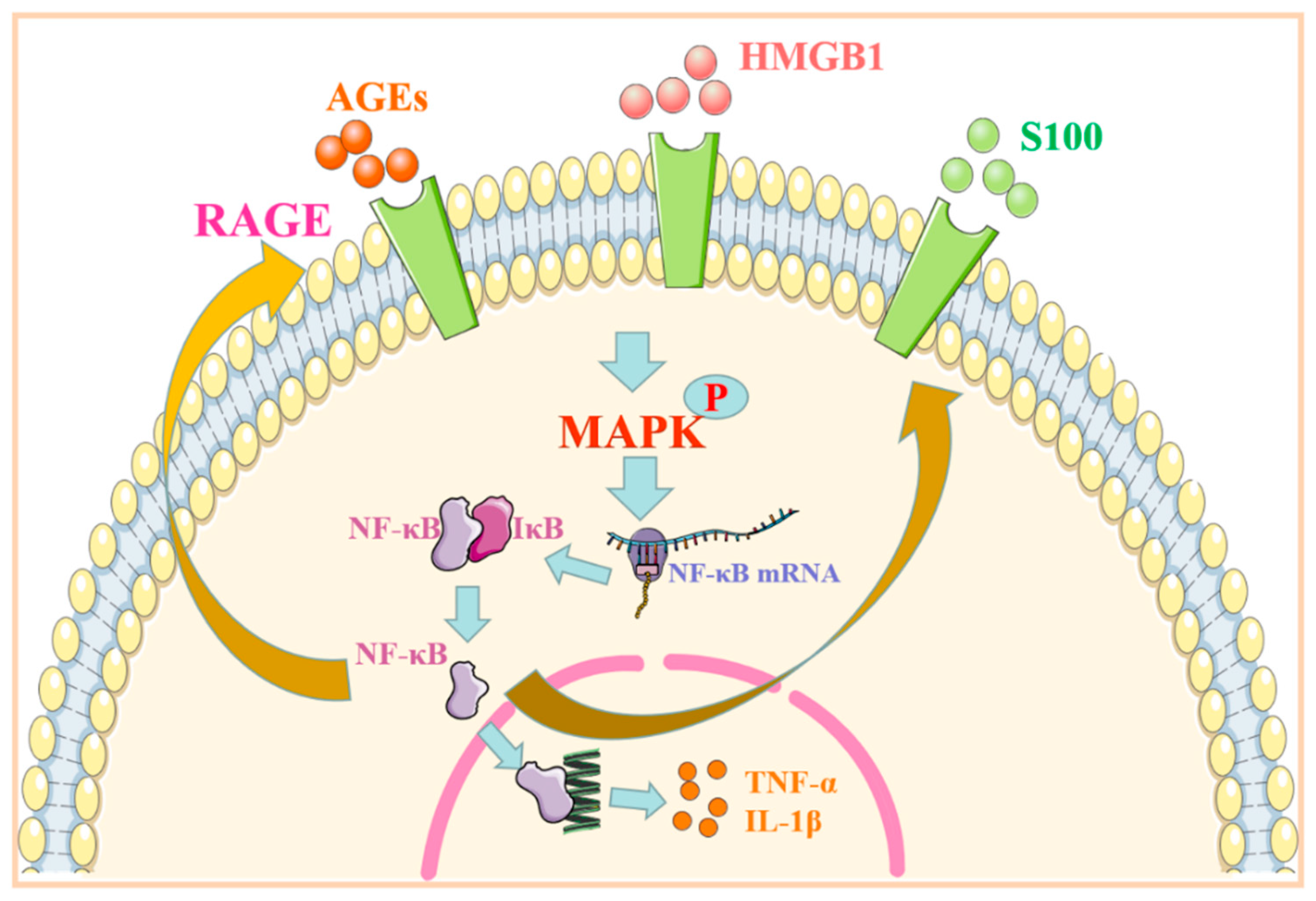
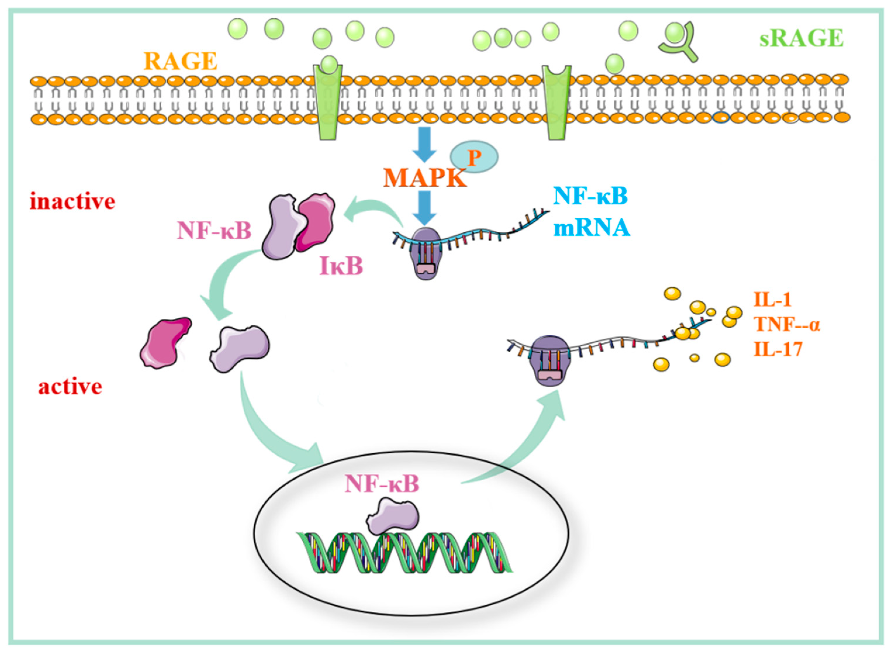
2. RAGE Regulates MAPK/NF-κB Signaling Pathway and Its Role in Immune-Associated Disease

As an essential protein complex, NF-κB can be found in almost all animal cells. It can affect cell growth, differentiation, and apoptosis by controlling critical physiological processes, such as DNA transcription and cytokine production. In resting cells, NF-κB usually binds to its protein inhibitor of NF-κB (IκB). Therefore, it is in a state of inhibition and does not exert the function of transcriptional activity. After activation, the MAPK pathway can hydrolyze IκB and dissociate it from NF-κB. The free NF-κB enters the nucleus to regulate the transcriptional expression of inflammatory factors [2]. The face of RAGE on the cell membrane is at a low level under normal physiological conditions. When the body’s inflammatory factors increase, the body is traumatized or suffers from abnormal conditions, such as diabetes. The content of ligands, such as advanced glycation end products (AGEs) and high-mobility group protein 1 (HMGB1), increase, and the expression of RAGE is upregulated [3]. When RAGE is combined with ligands, it promotes the downstream phosphorylation of the p38 MAPK protein, thus increasing the expression of the NF-κB signaling path [4]. NF-κB encourages the expression of inflammatory factors, such as TNF-α and IL-1β, by regulating the target genes and triggering related inflammatory and autoimmune responses, resulting in persistent tissue damage [5]. When NF-κB is activated, it can promote the binding of RAGE to its ligand and further encourage the continuous expression of cytokines and tissue factors [6] (Figure 2).
Figure 2. RAGE regulates the MAPK/NF-κB signaling pathway. After binding to the ligand, RAGE phosphorylates its downstream MAPK and activates NF-κB protein. NF-κB enters the nucleus to promote the transcriptional expression of inflammatory factors. Abbreviations: AGEs: advanced glycation end products; HMGB1: high-mobility group protein 1; RAGE: receptor for advanced glycation end products; MAPK: mitogen-activated protein kinase; NF-κB: nuclear factor kappa-B; TNF-α: tumor necrosis factor α; IL-1β: interleukin-1β.
Table 1 summarizes the related ligands and mechanisms of the RAGE-MAPK/NF-κB pathway in SLE, RA, pulmonary fibrosis, and AD.
Table 1. The role of RAGE-related signaling pathways in immune-associated diseases.

2.1. RAGE Regulates MAPK/NF-kB Signaling Pathway and Its Role in Mediating Systemic Lupus Erythematosus

SLE is an autoimmune disease involving various systems and organs, with diverse and variable clinical manifestations. The pathogenesis of SLE is complicated. The interaction between genetic and environmental factors, the production of pathogenic antibodies, and the deposition of immune complexes (ICs) formed by combining autoantibodies and antigens are all causes of SLE [20]. HMGB1 binds to Toll-like receptors, such as TRL-2, TRL4, and RAGE, to regulate the immunoinflammatory response by mediating the MAPK/NF-κB signal pathway. In addition, it has been found that the immune complex formed by the combination of HMGB1 and DNA plays a vital role in the pathogenesis of SLE [21][22]. These findings suggest that HMGB1 plays a crucial role in the occurrence and development of SLE.
To confirm the above conjecture, some studies have further examined the level of HMGB1 in SLE patients and animal models. The results showed that the expression level of HMGB1 in patients and animals increased, and the inflammatory response of related immune cells increased significantly, which led to the occurrence of SLE [7]. Immune complexes can activate the expression of RAGE in human endothelial cells, participate in the response of the HMGB1-RAGE axis in promoting SLE vasculitis, and induce the production of TNF-α and B-cell-activating factors. Some studies have revealed that plasma HMGB1 levels in patients with SLE are significantly higher than those in healthy subjects and positively correlated with the concentration of plasma antinuclear antibody [23]. In addition, the concentration of AGEs in skin cells and serum, as well as the expression of RAGE mRNA in peripheral blood monocytes, are significantly increased in patients with SLE. Immunohistochemical detection revealed almost no expression of RAGE in normal glomerular cells of normal subjects. However, prominent expression still occurs in glomeruli patients with SLE [8]. In summary, the researchers can speculate that the possible mechanism of RAGE involves binding to ligands and activation of transcription factor NF-κB through the MAPK pathway to enhance inflammatory response and promote SLE. At present, direct evidence about the involvement of RAGE in SLE is rare, whereas the role of its ligands in SLE is more prominent; therefore, the mechanism of RAGE in SLE requires further investigation.

2.2. The Role of the MAPK/NF-kB Signaling Pathway Regulated by RAGE in Mediating Rheumatoid Arthritis

RA is characterized by inflammatory synovitis, leading to joint injury and deformity. Although the formation of RA is not precise, the overexpression of many cytokines is a significant cause of bone destruction. The cytokines that play a critical role in this process are interleukin-1β (IL-1β) and TNF-α [24]. Previous studies have confirmed that HMGBl is highly expressed in the synovium of patients with RA, and anti-HMGB1 antibody can inhibit the inflammatory response caused by HMGB1, reduce the damage to joint tissue, and effectively alleviate arthritis caused by HMGB1 [25]. As one of the ligands with the highest affinity for RAGE, HMGB1 activates NF-κB mainly through an RAS/MAPK kinase cascade reaction after binding to each other and induces the production of a large number of cytokines and inflammatory factors [26].
Comparison of the expression of RAGE mRNA, TNF-α, and IL-1β in synovial tissue of patients with rheumatoid arthritis and osteoarthritis revealed that the concentration of TNF-α and IL-1β in synovial tissue of the former was higher than that of the latter. There was a significant positive correlation between the attention of two inflammatory factors and the expression of RAGE. These results suggest that HMGBl-RAGE can promote the expression of inflammatory cytokines by activating NF-κB [9]. It can be inferred that HMGBl-RAGE upregulates the expression of inflammatory factors in RA by activating NF-κB. In addition, the test revealed that patients positive for serum rheumatoid factor had lower serum sRAGE levels than patients with RA disease without a rheumatoid factor [27]; a number of studies have corroborated these results [28][29][30]. Recent studies have revealed that compared with healthy controls, RA patients have higher serum sRAGE levels, which is positively correlated with disease activity [31]. This is phenomenon is contrary to the results reported in previous studies. The researchers speculate that the increase in serum sRAGE levels is most likely due to the decrease in proinflammatory RAGE–ligand binding when disease activity is controlled. High levels of sRAGE production and secretion may indicate the response of compensatory anti-inflammatory mechanisms to tissue damage by acting as bait receptors and proinflammatory ligands and preventing them from transmitting harmful signals. This theory further supports the hypothesis that decreased sRAGE levels in RA patients may increase inflammatory tendencies. In summary, HMGB1/RAGE can damage joints by regulating the MAPK/NF-κB signal pathway, resulting in RA-related symptoms. Furthermore, by competitively binding RAGE ligands, sRAGE inhibits the entry of inflammatory cells into the joint cavity (Figure 3).
Figure 3. The mechanism of action of RAGE–ligand binding in RA. The binding of RAGE with the ligand activates the MAPK/NF-κB signaling pathway and induces the production of inflammatory factors. sRAGE can block the ligand–RAGE interaction on the cell surface, reducing the entry of inflammatory cells into the joint cavity. Abbreviations: RAGE: receptor for advanced glycation end products; sRAGE: soluble receptor for advanced glycation end products; MAPK: mitogen-activated protein kinase; NF-κB: nuclear factor kappa-B; IκB: inhibitor of NF-κB; TNF-α: tumor necrosis factor α; IL-1: interleukin-1; IL-17: interleukin-17.

2.3. The Role of the MAPK/NF-kB Signaling Pathway Regulated by RAGE in Pulmonary Fibrosis

When the lungs are injured, immune cells (such as monocytes, macrophages, T cells, B cells, and NK cells) and some non-immune cells (such as endothelial cells, epidermal cells, and fibroblasts) secrete a variety of inflammatory, profibrotic cytokines and chemokines, leading to persistent inflammation in the lungs and accumulation of extracellular matrix, ultimately leading to pulmonary fibrosis disease. In the pathogenesis of idiopathic pulmonary fibrosis, it is indispensable for epithelial cells to form myofibroblasts through epithelial–mesenchymal transition (EMT) [32]. RAGE was found to be significantly expressed on the membrane of alveolar macrophages, vascular smooth muscle cells, pulmonary endothelia, and epithelial cells [33][34]. Considerable evidence has shown that the interaction between RAGE and ligands is essential in pathological processes, such as inflammation and fibrosis [35][36]. S100A12 is reported to be highly expressed in the lungs of patients with acute respiratory distress syndrome, which activates pulmonary inflammation and endothelial cells through the binding of RAGE, leading to an increase in fibrogenic growth factor and the induction of epithelial–mesenchymal transformation [10][37]. A large amount of HMGB1 can induce EMT in normal alveolar type II epithelial cells, although it is difficult for alveolar type II epithelial cells without the RAGE genotype to produce EMT, suggesting that HMGB1–RAGE–ligand interaction can induce the EMT process of alveolar type II epithelial cells [38]. Through the stimulation of pulmonary interstitial cells in vitro, it was found that S100A9 stimulation of fibroblasts can significantly increase the expression of inflammatory factors in a dose-dependent manner. Treatment with ERK1/2MAPK inhibitors, anti-RAGE antibodies, and NF-κB inhibitors significantly downregulates S100A9-induced fibroblast proliferation and inflammatory cytokine secretion. It was confirmed that S100A9 and RAGE can activate fibroblasts through ERK1/2, MAPK, and NF-κB signaling pathways [11]. In summary, S100 protein RAGE can mediate the secretion of inflammatory factors and promote fibrosis through the MAPK pathway.
MAPK/NF-KB, as the first confirmed RAGE regulatory pathway, is also a classic inflammatory pathway. Whether AGEs, HMGB1, or S100 protein family members, RAGE classic ligands can activate the MAPK/NF-KB pathway and promote the occurrence and development of SLE, RA, pulmonary fibrosis, and other immune diseases.

3. The Role of RAGE-Related Signaling Pathways in Alzheimer’s Disease

3.1. β-Amyloid Protein Regulates the RAGE-Related Signaling Pathway and Its Role in Alzheimer’s Disease

AD is a neurodegenerative disease characterized by memory impairment, executive dysfunction, and personality and behavioral changes. In recent years, an increasing number of studies have found that AD is also an immune-related disease, and the inflammation caused by the brain immune system can drive the deterioration of AD. However, its pathogenesis has not been studied. The mainstream theory is that β-secretase and γ-secretase on the mass membrane are cut sequentially at the N and C ends of the amyloid prebiotic protein (APP) to form a β-amyloid peptide. The primary forms are Aβ40 and Aβ42 [39]. There are three different soluble forms of Aβ in the brain: haplotype Aβ, Aβ aggregate, and filamentous Aβ [40]. The proportion of Aβ42 content increases and accumulates in the patient’s brain to form neurofilaments and Aβ aggregates. Increasing evidence in recent years shows that Aβ aggregates are the “culprit” of this neurodegenerative disease. The deposited Aβ aggregates cause neurofibrillary tangles (NFTs) and senile plaques to form in the brain. In contrast, NFTs and senile plaques can reduce the activity of neurons and cause neurotoxic damage, leading to inflammation of the nervous system. Studies have shown that deposited Aβ aggregates can activate the expression of RAGE. Compared with non-AD patients, the expression of RAGE in hippocampal neurons, astrocytes, microglia, and endothelial cells was found to be significantly increased in AD patients [12][41]. In addition, a series of experiments have shown that Aβ can cause neurotoxicity in mice, inducing nerve apoptosis, which is enhanced by the coaction of Aβ and RAGE [13].
In experiments, Cuevas and colleagues found that after injecting Aβ into the hippocampus of rats, the expression of RAGE increased [14], and the downstream pathway protein NF-κB was also activated. Guglielmotto et al. found that two different AGEs can upregulate the expression of Aβ-secretase BACE1 by binding to RAGE through inhibitor treatment of neuroblastoma cells differentiated by SK-N-BE [42]. In Neuro-2a cells transfected with RAGE-EGFP, RAGE-Aβ can activate signal transduction pathways, including p38MAPK, and activate NF-κB and AP-1 proteins and promote the production of inflammatory cytokines IL-6, TNF-α, and macrophage colony-stimulating factor (M-CSF), eventually leading to an inflammatory response. In summary, the combination of RAGE on the nerve cell membrane and Aβ can activate NF-κB to mediate the inflammatory response and play an essential role in developing Alzheimer’s disease (Figure 4).
Figure 4. RAGE-related signaling pathways in Alzheimer’s disease. After APP is converted to Aβ, on the one hand, it combines with RAGE to activate the downstream MAPK/NF-κB pathway to release inflammatory factors and damages nerve cells. On the other hand, Aβ binds to RAGE on the surface of BBB cells, causing ERS, altering the permeability of the BBB, increasing the concentration of Aβ in the central nervous system, and aggravating AD-related symptoms. Abbreviations: APP: amyloid prebiotic protein; Aβ: amyloid β-protein; RAGE: receptor for advanced glycation end products; GSK-3β: glycogen synthase kinase 3β; PERK: protein kinase R-like endoplasmic reticulum kinase; eIF2α: eukaryotic translation initiation factor 2α; NOX: nitrogen oxide; NF-κB: nuclear factor kappa-B; IκB: inhibitor of NF-κB; TNF-α: tumor necrosis factor α; IL-1β: interleukin-1β; IL-6: interleukin-6; M-CSF: macrophage colony-stimulating factor.

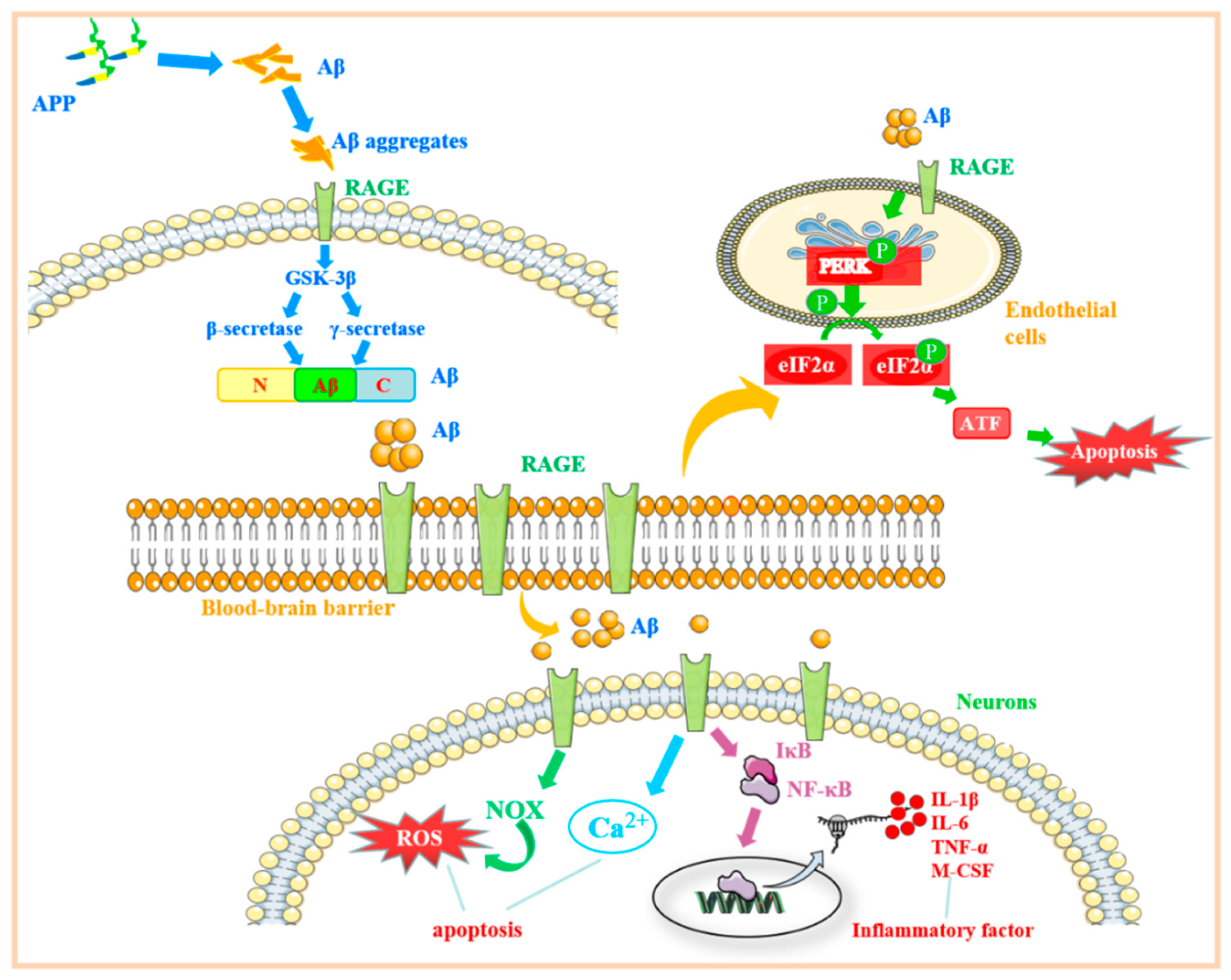
3.2. RAGE Regulates the Mechanisms Associated with Endoplasmic Reticulum Stress and Its Role in Alzheimer’s Disease

Under normal circumstances, the blood–brain barrier (BBB) can play a protective role and prevent Aβ in the blood from entering the central nervous system [15][18]. When AD occurs, the expression of RAGE on cerebral microvessels increases significantly. After injecting Aβ into the blood of AD model mice, it was found that Aβ was transported to brain tissue in a process mediated by RAGE. Following anti-RAGE action, the transport process was blocked. Through an LG-RAGE particular antibody blocking test and ap radiotracer assay, it was ultimately confirmed that Aβ penetrated the BBB in a process mediated by RAGE, a specific receptor on the vascular wall [43]. It was found that the potential mechanism of AD blood–brain barrier destruction is the activation of endoplasmic reticulum stress (ERS) in a dose-dependent manner [16].
The endoplasmic reticulum is an important location of protein processing in cells and plays a vital role in the synthesis, folding, assembly, and modification of soluble proteins and membrane proteins. When exogenous harmful substances disrupt the normal folding reaction of proteins, many abnormal proteins accumulate in the endoplasmic reticulum, triggering ERS.
Some studies have revealed that the RAGE-related signaling pathway can activate the protein kinase R-like endoplasmic reticulum kinase (PERK) pathway in ERS to control the apoptosis of human endothelial progenitor cells and affect the pathogenesis of cardiovascular disease [44]. PERK is a type I transmembrane protein that exists in the endoplasmic reticulum. It can phosphorylate the alpha subunit of eukaryotic translation initiation factor 2α (eIF2α) protein kinase. When unfolded or misfolded, proteins accumulate in the cell, and the immunoglobulin heavy-chain binding protein (BiP) is released from PERK and phosphorylates eIF2α. After phosphorylation, eIF2α can induce the expression of activating transcription factor 4 (ATF4), which activates the expression of unfolded protein response (UPR) target genes and promotes apoptosis [17]. Chen also studied the potential mechanism of the destruction of the blood–brain barrier in Alzheimer’s disease and found that Aβ42-induced RAGE activates ERS in a dose-dependent manner [15]. When cells were transfected with RAGE-siRNA, the endoplasmic reticulum stress markers decreased under the induction of Aβ42. In summary, Aβ can activate endothelial cell ERS through RAGE and increase the permeability of the blood–brain barrier. Therefore, the concentration of Aβ in the central nervous system increases, aggravating the AD condition (Figure 4).

3.3. Regulation of RAGE-Related Signaling Pathway by S100B Protein and Its Role in Alzheimer’s Disease

S100B is an essential member of the S100 protein family and is highly expressed in astrocytes. Under the induction of some stimuli, astrocytes can release a certain amount of S100B [19]. Through immunoblotting, EMSA ultrasonography, and reverse transcriptase-polymerase chain reaction techniques, experiments have proven that micromolar S100B can stimulate c-Jun N-terminal kinase (cJNK) phosphorylation by binding to RAGE and activating nuclear AP-1/cJun in cultured human neural stem cell transcription. In addition, Western blot, siRNA, and immunofluorescence analyses have shown that phosphorylated cJNK induced by S100B can promote the formation of NFTs in AD.
Recent studies have revealed that S100B can protect LAN-5 nerve cells at the nanomolecular level by upregulating antiapoptotic factor Bcl-2 and inhibiting the neurotoxic Aβ25–35 peptide, which can reduce the level of Bcl-2 [45]. At this concentration, S100B binds to RAGE and activates the downstream MEK-ERK1/2 pathway to stimulate the expression of survival-related genes and produce an antiapoptosis effect. After adding the MEK inhibitor, the ability of S100B to upregulate the expression of Bcl-2 was inhibited, which further confirmed the above pathway. At high concentrations, for example, due to astrocyte death, damaged astrocyte leakage, and protein clearance defects, when the intercellular concentration of S100B is expected to be higher than that of 500 nM, S100B not only does not protect LAN-5 cells from neurotoxicity from Aβ 25–35 peptide but also exerts neurotoxicity itself, which could further increase the neurotoxicity of Aβ 25–35 peptide. Furthermore, S100B was found to overactivate RAGE and its downstream ERK1/2 pathway, causing oxidative stress and inducing neuronal death. In summary, varying concentrations of S100B combined with RAGE play an opposite role in AD by activating different signal pathways. A low concentration of S100B can protect nerve cells, whereas low and high concentrations of S100B result in neurotoxicity.

3.4. The Mechanism of the RAGE-Related Pathway in AD

In addition to its neurotoxicity, which can activate inflammatory factors in nerve cells through RAGE and induce nerve cell apoptosis, Aβ can also combine with RAGE on the surface of endothelial cells to cause endoplasmic reticulum stress, which pathologically increases the permeability of the blood–brain barrier, increases the concentration of Aβ in the central nervous system, and further aggravates the injury of nerve cells. In addition, a high concentration of S100B was found to produce certain neurotoxicity in combination with RAGE. The combination of Aβ and S100B with RAGE shows that RAGE plays an important role in the occurrence and development of AD.

References

  1. Oh, S.; Son, M.; Choi, J.; Lee, S.; Byun, K. sRAGE prolonged stem cell survival and suppressed RAGE-related inflammatory cell and T lymphocyte accumulations in an Alzheimer’s disease model. Biochem. Biophys. Res. Commun. 2018, 495, 807–813.
  2. Li, S.T.; Dai, Q.; Zhang, S.X.; Liu, Y.J.; Yu, Q.Q.; Tan, F.; Lu, S.H.; Wang, Q.; Chen, J.W.; Huang, H.Q.; et al. Ulinastatin attenuates LPS-induced inflammation in mouse macrophage RAW264.7 cells by inhibiting the JNK/NF-kappaB signaling pathway and activating the PI3K/Akt/Nrf2 pathway. Acta Pharm. Sin. 2018, 39, 1294–1304.
  3. Hudson, B.I.; Lippman, M.E. Targeting RAGE Signaling in Inflammatory Disease. Annu. Rev. Med. 2018, 69, 349–364.
  4. Ahmad, S.; Khan, M.Y.; Rafi, Z.; Khan, H.; Siddiqui, Z.; Rehman, S.; Shahab, U.; Khan, M.S.; Saeed, M.; Alouffi, S.; et al. Oxidation, glycation and glycoxidation-The vicious cycle and lung cancer. Semin. Cancer Biol. 2018, 49, 29–36.
  5. Laczko, R.; Chang, A.; Watanabe, L.; Petelo, M.; Kahaleua, K.; Bingham, J.P.; Csiszar, K. Anti-inflammatory activities of Waltheria indica extracts by modulating expression of IL-1B, TNF-alpha, TNFRII and NF-kappaB in human macrophages. Inflammopharmacology 2020, 28, 525–540.
  6. Gao, X.; Zhang, H.; Schmidt, A.M.; Zhang, C. AGE/RAGE produces endothelial dysfunction in coronary arterioles in type 2 diabetic mice. Am. J. Physiol. Heart Circ. Physiol. 2008, 295, H491–H498.
  7. Maillard-Lefebvre, H.; Boulanger, E.; Daroux, M.; Gaxatte, C.; Hudson, B.I.; Lambert, M. Soluble receptor for advanced glycation end products: A new biomarker in diagnosis and prognosis of chronic inflammatory diseases. Rheumatology 2009, 48, 1190–1196.
  8. Martens, H.A.; Nienhuis, H.L.; Gross, S.; van der Steege, G.; Brouwer, E.; Berden, J.H.; de Sevaux, R.G.; Derksen, R.H.; Voskuyl, A.E.; Berger, S.P.; et al. Receptor for advanced glycation end products (RAGE) polymorphisms are associated with systemic lupus erythematosus and disease severity in lupus nephritis. Lupus 2012, 21, 959–968.
  9. Cerezo, L.A.; Remakova, M.; Tomcik, M.; Gay, S.; Neidhart, M.; Lukanidin, E.; Pavelka, K.; Grigorian, M.; Vencovsky, J.; Senolt, L. The metastasis-associated protein S100A4 promotes the inflammatory response of mononuclear cells via the TLR4 signalling pathway in rheumatoid arthritis. Rheumatology 2014, 53, 1520–1526.
  10. Lu, C.; Liu, J.; Yao, M.; Li, L.; Li, G. Downregulation of S100 calcium binding protein A12 inhibits the growth of glioma cells. BMC Cancer 2020, 20, 261.
  11. Xu, X.; Chen, H.; Zhu, X.; Ma, Y.; Liu, Q.; Xue, Y.; Chu, H.; Wu, W.; Wang, J.; Zou, H. S100A9 promotes human lung fibroblast cells activation through receptor for advanced glycation end-product-mediated extracellular-regulated kinase 1/2, mitogen-activated protein-kinase and nuclear factor-kappaB-dependent pathways. Clin. Exp. Immunol. 2013, 173, 523–535.
  12. Chakraborty, S.; ThimmaReddygari, J.; Selvaraj, D. G-Lymphatic, Vascular and Immune Pathways for Abeta Clearance Cascade and Therapeutic Targets For Alzheimer’s Disease. Comb. Chem. High Throughput Screen 2021, 24, 1083–1092.
  13. Da Costa Dias, B.; Jovanovic, K.; Gonsalves, D.; Weiss, S.F. Structural and mechanistic commonalities of amyloid-beta and the prion protein. Prion 2011, 5, 126–137.
  14. Cuevas, E.; Lantz, S.M.; Tobon-Velasco, J.C.; Newport, G.D.; Wu, Q.; Virmani, A.; Ali, S.F.; Santamaria, A. On the in vivo early toxic properties of A-beta 25–35 peptide in the rat hippocampus: Involvement of the Receptor-for-Advanced Glycation-End-Products and changes in gene expression. Neurotoxicol. Teratol. 2011, 33, 288–296.
  15. Chen, W.; Chan, Y.; Wan, W.; Li, Y.; Zhang, C. Abeta1–42 induces cell damage via RAGE-dependent endoplasmic reticulum stress in bEnd.3 cells. Exp. Cell Res. 2018, 362, 83–89.
  16. Cai, Y.; Arikkath, J.; Yang, L.; Guo, M.L.; Periyasamy, P.; Buch, S. Interplay of endoplasmic reticulum stress and autophagy in neurodegenerative disorders. Autophagy 2016, 12, 225–244.
  17. Bettigole, S.E.; Glimcher, L.H. Endoplasmic reticulum stress in immunity. Annu. Rev. Immunol. 2015, 33, 107–138.
  18. Nation, D.A.; Sweeney, M.D.; Montagne, A.; Sagare, A.P.; D’Orazio, L.M.; Pachicano, M.; Sepehrband, F.; Nelson, A.R.; Buennagel, D.P.; Harrington, M.G.; et al. Blood-brain barrier breakdown is an early biomarker of human cognitive dysfunction. Nat. Med. 2019, 25, 270–276.
  19. Prasad, K. AGE-RAGE stress: A changing landscape in pathology and treatment of Alzheimer’s disease. Mol. Cell Biochem. 2019, 459, 95–112.
  20. Narvaez, J. Systemic lupus erythematosus 2020. Med. Clin. 2020, 155, 494–501.
  21. Wang, X.; Xiang, L.; Li, H.; Chen, P.; Feng, Y.; Zhang, J.; Yang, N.; Li, F.; Wang, Y.; Zhang, Q.; et al. The Role of HMGB1 Signaling Pathway in the Development and Progression of Hepatocellular Carcinoma: A Review. Int. J. Mol. Sci. 2015, 16, 22527–22540.
  22. Pisetsky, D.S. The complex role of DNA, histones and HMGB1 in the pathogenesis of SLE. Autoimmunity 2014, 47, 487–493.
  23. Li, J.; Xie, H.; Wen, T.; Liu, H.; Zhu, W.; Chen, X. Expression of high mobility group box chromosomal protein 1 and its modulating effects on downstream cytokines in systemic lupus erythematosus. J. Rheumatol. 2010, 37, 766–775.
  24. Yan, L.; Liang, M.; Yang, T.; Ji, J.; Jose Kumar Sreena, G.S.; Hou, X.; Cao, M.; Feng, Z. The Immunoregulatory Role of Myeloid-Derived Suppressor Cells in the Pathogenesis of Rheumatoid Arthritis. Front. Immunol. 2020, 11, 568362.
  25. Park, S.Y.; Lee, S.W.; Kim, H.Y.; Lee, W.S.; Hong, K.W.; Kim, C.D. HMGB1 induces angiogenesis in rheumatoid arthritis via HIF-1alpha activation. Eur. J. Immunol. 2015, 45, 1216–1227.
  26. Rojas, A.; Delgado-Lopez, F.; Perez-Castro, R.; Gonzalez, I.; Romero, J.; Rojas, I.; Araya, P.; Anazco, C.; Morales, E.; Llanos, J. HMGB1 enhances the protumoral activities of M2 macrophages by a RAGE-dependent mechanism. Tumour Biol. 2016, 37, 3321–3329.
  27. Pullerits, R.; Bokarewa, M.; Dahlberg, L.; Tarkowski, A. Decreased levels of soluble receptor for advanced glycation end products in patients with rheumatoid arthritis indicating deficient inflammatory control. Arthritis Res. Ther. 2005, 7, R817–R824.
  28. Chen, Y.S.; Yan, W.; Geczy, C.L.; Brown, M.A.; Thomas, R. Serum levels of soluble receptor for advanced glycation end products and of S100 proteins are associated with inflammatory, autoantibody, and classical risk markers of joint and vascular damage in rheumatoid arthritis. Arthritis Res. Ther. 2009, 11, R39.
  29. Myles, A.; Viswanath, V.; Singh, Y.P.; Aggarwal, A. Soluble receptor for advanced glycation endproducts is decreased in patients with juvenile idiopathic arthritis (ERA category) and inversely correlates with disease activity and S100A12 levels. J. Rheumatol. 2011, 38, 1994–1999.
  30. Knani, I.; Bouzidi, H.; Zrour, S.; Bergaoui, N.; Hammami, M.; Kerkeni, M. Methylglyoxal: A Relevant Marker of Disease Activity in Patients with Rheumatoid Arthritis. Dis. Markers 2018, 2018, 8735926.
  31. Jafari Nakhjavani, M.R.; Jafarpour, M.; Ghorbanihaghjo, A.; Abedi Azar, S.; Malek Mahdavi, A. Relationship between serum-soluble receptor for advanced glycation end products (sRAGE) and disease activity in rheumatoid arthritis patients. Mod. Rheumatol. 2019, 29, 943–948.
  32. Inui, N.; Sakai, S.; Kitagawa, M. Molecular Pathogenesis of Pulmonary Fibrosis, with Focus on Pathways Related to TGF-beta and the Ubiquitin-Proteasome Pathway. Int. J. Mol. Sci. 2021, 22, 6107.
  33. Brett, J.; Schmidt, A.M.; Yan, S.D.; Zou, Y.S.; Weidman, E.; Pinsky, D.; Nowygrod, R.; Neeper, M.; Przysiecki, C.; Shaw, A.; et al. Survey of the distribution of a newly characterized receptor for advanced glycation end products in tissues. Am. J. Pathol. 1993, 143, 1699–1712.
  34. Oldfield, M.D.; Bach, L.A.; Forbes, J.M.; Nikolic-Paterson, D.; McRobert, A.; Thallas, V.; Atkins, R.C.; Osicka, T.; Jerums, G.; Cooper, M.E. Advanced glycation end products cause epithelial-myofibroblast transdifferentiation via the receptor for advanced glycation end products (RAGE). J. Clin. Investig. 2001, 108, 1853–1863.
  35. Yamaguchi, K.; Iwamoto, H.; Horimasu, Y.; Ohshimo, S.; Fujitaka, K.; Hamada, H.; Mazur, W.; Kohno, N.; Hattori, N. AGER gene polymorphisms and soluble receptor for advanced glycation end product in patients with idiopathic pulmonary fibrosis. Respirology 2017, 22, 965–971.
  36. Grauen Larsen, H.; Marinkovic, G.; Nilsson, P.M.; Nilsson, J.; Engstrom, G.; Melander, O.; Orho-Melander, M.; Schiopu, A. High Plasma sRAGE (Soluble Receptor for Advanced Glycation End Products) Is Associated With Slower Carotid Intima-Media Thickness Progression and Lower Risk for First-Time Coronary Events and Mortality. Arter. Thromb. Vasc. Biol. 2019, 39, 925–933.
  37. Wittkowski, H.; Sturrock, A.; van Zoelen, M.A.; Viemann, D.; van der Poll, T.; Hoidal, J.R.; Roth, J.; Foell, D. Neutrophil-derived S100A12 in acute lung injury and respiratory distress syndrome. Crit. Care Med. 2007, 35, 1369–1375.
  38. Mulrennan, S.; Baltic, S.; Aggarwal, S.; Wood, J.; Miranda, A.; Frost, F.; Kaye, J.; Thompson, P.J. The role of receptor for advanced glycation end products in airway inflammation in CF and CF related diabetes. Sci. Rep. 2015, 5, 8931.
  39. Ray, B.; Maloney, B.; Sambamurti, K.; Karnati, H.K.; Nelson, P.T.; Greig, N.H.; Lahiri, D.K. Rivastigmine modifies the alpha-secretase pathway and potentially early Alzheimer’s disease. Transl. Psychiatry 2020, 10, 47.
  40. Busche, M.A.; Hyman, B.T. Synergy between amyloid-beta and tau in Alzheimer’s disease. Nat. Neurosci. 2020, 23, 1183–1193.
  41. Nan, K.; Han, Y.; Fang, Q.; Huang, C.; Yu, L.; Ge, W.; Xiang, F.; Tao, Y.X.; Cao, H.; Li, J. HMGB1 gene silencing inhibits neuroinflammation via down-regulation of NF-kappaB signaling in primary hippocampal neurons induced by Abeta25–35. Int. Immunopharmacol. 2019, 67, 294–301.
  42. Esposito, G.; Scuderi, C.; Lu, J.; Savani, C.; De Filippis, D.; Iuvone, T.; Steardo, L., Jr.; Sheen, V.; Steardo, L. S100B induces tau protein hyperphosphorylation via Dickopff-1 up-regulation and disrupts the Wnt pathway in human neural stem cells. J. Cell Mol. Med. 2008, 12, 914–927.
  43. Deane, R.; Du Yan, S.; Submamaryan, R.K.; LaRue, B.; Jovanovic, S.; Hogg, E.; Welch, D.; Manness, L.; Lin, C.; Yu, J.; et al. RAGE mediates amyloid-beta peptide transport across the blood-brain barrier and accumulation in brain. Nat. Med. 2003, 9, 907–913.
  44. Huang, Q.; Yang, Z.; Zhou, J.P.; Luo, Y. HMGB1 induces endothelial progenitor cells apoptosis via RAGE-dependent PERK/eIF2alpha pathway. Mol. Cell Biochem. 2017, 431, 67–74.
  45. Businaro, R.; Leone, S.; Fabrizi, C.; Sorci, G.; Donato, R.; Lauro, G.M.; Fumagalli, L. S100B protects LAN-5 neuroblastoma cells against Abeta amyloid-induced neurotoxicity via RAGE engagement at low doses but increases Abeta amyloid neurotoxicity at high doses. J. Neurosci. Res. 2006, 83, 897–906.
More
Information
Contributors MDPI registered users' name will be linked to their SciProfiles pages. To register with us, please refer to https://encyclopedia.pub/register : , , , , ,
View Times: 690
Revisions: 3 times (View History)
Update Date: 25 Aug 2022
1000/1000