Submitted Successfully!
To reward your contribution, here is a gift for you: A free trial for our video production service.
Thank you for your contribution! You can also upload a video entry or images related to this topic.
Version Summary Created by Modification Content Size Created at Operation
1 -- 7398 2022-08-11 09:57:52 |
2 format correct -4573 word(s) 2825 2022-08-12 03:32:30 |

Video Upload Options

Do you have a full video?

Confirm

Are you sure to Delete?
Cite
If you have any further questions, please contact Encyclopedia Editorial Office.
Akinyemi, K.O.;  Fakorede, C.O.;  Amisu, K.O.;  Wareth, G. Human and Animal Brucellosis in Nigeria. Encyclopedia. Available online: https://encyclopedia.pub/entry/26079 (accessed on 29 March 2024).
Akinyemi KO,  Fakorede CO,  Amisu KO,  Wareth G. Human and Animal Brucellosis in Nigeria. Encyclopedia. Available at: https://encyclopedia.pub/entry/26079. Accessed March 29, 2024.
Akinyemi, Kabiru O., Christopher O. Fakorede, Kehinde O. Amisu, Gamal Wareth. "Human and Animal Brucellosis in Nigeria" Encyclopedia, https://encyclopedia.pub/entry/26079 (accessed March 29, 2024).
Akinyemi, K.O.,  Fakorede, C.O.,  Amisu, K.O., & Wareth, G. (2022, August 11). Human and Animal Brucellosis in Nigeria. In Encyclopedia. https://encyclopedia.pub/entry/26079
Akinyemi, Kabiru O., et al. "Human and Animal Brucellosis in Nigeria." Encyclopedia. Web. 11 August, 2022.
Human and Animal Brucellosis in Nigeria
Edit

Brucellosis caused by Brucella spp. is transmitted by direct or indirect contact with infected animals or their secretions and through the consumption of infected animal meat and unpasteurized milk/milk products. Brucellosis is classified as one of the top neglected zoonosis by the World Health Organization (WHO), and despite this, it does not attract the appropriate attention it requires from both the Federal and State ministries of health in Nigeria. 15.8% (7178/45,363) seroprevalence of brucellosis was recorded in northern Nigeria as against 8.7% (1902/21,740) in the southern part. Brucella abortus, B. melitensis, B. suis, and B. canis were reported in 27 of the 36 states.

human and animal brucellosis Brucella Nigeria prevalence and seroprevalence systemic review and meta-analysis

1. Introduction

Brucellosis, which is also referred to as the travel-related infectious disease “undulant fever”, “Mediterranean fever”, “gastric remittent fever”, or “Malta fever”, is a zoonotic disease caused by intracellular Gram-negative coccobacilli bacteria of the genus Brucella [1][2][3]. The disease is distributed globally, affecting humans, a wide range of wild animals, and economically viable domestic livestock such as cattle, goats, sheep, donkeys, camels, swine, dogs, etc. [4]. Currently, 12 species of the genus Brucella are accepted; however, only B. melitensisB. abortusB. suis, and in rare cases, B. canis are characterized as human pathogens. The global burden of human brucellosis remains very large. The organism causes more than 500,000 infections per year worldwide [3]. The socio-economic impact of brucellosis is enormous and is higher in developing countries than the developed countries, with an estimated 3.5 billion individuals at risk of being infected with one or more Brucella spp. and a high morbidity rate in both humans and animals [5][6]. Brucellosis is highly contagious. It is transmitted by direct or indirect contact with infected animals or their secretions and through the consumption of infected animal meat and products such as unpasteurized milk/milk products [1][7]. The risk of acquiring the disease has been attributed to a certain occupation (occupational hazard) but most especially among livestock caregivers. People who work with animals and are constantly in contact with blood, placenta, foetuses, and uterine secretions have an increased risk of contracting the disease. This method of transmission primarily affects farmers, butchers, hunters, veterinarians, and laboratory personnel [4]. However, in endemic areas, human brucellosis has serious public health consequences. The organism can enter the human body through breaks in the skin, mucous membranes, conjunctiva, respiratory and gastrointestinal tracts, resulting in systemic infection with acute and chronic phases [3][8]. The disease may persist as relapse, chronic localized infection, or delayed convalescence. Symptoms of the disease include but are not limited to fever or chill, arthralgia or arthritis, sweating, hepatomegaly, splenomegaly, anorexia, asthenia, fatigue, weakness, pallor, diarrhea, jaundice, lymphadenopathy, rash, and malaise [1][8]. Urbanization and the subsequent expansion of animal industries, coupled with a lack of good hygienic practices, especially in animal husbandry and food handling, partly account for brucellosis remaining a public health problem [1]. It affects people of all races, age groups, and both sexes. Although many countries have made great progress in controlling the disease, Brucella infection persists in domestic animals in some regions, and consequently, transmission to the human population is imminent. Brucellosis in livestock is mostly a reproductive disease characterized by infertility, late abortion, retained foetal membranes, and impaired productivity [9]. Brucellosis infects many species, especially cattle, sheep, goats, and pigs. Different Brucella types infect different species preferentially. The disease is still widely distributed in Africa, especially in areas with large animal populations [10].
Brucellosis, currently classified as one of the top neglected zoonosis by the World Health Organization (WHO), does not attract the appropriate attention it requires from both Federal and State ministries of health in Nigeria. According to the United Nations Department of Economic and Social Affairs, Population Division 2019 [11], Nigeria is the most populous country in Africa and ranked 7th in the world with over 201 million people. A projection of its population is expected to rise to about 401 million people by 2050, with an estimated livestock population of 20.49 million cattle, 23.07 million sheep, 28.07 million goats, 6.54 million pigs, 18,200–90,000 camels, and 210,000 horses [12][13]. Livestock slaughtered at the different abattoirs in Nigeria for human consumption are not usually screened for brucellosis. Free-range domestic animals are also commonplace in Nigeria; pet animals, i.e., dogs, cats, and livestock, i.e., goats, sheep, and cattle, move freely amongst the people. The incidence of brucellosis in Nigeria is under-reported. Currently, there is insufficient epidemiological data on Nigeria’s prevalence and distribution of human and animal brucellosis. This entry on reported cases of brucellosis in Nigeria was undertaken to determine the true prevalence and distribution of the disease across the country and to make valuable suggestions that will serve as a guide for the implementation of measures for sustainable management of this disease in Nigeria.

2. Human and Animal Brucellosis in Nigeria

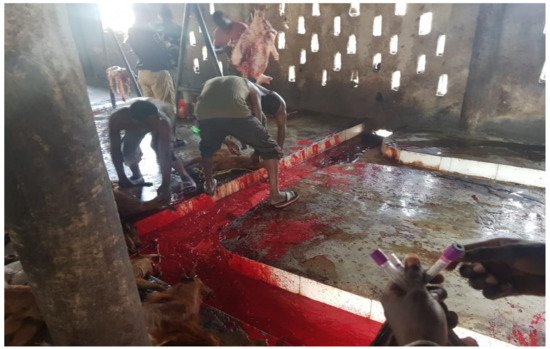
The economic burden in the livestock industry arising from brucellosis and its morbidity in humans has made this zoonotic disease a global public health challenge [14]. The main objective of this entry was to systematically review the literature reporting brucellosis and perform a meta-analysis to estimate the national prevalence of brucellosis in Nigeria. Of the 99 publications accessed, 14 publications reported Brucella infection in humans from 11 states of the 36 states in Nigeria, including the federal capital territory. The remaining 25 states either do not have reports because of the lack of interest in brucellosis, or the reported cases do not meet the inclusion criteria of this entry. Most of the studies on human brucellosis were from high-risk occupational groups, especially abattoir workers. The national prevalence of human brucellosis in Nigeria for all methods revealed in this entry was 17.6%. However, based on the Rose Bengal plate test (RBPT), as reported by about 80% of the publications accessed for human brucellosis, the seroprevalence of the disease in Nigeria was 15.7% (493/3144). This is similar to the 15.8% recorded in Cameroon [15], but lower when compared to the sub-national study in other sub-Saharan African countries, such as the 44% prevalence recorded in Kenya [16], 31.5% prevalence in Ethiopia [17] and 17% in Uganda [18]. Similarly, the prevalence of human brucellosis is also higher in northern African countries such as Egypt, with a prevalence of 23.9% [19]. The prevalence is higher in Nigeria compared to the prevalence of 1.41% reported in Tanzania [20] and the 3.0% national prevalence in Kenya [21]. The high prevalence of human brucellosis from this entry has demonstrated the fact that brucellosis is endemic with a high burden in Nigeria and requires the attention of policymakers and stakeholders in the health sector. The high burden of human brucellosis in Nigeria, as seen in this entry, can be attributed to several factors, which include nomadic pastoralists who run open grazing, abattoir workers, especially those slaughtering animals who are in constant contact with animal blood without personal protective equipment (PPE) (Figure 1), and the love of Nigerians for the consumption of animals’ intestinal parts, which are the most likely source of zoonotic transmission of the disease in Nigeria. Unfortunately, the animals that are ready for slaughter in Nigeria’s abattoirs are sometimes not screened for brucellosis. The spectrum of clinical presentation of human brucellosis, which mimics several other febrile illnesses such as rheumatic fever, typhoid, and malaria, has resulted in several misdiagnoses of this disease since malaria and typhoid fever are also endemic in Nigeria. Additionally, brucellosis is not routinely screened in private and public health care facilities, which obscures the detection and true prevalence of the disease.
Figure 1. Typical abattoir in Nigeria showing butcher slaughtering animals without personal protective equipment.
Although the prevalence of the brucellosis reported in this entry was based on serological investigation, no publication reported human brucellosis based on culture and molecular methods. Nevertheless, the overall prevalence of brucellosis was higher at 15.6% (7178/46,022) in Northern Nigeria than in the Southern part at 8.7% (1902/21,740). The differences in the prevalence rate may be due to the number of research articles reporting brucellosis in Northern Nigeria being higher when compared to the reportage in the Southern part of Nigeria. Moreover, easy access to and frequency of contact of abattoir workers with animals, e.g., cattle, donkeys, horses, goats, and sheep and animal products, as well as consumption of raw and untreated fermented animal products, especially milk (nono), a habit that is very common in northern Nigeria, contribute to the prevalence rates. Furthermore, the uncontrolled movement of nomadic Fulani (herdsmen) from northern Nigeria has contributed significantly to the spread of the disease to other parts of the country, a fact that has long been documented by other authors [22][23].
The burden of brucellosis in animals, as observed in this entry, was highest in pigs, at 28.3%, compared to in other animals such as sheep, at 23.3% and camels, at 20.9%. The lowest prevalence was among chickens, with an 8.4% prevalence recorded. The higher prevalence of the disease in pigs could be attributed to multiple Brucella spp. detected from the samples investigated in the SS as reported by Bello-Onaghise et al. [24] based on the culture method. From 30 blood samples, 81 Brucella isolates were documented (24 B. abortus, 27 B. melitensis, and 30 B. suis), as well as 68 isolates (19 B. abortus, 24 B. melitensis, and 25 B. suis) from 25 vaginal swabs, giving an overall prevalence of 100% (55/55) as reported in that study. Although the report of co-infection among different Brucella species is very rare in literature, only Bello-Onaghise et al. [24] have been able to document this in Nigeria to the best of researchers' knowledge. In this entry, the occurrence of brucellosis in animals varied from region to region and animal to animal. Interestingly, despite the higher occurrence of the disease in Northern Nigeria, the SS geopolitical zone of Nigeria reported the highest prevalence of brucellosis at 48.6% (206/424) in animals from four publications. The lowest occurrence was observed in the SE, with a prevalence of 3.9%, while the SW was 7%. Other studies included the NC at 16.6%, the NE at 18%, and the NW at 14.2%. This entry also showed evidence of geographical variations in the prevalence of brucellosis not only in the sources of samples but also in the types of samples evaluated. Investigation of brucellosis from different samples across the country revealed the prevalence of the disease estimated to range from 5% in the aborted foetus to 50% in hygroma fluid. Bloodstream infection with brucellosis was estimated at 13.5%, and its prevalence in milk was estimated at 10.2%. The prevalence of brucellosis from vaginal swabs obtained from different animals was 12.4%, while a prevalence of 29.4% was observed in the lymph node of cattle from NW Nigeria. Thus far, only 7 out of the 104 publications reported culture detection methods [24][25][26][27][28][29][30], while a single publication reported molecular methods [31]. The Rose Bengal rapid test is the most popular diagnostic method reported by 11/14 publications on the detection of human brucellosis, while 95/107 publications reported RBPT as the detection method in animal samples. Several publications also reported other diagnostic methods across the country. Although in this entry, it was observed that several publications reported funding from donors, grants, and/or direct receipt of diagnostic kits from some laboratories for their studies, unfortunately, researchers were unable to determine if the donors or the providers of those kits have any influence on the outcomes of their studies.
Four different Brucella species were recorded, including B. abortusB. melitensisB. suis, and B. canis. However, B. abortus biovar 1 has been documented as the most frequently encountered brucellosis in cattle globally [32][33] , while biovar 3 has been reported to be a common biovar in Côte d’Ivoire [34] and Nigeria [27]B. melitensis was mostly reported in humans, and B. abortus with biovars 1 and 3 was frequently detected in animals from Nigeria [27][28]B. melitensis was mostly encountered in sheep, goats, and pigs, with few reports in cattle. This agreed with a study conducted in China by Liu et al. [35] in which B. melitensis infection was common in sheep and goats. Meanwhile, B. suis was observed only in sheep, goats, and pigs. This entry also revealed the detection of B. canis only in dogs in Nigeria. In Nigeria, a similar distribution pattern of the biodiversity of brucellosis in the different regions occurred. From the Northern to the Southern region, B. abortus is the most encountered species in animals, followed by B. melitensis. Only the NC and the SS reported B. suis, while 99% of the B canis detected in dogs were observed in the SE, with only 1% from SW Nigeria. Brucellosis in Nigeria, as seen in this entry, is spread across the country and has been responsible for reproductive disease in animals associated with abortion, stillbirth, death of young animals, placenta previa, the birth of immature calves, delayed calving, male infertility, and heavy reductions in milk output.
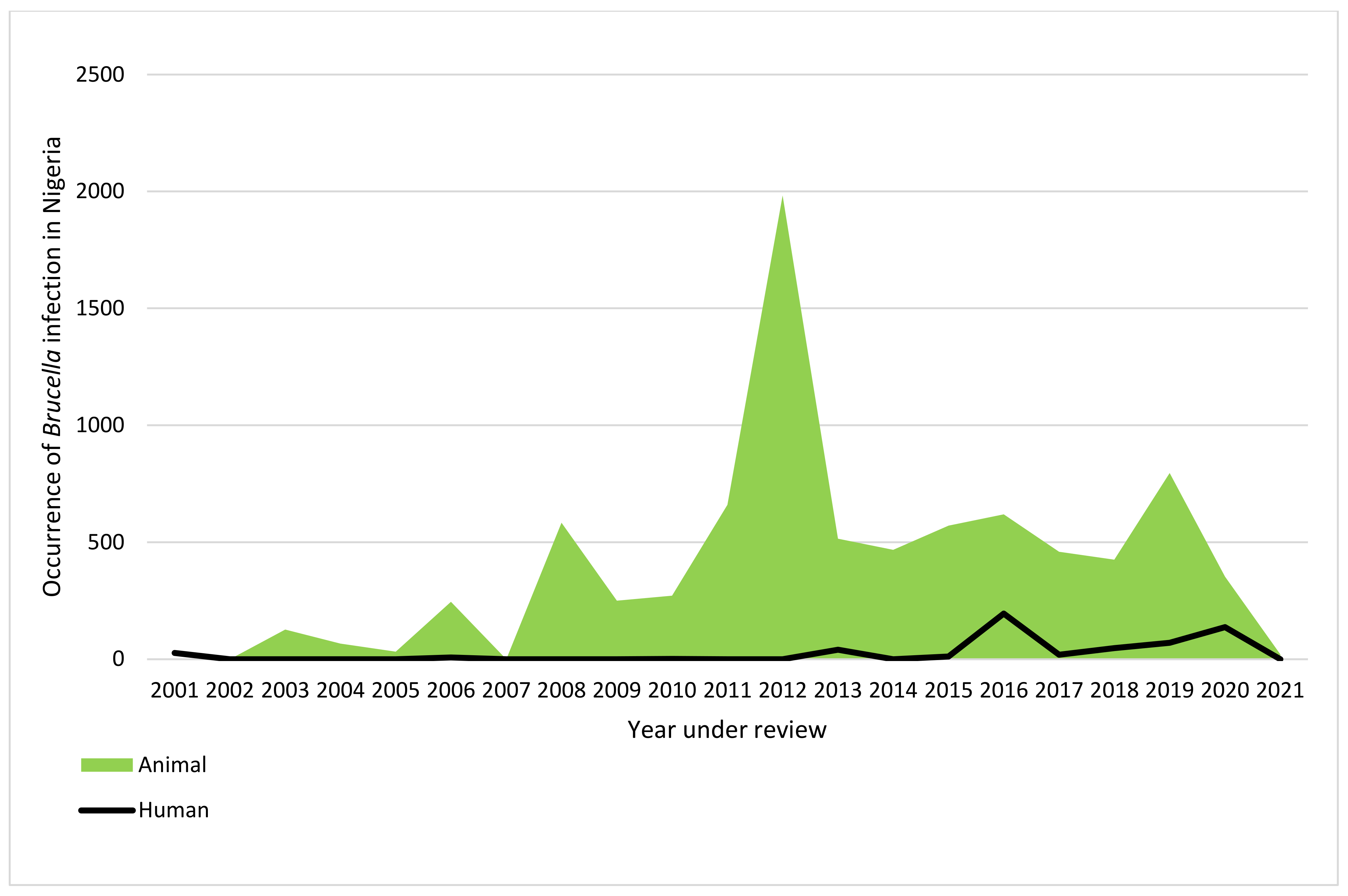
Most phenotypic detection methods of Brucella consist of bacteriological isolation and biochemical identification, which solely relies on a combination of some investigating parameters to characterize suspicious colonies [36]. Unfortunately, this typing method often fails to correctly differentiate some strains, which was seen in the case of the strains reported in Plateau State by Bertu et al. [27]. Thus, there is a need for other combined identification methods, such as the use of molecular tools to characterize Brucella species with good discriminatory capacity and the potential to evaluate relationships between species, such as the multiple-locus variable of tandem repeat analysis (MLVA) [37][38]. Future studies on brucellosis should also focus more on the proper use and interpretation of diagnostic testing for animals as recommended by the WHO, FAO, and OIE [1][39][40]. There was stability in the trend of human brucellosis from 2001 to 2012. The trend began to fluctuate from 2013 to 2020, with a decline in 2021. The observed decline in human Brucella infection may be connected to the improved abattoir environment in some states in Nigeria when compared to previous years. The peak of human Brucella infection in Nigeria was observed in 2016 from SE Nigeria [41] (Table 1 and Figure 2). There was a fluctuation in the trend of the disease in animals from 2003 to 2021, with a remarkable increase in the trend of the disease from 2008 to 2019 and a sharp decline by 2020. The peak of the disease in animals was observed in 2012, though there was no report on the outbreak of the disease in Nigeria in 2012. Furthermore, the number of articles reporting Brucella infection in 2012 was also lower than in some of the years under review, while the number of animals screened for brucellosis in 2012 was also lower than the number of samples screened for some years under review. Hence, the increase cannot be attributed to sampling size. Despite the problem of antibiotic resistance being on the rise in brucellae [8], unfortunately, there was no single report on antimicrobial susceptibility testing on the Brucella species isolated from both humans and animals, thus making it difficult to determine the pattern of resistance of Brucella spp. in Nigeria. The use of quality molecular tools in detecting Brucella infection in Nigeria is also lacking and has contributed to many reported un-typed Brucella species. There is a need to focus more on culture and molecular methods to determine the epidemiological link between Brucella species and its biodiversity from humans and animals, to know the prevailing biovars, and for prompt interventions. The findings from this entry would serve as baseline information on the national prevalence of Brucella infection in Nigeria. The generated data would help estimate the true burden of Brucella infections in Nigeria. As it is today, national data on human and animal Brucella infection are lacking in both State and Federal Ministries.

Limitations: Researchers' inability to determine the prevalence of the disease based on certain variables such as age, sex, cattle herds, breeds of animals, and mortality rate both in humans and animals are part of the limitations of this entry. The use of serological methods, as seen in this entry, is of national importance because of their usefulness in detecting antibodies against Brucella species. However, the results are only reliable when the diagnostic methods and procedures are properly conducted and the outcomes are well interpreted. Interestingly, serological methods have several shortcomings, including the antisera’s ability to cross-react with other bacterial pathogens such as Escherichia coli 0157, Salmonella Urbana group N, Francisella tularensis, and Yersinia enterocolitica 0:9 [130].

Conclusions: This entry revealed a national prevalence of 19.2% of human Brucella infection and 13.1% prevalence of Brucella infection in common domestic animals, respectively, from 2001 to 2021. This entry also indicated that only 27 states, including the FCT, reported brucellosis in Nigeria across the six geopolitical zones. Researchers' study also revealed the highest occurrence of brucellosis is in Northern Nigeria. Four Brucella species, B. abortus, B. melitensis, B. suis, and B. canis, have been reported in Nigeria. Culture and molecular methods of detection of brucellosis and reports of antimicrobial susceptibility testing remain a conjecture. This entry will help researchers redirect their research focus and serve as a guide for policymakers on measures for managing brucellosis in Nigeria and other sub-Saharan countries.The need for improved sanitary conditions of the abattoirs, the use of personal protective equipment by animal handlers, vaccination of animals against bovine brucellosis, and ranching of animals to curb the spread of the disease should be paramount to all the stakeholders.

Table 1. Seroprevalence of reported human brucellosis in Nigeria (2001–2021).
Region State No of Samples Tested No of Positive Sample Seroprevalence
%
Detection Methods Brucella Type Reference
RBPT SAT cELISA iELISA IgM IgG IgG/IgM
ELISA
Brucella spp. Brucella abortus Brucella
melitensis
 
NC Kwara 189 42 22.2% 42 0 0 0 0 0 0 42 0 0 [42]
  Abuja 224 40 17.9% 40 0 0 0 22 18 0 40 0 0 [43]
  Nasarawa 160 16 10% 16 0 0 0 0 0 0 16 0 0 [44]
Total 573 98 17.1% 98 0 0 0 22 18 0 98 0 0  
NE Bauchi 285 95 33.3% 95 0 0 0 6 18 0 95 0 0 [45]
  Not indicated 500 26 5.2% 26 0 0 0 0 0 0 26 0 0 [46]
  Borno 106 4 3.8% 4 0 0 0 0 0 0 4 0 0 [47]
  Borno 100 11 11% 11 0 0 0 0 0 0 0 11 0 [48]
Total 990 136 13.7% 136 0 0 0 6 18 0 125 11 0  
NW Kaduna 1 1 100% 0 1 0 0 0 0 0 0 1 0 [49]
  Kaduna 100 19 19% 0 0 0 19 0 0 0 19 0 0 [50]
  Sokoto 137 1 0.7% 1 0 0 0 0 0 0 0 1 0 [51]
Total 238 21 8.8% 1 1 0 19 0 0 0 19 2 0  
SE Enugu 682 195 28.6% 195 0 0 0 0 0 0 195 0 0 [41]
  Total 682 195 28.6% 195 0 0 0 0 0 0 195 0 0  
SS Akwa Ibom 228 70 30.7% 29 0 0 0 0 41 0 0 0 70 [52]
Total 228 70 30.7% 29 0 0 0 0 41 0 0 0 70  
SW Lagos 422 27 6.4% 27 0 0 3 0 0 0 27 0 0 [53]
  Oyo 11 7 63.6% 7 0 0 0 0 0 0 7 0 0 [54]
Total 433 34 7.9% 34 0 0 3 0 0 0 34 0 0  
Grand Total 3144 554 17.6% 493 1 0 22 28 77 0 471 13 70  
NC: North-Central, NE: North-East, NW: North-West, SE: South-East, SS: South-South, SW: South-West, RBPT: Rose Bengal plate test, SAT: serum agglutination test, cELISA: competitive enzyme-linked immunosorbent assay, iELISA: indirect enzyme-linked immunosorbent assay, IgM: immunoglobulin M, IgG: immunoglobulin.
Figure 2. Trend of human and animal brucellosis in Nigeria from 2001 to 2021.

References

  1. Corbel, M.J. Brucellosis in Humans and Animals; World Health Organization: Geneva, Switzerland, 2006.
  2. Ducrotoya, M.; Bertub, W.J.; Matopec, G.; Cadmusd, S.; Conde-Álvareze, R.; Gusib, A.M.; Welburna, S.; Ocholib, R.; Blascof, J.M.; Moriyón, I. Brucellosis in Sub-Saharan Africa: Current challenges for management, diagnosis and control. Acta Tropica. 2017, 165, 179–193.
  3. Bennett, N.J.; Bronze, M.S. Brucellosis. Available online: https://emedicine.medscape.com/article/213430-print (accessed on 20 April 2022).
  4. World Health Organization. Home/Newsroom/Fact sheets/Detail/Brucellosis; World Health Organization: Geneva, Switzerland, 2020.
  5. Rossetti, C.A.; Arenas-Gamboa, A.M.; Maurizio, E. Caprine brucellosis: A historically neglected disease with significant impact on public health. PLOS Negl. Trop. Dis. 2017, 11, e0005692.
  6. Bosilkovski, M.; Keramat, F.; Arapovic, J. The current therapeutic strategies in human brucellosis. Infection 2021, 49, 823–832.
  7. Cadmus, S.I.; Akporube, K.A.; Ola-Daniel, F.; Adelakun, O.D.; Akinseye, V.O. Seroprevalence and associated factors of brucellosis and Q-fever in cattle from Ibarapa area, Oyo state, south-western Nigeria. Pan Afr. Med. J. 2020, 36, 370.
  8. Wareth, G.; Dadar, M.; Ali, H.; Hamdy, M.E.R.; Al-Talhy, A.M.; Elkharsawi, A.R.; El Tawab, A.A.A.; Neubauer, H. The perspective of antibiotic therapeutic challenges of brucellosis in the Middle East and North African countries: Current situation and therapeutic management. Transbound. Emerg. Dis. 2022, 1, 1–16.
  9. El-Diasty, M.; El-Said, R.; Abdelkhalek, A. Seroprevalence and molecular diagnosis of sheep brucellosis in Dakahlia governorate, Egypt. Ger. J. Vet. Res. 2021, 1, 34–39.
  10. McDermott, J.J.; Arimi, S. Brucellosis in sub-Saharan Africa: Epidemiology, control and impact. Vet. Microbiol. 2002, 90, 111–134.
  11. United Nations, Department of Economic and Social Affairs, Population Division. World Population Prospects 2019: Highlights; ST/ESA/SER.A/423; United Nations: New York, NY, USA, 2019.
  12. Adamu, N.N.; Ajogi, I. Serological investigations of camels (Camelus dromedarius) slaughtered at Kano municipal abattoir for evidence of brucellosis. Trop. Vet. 1999, 18, 45–48.
  13. Ducrotoy, M.J.; Bertu, W.J.; Ocholi, R.A.; Gusi, A.M.; Bryssinckx, W.; Welburn, S.; Moriyón, I. Brucellosis as an Emerging Threat in Developing Economies: Lessons from Nigeria. PLoS Negl. Trop. Dis. 2014, 8, e3008.
  14. Dean, A.S.; Crump, L.; Greter, H.; Hattendorf, J.; Schelling, E.; Zinsstag, J. Clinical Manifestations of Human Brucellosis: A Systematic Review and Meta-Analysis. PLOS Negl. Trop. Dis. 2012, 6, e1929.
  15. Laine, C.G.; Wade, A.; Scott, H.M.; Krecek, R.C.; Arenas-Gamboa, A.M. Scoping review of brucellosis in Cameroon: Where do we stand, and where are we going? PLoS ONE 2020, 15, e0239854.
  16. Kahariri, S.M.; Kitala, P.M.; Muchemi, G.M.; Njenga, K.; Nanyingi, M. Sero-prevalence and risk factors for human brucellosis in Marsabit county, Kenya (2014). PAMJ One Health 2021, 4, 9.
  17. Mehari, S.; Zerfu, B.; Desta, K. Prevalence and risk factors of human brucellosis and malaria among patients with fever in malaria-endemic areas, attending health institutes in Awra and Gulina district, Afar Region, Ethiopia. BMC Infect. Dis. 2021, 21, 942.
  18. Tumwine, G.; Matovu, E.; Kabasa, J.D.; Owiny, D.O.; Majalija, S. Human brucellosis: Sero-prevalence and associated risk factors in agro-pastoral communities of Kiboga District, Central Uganda. BMC Public Health 2015, 15, 900.
  19. Diab, M.S.; Zidan, S.A.A.; Hassan, N.A.A.; Elaadli, H.; Bayoumi, A.M. Seroprevalence and Associated Risk Factors of Brucellosis in Livestock and Residents of New Valley Governorate, Egypt. World’s Vet. J. 2020, 10, 531–539.
  20. Sagamiko, F.D.; Mfune, R.L.; Hang’Ombe, B.M.; Karimuribo, E.D.; Mwanza, A.M.; Sindato, C.; Muma, J.B. Seroprevalence of human Brucellosis and associated risk factors among high-risk occupations in Mbeya Region of Tanzania. J. Epidemiol. Res. 2020, 6, 1.
  21. Njeru, J.; Wareth, G.; Melzer, F.; Henning, K.; Pletz, M.W.; Heller, R.; Neubauer, H. Systematic review of brucellosis in Kenya: Disease frequency in humans and animals and risk factors for human infection. BMC Public Health 2016, 16, 853.
  22. Bale, J.O.; Kumi-Diaka, J. Serological and bacteriological study of bovine Brucellae from livestock investigation and breeding centers in Nigeria. Br. Vet. J. 1981, 37, 256–261.
  23. Gameel, S.E.A.M.; Mohamed, S.O.; Mustafa, A.A.; Azwai, S.M. Prevalence of camel brucellosis in Libya. Trop. Anim. Health Prod. 1993, 25, 91–93.
  24. Bello-Onaghise, G.; Vaikosen, S.E.; Evivie, S.E. Abortion cases in pig farms in Benin city and some surrounding communities in Edo State, Nigeria. Niger. J. Agri. Food Env. 2012, 8, 37–42.
  25. Ogugua, A.; Akinseye, V.; Ayoola, M.; Oyesola, O.; Shima, F.; Tijjani, A.; Musa, A.N.A.; Adesokan, H.; Perrett, L.; Taylor, A.; et al. Seroprevalence and risk factors of brucellosis in goats in selected states in Nigeria and the public health implications. Afr. J. Med. Med Sci. 2014, 43 (Suppl. 1), 121–129.
  26. Bertu, W.; Ocholi, R.; Gusi, A.; Ngulukun, S.; Ducrotoy, M.; Moriyon, I. Isolation of brucella strains in cattle from sedentary and nomadic communities and its public health implication. Int. J. Infect. Dis. 2020, 101, 533.
  27. Bertu, W.J.; Ocholi, R.A.; Gusi, A.M.; Abdullahi, S.; Zwandor, N.J.; Durbi, I.A.A.; Opara, J.; Okewole, P.A. Brucella abortus infection in a multispecies livestock farm in Nigeria. Int. J. Biotec. Food Sci. 2015, 3, 36–40.
  28. Ocholi, R.; Kwaga, J.; Ajogi, I.; Bale, J. Phenotypic characterization of Brucella strains isolated from livestock in Nigeria. Vet. Microbiol. 2004, 103, 47–53.
  29. Ocholi, R.A.; Kwaga, J.K.; Ajogi, I.; Bale, J.O. Abortion due to Brucella abortus in sheep in Nigeria. Rev. Sci. Tech. 2005, 24, 973–979.
  30. Maurice, N.A. Bacteriological and serological studies of bovine brucellosis in Obudu cattle ranch, Cross River State. Nigeria. Eur. J. Exp. Biol. 2013, 3, 484–488.
  31. Yusuf, Y.; Abdulrasheed, A. Survey on Bovine Brucellosis in Sokoto Metropolitan Abattoir, Nigeria. J. Anim. Vet. Adv. 2019, 18, 143–146.
  32. Lucero, N.E.; Ayala, S.M.; Escobar, G.I.; Jacob, N.R. Brucella isolated in humans and animals in Latin America from 1968 to 2006. Epidemiol. Infect. 2008, 136, 496–503.
  33. Minharro, S.; Mol, J.P.S.; Dorneles, E.M.S.; Pauletti, R.B.; Neubauer, H.; Melzer, F.; Poester, F.P.; Dasso, M.G.; Pinheiro, E.S.; Filho, P.M.S.; et al. Biotyping and Genotyping (MLVA16) of Brucella abortus Isolated from Cattle in Brazil, 1977 to 2008. PLoS ONE 2013, 8, e81152.
  34. Sanogo, M.; Thys, E.; Achi, Y.L.; Fretin, D.; Michel, P.; Abatih, E.; Berkvens, D.; Saegerman, C. Bayesian estimation of the true prevalence, sensitivity and specificity of the Rose Bengal and indirect ELISA tests in the diagnosis of bovine brucellosis. Vet. J. 2013, 195, 114–120.
  35. Liu, F.; Li, J.-M.; Zeng, F.-L.; Zong, Y.; Leng, X.; Shi, K.; Diao, N.-C.; Li, D.; Li, B.-Y.; Zhao, Q.; et al. Prevalence and Risk Factors of Brucellosis, Chlamydiosis, and Bluetongue Among Sika Deer in Jilin Province in China. Vector Borne Zoonotic Dis. 2018, 18, 226–230.
  36. Alton, G.G.; Jones, L.M.; Angus, R.D.; Verger, J.M. Techniques for the Brucellosis Laboratory; INRA: Paris, France, 1988.
  37. Bricker, B.J.; Ewalt, D.R.; Halling, S.M. Brucella ‘HOOF-Prints’: Strain typing by multi-locus analysis of variable number tandem repeats (VNTRs). BMC Microbiol. 2003, 3, 15.
  38. Le Flèche, P.; Jacques, I.; Grayon, M.; Al Dahouk, S.; Bouchon, P.; Denoeud, F.; Nöckler, K.; Neubauer, H.; Guilloteau, L.A.; Vergnaud, G. Evaluation and selection of tandem repeat loci for a Brucella MLVA typing assay. BMC Microbiol. 2006, 6, 9–14.
  39. Robinson, R. Emergency Prevention System for Transboundary Animal and Plant Pests and Diseases. In Guidelines for Coordinated Human and Animal Brucellosis Surveillance; Food and Agriculture Organization of the United Nations: Rome, Italy, 2003.
  40. World Organization for Animal Health (OIE). Bovine Brucellosis. In Manual of Diagnostic Test and Vaccines for Terrestrial Animals; OIE: Paris, France, 2009; Chapter 2.4.3.; pp. 1–35.
  41. Ogbodo, S.O.; Isiofia, O.P.; Uzodinma, B.A. Co-existence and seroprevalence of brucellosis in a malaria-endemic metropolis of south-eastern Nigeria. J. Exp. Res. 2016, 4, 2.
  42. Bamidele, F.; Gidado, S.; Edukugho, A.; Cadmus, S. Seroprevalence of Brucellosis in abattoir workers and slaughtered cattle in Ilorin metropolis Kwara State Nigeria. Int. J. Infect. Dis. 2020, 101, 532–533.
  43. Aworh, M.; Okolocha, E.; Kwaga, J.; Fasina, F.; Lazarus, D.; Suleman, I.; Poggensee, G.; Nguku, P.; Nsubuga, P. Human brucellosis: Seroprevalence and associated exposure factors among abattoir workers in Abuja, Nigeria—2011. Pan Afr. Med. J. 2013, 16, 103.
  44. Agada, C.; Mohammed, J.; Okoh, A.E.J.; Ogugua, J.A. Prevalence and risk factors associated with brucellosis among high-risk individuals in Lafia, Nasarawa state, Nigeria. Int. J. One Health 2018, 4, 45–51.
  45. Igawe, P.B.; Okolocha, E.; Kia, G.S.; Irmiya, I.B.; Balogun, M.S.; Nguku, P.; Bugun, I.I. Seroprevalence of brucellosis and associated exposure factors among in Bauchi state, Nigeria. Pan Afr. Med. J. 2020, 35, 33.
  46. Baba, M.M.; Sarkindared, S.E.; Brisibe, F. Serological evidence of brucellosis among predisposed patients with pyrexia of unknown origin in the north eastern Nigeria. Central Eur. J. Public Health 2001, 9, 158–161.
  47. Atsanda, N.; Liba, J.; Francis, M.; Malgwi, H. Serological survey of brucellosis among internally displaced persons in Maiduguri, North eastern Nigeria. Sokoto J. Vet. Sci. 2018, 16, 61–65.
  48. Adamu, N.B.; Adeniyi, S.O.; Adamu, S.G.; Bale, J.O.O.; Okoh, A.E.J.; Umaru, G.A.; Umar, Y.A. Seroprevalence of brucellosis among livestock workers at Maiduguri cattle market, Borno State, North-Eastern, Nigeria. J. Public Health Epidemiol. 2015, 7, 253–257.
  49. Obiako, O.R.; Ogoina, D.; Danbauchi, S.S.; Kwaifa, S.I.; Chom, N.D.; Nwokorie, E. Neurobrucellosis—a case report and review of literature. Niger. J. Clin. Pract. 2010, 13, 347–350.
  50. Adesiyun, A.G.; Folagbade, O.B.; Olayinka, A.T.; Randawa, A.; Bawa, U. Seroprevalence of brucellosis among women with miscarriage at Ahmadu Bello University Teaching Hospital, Zaria. Trop. J. Obstet. Gynaecol. 2017, 34, 145–151.
  51. Cadmus, S.; Salam, S.P.; Adesokan, H.K.; Akporube, K.; Ola-Daniel, F.; Awosanya, E.J. Seroprevalence of brucellosis and Q fever infections amongst pastoralists and their cattle herds in Sokoto State, Nigeria. PLoS ONE 2021, 16, e0254530.
  52. Owowo, E.E.; Antia, U.E.; Christopher, M.A.; Okon, I.E. Sero-Prevalence of Brucellosis among Nomadic Herdsmen, Abattoir and Livestock Workers in Niger-Delta Region, Nigeria. J. Biosci. Med. 2019, 7, 32–41.
  53. Adeyemi, A.K. Survey of Brucellosis among People at Risk in Lagos, Nigeria. Master’s Dissertation, University of South Africa, Pretoria, South Africa, 2018.
  54. Cadmus, S.; Ijagbone, I.; Oputa, H.; Adesokan, H.; Stack, J. Serological survey of Brucellosis in livestock animals and workers in Ibadan, Nigeria. Afr. J. Biomed. Res. 2009, 9, 3.
More
Information
Subjects: Microbiology
Contributors MDPI registered users' name will be linked to their SciProfiles pages. To register with us, please refer to https://encyclopedia.pub/register : , , ,
View Times: 524
Revisions: 2 times (View History)
Update Date: 12 Aug 2022
1000/1000