Submitted Successfully!
To reward your contribution, here is a gift for you: A free trial for our video production service.
Thank you for your contribution! You can also upload a video entry or images related to this topic.
Version Summary Created by Modification Content Size Created at Operation
1 -- 4054 2022-08-11 07:32:52 |
2 update references and layout + 6 word(s) 4060 2022-08-11 07:43:03 | |
3 update description -3 word(s) 4057 2022-08-19 05:06:06 |

Video Upload Options

Do you have a full video?

Confirm

Are you sure to Delete?
Cite
If you have any further questions, please contact Encyclopedia Editorial Office.
Cruz-Hernández, C.D.;  Rodríguez-Martínez, G.;  Cortés-Ramírez, S.A.;  Morales-Pacheco, M.;  Cruz-Burgos, M.;  Losada-García, A.;  Reyes-Grajeda, J.P.;  González-Ramírez, I.;  González-Covarrubias, V.;  Camacho-Arroyo, I.; et al. Aptamers as Theragnostic Tools in Prostate Cancer. Encyclopedia. Available online: https://encyclopedia.pub/entry/26064 (accessed on 24 April 2024).
Cruz-Hernández CD,  Rodríguez-Martínez G,  Cortés-Ramírez SA,  Morales-Pacheco M,  Cruz-Burgos M,  Losada-García A, et al. Aptamers as Theragnostic Tools in Prostate Cancer. Encyclopedia. Available at: https://encyclopedia.pub/entry/26064. Accessed April 24, 2024.
Cruz-Hernández, Carlos David, Griselda Rodríguez-Martínez, Sergio A. Cortés-Ramírez, Miguel Morales-Pacheco, Marian Cruz-Burgos, Alberto Losada-García, Juan Pablo Reyes-Grajeda, Imelda González-Ramírez, Vanessa González-Covarrubias, Ignacio Camacho-Arroyo, et al. "Aptamers as Theragnostic Tools in Prostate Cancer" Encyclopedia, https://encyclopedia.pub/entry/26064 (accessed April 24, 2024).
Cruz-Hernández, C.D.,  Rodríguez-Martínez, G.,  Cortés-Ramírez, S.A.,  Morales-Pacheco, M.,  Cruz-Burgos, M.,  Losada-García, A.,  Reyes-Grajeda, J.P.,  González-Ramírez, I.,  González-Covarrubias, V.,  Camacho-Arroyo, I.,  Cerbón, M., & Rodríguez-Dorantes, M. (2022, August 11). Aptamers as Theragnostic Tools in Prostate Cancer. In Encyclopedia. https://encyclopedia.pub/entry/26064
Cruz-Hernández, Carlos David, et al. "Aptamers as Theragnostic Tools in Prostate Cancer." Encyclopedia. Web. 11 August, 2022.
Aptamers as Theragnostic Tools in Prostate Cancer
Edit

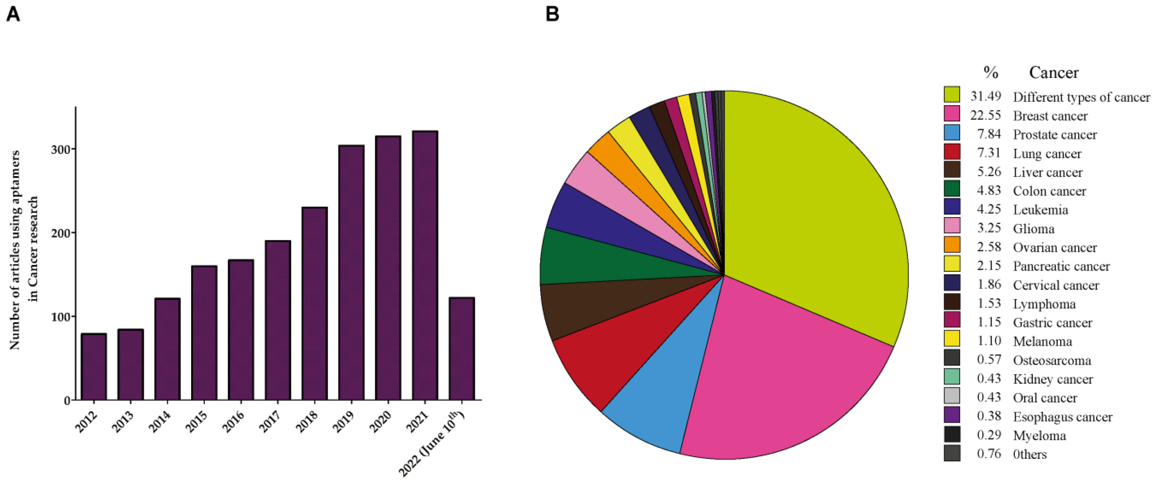
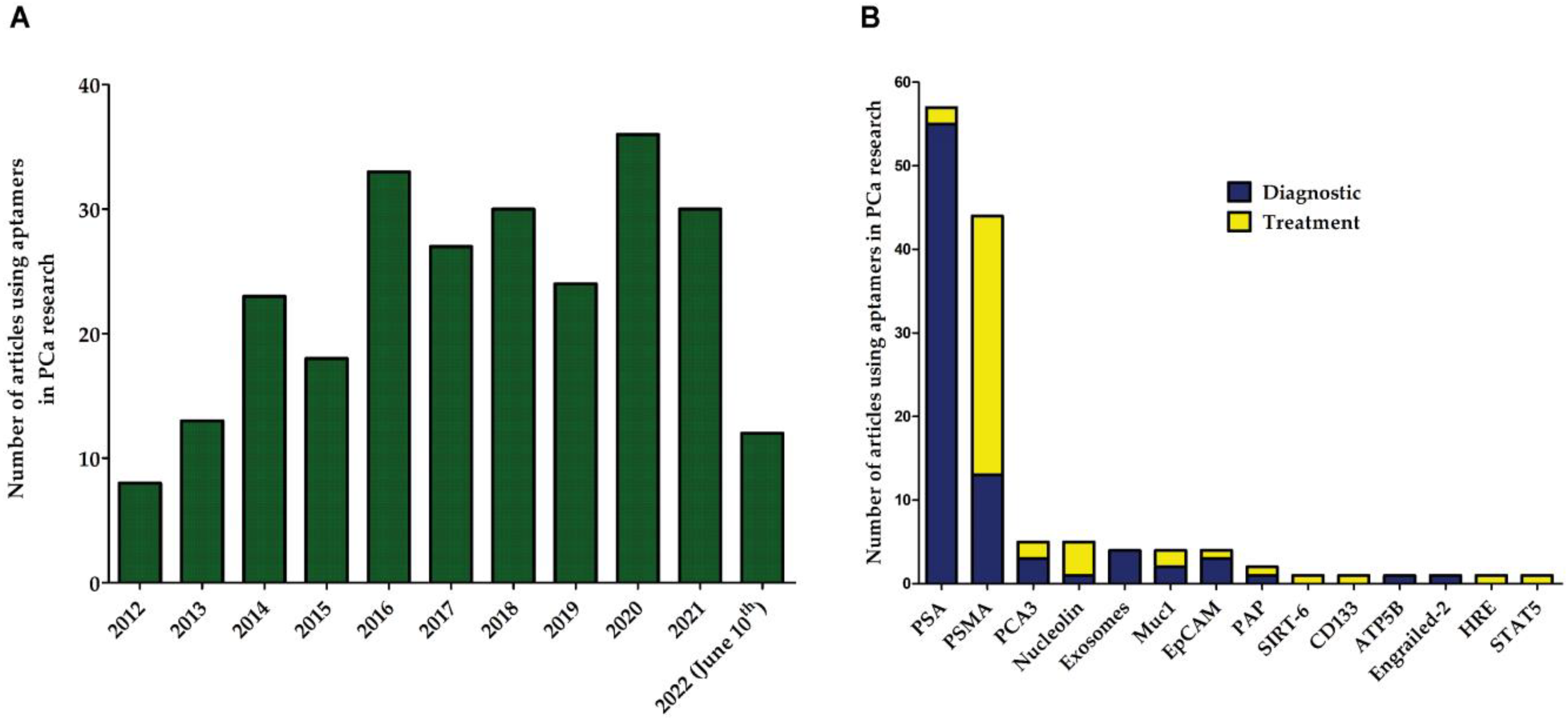
Aptamers are DNA and RNA oligonucleotides that can adopt tridimensional structures that enable them to join specifically to any desired target. Aptamers are capable of binding to specific molecules including drugs, proteins, carbohydrates, cells, and viruses. Aptamers were first described in 1990, and since then several groups have used their binding properties to isolate a diversity of specific aptamers. Aptamers have been studied for treatment and detection of many diseases including cancer. In Prostate Cancer, numerous works have reported their use in the development of new approaches in diagnostics and treatment strategies. Aptamers have been joined with drugs or other specific molecules such as silencing RNAs (aptamer–siRNA chimeras) to specifically reduce the expression of oncogenes in prostate cancer (PCa) cells. These studies have shown good results in the early stages, more research is still needed to demonstrate the clinical value of aptamers in PCa. 

prostate cancer aptamers PCa diagnosis PCa treatment

1. Aptamers in Cancer

Since aptamers can recognize targets and modulate biological activities with higher specificity and affinity than antibodies, it is not surprising that these molecules are considered promising therapeutic tools for the treatment of different types of cancer. Importantly, unlike antibodies, aptamers have low toxicity, are non-immunogenic, and they can easily penetrate the tumor core because of their smaller size [1]. Therefore, a 3-fold increase in the number of articles on aptamers in cancer research has been reported in the last four years (Figure 1A). Between 2012 and 2013 the number of articles using aptamers was less than 100, whereas from 2019 to 2021 it was higher than 300. Notably, more than 22% of the articles reporting the use of aptamers are used in breast cancer, followed by aptamer applications in prostate cancer (PCa) and lung cancer (Figure 1B).
Figure 1. Aptamers in cancer research. (A) Yearly trend of published articles reporting the application of aptamers in cancer research. (B) Proportion of types of cancer in which aptamers are most frequently used for therapeutic or diagnostic purposes.
Numerous works have suggested that aptamers can be used in cancer therapy as inhibitors of growth factors or oncoproteins, as delivery methods for anti-cancer drugs or siRNAs into tumor cells, or even as immune stimulators to fight cancer [2][3][4]; however, most of them are still in a preclinical stage. Noteworthy, the aptamers AS1411 and NOX-A12 are the most advanced aptamer-based therapies for leukemia undergoing clinical research [5]. AS1411 is a guanine-rich 26-base quadruplex DNA aptamer targeting nucleolin (also called C23) [6]. Although nucleolin is found in the cell membrane, it is mainly present in the nucleolus, and high expression of cell surface nucleolin is associated with poor prognosis and higher risk of metastasis [7]. The binding of AS1411 to nucleolin inhibits DNA synthesis that leads to destabilization of BCL2 mRNA and apoptosis [8]. AS1411 is currently undergoing phase II clinical trials for acute myeloid leukemia (AML) and metastatic renal cell carcinoma [9][8][10]. The NOX-A12 aptamer is capable of targeting CXCL12, a chemokine that promotes homing and retention of leukemia cells [10]. Treatment with NOX-A12 sensitizes leukemia cells to conventional therapies and, in combination with bendamustine/rituximab, it improves the therapeutic response in patients with chronic lymphocytic leukemia and multiple myeloma [10].
In addition, the aptamer A30, which binds to the extracellular domain of the human epidermal growth factor receptor-3 (HER3) and does reduce cell proliferation by inhibiting heregulin (HRG) signaling, is under investigation in breast cancer therapy. The combination of A30 aptamer with siRNAs against EEF2, PLK-1, GRK4, and SKIP5, induced specific gene silencing and suppressed cell proliferation. Since the aptamer–siRNA chimera was taken up specifically by HER3-expressing breast cancer cells [11], this aptamer is a promising candidate in breast cancer treatment.
To note, the capacity of aptamers as drug carriers for cancer cells is also a matter of extensive research. Thus, the A10 aptamer that binds to Prostate-Specific Membrane Antigen (PSMA) has been conjugated with doxorubicin to confer both high affinity and specificity against prostate cancer cells. This efficient drug-delivery aptamer significantly inhibited cell proliferation of PSMA-positive cells [12]. Similarly, for AML, aptamer–drug conjugates have been developed. Specifically, aptamer–drug conjugates with methotrexate (Apt-MTX) were able to inhibit AML cell growth, trigger cell apoptosis, and induce cell cycle arrest in the G1 phase in a highly specific manner. Trials with human bone marrow specimens demonstrated that this aptamer–drug conjugate induced selective growth inhibition of primary AML cells without toxicity in normal marrow cells after Apt-MTX exposure. Overall, these findings demonstrate the potential clinical value of Apt-MTX for targeting AML [13]. Another chimeric aptamer siRNA targeting BCL-2 was bound to doxorubicin (siRNA-Dox). siRNA-Dox increased sensitivity of cells to apoptosis and, in turn, decreased cell viability in multi-drug-resistant MCF-7 breast cancer cells [14].
Moreover, aptamers can be used as biosensors for cancer detection. For example, an electrochemical apta-sensor against mucin-1 (MUC1) was recently developed. MUC1 is a surface glycan highly expressed in cancer cells. Voltage changes induced by the chemical reaction between the aptamer conjugated to magnetic beads and gold reduction allow cancer detection by electrochemical analysis [15]. Aptamers can also be used in the field of circulating tumor cells (CTCs). The aptamer BC-15 has been used to specifically identify rare CTCs out of background nucleated cells. This aptamer showed high affinity for nuclei of different human cancer cell lines as well as CTCs isolated from pancreatic cancer patients. The target of the BC-15 aptamer is the heterogeneous nuclear ribonucleoprotein A1 (hnRNP A1). Overexpression of hnRNP A1 has been reported in breast, small cell lung, ovarian, colorectal carcinoma, and pancreatic cancer [16][17][18]. Such results establish a novel way to identify CTCs by using a synthetic aptamer probe [19]. In addition, aptamers can be used in microfluidic systems to capture cells with high affinity. In ovarian cancer, the CX-BG1-10-A aptamer captures CTCs faster than with antibodies in whole blood [20]. Overall, these works show that aptamers are promising candidates to be used in cancer diagnosis and therapy.

2. Aptamers in Prostate Cancer (PCa)

Prostate cancer (PCa) is the most frequent cause of cancer-related death in men [21]. The main factors involved in disease etiology are age, lifestyle, and diet. As aging populations worldwide are increasing, the development of new tools to achieve quick and safe diagnosis and treatment in PCa has become highly relevant [22]. The diagnosis and treatment of PCa is a challenge due to the lack of biomarkers and therapeutic targets specific to the disease. For the diagnosis, the PSA-specific prostate antigen is the biomarker used for excellence in the clinic, but it lacks specificity [23][24].
Although most clinicians agree with a PSA threshold of 4.0 ng/mL for men over 50 years old as normal, several factors can produce PSA fluctuations, for example, prostatitis and benign prostatic hyperplasia (BPH) increase PSA levels [25]. Thus, men with PSA levels of 4–10 ng/mL have a 1 in a 4 chance of having PCa, whereas in the cases that PSA is superior to 10, the probability increases to 50% [26][27][28]. Whereas when elevated PSA levels are found, but no symptoms of PCa are present, another PSA test may be recommended to confirm the original finding. If the PSA level is still high, the test must be supplemented with digital rectal exams, imaging tests, and/or prostate biopsy; highlighting the importance of searching for more precise and specific markers in PCa. Hence, other molecules are being studied for the diagnosis of PCa, such as the lncRNA PCA3, a set of kallikreins including klk2 and klk3, and fusion genes such as TMPRSS2-ERG [29][30][31]. Regarding the treatment of PCa, classic chemotherapy consists of blocking androgen receptor activity also called chemical castration. This therapy has good effects at the beginning of the treatment; however, PCa cells become resistant to castration as time progresses [32].
PCa represents a heterogeneous variety of tumors, and numerous studies have reported the use of aptamers in PCa to target these different types of tumors. The yearly average of reported works using aptamers in PCa research has been 28 in the last 10 years (Figure 2B). In these articles, more than 20 proteins have been used as targets in aptamer research in either diagnostic or therapeutic approaches (Figure 2B). In the field of aptamer-based diagnostics, PSA is the most frequently used protein as the target in aptamer technology, whereas PSMA is highly used as the target in therapeutic approaches (Figure 2B).
Figure 2. Aptamer publications in PCa research. (A) Yearly trend of published articles reporting the use of aptamers in PCa. (B) Proteins used as targets of the aptamers in PCa research. Blue bars represent the number of articles using aptamers directed against each protein in diagnosis, whereas yellow bars refer to articles using aptamers in PCa therapy. PCA3 = prostate-cancer-associated 3; Muc1 = mucin 1; EpCAM = epithelial cellular adhesion molecule; PAP = prostatic acid phosphatase; SIRT-6 = sirtuin 6; CD133 = prominin-1; ATP5B = ATP synthase F1 subunit beta; HRE = hormone response element; STAT5 = signal transducer and activator of transcription 5A.

3. Aptamers against Prostate-Specific Membrane Antigen (PSMA)

PSMA is a type II membrane protein expressed in all forms of prostate tissues, including carcinoma [33]. PSMA is coded by FOLH1, a gene located at chromosome 11 in a region that is not commonly deleted in prostate cancer. PSMA has peptidase and hydrolase activity and digests dietary folates [34]. As PSMA expression is increased in PCa [35], it has been assayed as a predictor of disease recurrence by using anti-PSMA monoclonal antibodies [36]. In addition, PSMA peptides have been used in PCa treatment for stimulating the immune response by infusing dendritic cells pulsed by these PSMA peptides [37]. Altogether, these works highlight the potential value of PSMA as a target in the development of new approaches to diagnose and treat PCa.

3.1. PSMA Aptamers in PCa Diagnosis

Aptamers have been also used as tools for the development of effective diagnostic methods in PCa. For example, an aptamer against PSMA has been used to improve the diagnosis from images by using a nano-crystal semiconductor known as quantum dots (QD’s). Bagalkot et al. developed a targeted QD imaging system with the A10 RNA aptamer capable of differential uptake and imaging of PCa cells expressing PSMA. Some of the advantages of this type of system are wide absorption with narrow photoluminescence, high quantum performance spectra, low photobleaching, and resistance to chemical degradation [38]. Although PSMA is a valuable marker in PCa, two types of cell lines, PSMA (+) and PSMA (−) cells, can be found in the prostate of patients with PCa. Thus, a new approach based on an RNA/peptide dual-aptamer probe was developed by Min et al. (2010) to detect both PSMA (+) and PSMA (−) prostate cancer cells [39]. Herein, two aptamers specific to prostate cancer cells, the A10 RNA aptamer (for the PSMA (+) cell line) and the DUP-1 peptide aptamer (for the PSMA (−) cell line), were conjugated to streptavidin to build the dual-aptamer probe and synchronously detected both prostate cancer cells with a high specificity by electrochemical impedance spectroscopy. Another approach uses lipid nanobubbles functionalized with the A10-3.2 aptamer that, when injected in the abdominal area of mice, the abdominal color Doppler blood flow imaging was significantly improved [40].
In addition, the detection of differentially expressed antigens (biomarkers) has proven to be important for PCa diagnosis and therapy. The main advantage of the use of aptamers over common techniques such as ELISA and tissue staining is that it does not require substantial amounts of starting material. For example, Pai and Ellington adapted the proximity ligation assay (PLA) to cell surface protein targets using modified RNA aptamers detecting and differentiating between cells that distinctively express PSMA tumor antigen [41]. Another application of PSMA aptamers in diagnosis is as biosensors, which are characterized by being ultra-sensitive. Farzin et al. (2017) developed an aptamer-based biosensor (aptasensor) to detect the tumor marker MUC 1 in serum from human samples [42].

3.2. PSMA Aptamers in PCa Therapy

The standard care for PCa treatment is prostatectomy or chemical castration to reduce the circulating levels of testosterone and induce the apoptosis of androgen-dependent tumor cells [43]. Although prostate cancer cells are highly sensitive to androgen ablation, prostate cancer tumors contain a population of cells resistant to the treatment, for instance, cells resistant to chemical castration, and prone to both maintenance and progression of the tumor toward metastatic events [44]. Actually, 177Lutetium-PSMA-617 has been approved in PCa therapy by the FDA. Although this radioligand therapy has been shown to be safe in patients resistant to chemotherapy, the median progression-free survival is still limited (3.8 months) [45]. Therefore, therapy with aptamers is an advantageous tool since it would allow the design of molecules capable of specifically recognizing various types of tumor populations to achieve an effective pharmacological treatment. Several examples of drug conjugates with aptamers exist in the scientific literature for PCa. Dhar et al. (2011) demonstrated enhanced in vivo pharmacokinetics (PK), tolerability, and efficacy of the cisplatin aptamer (Pt-PLGA-b-PEG-Apt-NP) when compared to cisplatin alone administered in a PSMA-expressing LNCaP subcutaneous xenograft mouse model of PCa [46]. In addition, docetaxel (Dtx), that is the drug of choice in PCa therapeutics, has also been used in aptamer technology to improve its pharmacological properties (administration route, solubility) and decrease its toxicity and side effects. Hence, docetaxel (Dtx)-encapsulated nanoparticles formulated with a biocompatible and biodegradable poly(D, L-lactic-co-glycolic acid)-block-poly (ethylene glycol) (PLGA-b-PEG) copolymer and surface functionalized with the A10 RNA aptamer improved the targeted delivery and uptake of drugs [47]. An additional work by Chen et al. (2016) also showed that the aptamer coupled to nanoparticles and Dtx (Dtx-apt-NPs) improved the antitumor effect in vivo on an LNCaP cell xenograft tumor model and was more effective in inducing LNCaP cell apoptosis or death through G2/M phase cell cycle arrest compared to Dtx-free nanoparticles [48].
Another application of aptamers in PCa has been as vehicles to direct drugs or simply to improve their bioavailability. This approach allows not only a better bioavailability but also a more specific recognition of cancer cells.  For example, the use of unimolecular micelles coupled to aptamers as vehicles for transporting doxorubicin to tumor cells of prostate cancer. This type of conjugated molecule induced a high accumulation in the tumor tissue when compared to those without the aptamer in their system [49]. The conjugation of aptamers with liposomes for PCa treatment has been a widely used tool in research. Bandekar et al. (2014) evaluated targeted liposomes loaded with Ac-225 to selectively kill prostate-specific membrane antigen (PSMA)-expressing cells with the aim to assess their potential as targeted antivascular radiotherapy [50].
Moreover, using gold nanoparticles for imaging and therapy of PCa coupled to aptamers has been tested. The latter is based on functionalization of the surface of gold nanoparticles (GNPs) with an RNA aptamer targeting PSMA. The resulting PSMA aptamer-conjugated GNP produced a 4-fold increase in the computed tomography (CT) intensity for targeted LNCaP cells in comparison to non-targeted PC3 cells. Furthermore, the conjugated aptamer was more potent against targeted LNCaP cells than non-targeted PC3 cells when doxorubicin was added to the system [51].
Another interesting application of aptamers is the sensitization of cancer cells to radiotherapy because radio-sensitization can occur by coupling physical agents that allow better absorption of radiation. In this regard, Ni et al. (2011) achieved radio-sensitization of cancer cells with aptamer–shRNA chimeras directed to PSMA for silencing the DNAPK protein [52]. It is well known that approximately 50% of PCa tumors do not express PSMA, some of them because ERG, a common overexpressed transcription factor in PCa, suppresses PSMA expression in tumors containing the TMPRSS2-ERG fusion [29]. To overcome the lack of PSMA, Jing et al. (2016) designed a dual recombinant adenovirus-aptamer system. The viral peptide DUP-1 is capable of recognizing the PSMA-negative cells, while the aptamer A10-3.2 recognizes the PSMA-positive cells. This system decreased the cell growth for both LNCaP (PSMA-positive) and PC3 (PSMA-negative) cells in vitro and in vivo [53].
Although it will be discussed in the aptamer–siRNA chimeras section, it is worth mentioning some examples of these conjugates in PCa therapy. Two anti-PSMA aptamers were designed by Wullner et al., in 2008, with specific cytotoxicity against PCa using siRNA-induced silencing of EEF2, resulting in enhanced cytotoxicity against cancer cells [54]. Dassie et al. (2009) showed that optimized aptamer–siRNA chimeras resulted in regression of PSMA-expressing tumors in athymic mice after systemic administration. This anti-tumor activity was enhanced by increasing the chimera’s half-life using polyethylene glycol [55]. In addition, RNA nanoparticles were constructed by bottom-up self-assembly containing the anti-PSMA aptamer as the targeting ligand and anti-miR17 or anti-miR21 as therapeutic systems. This conjugate was able to strongly bind to the tumors and repressed the tumor growth in mice that received low doses of the conjugate by systemic injection [56]. Finally, an approach that integrates several systems in one is a novel prostate surface membrane antigen (PSMA) aptamer-cationic liposome-double siRNA complex that targets prostate cancer cells to inhibit cell proliferation. The delivery system showed synergism in inhibiting the growth of tumor cells indicating the potential application of the double functional siRNA delivery system for gene therapy in PCa [57].

3.3. Chimeras of PSMA

In addition to the traditional use of PCa aptamers in either target recognition or inhibition, aptamers have also been used to mediate targeted delivery of small interfering RNA (siRNAs). This system is called the aptamer–siRNA chimera and allows the delivery of siRNAs in a cell-type-specific manner. McNamara et al. (2006) developed a system based on an aptamer–siRNA chimera whose aptamer portion mediated the binding to PSMA in prostate cancer cells, and the siRNA portion targeted the expression of two survival genes (PLK1 and BCL2) overexpressed in most human tumors [58]. Lupold et al. (2002) characterized two aptamers that bind the extracellular membrane fraction of the PSMA membrane protein [59]. After this, the use of aptamer–siRNA chimeras has been extended to recognize more than one molecular target on PSMA. Mathieu et al. (2017) designed an aptamer–siRNA capable of simultaneously inhibiting EGFR and survivin [60]. The use of chimeras allows not only the fusion of aptamers with other types of molecules such as siRNAs, liposomes, or viruses but also aptamer–aptamer chimeras are possible. This is the case of the RNA aptamer–aptamer chimera designed for transporting both paramagnetic iron oxide and doxorubicin simultaneously, this chimera being more cytotoxic to the targeted cells [61].
In recent years, the gene editing approaches have gained great importance in the field of cancer research. Besides the use of the well-known CRISPR-Cas system, aptamers both activate and repress gene expression. Li and Li (2017) succeeded in designing a system that fuses a PSMA aptamer with a small activation RNA known as saRNAs [62]. This model promoted the expression of the DPYSL3 protein, decreasing cell migration in cancer cells. Another report shows that pre-treatment of animals bearing PSMA-positive tumors with chemically synthesized and systemically administered aptamer–siRNA chimeras (two days before ionizing radiation therapy) can significantly enhance tumor response to IR [63]. This type of system can be used for different applications as the new multifunctional probe comprising a cell-specific internalization aptamer, fluorescent silver nanoclusters (Ag Ncs), and therapeutic siRNA encompassing one system [62].
Although aptamer–siRNA chimeras have effectiveness in the cell-specific delivery of siRNAs, improvements can be made. One example could contemplate the binding to other peptides that are known to be a fundamental part of the cell or that intervene in some important cell proliferation processes. A biotinylated PSMA-specific aptamer A10 and SURVIVIN-siRNA were linked to a Streptavidin-Trans-Activator of Transcription- Double strand RNA binding domain fusion protein (STD protein) to form a therapeutic complex. This complex demonstrated higher efficiency in delivering siRNA into target cells and increasing apoptosis compared to lipofectamine and A10–siRNA chimera [64]. Furthermore, Jiao et al. (2022) developed a 99mTc–Aptamer–siRNA chimera to both diagnose and treat PSMA-positive PCa in vivo [65]. This chimera was composed by the PSMA aptamer A10 to specifically deliver the siRNA against the Mouse double minute 2 homolog (MDM2) in PCa cells. The 99mTc-A10 Aptamer–MDM2 siRNA chimera decreased MDM2 expression in PSMA-positive PCa cell lines. The inclusion of the technetium radionuclide (99mTc) in the chimera allowed a good labeling rate and targeting to the tumor, indicating that the 99mTc-labeled MDM2 siRNA-Apt chimera can be used not only as a nucleic acid treatment drug, but also as an imaging probe [65]. Although further research is necessary, it was demonstrated that the potential of aptamers to develop new approaches that integrate diagnosis and treatment of PSMA-positive PCa to provide clinical support to PCa patients.

3.4. PSMA Aptamers as Vehicles in PCa

Aptamers can also be used to selectively deliver drugs to PCa cells and enhance their effects at lower concentrations. The use of PSMA aptamers has resulted very efficient for this application because many PCa cells and tissues have high expression of this protein. Dhar et al. (2008) developed cisplatin(IV)-encapsulated nanoparticles with targeting aptamers to effectively deliver cisplatin to PCa cells [66]. These PSMA aptamers showed greater effectiveness (one order of magnitude higher than free cisplatin) of cisplatin on the PCa cell line, LNCaP [66]. This same system has been used to deliver a variety of anticancer drugs such as docetaxel using biocompatible and biodegradable co-polymers such as poly (D, L-lactic-co-glycolic acid)-block-poly (ethylene glycol) (PLGA-b-PEG) functionalized with the A10 aptamer that binds to PSMA [67]. In addition, Singh et al. (2020) encapsulated the A10 RNA aptamer in polysaccharide nanoparticles containing the natural compound thymoquinone (TQ) to inhibit the Hedgehog signaling pathway [68]. The resulting aptamer-based nanoparticles carrying TQ were more effective in both inhibiting the Hh signaling in low drug concentrations and delivering the agent to the PCa cells [68].
PSMA aptamers can further improve their capacity as vehicles when conjugated to liposomal complexes, known as aptosomes. An example for this is an RNA micelle aptamer-conjugated liposome that specifically binds to LNCaP cells expressing PSMA. This aptamosome demonstrated in vivo an anticancer efficacy of the doxorrubicin-encapsulating PSMA-aptamosomes on tumor size regression in LNCaP xenograft mice [69].
The generation of aptamers conjugated to drugs that target PSMA is a growing area of interest in PCa therapy. To mention some seminal research, some involve the co-delivery of shRNAs against Bcl simultaneously with the delivery of doxorubicin. Another example is the encapsulation of cisplatin in positive nanoparticles with PSMA targeting aptamers on the surface of the nanoparticles to specifically deliver cisplatin to PCa cells [70]. The concomitant diagnostic and therapeutic advantages of aptamers have been reflected in the work by Wu et al. (2017), where poly (lactide-co-glycolic acid) nanobubbles (PLGA) modified with the aptamer A-10-3.2 were loaded with paclitaxel, producing tumor regression and diminishing neoplasic characteristics of cells in vitro and in vivo [71].

4. Other Aptamers in PCa

Although aptamers targeting PSMA are the second most used aptamers in PCa research (Figure 2B), there are others under study and have presented interesting results. Figure 2B summarize reported aptamers in diagnosis and treatment of PCa whose targets are different to PSMA. In the field of aptamers targeting cells, a pluri-targeting DNA aptamer called DML-7 was first designed for recognizing the human PCa cell line DU145. This aptamer internalized into the target cells and exhibited high binding affinity with dissociation constants (Kd) in the nanomolar range. Interestingly, DML-7 bound to DU145 and PC-3 cells but not to LNCaP or 22Rv1 cells [72].
There are many examples of aptamers used as delivery tools. The MUC1 aptamer was explored as a vehicle for delivering doxorubicin to cancer cells. This 86-base DNA aptamer (MA3) bound to the epitope of MUC1 with a Kd= 38.3 nM. The cancer cell lines, A549 (lung) and MCF-7 (breast) express MUC1, the aptamer MA3 preferentially bound to MUC1-positive but not MUC1-negative cells, suggesting that the MUC1 aptamer may have a potential utility as a targeting ligand for selective delivery of cytotoxic agents to MUC1-expressing tumors including prostate [73].
In diagnosis, aptamers have the potential to improve PSA-based tests as they show more sensibility and specificity and are less expensive than ELISA-based tests. The AS1411 aptamer, a 26-base guanine-rich oligonucleotide aptamer, has high affinity to nucleolin on tumor cell surfaces. The AS1411 aptamer labeled with (99m)Tc was stable in normal saline, human serum, and cellular experiments demonstrating specific binding. Since tumors had higher accumulation of radioactivity with this labeled aptamer, it could be a potential tool for use in molecular imaging of PCa [74]. In this line, aptamers have been modified to serve in diagnosis, for example, the mA4 aptamer was modified in the 2′ hydroxyl groups of RNA and poly-T in the 5′ sequence was added to increase its resistance to degradation by nucleases [75]. Other types of aptamers used in the diagnosis of PCa are based on electrochemical detection of PSA. For example, labeled free DNA aptamers coupled to gold particles showed to be useful in PSA detection after electrochemical impedance spectroscopy (EIS) with a range of 1–200 pg mL−1. A similar study using a screen-printed carbon electrode (SPCE) showed improved results with a remarkably lower limit of detection of 0.077 pg/mL [76].

References

  1. Dongxi Xiang; Conglong Zheng; Shu-Feng Zhou; Shuxi Qiao; Phuong Tran; Chunwen Pu; Yong Li; Lingxue Kong; Abbas Z. Kouzani; Jia Lin; et al.Ke LiuLianhong LiSarah ShigdarWei Duan Superior Performance of Aptamer in Tumor Penetration over Antibody: Implication of Aptamer-Based Theranostics in Solid Tumors. Theranostics 2014, 5, 1083-1097, 10.7150/thno.11711.
  2. Guizhi Zhu; Xiaoyuan Chen; Aptamer-based targeted therapy. Advanced Drug Delivery Reviews 2018, 134, 65-78, 10.1016/j.addr.2018.08.005.
  3. Weihong Tan; Hui Wang; Yan Chen; Xiaobing Zhang; Haizhen Zhu; Chaoyong Yang; Ronghua Yang; Chen Liu; Molecular aptamers for drug delivery. Trends in Biotechnology 2011, 29, 634-640, 10.1016/j.tibtech.2011.06.009.
  4. Fernando Pastor; Pedro Berraondo; Iñaki Etxeberria; Josh Frederick; Ugur Sahin; Eli Gilboa; Ignacio Melero; An RNA toolbox for cancer immunotherapy. Nature Reviews Drug Discovery 2018, 17, 751-767, 10.1038/nrd.2018.132.
  5. Sridharan Soundararajan; Li Wang; Vijayalakshmi Sridharan; Weiwei Chen; Nigel Courtenay-Luck; David Jones; Eleanor K. Spicer; Daniel J. Fernandes; Plasma Membrane Nucleolin Is a Receptor for the Anticancer Aptamer AS1411 in MV4-11 Leukemia Cells. Molecular Pharmacology 2009, 76, 984-991, 10.1124/mol.109.055947.
  6. Pooria Safarzadeh Kozani; Pouya Safarzadeh Kozani; Mohammad Tariq Malik; AS1411-functionalized delivery nanosystems for targeted cancer therapy. Exploration of Medicine 2021, 2, 146-166, 10.37349/emed.2021.00039.
  7. Caroline Madeleine Berger; Xavier Gaume; Philippe Bouvet; The roles of nucleolin subcellular localization in cancer. Biochimie 2015, 113, 78-85, 10.1016/j.biochi.2015.03.023.
  8. Sridharan Soundararajan; Weiwei Chen; Eleanor K. Spicer; Nigel Courtenay-Luck; Daniel J. Fernandes; The Nucleolin Targeting Aptamer AS1411 Destabilizes Bcl-2 Messenger RNA in Human Breast Cancer Cells. Cancer Research 2008, 68, 2358-2365, 10.1158/0008-5472.can-07-5723.
  9. Man Chen; Yuanyuan Yu; Feng Jiang; Junwei Zhou; Yongshu Li; Chao Liang; Lei Dang; Aiping Lu; Ge Zhang; Development of Cell-SELEX Technology and Its Application in Cancer Diagnosis and Therapy. International Journal of Molecular Sciences 2016, 17, 2079, 10.3390/ijms17122079.
  10. Julia Hoellenriegel; Dirk Zboralski; Christian Maasch; Nathalie Y. Rosin; William G. Wierda; Michael J. Keating; Anna Kruschinski; Jan A. Burger; The Spiegelmer NOX-A12, a novel CXCL12 inhibitor, interferes with chronic lymphocytic leukemia cell motility and causes chemosensitization. Blood 2014, 123, 1032-1039, 10.1182/blood-2013-03-493924.
  11. Inga Nachreiner; Ahmad Fawzi Hussain; Ulrich Wullner; Nikolaus Machuy; Thomas F. Meyer; Rainer Fischer; Ivo Meinhold‑Heerlein; Stefan Barth; Mehmet Kemal Tur; Elimination of HER3‑expressing breast cancer cells using aptamer‑siRNA chimeras. Experimental and Therapeutic Medicine 2019, 18, 2401-2412, 10.3892/etm.2019.7753.
  12. Vaishali Bagalkot; Omid C. Farokhzad; Robert Langer; Sangyong Jon; An Aptamer–Doxorubicin Physical Conjugate as a Novel Targeted Drug-Delivery Platform. Angewandte Chemie International Edition 2006, 45, 8149-8152, 10.1002/anie.200602251.
  13. Nianxi Zhao; Sung-Nan Pei; Jianjun Qi; Zihua Zeng; Swaminathan P. Iyer; Pei Lin; Ching-Hsuan Tung; Youli Zu; Oligonucleotide aptamer-drug conjugates for targeted therapy of acute myeloid leukemia. Biomaterials 2015, 67, 42-51, 10.1016/j.biomaterials.2015.07.025.
  14. Hyosook Jeong; Soo Hyeon Lee; Yeonju Hwang; Hyundong Yoo; Heesun Jung; Sun Hwa Kim; Hyejung Mok; Multivalent Aptamer-RNA Conjugates for Simple and Efficient Delivery of Doxorubicin/siRNA into Multidrug-Resistant Cells. Macromolecular Bioscience 2016, 17, 1600343, 10.1002/mabi.201600343.
  15. Jing-Jing Zhang; Fang-Fang Cheng; Ting-Ting Zheng; Jun-Jie Zhu; Versatile aptasensor for electrochemical quantification of cell surface glycan and naked-eye tracking glycolytic inhibition in living cells. Biosensors and Bioelectronics 2017, 89, 937-945, 10.1016/j.bios.2016.09.087.
  16. Mitsunori Ushigome; Tsuneyuki Ubagai; Hirokazu Fukuda; Naoto Tsuchiya; Takashi Sugimura; Jun Takatsuka; Hitoshi Nakagama; Up-regulation of hnRNP A1 gene in sporadic human colorectal cancers. International Journal of Oncology 2005, 26, 635-640, 10.3892/ijo.26.3.635.
  17. Laura K. Zerbe; Irene Pino; Ruben Pio; Pippa Cosper; Lori D. Dwyer-Nield; Amy M. Meyer; J. David Port; Luis Montuenga; Alvin M. Malkinson; Relative amounts of antagonistic splicing factors, hnRNP A1 and ASF/SF2, change during neoplastic lung growth: Implications for pre-mRNA processing. Molecular Carcinogenesis 2004, 41, 187-196, 10.1002/mc.20053.
  18. Yan Yan-Sanders; George J Hammons; Beverly D Lyn-Cook; Increased expression of heterogeneous nuclear ribonucleoprotein A2/B1 (hnRNP) in pancreatic tissue from smokers and pancreatic tumor cells. Cancer Letters 2002, 183, 215-220, 10.1016/s0304-3835(02)00168-4.
  19. Jinqiang Zhang; Shaohua Li; Fang Liu; Lanping Zhou; Ningsheng Shao; Xiaohang Zhao; SELEX Aptamer Used as a Probe to Detect Circulating Tumor Cells in Peripheral Blood of Pancreatic Cancer Patients. PLOS ONE 2015, 10, e0121920-e0121920, 10.1371/journal.pone.0121920.
  20. Sung-Chi Tsai; Lien-Yu Hung; Gwo-Bin Lee; An integrated microfluidic system for the isolation and detection of ovarian circulating tumor cells using cell selection and enrichment methods. Biomicrofluidics 2017, 11, 034122, 10.1063/1.4991476.
  21. Rebecca L. Siegel; Kimberly D. Miller; Ahmedin Jemal; Cancer statistics, 2016. CA: A Cancer Journal for Clinicians 2016, 66, 7-30, 10.3322/caac.21332.
  22. Michael E Stokes; Jack Ishak; Irina Proskorovsky; Libby K Black; Yijian Huang; Lifetime economic burden of prostate cancer. BMC Health Services Research 2011, 11, 349-349, 10.1186/1472-6963-11-349.
  23. W. J. Catalona; Evaluation of percentage of free serum prostate-specific antigen to improve specificity of prostate cancer screening. JAMA 1995, 274, 1214-1220, 10.1001/jama.274.15.1214.
  24. Per-Anders Abrahamsson; Hans Lilja; Joseph E. Oesterling; MOLECULAR FORMS OF SERUM PROSTATE-SPECIFIC ANTIGEN: The Clinical Value of Percent Free Prostate-Specific Antigen. Urologic Clinics of North America 1997, 24, 353-365, 10.1016/s0094-0143(05)70382-7.
  25. Eric H Kim; Gerald L Andriole; Prostate-specific antigen-based screening: controversy and guidelines. BMC Medicine 2015, 13, 61-61, 10.1186/s12916-015-0296-5.
  26. Alan W. Partin; H. Ballentine Carter; Daniel W. Chan; Jonathan I. Epstein; Joseph E. Oesterling; Robert C. Rock; Jed P. Weber; Patrick C. Walsh; Prostate Specific Antigen in the Staging of Localized Prostate Cancer: Influence of Tumor Differentiation, Tumor Volume and Benign Hyperplasia. Journal of Urology 1990, 143, 747-752, 10.1016/s0022-5347(17)40079-6.
  27. H. B. Carter; Longitudinal evaluation of prostate-specific antigen levels in men with and without prostate disease. JAMA 1992, 267, 2215-2220, 10.1001/jama.267.16.2215.
  28. Joseph A. Smith; Prevalence of prostate cancer among men with a prostate-specific antigen level ⩽4.0 ng per milliliter: Thompson IM, Pauler DK, Goodman PJ, Tangen CM, Lucia, MS, Parnes HL, Minasian LM, Ford LG, Lippman SM, Crawford ED, Crowley JJ, Coltman CA Jr., Division of Urology, Department of Surgery, University of Texas Health Science Center at San Antonio, San Antonio, TX. N Engl J Med 2004;350:2239–46. Urologic Oncology: Seminars and Original Investigations 2004, 22, 493-493, 10.1016/j.urolonc.2004.08.008.
  29. S.A. Tomlins; D.R. Rhodes; S. Perner; S.M. Dhanasekaran; R. Mehra; X.-W. Sun; S. Varambally; X. Cao; J. Tchinda; R. Kuefer; et al.C. LeeJ.E. MontieR.B. ShahK.J. PientaM.A. RubinA.M. Chinnaiyan Recurrent Fusion of TMPRSS2 and ETS Transcription Factor Genes in Prostate Cancer. Journal of Urology 2006, 175, 1707-1707, 10.1016/s0022-5347(06)00096-6.
  30. Grégoire Robert; Sander Jannink; Frank Smit; Tilly Aalders; Daphne Hessels; Ruben Cremers; Peter F. Mulders; Jack A. Schalken; Rational basis for the combination of PCA3 and TMPRSS2:ERG gene fusion for prostate cancer diagnosis. The Prostate 2012, 73, 113-120, 10.1002/pros.22546.
  31. Indu Kohaar; Gyorgy Petrovics; Shiv Srivastava; A Rich Array of Prostate Cancer Molecular Biomarkers: Opportunities and Challenges. International Journal of Molecular Sciences 2019, 20, 1813, 10.3390/ijms20081813.
  32. Philip A. Watson; Vivek K. Arora; Charles L. Sawyers; Emerging mechanisms of resistance to androgen receptor inhibitors in prostate cancer. Nature Cancer 2015, 15, 701-711, 10.1038/nrc4016.
  33. Jeffrey S Ross; Christine E Sheehan; Hugh A G Fisher; Ronald P Kaufman; Prabhjot Kaur; Karen Gray; Iain Webb; Gary S Gray; Rebecca Mosher; Bhaskar V S Kallakury; et al. Correlation of primary tumor prostate-specific membrane antigen expression with disease recurrence in prostate cancer.. Clinical Cancer Research 2003, 9, 6357–6362.
  34. Denise S. O’Keefe; Sai L. Su; Dean J. Bacich; Yutaka Horiguchi; Ying Luo; C.Thomas Powell; Dorothea Zandvliet; Pamela Russell; Peter Molloy; Norma J. Nowak; et al.Thomas B. ShowsCami MullinsRaymond A. Vonder HaarWilliam R. FairWarren D.W. Heston Mapping, genomic organization and promoter analysis of the human prostate-specific membrane antigen gene. Biochimica et Biophysica Acta (BBA) - Gene Structure and Expression 1998, 1443, 113-127, 10.1016/s0167-4781(98)00200-0.
  35. Sam S Chang; Victor E Reuter; W.D.W Heston; Paul B Gaudin; Comparison of anti-prostate-specific membrane antigen antibodies and other immunomarkers in metastatic prostate carcinoma. Urology 2001, 57, 1179-1183, 10.1016/s0090-4295(01)00983-9.
  36. M L Beckett; Lisa Cazares; Antonia Vlahou; P F Schellhammer; G L Wright; Prostate-specific membrane antigen levels in sera from healthy men and patients with benign prostate hyperplasia or prostate cancer.. Clinical Cancer Research 1999, 5, 4034–4040.
  37. Michael L. Salgaller; Patricia A. Lodge; Joanne G. McLean; Ben A. Tjoa; Douglas J. Loftus; Haakon Ragde; Gerald M. Kenny; Mary Rogers; Alton L. Boynton; Gerald P. Murphy; et al. Report of immune monitoring of prostate cancer patients undergoing T-cell therapy using dendritic cells pulsed with HLA-A2-specific peptides from prostate-specific membrane antigen (PSMA). The Prostate 1998, 35, 144-151, 10.1002/(sici)1097-0045(19980501)35:2<144::aid-pros8>3.0.co;2-j.
  38. Vaishali Bagalkot; Liangfang Zhang; Etgar Levy-Nissenbaum; Sangyong Jon; Philip W. Kantoff; And Robert Langer; Omid C. Farokhzad; Quantum Dot−Aptamer Conjugates for Synchronous Cancer Imaging, Therapy, and Sensing of Drug Delivery Based on Bi-Fluorescence Resonance Energy Transfer. Nano Letters 2007, 7, 3065-3070, 10.1021/nl071546n.
  39. Kyoungin Min; Kyung-Mi Song; Minseon Cho; Yang-Sook Chun; Yoon-Bo Shim; Ja Kang Ku; Changill Ban; Simultaneous electrochemical detection of both PSMA (+) and PSMA (−) prostate cancer cells using an RNA/peptide dual-aptamer probe. Chemical Communications 2010, 46, 5566-5568, 10.1039/c002524k.
  40. Xiaozhou Fan; Yanli Guo; Luofu Wang; Xingyu Xiong; Lianhua Zhu; Kejing Fang; Diagnosis of prostate cancer using anti-PSMA aptamer A10-3.2-oriented lipid nanobubbles. International Journal of Nanomedicine 2016, ume 11, 3939-3950, 10.2147/ijn.s112951.
  41. Supriya S. Pai; Andrew D. Ellington; Using RNA Aptamers and the Proximity Ligation Assay for the Detection of Cell Surface Antigens. null 2007, 504, 385-398, 10.1007/978-1-60327-569-9_21.
  42. Leila Farzin; Mojtaba Shamsipur; Recent advances in design of electrochemical affinity biosensors for low level detection of cancer protein biomarkers using nanomaterial-assisted signal enhancement strategies. Journal of Pharmaceutical and Biomedical Analysis 2017, 147, 185-210, 10.1016/j.jpba.2017.07.042.
  43. Annika Spruessel; Garnet Steimann; Mira Jung; Sung A. Lee; Theresa Carr; Anne-Kristin Fentz; Joerg Spangenberg; Carsten Zornig; Hartmut H. Juhl; Kerstin A. David; et al. Tissue ischemia time affects gene and protein expression patterns within minutes following surgical tumor excision. BioTechniques 2004, 36, 1030-1037, 10.2144/04366rr04.
  44. Matias Knuuttila; Emrah Yatkin; Jenny Kallio; Saija Savolainen; Teemu D. Laajala; Tero Aittokallio; Riikka Oksala; Merja Häkkinen; Pekka Keski-Rahkonen; Seppo Auriola; et al.Matti PoutanenSari Mäkelä Castration Induces Up-Regulation of Intratumoral Androgen Biosynthesis and Androgen Receptor Expression in an Orthotopic VCaP Human Prostate Cancer Xenograft Model. The American Journal of Pathology 2014, 184, 2163-2173, 10.1016/j.ajpath.2014.04.010.
  45. Nicolai Mader; Daniel Groener; Nikolaos Tselis; Séverine Banek; James Nagarajah; Frank Grünwald; Amir Sabet; Outcome of 177Lu-PSMA-617 Radioligand Therapy in Chemo-Refractory Patients with Metastatic Castration-Resistant Early-Onset Prostate Cancer. Cancers 2021, 13, 4193, 10.3390/cancers13164193.
  46. Shanta Dhar; Nagesh Kolishetti; Stephen J. Lippard; Omid C. Farokhzad; Targeted delivery of a cisplatin prodrug for safer and more effective prostate cancer therapy in vivo. Proceedings of the National Academy of Sciences 2011, 108, 1850-1855, 10.1073/pnas.1011379108.
  47. Omid C. Farokhzad; Jianjun Cheng; Benjamin A. Teply; Ines Sherifi; Sangyong Jon; Philip W. Kantoff; Jerome P. Richie; Robert Langer; Targeted nanoparticle-aptamer bioconjugates for cancer chemotherapy in vivo. Proceedings of the National Academy of Sciences 2006, 103, 6315-6320, 10.1073/pnas.0601755103.
  48. Zhongjian Chen; Zongguang Tai; Fenfen Gu; Chuling Hu; Quangang Zhu; Shen Gao; Aptamer-mediated delivery of docetaxel to prostate cancer through polymeric nanoparticles for enhancement of antitumor efficacy. European Journal of Pharmaceutics and Biopharmaceutics 2016, 107, 130-141, 10.1016/j.ejpb.2016.07.007.
  49. Wenjin Xu; Imtiaz A. Siddiqui; Minakshi Nihal; Srikanth Pilla; Kimberly Rosenthal; Hasan Mukhtar; Shaoqin Gong; Aptamer-conjugated and doxorubicin-loaded unimolecular micelles for targeted therapy of prostate cancer. Biomaterials 2013, 34, 5244-5253, 10.1016/j.biomaterials.2013.03.006.
  50. Bandekar, A.; Zhu, C.; Jindal, R.; Bruchertseifer, F.; Morgenstern, A.; Sofou, S.; Anti-prostate-specific membrane antigen liposomes loaded with 225Ac for potential targeted antivascular alpha-particle therapy of cancer.. J. Nucl. Med. 2014, 55, 107-114, https://doi.org/10.2967/jnumed.113.125476.
  51. Dongkyu Kim; Yong Yeon Jeong; Sangyong Jon; A Drug-Loaded Aptamer−Gold Nanoparticle Bioconjugate for Combined CT Imaging and Therapy of Prostate Cancer. ACS Nano 2010, 4, 3689-3696, 10.1021/nn901877h.
  52. Xiaohua Ni; Yonggang Zhang; Kenji Zennami; Mark Castanares; Amarnath Mukherjee; Raju R. Raval; Haoming Zhou; Theodore L. DeWeese; Shawn E. Lupold; Systemic Administration and Targeted Radiosensitization via Chemically Synthetic Aptamer–siRNA Chimeras in Human Tumor Xenografts. Molecular Cancer Therapeutics 2015, 14, 2797-2804, 10.1158/1535-7163.mct-15-0291-t.
  53. Pei Jing; Shousong Cao; Shuangli Xiao; Xiaoqin Zhang; Siyun Ke; Famin Ke; Xin Yu; Li Wang; Shurong Wang; Yuling Luo; et al.Zhirong Zhong Enhanced growth inhibition of prostate cancer in vitro and in vivo by a recombinant adenovirus-mediated dual-aptamer modified drug delivery system. Cancer Letters 2016, 383, 230-242, 10.1016/j.canlet.2016.10.003.
  54. Ulrich Wullner; Inga Neef; Andreas Eller; Michael Kleines; Mehmet Kemal Tur; Stefan Barth; Cell-specific induction of apoptosis by rationally designed bivalent aptamer-siRNA transcripts silencing eukaryotic elongation factor 2.. Current Cancer Drug Targets 2008, 8, 554-565, 10.2174/156800908786241078.
  55. Justin P Dassie; Xiu-Ying Liu; Gregory S Thomas; Ryan M Whitaker; Kristina W Thiel; Katie R Stockdale; David Meyerholz; Anton P McCaffrey; James O McNamara II; Paloma H Giangrande; et al. Systemic administration of optimized aptamer-siRNA chimeras promotes regression of PSMA-expressing tumors. Nature Biotechnology 2009, 27, 839-846, 10.1038/nbt.1560.
  56. Daniel W Binzel; Yi Shu; Hui Li; Meiyan Sun; Qunshu Zhang; Dan Shu; Bin Guo; Peixuan Guo; Specific Delivery of MiRNA for High Efficient Inhibition of Prostate Cancer by RNA Nanotechnology. Molecular Therapy 2016, 24, 1267-1277, 10.1038/mt.2016.85.
  57. 119. Jie, F.; Xiao-Lin, W.; Yu-Jie, L.; Jing, Z.; Fang, W.; Shen-Si, X.; Xin, G.; Qing-Qing, W.; Hai-Feng, S.; In vivo analysis of treatment of prostate cancer with prostate surface membrane antigen aptamer-cationic liposome-double siRNA complex.. Chin. J. Biol. 2016, 29, 151-156.
  58. James O McNamara Ii; Eran Andrechek; Yong Wang; Kristi D Viles; Rachel E Rempel; Eli Gilboa; Bruce A Sullenger; Paloma H Giangrande; Cell type–specific delivery of siRNAs with aptamer-siRNA chimeras. Nature Biotechnology 2006, 24, 1005-1015, 10.1038/nbt1223.
  59. Shawn E Lupold; Brian J Hicke; Yun Lin; Donald S Coffey; Identification and characterization of nuclease-stabilized RNA molecules that bind human prostate cancer cells via the prostate-specific membrane antigen.. Cancer Research 2002, 62, 4029-4033.
  60. Romain Mathieu; Ilaria Lucca; Mihai D. Vartolomei; Aurélie Mbeutcha; Tobias Klatte; Christian Seitz; Pierre I. Karakiewicz; Harun Fajkovic; Maxine Sun; Yair Lotan; et al.Francesco MontorsiAlberto BrigantiMorgan RouprêtVitaly MargulisMichael RinkMalte RiekenLukas KennerMartin SusaniLoidl WolgangShahrokh F. Shariat Role of survivin expression in predicting biochemical recurrence after radical prostatectomy: a multi-institutional study. BJU International 2016, 119, 234-238, 10.1111/bju.13472.
  61. John C. Leach; Andrew Wang; Kaiming Ye; Sha Jin; A RNA-DNA Hybrid Aptamer for Nanoparticle-Based Prostate Tumor Targeted Drug Delivery. International Journal of Molecular Sciences 2016, 17, 380, 10.3390/ijms17030380.
  62. Benyi Li; Changlin Li; Suppression of Prostate Cancer Metastasis by DPYSL3-Targeted saRNA. null 2017, 983, 207-216, 10.1007/978-981-10-4310-9_15.
  63. Xiaohua Ni; Yonggang Zhang; Kenji Zennami; Mark Castanares; Amarnath Mukherjee; Raju R. Raval; Haoming Zhou; Theodore L. DeWeese; Shawn E. Lupold; Systemic Administration and Targeted Radiosensitization via Chemically Synthetic Aptamer–siRNA Chimeras in Human Tumor Xenografts. Molecular Cancer Therapeutics 2015, 14, 2797-2804, 10.1158/1535-7163.mct-15-0291-t.
  64. 125. Diao, Y.; Liu, J.; Ma, Y.; Su, M.; Zhang, H.; Hao, X.; A specific aptamer-cell penetrating peptides complex delivered siRNA efficiently and suppressed prostate tumor growth in vivo. . Cancer Biol. Ther. 2016, 17, 498–506, https://doi.org/10.1080/15384047.2016.1156266.
  65. Yuying Jiao; Peng Xu; Sha Luan; Xinyu Wang; Yue Gao; Changjiu Zhao; Peng Fu; Molecular imaging and treatment of PSMA-positive prostate cancer with 99mTc radiolabeled aptamer-siRNA chimeras. Nuclear Medicine and Biology 2021, 104-105, 28-37, 10.1016/j.nucmedbio.2021.11.003.
  66. A.S. Kibel; Targeted delivery of cisplatin to prostate cancer cells by aptamer functionalized Pt(IV) prodrug-PLGA–PEG nanoparticles. Yearbook of Urology 2008, 2009, 157-158, 10.1016/s0084-4071(09)79258-9.
  67. 128. Farokhzad, O.C.; Jon, S.; Khademhosseini, A.; Tran, T.N.; Lavan, D.A.; Langer, R.; Nanoparticle-aptamer bioconjugates: A new approach for targeting prostate cancer cells. Cancer Res. 2004, 64, 7668–7672, https://doi.org/10.1158/0008-5472.CAN-04-2550.
  68. Santosh Kumar Singh; Jennifer B. Gordetsky; Sejong Bae; Edward P. Acosta; Jr. James W. Lillard; Rajesh Singh; Selective Targeting of the Hedgehog Signaling Pathway by PBM Nanoparticles in Docetaxel-Resistant Prostate Cancer. Cells 2020, 9, 1976, 10.3390/cells9091976.
  69. Si Eun Baek; Kwang Hyun Lee; Yong Serk Park; Deok-Kun Oh; Sangtaek Oh; Keun-Sik Kim; Dong-Eun Kim; RNA aptamer-conjugated liposome as an efficient anticancer drug delivery vehicle targeting cancer cells in vivo. Journal of Controlled Release 2014, 196, 234-242, 10.1016/j.jconrel.2014.10.018.
  70. A.S. Kibel; Targeted delivery of cisplatin to prostate cancer cells by aptamer functionalized Pt(IV) prodrug-PLGA–PEG nanoparticles. Yearbook of Urology 2008, 2009, 157-158, 10.1016/s0084-4071(09)79258-9.
  71. Meng Wu; Ying Wang; Yiru Wang; Mingbo Zhang; Yukun Luo; Jie Tang; Zhigang Wang; Dong Wang; Lan Hao; Zhibiao Wang; et al. Paclitaxel-loaded and A10-3.2 aptamer-targeted poly(lactide-co-glycolic acid) nanobubbles for ultrasound imaging and therapy of prostate cancer. International Journal of Nanomedicine 2017, ume 12, 5313-5330, 10.2147/ijn.s136032.
  72. Minlan Duan; Yuqian Long; Cai Yang; Xiaoqiu Wu; Yang Sun; Jianglin Li; Xiaoxiao Hu; Wei Lin; Dongmei Han; Yifan Zhao; et al.Jing LiuMao YeWeihong Tan Selection and characterization of DNA aptamer for metastatic prostate cancer recognition and tissue imaging. Oncotarget 2016, 7, 36436-36446, 10.18632/oncotarget.9262.
  73. Yan Hu; Jinhong Duan; Qimin Zhan; Fengdan Wang; Xin Lu; Xian-Da Yang; Novel MUC1 Aptamer Selectively Delivers Cytotoxic Agent to Cancer Cells In Vitro. PLOS ONE 2012, 7, e31970, 10.1371/journal.pone.0031970.
  74. Zohreh Noaparast; Seyed Jalal Hosseinimehr; Majid Piramoon; Seyed Mohammad Abedi; Tumor targeting with a99mTc-labeled AS1411 aptamer in prostate tumor cells. Journal of Drug Targeting 2015, 23, 497-505, 10.3109/1061186x.2015.1009075.
  75. Esther Campos-Fernández; Letícia S. Barcelos; Aline G. Souza; Luiz R. Goulart; Vivian Alonso-Goulart; Post-SELEX Optimization and Characterization of a Prostate Cancer Cell-Specific Aptamer for Diagnosis. ACS Omega 2020, 5, 3533-3541, 10.1021/acsomega.9b03855.
  76. Shokoufeh Hassani; Armin Salek Maghsoudi; Milad Rezaei Akmal; Soheila Rahmani Rahmani; Pouria Sarihi; Mohammad Reza Ganjali; Parviz Norouzi; Mohammad Abdollahi; A Sensitive Aptamer-Based Biosensor for Electrochemical Quantification of PSA as a Specific Diagnostic Marker of Prostate Cancer. Journal of Pharmacy & Pharmaceutical Sciences 2020, 23, 243-258, 10.18433/jpps31171.
More
Information
Contributors MDPI registered users' name will be linked to their SciProfiles pages. To register with us, please refer to https://encyclopedia.pub/register : , , , , , , , , , , ,
View Times: 616
Revisions: 3 times (View History)
Update Date: 19 Aug 2022
1000/1000