Submitted Successfully!
To reward your contribution, here is a gift for you: A free trial for our video production service.
Thank you for your contribution! You can also upload a video entry or images related to this topic.
Version Summary Created by Modification Content Size Created at Operation
1 -- 3330 2022-08-10 23:10:47 |
2 Adding the references + 920 word(s) 10125 2022-08-10 23:20:12 | |
3 update references and layout -5331 word(s) 4794 2022-08-11 03:54:27 | |
4 update layout Meta information modification 4794 2022-08-12 08:40:54 |

Video Upload Options

Do you have a full video?

Confirm

Are you sure to Delete?
Cite
If you have any further questions, please contact Encyclopedia Editorial Office.
Rishmawi, S.;  Haddad, F.;  Dokmak, G.;  Karaman, R. Anti-Cancer Effects of Oleuropein. Encyclopedia. Available online: https://encyclopedia.pub/entry/26057 (accessed on 27 August 2024).
Rishmawi S,  Haddad F,  Dokmak G,  Karaman R. Anti-Cancer Effects of Oleuropein. Encyclopedia. Available at: https://encyclopedia.pub/entry/26057. Accessed August 27, 2024.
Rishmawi, Sabreen, Fatma Haddad, Ghadeer Dokmak, Rafik Karaman. "Anti-Cancer Effects of Oleuropein" Encyclopedia, https://encyclopedia.pub/entry/26057 (accessed August 27, 2024).
Rishmawi, S.,  Haddad, F.,  Dokmak, G., & Karaman, R. (2022, August 10). Anti-Cancer Effects of Oleuropein. In Encyclopedia. https://encyclopedia.pub/entry/26057
Rishmawi, Sabreen, et al. "Anti-Cancer Effects of Oleuropein." Encyclopedia. Web. 10 August, 2022.
Anti-Cancer Effects of Oleuropein
Edit

Longevity and lower morbidity and mortality have long been associated with olive oil use in the Mediterranean diet. Olive leaves have been used to treat malaria fever since ancient times, and numerous studies have shown that olive oil and olive leaves can enhance health by reducing cardiovascular and neurological illnesses. Oleuropein (Ole) is the principal phenolic chemical found in all sections of the olive tree Olea europaea L., and their health advantages are described below, as it is found in all parts of the tree, particularly in raw olive fruit and leaves. Ole is an ester of oleanolic acid and hydroxytyrosol (HT), which was discovered in 1908. Ole aglycone is generated when Ole is hydrolyzed during the mechanical extraction of green olives, and it has a bitter taste. Ole is broken down into HT, which is found in abundance in processed olive oil and fruit. Ole can be degraded chemically or enzymatically. Many factors influence the amount of Ole in olive trees, including cultivar and production area, as well as soil moisture content, pollutants, and atmospheric conditions.

Oleuropein olive oil cancer Olea europaea antioxidant

1. Introduction

Longevity and lower morbidity and mortality have long been associated with olive oil use in the Mediterranean diet [1]. Olive leaves have been used to treat malaria fever since ancient times [2], and numerous studies have shown that olive oil and olive leaves can enhance health by reducing cardiovascular and neurological illnesses [3]. Oleuropein (Ole) is the principal phenolic chemical found in all sections of the olive tree Olea europaea L., and their health advantages are described below [4], as it is found in all parts of the tree [5], particularly in raw olive fruit and leaves [1]. Ole is an ester of oleanolic acid and hydroxytyrosol (HT) [1][3], which was discovered in 1908 [2][3]. Ole aglycone is generated when Ole is hydrolyzed during the mechanical extraction of green olives [1][5], and it has a bitter taste [1][2]. Ole is broken down into HT, which is found in abundance in processed olive oil and fruit [3]. Ole can be degraded chemically or enzymatically [3]. Many factors influence the amount of Ole in olive trees, including cultivar and production area [1], as well as soil moisture content, pollutants, and atmospheric conditions [2].
Many studies have shown tat olive oil consumption reduces the incidence of cancer of any kind, particularly breast and digestive system tumors [5]. Many recent studies have suggested that Ole may play a role in cancer [1]; this impact is thought to be owing to Ole’s significant antioxidant qualities [2]. The effects of Ole on cancer cells are influenced by Ole concentration, exposure time, and cancer cell type, which explain the various processes by which Ole works [6].

2. Pharmacokinetics of Ole

2.1. Dose of Ole

Ole or olive leaf extract is available in a variety of formats as a dry extract [7]. Some studies suggest that 7.5 g of Ole for a 70 kg human may have an anti-tumor effect by decreasing mitosis and by increasing apoptosis [8][9], but this high dose may be impossible to achieve because the commercially available supplement of the olive leave extract contains approximately 20 mg−50 mg of Ole [10][11][12]. Ole at high levels, such as 50 mg/kg, is non-toxic [13].

2.2. Metabolism of Ole

Ole is absorbed in the small intestine and colon of animals and humans [14], undergoes high first-pass metabolism, forms sulfate and glucuronide conjugations [15], is degraded to HT in the large intestine, binds circulating human lipoproteins, and is excreted in urine as glucuronide conjugates [14]. The free form of Ole cannot be detected since it is quickly transformed into HT [15]. Because ole is resistant to stomach acidity, it does not undergo hydrolysis in the stomach [16].
It is poorly absorbed in the isolated perfused rat gut, according to Ole’s in situ test, whereas HT absorption is greater [17]. It is worth noting that, according to one study, Ole is not the primary source of HT [12]. In an experiment on freely moving rats, Ole showed the slowest absorption profile compared with HT, with just a little amount of Ole detected in plasma and urine following an oral dose (300 mg/kg; 555.02 µmol/kg) [17]. Ole was found in the plasma 5 min after receiving IV therapy (10 mg/kg; 18.50 µmol/kg) and was not found in urine; however, it was found in bile [17]. Ole metabolism is mediated by a number of metabolic processes, including de-glycosylation, hydrolysis, oxygenation, and methylation, according to that study, which explains why Ole was detected in bile [18].
Some published human data on Ole were found, one of which looked at the excretion and metabolism of Ole after acute and chronic ingestion of olive leaf supplement [12][19]. The supplement came in the form of a pill with 20 mg of Ole and a liquid with 22 mg/5 mL of Ole. Three capsules or 15 mL were taken three times daily in the acute consumption [12]. It was discovered that phase II metabolites were detected in urine samples after chronic intake but not after acute ingestion, when five different Ole aglycone metabolites but no HT in any sample were detected [12]. Ole in its glycosylated form is absorbed through the small intestine wall [20], perhaps due to its polarity [15] or maybe through the use of a glucose transporter [20], according to the findings. It functions as a glucose carrier by facilitated and active diffusion [15][19]. In contrast to a prior rat study, which found that bacteria in the large intestine converts Ole into HT [1], in vivo observations have demonstrated that this is not the case [20].
Five male and five female middle-aged human volunteers were given two single doses of Ole, one week apart, with different doses of Ole, lower (51.1 mg) and higher (76.6 mg), so that at the end of the trial, each subject received the same quantity of Ole. There were no side effects or impaired liver function found in the trial [21]. The maximal concentration of Ole plasma in liquid form was six times greater than in capsule form. Gender, enzymatic makeup, and preparation form are all aspects that influence Ole metabolism and absorption, according to the study in [22]. There was a variation in Ole metabolism between post-menopausal and pre-menopausal women, according to another study [23]. To better understand pharmacokinetics, more human studies are needed to discover the various factors influencing Ole metabolism and absorption.

3. Role of Ole in Cancer

3.1. Anti-Proliferative Effects of Ole

Researchers have discovered that, following Ole treatment at various IC50 doses (0.5× IC50, IC50, and 2× IC50), the proliferation of in vitro MCF-7 breast cancer cells diminishes in a time-dependent way [24]. Ole’s anti-proliferative action has been established in numerous research using MCF-7 cell lines. Researchers have noted that the benefits of Ole may be preventative rather than therapeutic [25]. In vivo studies on mice that were subcutaneously injected with MCF-7 and given 125 mg/kg of Ole in their food revealed that it suppresses peri pulmonary and parenchymal lung metastases [26].
Ole (100 µM) was found to suppress the nuclear factor-light-chain-enhancer of activated B (NF-kB) and its downstream targets cyclin D1 and cyclooxygenase-2 (COX2) in the MDA-MB-231 breast cancer cell line. This impact is thought to be connected to the fact that Ole reduces the expression of serine/threonine kinase (Akt) and IB [27], which are components of the NF-kB activation cascade (Figure 1) [28]. In vitro, Ole induced anti-proliferative effects on both TCAM-2 and SEM-1 cells by inhibiting the NF-κB pathway [29]. Ole was also reported to not affect IB in HT 29 colon cancer at concentrations of 400 and 800 µM [30]. The standard mechanism of NF-kB activation necessitates the phosphorylation of its inhibitor proteins, such as IB, but the non-canonical pathway does not [31].
Figure 1. Proposed anti-proliferative mechanisms of Ole.
The COX2 pathway is linked to colon cancer because it promotes proliferation and angiogenesis [32]. This is due to increased prostaglandin production. Ole was demonstrated to downregulate COX2. The suppression of the transcription factor cAMP response element-binding protein (CREB) is linked to this effect [15]. The downregulation of COX2 in colon cancer could be linked to the downregulation of the wnt/-catenin pathway by Ole [33]. Many malignancies, including gastric [34], colon [33], and endometrial [35], were linked to hyperactivation of the wnt/-catenin pathway. Because of the capacity of the wnt/-catenin pathway to activate COX2, studies on the use of non-steroidal anti-inflammatory drugs (NSAIDs) in cancer prevention and treatment have been conducted [36].
B-cell lymphoma 2 (Bcl2); NF-kB; the wnt/-catenin pathway [37]; and peroxisome proliferator-activated receptor (PPAR), which plays a key role in adipogenesis and is linked to obesity, insulin-dependent treatment [38], and osteoporosis [37], were discovered to modulate many gene expressions that promote adipogenesis but are also linked to cancer.
Because of its high expression in prostate, ovarian, cervical, follicular thyroid, esophageal, and testicular cancers, new studies looked at the role of PPAR in cancer [39]. According to one study, PPAR agonists may increase the risk of colorectal cancer [30]. High levels of PPAR expression have been associated with cell proliferation and tumor development inhibition [39], but it is yet unclear whether this is beneficial. It was discovered that HT, not Ole, is responsible for upregulating PPAR gene expression in HT-29 colon cancer cells in vitro [30].
Because several of these pathways are linked to the epithelial–mesenchymal transition [40], evidence of Ole’s ability in preventing osteoporosis has led to the hypothesis that Ole can also prevent bone cancer proliferation [6]. Ole can influence numerous pathways, including wnt/-catenin [40] and MMP and their inhibitors, as well as upstream activators of NF-kB [41] such as mitogen-activated protein kinases (p38 MAPK), extracellular signal-regulated kinase (ERK), and AKT [42]. Therefore, it is possible that Ole can prevent bone tumor formation by altering the tumor microenvironment [40]. The properties of the tumor microenvironment, or premetastatic niche, are significant aspects of cancer [43]. Many of those influenced by Ole, including VEGF, MMP, and NF-kB, are involved in this niche.
Ole (370 µM) possesses an anti-metastatic effect in MDA-MB-231 cells, which is thought to be attributable to the increased tissue suppression of metalloproteinase activity, which improves apoptosis and inhibits the activity of matrix metalloproteinase, (MMP) (promotes tumor cell metastasis) [5]. In glioma cells, the upregulation of MMP, which promotes invasion, was also observed. Ole (200/400 µM) considerably reduces the expressions of MMP-2 and MMP-9 in U251 and A172 hepatocarcinoma cells [44], while Ole (400 µM) dramatically reduces MMP-7 in HepG2 hepatocarcinoma cells [45]. MMP-2 plays a variety of roles in cancer angiogenesis, invasion, and tolerance [46]. MMP-7 and MMP-9, on their own or in combination, can induce tumor angiogenesis [47]. The mechanism by which MMP-9 and MMP-7 exert both angiogenic and pro-angiogenic effects is currently unknown, and more research is needed [47]. The activation of the NF-kB pathway has been shown to enhance MMP-9 production [26], and because Ole affects multiple components of the NF-kB pathway, the effects on MMP might be attributable to this [27][33].
In thyroid cancer cell lines, TPC-1 and BCPAP were used to confirm the antiproliferative activity of Ole and peracetylated Ole compared with non-tumor TAD-2 cell lines [48]. It was demonstrated that the antiproliferative activity was seen at concentrations below 100 M. The reduced phosphorylation of ERK and Akt [48], which are critical in cancer dissemination and invasion [49][50], was linked to this activity. Ole (100 and 500 µM) also inhibits LNCaP and DU145 prostate cell proliferation by causing necrosis [51].
AMPK (5′ adenosine monophosphate-activated protein kinase), CK1 or p21 are Cyclin-dependent kinase inhibitors, GF (growth factor), GRB2 (growth factor receptor-bound protein 2), MEK (mitogen-activated protein kinase), MDM2 (murine double minute 2), RAF (rapidly accelerated fibrosarcoma), RTK (receptor tyrosine kinase), p53 (tumor protein p53), and p70S6K (70-kDa ribosomal protein S6 kinase). The figure highlights Oleuropein’s main signaling effects in reducing cancer cell proliferation and survival as well as its influence on these molecules as a chemotherapeutic drug that induces apoptosis by inhibiting the AKT signaling cascade and p53. Oleuropein raises the expression of the proapoptotic proteins p53 and Bax while decreasing the expression of the antiapoptotic proteins Bcl-2 and HIF-1. It also targets particular pathways that inhibit AKT [5].
Cannabinoid receptors (CB) are considered new anti-cancer targets because of their relevance to cancer progression and proliferation. In particular, CB1 is considered a tumor suppressor, and activation of this receptor initiates many cascades that prevent cancer onset, progression, and proliferation due to diverse mechanisms in multiple cancer cells, such as GI, lung, breast, and brain, prostate, pancreas, and thyroid [52]. The proliferative effect of lowering CB1 levels in cancer is inhibited by the upregulation of this receptor through Ole (50 µM) administration in human colon Caco-2 cells [53]. Ole (50 µM) also prevents the onset of colon cancer progression in vitro and in vivo through upregulation of the gene coding for CB1 [15]. In SH-SY5Y neuroblastoma cells, Ole (350 µM) significantly inhibits cell migration in vitro [54], and the mechanism behind this is still unknown.

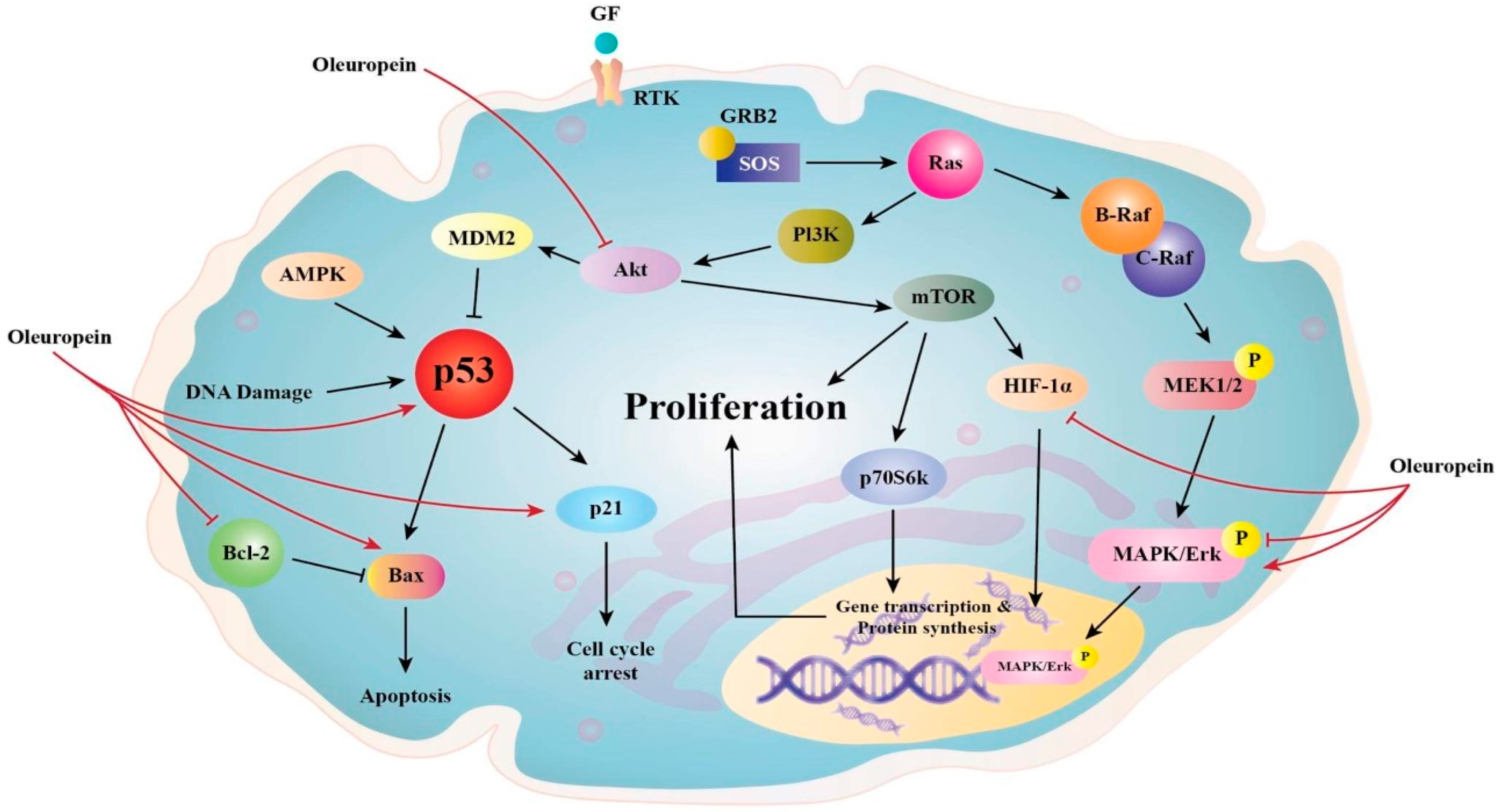
3.2. Anti-Angiogenic and Apoptotic of Ole

Ole’s anti-angiogenic effect in in vivo breast cancer cell lines may be mediated by lowering the vascular endothelial growth factor (VEGF) at doses of 225 mg/kg/day for 3 weeks, administered in distilled water via gastric lavage [8]. This conclusion is supported by the fact that Ole (150 and 225 mg/kg/day), in vivo, decreases breast tumor volume, and this is related to the increase in endostatin expression [8].
In vivo and in vitro studies indicate that endostatin competes with VEGF to bind its receptor, which further prevents its phosphorylation and downstream pathway activation [8][55]. In the in vitro MDA-MB-321 breast cancer cell line, Ole induced cysteine proteases with aspartate specificity (caspase)-3 cleavage at a concentration of 200 µM, which resulted in the induction of cancer cell apoptosis [13]. The same was also observed in the in vitro NSCLC H1299 lung cancer cell line in which Ole, at the same concentration, caused a significant increase in cytochrome c and thus caspase-3 [56]. Ole’s effects on PARP cleavage might be modulated in part by caspase-3 cleavage at 100 µM concentration [27]. A study conducted to elucidate the effects of Ole on gene expression of breast cancer cells showed that in MIDA-MB-468 in vitro cells, Ole (250 µM in water) increased the expression of many caspases including caspase 1 and 14, which have a role in initiating apoptosis [57], while in MDA-MB-231 in vitro cells, Ole (500 µM in water) caused an increase in caspase 4 expression, which also promotes apoptosis [57].
Bcl-2-associated X protein (Bax) and Bcl2 have been demonstrated to promote apoptosis in U251 and A172 glioma cells by increasing caspase-3 and 9 expressions [44]. Bax and Bcl2 are proteins that regulate the apoptotic pathway in mitochondria through caspases. Bax is pro-apoptotic (promotes death), whereas Bcl2 is anti-apoptotic (prevents apoptosis and promotes survival).
Ole (200 µM) raised the ratio of Bax/Bcl2, favoring the apoptotic pathway in MIA PaCa-2 pancreatic cancer cells better than HT (100 µM), although this is not found in healthy cell lines [58]. Similar effects were observed in NSCLC H1299 lung cancer cells in vitro at 200 µM [56], breast neuroblastoma in vitro at 350 µM [54], and Hela cervical cancer cells in vitro at 200 µM [23]. Evidence suggests that Ole (200 µM) may affect the proapoptotic gene tumor protein P53 (p53) as well as the Bax/bcl2 ratio, which favors apoptosis. P53 and Bax are both proapoptotic, according to studies on MCF-7 in in vitro breast cancer cells [24]. Ole caused apoptosis in breast [24] and colon tumors at 400 and 800 µM [30] but not in U251 and A172 in in vitro glioma cancer cells at 200 or 400 µM [44]. Ole (200 µM) enhanced apoptosis in in vitro MIA PaCa-2 pancreatic cancer cells via the dimerization of c-Jun and c-Fos into API [58]. In addition to Bax/Bcl2, Ole (200 µM) promoted apoptosis in in vitro MIA PaCa-2 pancreatic cancer cells via the dimerization of c-Jun and c-Fos into AP1 [58]. In in vitro, Ole promoted cell apoptosis in TCAM-2 and SEM-1 cells by concomitantly enhancing the pro-apoptotic potential in these cells by the overexpression of BAX [29].
Another important mechanism in which Ole has a role in apoptosis is the p38 MAPK pathway [56]. One study found that Ole’s (200 µM) apoptotic ability is mediated by the p38/ATF-2 pathway in in vitro NSCLC H1299 lung cancer [56]. Similar results were seen in MCF-7 breast cancer [24], Hela cervical cancer cell [24], in vitro A549 lung cancer (200 µM) [59], and in vitro neuroblastoma SH-SY5Y [54] cell lines. However, treatment with Ole at doses of 200 or 400 in glioma cancer cells in vitro had no effect on the p38, ERK, or Jun N-terminal kinase (JNK) pathways [44].

3.3. Antioxidant Properties of Ole

The antioxidant effects of Ole and its derivatives are mediated by the breakdown of the radical chain [60]. Ole (100 µmol/L dissolved in dimethyl sulfoxide) showed partial antioxidant and pro-oxidant characteristics in HepG2 hepatocarcinoma cells in vitro, with no harmful effects, according to one study. The activation of reactive oxygen species (ROS) is responsible for this pro-oxidant property [14]. Furthermore, the antioxidant impact of Ole is dependent on the cell type, time, and concentration of exposure [14][58]. Antioxidant effects were identified in non-malignant tissues; however, pro-oxidant stimulating ROS activation was seen in cancer tissue such as breast, prostate, and leukemia HL-60 cells [14]. Furthermore, a concentration larger than 100 µmol/l caused cell death, whereas a concentration less than that had an antioxidant effect [14]. In vitro, Ole (500 µM) possesses antioxidant and pro-oxidant effects on non-tumorous BPH-1 prostate cells as well as LNCaP and DU145 tumor cells [51]. Furthermore, an increase in the heme-oxygenase 1 (HO-1) enzyme at doses of 100 and 500 µM, which is a potent antioxidant containing thiol groups, is thought to be the mechanism by which the antioxidant action is exclusive to BPH-1 cells [51]. Several antioxidants, including glutathione (GSH) and others, need the presence of thiol groups [61]. Furthermore, rather than a change in GSH production via glutamyl cysteine synthetase (GCS) expression, the antioxidant impact in the in vitro prostate DU145 cell line is likely to be associated with a reduction in ROS [51]. According to certain studies, Ole’s antioxidant action has a chemo-protective effect, as evidenced by the fact that it slows colon cancer progression [15]. In in vitro TPC-1 and BCPAP thyroid cancer cell lines, the antioxidant effect of Ole and peracetylated Ole at concentrations of 100 µM was also detected [48]. Ole (as a dissolved solution in culture conditions) is thought to minimize oxidative stress by modulating intracellular GSH, a powerful antioxidant molecule. This was seen in vitro in human glioblastoma cells (U87) by Ole at 10 µM. Furthermore, Ole pre-treatment dramatically reduced NO and inducible nitric oxide synthase iNOS gene expression in these cells [62], supporting the chemo-protective role. The capacity of ROS to activate the Akt pathway could explain why giving Ole to cancer cells causes cell cycle arrest. Because ROS plays such an important part in cancer [63], it may potentially play a function in malignancy. Ole promotes ROS production in cancerous tissues while also inhibiting the Akt pathway, as previously mentioned. This dual impact could lead to an increase in intracellular ROS, which could lead to cell arrest [63][64].

3.4. Ole and Cell Viability

Breast cancer is divided into three subtypes based on chemotherapeutic sensitivity: (1) estrogen receptor-positive (ER+); (2) overexpressing human epidermal growth factor receptor 2 (HER2+), which can be ER+ or ER− and (3) triple-negative (TN), which lacks estrogen, progesterone, and HER2 receptor expression [5]. In HER2+ breast cancer cells, Ole aglycone (6.25–100 µM) was observed to reduce cell viability and apoptosis. Reduced extracellular domain cleavage, autophosphorylation, and HER2 expression are all linked to this action [5]. Ole failed to diminish cell viability in vitro hepatic cancer cells [14], but it did reduce cell viability in pancreatic cancer cells [58]. Furthermore, this effect was found to be specific to tumor cells and not to healthy cells [58]. A study comparing the effects of Ole on ER-negative MDA-MB-231 and ER-positive MCF-7 in in vitro cells found that the former is more responsive to Ole treatment than the latter at concentrations of 100 µM [27]. Most recently, a study carried out by Bossio et al. demonstrated that Ole (15–200 μM) has an inhibition effect on the cell viability assay in a dose-dependent manner in both intra- and extragonadal TCAM-2 and SEM-1 seminoma cells [29].

3.5. Ole and Cell Cycle Arrest

The effects of Ole on the cell cycle have been verified in numerous research. Ole (100 µg/mL) was shown to be more effective than its derivative HT (25 µg/mL) in slowing the transition from G1 to S phase in MCF-7 breast cancer cell lines [5][25]. Ole also produced a delay in the S phase cell cycle in MDA-MB-231 breast cancer cells in another in vitro investigation by upregulating p21 [13][65][66]. The effect of Ole on the MDA-MB-231 cell cycle was shown to be at the sub-G1 phase [53].

Ole also slowed mitosis, which is thought to be related to a reduction in COX-2, which interrupts the cell cycle in the G2/M phase. This effect was detected in mice given 150 and 225 mg/kg/day of Ole diluted in distilled water by gastric lavage [8]. The down-regulation of cyclin-D1, 2 and 3, and CDK4 and 6 gene expression, as well as the up-regulation of p53 [58] and cyclin-dependent kinase inhibitor gene expression are likely to be responsible for the cell cycle arrest effects of Ole (350 µM) on in vitro neuroblastoma [54]. In neuroblastoma, Ole has a particular effect on the G1/S phase [57]. Cyclins and their allosteric activators, cyclin-dependent kinases (CDK), play critical roles in cell cycle control. The overexpression of cyclins, particularly cyclin D1, has been confirmed to have a role in cancer. Cyclin D1 can regulate the G1/S phase of the cell cycle via retinoblastoma (Rb) phosphorylation. Pancreatic cancer; non-small cell lung carcinoma; breast, NSCLC, head, and neck squamous cell carcinoma; melanoma; and endometrial, colorectal, and mantle cell lymphoma malignancies all have hyperphosphorylation of Rb [65].
Furthermore, Ole (200 µM) or its metabolite HT (100 µM) suppresses the cell cycle in the G2 phase in MIA PaCA-2 pancreatic cancer cells in vitro [58].

3.6. Ole as a Cytoskeleton Disruptor

Ole disrupted actin filaments in the cytoskeleton of breast cancer cells (MCF-7) within two hours in vivo. Interestingly, when Ole was combined with D-glucose, the potential of Ole to disrupt the cytoskeleton was reduced, suggesting the involvement of the glucose transporters (GLUTs) [67]. Ole’s capacity to cause cell rounding in ovarian cancer cells is attributed to the disruption of the actin cytoskeleton, which hinders these cells from replicating and invasiveness [13].

3.7. Ole and Fatty Acid Synthase

Ole’s anti-cancer benefits could be attributed to its capacity to inhibit the fatty acid synthase enzyme (FASN), which could be linked to the fact that Ole modifies gene expression and enzymatic activity of this enzyme, as seen in colon cancer cell lines SW620 and HT-29 in vitro [15]. FASN is overexpressed in a variety of cancers, including prostate, ovarian, breast, endometrial, thyroid, colorectal, bladder, lung, thyroid, oral, tongue, esophageal, hepatocellular, pancreatic, and gastric carcinomas; malignant melanoma; mesothelioma; nephroblastoma; and retinoblastoma; as well as soft tissue sarcoma [68]. In tumor cells, the overexpression of this enzyme impacts mitochondrial activity as well as peroxisomes, nuclei, and endoplasmic reticula. Furthermore, unlike healthy cells, cancers use fatty acid in de novo synthesis regardless of circulating lipid levels [69].

3.8. Ole and Inflammation

Inflammation is a condition that occurs in injured cells and is characterized by the production of pro-inflammatory cytokines such as IL-6 and IL-1, as well as the activation of COX and iNOS [1]. The activation of the NF-kB pathway by these cytokines, as well as the overexpression of MMP are all part of the inflammation process [1]. There is a well-established relationship between chronic inflammation and cancer progression and development. At various phases of cancer progression, all immune cells are implicated. This persistent inflammation as well as the proinflammatory cytokines IL-1 and IL-6 promote tumor formation in lung and breast cancer models [41]. Many proinflammatory cytokines, including IL-6, IL-8, IL-1, and TNF-, have been identified as altering bone tumor angiogenesis, progression, and tumor microenvironment [40]. One of the most important properties of a premetastatic niche is inflammation [44]. Ole aglycone (10 µM for 5 weeks) suppresses the establishment of a premetastatic niche by lowering the release of IL-8 and MMP in senescence-associated secretory-phenotype cells [1].
In vitro (human whole blood) investigations have found that Ole (104 µM) reduces IL-1 relative to the other olive oil phenols and induces a decrease in the expressions of IL-6, iNOS, NF-kB, and JNK in murine RAW 264.7 cells at 300 µM [1], while the effects of Ole on colonic biopsies taken from human patients with ulcerative colitis showed that Ole decreases COX and IL-17, which are upregulated in this disease. This may be questionable because the metabolism of Ole in these models may be different than in humans, as stated in the Metabolism of Ole section. However, the effects of Ole on colonic biopsies taken from human patients with ulcerative colitis showed that Ole decreases COX and IL-17, which are upregulated in this disease [1].
The only olive phenol that has anti-inflammatory properties in polymorphonuclear cells isolated from human samples was Ole (80 µg/mL) [70]. TNF secretion is reduced, and this impact is apparent at greater concentrations (320 µg/mL) [70].

4. Derivatives of Ole

Due to Ole’s hydrophilic character, researchers found that more stable hydrophobic derivatives of Ole had stronger anti-proliferative and antioxidant effects in MCF-7 and T-47D breast cancer cells in vitro [60]. The improved efficacy of the derivatives, according to the researchers, is related to the creation of better antioxidants by enzymatic breakdown of the derivatives; additionally, the compounds are more hydrophobic, which facilitates membrane integration [60]. A recent study found that liposomes containing Ole and HT are an optimal delivery strategy for delivering Ole directly into cells without the complications and variables associated with Ole metabolism [71]. The antioxidant activity of the more lipophilic Ole acetylated derivatives, particularly acetylated Ole aglycone, was found to be higher in mice given Ole, Ole aglycone, and their semisynthetic acetylated derivatives [72]. In another study, the acetylation of Ole’s glucose moiety increased lipophilicity and permeability as well as improved anti-inflammatory and antioxidant profiles in murine peritoneal macrophages [71].
Many researchers conducted trials employing different drug delivery technologies to better distribute Ole inside infected cells since Ole has a variable metabolism and bioavailability profile. Nanostructured lipid carriers loaded with Ole were used in one of these studies. These carriers showed good encapsulation efficiency and effective Ole loading. Ole showed sustained release kinetics after assessing the features of the delivery system, indicating that Ole was stable inside the lipid carriers and did not experience any degradation, which is further verified by antioxidation activity against A549 lung cancer cells [72].

5. Ole in Combination with Chemotherapy

A combination of Ole with other anticancer agents may be beneficial in chemotherapeutic resistance. This was observed from the in vitro and in vivo experiments of the combinations of SKBR3/Tzb100 in SKBR3/Tzb100 (trastuzumab with Ole aglycone) [5][73] and doxorubicin with Ole, [5][13]. In in vivo models of breast cancer mice, the combination of Ole (50 mg/kg) and doxorubicin (1.5 mg/kg) decreased tumor growth and size, caused apoptosis, and affected the protein cytoskeleton of tumor cells [13][73]. This combination also suppressed Bcl2 and survival expression, both of which prevent apoptosis [13]. It also downregulated NF-kB, cyclin D1, and COX2; enhanced capsapse-3 cleavage and Bax expression; and boosted capsapse-3 cleavage and Bax expression. In in vivo male Winstar rats, Ole showed a protective effect when combined with cyclophosphamide [73].
In the case of cisplatin-induced renal impairment, combining Ole with cisplatin treatment could be beneficial. The ERK pathway has been shown to mediate Ole’s reno-protective action [74]. In vivo and in vitro investigations verified that ERK activation is linked to inflammation and apoptosis, as evidenced by the activation of downstream targets such as COX and TNFa [74]. In contrast to Ole monotherapy, adding Ole to 50 µM cisplatin significantly reduces NO levels and cell survival rate while also inducing cytotoxicity in in vitro HepG2 hepatocarcinoma cells [45]. Furthermore, Ole (100–400 µM) improved the efficacy of cisplatin (50 µM) to inhibit MMP-7 gene expression [39]. The same protective effect was observed in the lungs, stomach, and pancreas [73]. In in vitro human melanoma cells (A375), Ole was found to have a synergistic impact when combined with Decarbonize and Everolimus, as well as Vemurafenib [73]. When Ole (100 µM) was combined with 2-metoxyestradiol (10 µM) in human osteosarcoma cells, a synergistic effect was seen [73].

6. Summary and Conclusions

Ole’s anticancer properties are numerous and encompass numerous mechanisms that are presently being explored. Ole inhibits cell proliferation by interacting with the AKT/NF-kB pathway and COX2, PPAR, MMP, and CB receptors. Ole’s potential to impact PARPs, the Bax/Bcl2 ratio, the P38 MAPK pathway, HIF-1α, and the Akt and Jnk pathways disrupts cancer’s anti-apoptotic and angiogenic capabilities. Ole decreases cell viability, causes cell cycle arrest, and functions as a cytoskeleton disruptor in many cancer types in addition to its antioxidant properties and unusual complex formation with copper.
As concluded from various studies, Ole has a variable metabolism and bioavailability profile and it undergoes extensive first-pass metabolism. Ole metabolism is mediated by several metabolic processes, including de-glycosylation, hydrolysis, oxygenation, and methylation. More human studies are needed to discover the various factors influencing Ole metabolism and absorption. Derivatives of Ole have improved its efficacy for example as an antioxidant and for anti-proliferation.
Ole have aided in cancer treatment over the years. Ole functions as an anti-cancer agent by having anti-proliferative, anti-angiogenic and apoptotic, antioxidant, fatty acid synthase inhibiting, cytoskeleton disrupting, and anti-inflammatory properties. Oleuropein effects on cancer cells are influenced by Ole concentration, exposure time, and cancer cell type. Ole has strong synergistic effects when combined with anticancer medicines such as doxorubicin and other. Ole is a prospective anticancer candidate that could be utilized as a supplement to existing anticancer therapy guidelines or as a recurrence prevention therapy.

References

  1. Nediani, C.; Ruzzolini, J.; Romani, A.; Calorini, L. Oleuropein, a bioactive compound from Olea europaea L., as a potential preventive and therapeutic agent in non-communicable diseases. Antioxidants 2019, 8, 578.
  2. Rahiman, S.; El-Metwally, T.H.; Shrivastava, D.; Tantry, M.N.; Tantry, B.A. Oleuropein and oleic acid: A novel emerging dietary target for human chronic diseases. Indian J. Biochem. Biophys. (IJBB) 2019, 56, 263–268.
  3. Bulotta, S.; Oliverio, M.; Russo, D.; Procopio, A. Biological Activity of Oleuropein and Its Derivatives; Springer: Berlin/Heidelberg, Germany, 2013.
  4. Al-Asmari, K.; Al-Attar, A.; Mohamed, I.; Zeid, A. Potential health benefits and components of olive oil: An overview. Biosci. Res. 2020, 17, 2673–2687.
  5. Shamshoum, H.; Vlavcheski, F.; Tsiani, E. Anticancer effects of oleuropein. Biofactors 2017, 43, 517–528.
  6. Moran, J.M.; Leal-Hernandez, O.; Canal-Macias, M.L.; Roncero-Martin, R.; Guerrero-Bonmatty, R.; Aliaga, I.; Zamorano, J.D.P. Antiproliferative properties of oleuropein in human osteosarcoma cells. Nat. Prod. Commun. 2016, 11, 491–492.
  7. Breakspear, I.; Guillaume, C. A quantitative phytochemical comparison of olive leaf extracts on the Australian market. Molecules 2020, 25, 4099.
  8. Milanizadeh, S.; Reza Bigdeli, M. Pro-apoptotic and anti-angiogenesis effects of olive leaf extract on spontaneous mouse mammary tumor model by balancing vascular endothelial growth factor and endostatin levels. Nutr. Cancer 2019, 71, 1374–1381.
  9. Emma, M.R.; Augello, G.; Di Stefano, V.; Azzolina, A.; Giannitrapani, L.; Montalto, G.; Cervello, M.; Cusimano, A. Potential uses of olive oil secoiridoids for the prevention and treatment of cancer: A narrative review of preclinical studies. Int. J. Mol. Sci. 2021, 22, 1234.
  10. Horcajada, M.-N.; Beaumont, M.; Sauvageot, N.; Poquet, L.; Saboundjian, M.; Costes, B.; Verdonk, P.; Brands, G.; Brasseur, J.; Urbin-Choffray, D. An oleuropein-based dietary supplement may improve joint functional capacity in older people with high knee joint pain: Findings from a multicentre-RCT and post hoc analysis. Ther. Adv. Musculoskelet. Dis. 2022, 14, 1–15.
  11. Carnevale, R.; Silvestri, R.; Loffredo, L.; Novo, M.; Cammisotto, V.; Castellani, V.; Bartimoccia, S.; Nocella, C.; Violi, F. Oleuropein, a component of extra virgin olive oil, lowers postprandial glycaemia in healthy subjects. Br. J. Clin. Pharmacol. 2018, 84, 1566–1574.
  12. Kendall, M.; Batterham, M.; Callahan, D.L.; Jardine, D.; Prenzler, P.D.; Robards, K.; Ryan, D. Randomized controlled study of the urinary excretion of biophenols following acute and chronic intake of olive leaf supplements. Food Chem. 2012, 130, 651–659.
  13. Elamin, M.H.; Elmahi, A.B.; Daghestani, M.H.; Al-Olayan, E.M.; Al-Ajmi, R.A.; Alkhuriji, A.F.; Hamed, S.S.; Elkhadragy, M.F. Synergistic anti-breast-cancer effects of combined treatment with oleuropein and doxorubicin in vivo. Altern. Ther. Health Med. 2017, 23, 17–24.
  14. Katsoulieris, E.N. The olive leaf extract oleuropein exerts protective effects against oxidant-induced cell death, concurrently displaying pro-oxidant activity in human hepatocarcinoma cells. Redox Rep. 2016, 21, 90–97.
  15. Imran, M.; Nadeem, M.; Gilani, S.A.; Khan, S.; Sajid, M.W.; Amir, R.M. Antitumor perspectives of oleuropein and its metabolite hydroxytyrosol: Recent updates. J. Food Sci. 2018, 83, 1781–1791.
  16. García-Villalba, R.; Larrosa, M.; Possemiers, S.; Tomás-Barberán, F.; Espín, J. Bioavailability of phenolics from an oleuropein-rich olive (Olea europaea) leaf extract and its acute effect on plasma antioxidant status: Comparison between pre-and postmenopausal women. Eur. J. Nutr. 2014, 53, 1015–1027.
  17. Kano, S.; Komada, H.; Yonekura, L.; Sato, A.; Nishiwaki, H.; Tamura, H. Absorption, metabolism, and excretion by freely moving rats of 3, 4-DHPEA-EDA and related polyphenols from olive fruits (Olea europaea). J. Nutr. Metab. 2016, 2016, 9104208.
  18. Annunziata, G.; Maisto, M.; Schisano, C.; Barrea, L.; Ciampaglia, R.; Novellinoet, E. Oleuropein as a novel anti-diabetic nutraceutical. Overv. Arch. Diabetes Obes. 2018, 1, 54–58.
  19. De Bock, M.; Thorstensen, E.B.; Derraik, J.G.; Henderson, H.V.; Hofman, P.L.; Cutfield, W.S. Human absorption and metabolism of oleuropein and hydroxytyrosol ingested as olive (Olea europaea L.) leaf extract. Mol. Nutr. Food Res. 2013, 57, 2079–2085.
  20. Gikas, E.; Papadopoulos, N.; Tsarbopoulos, A. Kinetic study of the acidic hydrolysis of oleuropein, the major bioactive metabolite of olive oil. J. Liq. Chromatogr. Relat. Technol. 2006, 29, 497–508.
  21. Galmés, S.; Reynés, B.; Palou, M.; Palou-March, A.; Palou, A. Absorption, distribution, metabolism, and excretion of the main olive tree phenols and polyphenols: A literature review. J. Agric. Food Chem. 2021, 69, 5281–5296.
  22. Bonechi, C.; Donati, A.; Tamasi, G.; Pardini, A.; Rostom, H.; Leone, G.; Lamponi, S.; Consumi, M.; Magnani, A.; Rossi, C. Chemical characterization of liposomes containing nutraceutical compounds: Tyrosol, hydroxytyrosol and oleuropein. Biophys. Chem. 2019, 246, 25–34.
  23. Rizzo, M.; Ventrice, D.; Giannetto, F.; Cirinnà, S.; Santagati, N.A.; Procopio, A.; Mollace, V.; Muscoli, C. Antioxidant activity of oleuropein and semisynthetic acetyl-derivatives determined by measuring malondialdehyde in rat brain. J. Pharm. Pharmacol. 2017, 69, 1502–1512.
  24. Liman, R.; Çoban, F.; Ciğerci, I.; Bulduk, İ.; Bozkurt, S. Antiangiogenic and apoptotic effects of Oleuropein on breast cancer cells. Br. J. Pharm. Res 2017, 16, 1–10.
  25. Han, J.; Talorete, T.P.; Yamada, P.; Isoda, H. Anti-proliferative and apoptotic effects of oleuropein and hydroxytyrosol on human breast cancer MCF-7 cells. Cytotechnology 2009, 59, 45–53.
  26. Ci, Y.; Qiao, J.; Han, M. Molecular mechanisms and metabolomics of natural polyphenols interfering with breast cancer metastasis. Molecules 2016, 21, 1634.
  27. Liu, L.; Ahn, K.S.; Shanmugam, M.K.; Wang, H.; Shen, H.; Arfuso, F.; Chinnathambi, A.; Alharbi, S.A.; Chang, Y.; Sethi, G. Oleuropein induces apoptosis via abrogating NF-κB activation cascade in estrogen receptor–negative breast cancer cells. J. Cell. Biochem. 2019, 120, 4504–4513.
  28. Brasier, A.R. The nuclear factor-κB–interleukin-6 signalling pathway mediating vascular inflammation. Cardiovasc. Res. 2010, 86, 211–218.
  29. Bossio, S.; Perri, A.; Malivindi, R.; Giordano, F.; Rago, V.; Mirabelli, M.; Salatino, A.; Brunetti, A.; Greco, E.A.; Aversa, A. Oleuropein Counteracts Both the Proliferation and Migration of Intra-and Extragonadal Seminoma Cells. Nutrients 2022, 14, 2323.
  30. Cárdeno, A.; Sánchez-Hidalgo, M.; Rosillo, M.A.; de la Lastra, C.A. Oleuropein, a secoiridoid derived from olive tree, inhibits the proliferation of human colorectal cancer cell through downregulation of HIF-1α. Nutr. Cancer 2013, 65, 147–156.
  31. Liu, T.; Zhang, L.; Joo, D.; Sun, S. Signal Transduct. Target. Ther 2017, 2, e17023.
  32. Sheng, J.; Sun, H.; Yu, F.-B.; Li, B.; Zhang, Y.; Zhu, Y.-T. The role of cyclooxygenase-2 in colorectal cancer. Int. J. Med. Sci. 2020, 17, 1095.
  33. Giner, E.; Recio, M.C.; Ríos, J.L.; Cerdá-Nicolás, J.M.; Giner, R.M. Chemopreventive effect of oleuropein in colitis-associated colorectal cancer in c57bl/6 mice. Mol. Nutr. Food Res. 2016, 60, 242–255.
  34. Nuñez, F.; Bravo, S.; Cruzat, F.; Montecino, M.; De Ferrari, G.V. Wnt/β-catenin signaling enhances cyclooxygenase-2 (COX2) transcriptional activity in gastric cancer cells. PLoS ONE 2011, 6, e18562.
  35. Deng, L.; Liang, H.; Han, Y. Cyclooxygenase-2 and β-catenin as potential diagnostic and prognostic markers in endometrial cancer. Front. Oncol. 2020, 10, 56.
  36. Vallée, A.; Lecarpentier, Y.; Vallée, J.-N. Targeting the canonical WNT/β-catenin pathway in cancer treatment using non-steroidal anti-inflammatory drugs. Cells 2019, 8, 726.
  37. Casado-Díaz, A.; Anter, J.; Müller, S.; Winter, P.; Quesada-Gómez, J.M.; Dorado, G. Transcriptomic analyses of the anti-adipogenic effects of oleuropein in human mesenchymal stem cells. Food Funct. 2017, 8, 1254–1270.
  38. Svobodova, M.; Andreadou, I.; Skaltsounis, A.-L.; Kopecky, J.; Flachs, P. Oleuropein as an inhibitor of peroxisome proliferator-activated receptor gamma. Genes Nutr. 2014, 9, 1–8.
  39. Gou, Q.; Gong, X.; Jin, J.; Shi, J.; Hou, Y. Peroxisome proliferator-activated receptors (PPARs) are potential drug targets for cancer therapy. Oncotarget 2017, 8, 60704.
  40. Leto, G.; Flandina, C.; Crescimanno, M.; Giammanco, M.; Sepporta, M.V. Effects of oleuropein on tumor cell growth and bone remodelling: Potential clinical implications for the prevention and treatment of malignant bone diseases. Life Sci. 2021, 264, 118694.
  41. Ritter, B.; Greten, F.R. Modulating inflammation for cancer therapy. J. Exp. Med. 2019, 216, 1234–1243.
  42. Razali, R.A.; Lokanathan, Y.; Yazid, M.D.; Ansari, A.S.; Saim, A.B.; Idrus, R.B. Modulation of epithelial to mesenchymal transition signaling pathways by Olea europaea and its active compounds. Int. J. Mol. Sci. 2019, 20, 3492.
  43. Liu, Y.; Cao, X. Characteristics and significance of the pre-metastatic niche. Cancer Cell 2016, 30, 668–681.
  44. Liu, M.; Wang, J.; Huang, B.; Chen, A.; Li, X. Oleuropein inhibits the proliferation and invasion of glioma cells via suppression of the AKT signaling pathway. Oncol. Rep. 2016, 36, 2009–2016.
  45. Sherif, I.O.; Al-Gayyar, M.M. Oleuropein potentiates anti-tumor activity of cisplatin against HepG2 through affecting proNGF/NGF balance. Life Sci. 2018, 198, 87–93.
  46. Winer, A.; Adams, S.; Mignatti, P. Matrix metalloproteinase inhibitors in cancer therapy: Turning past failures into future successes. Mol. Cancer Ther. 2018, 17, 1147–1155.
  47. Quintero-Fabián, S.; Arreola, R.; Becerril-Villanueva, E.; Torres-Romero, J.C.; Arana-Argáez, V.; Lara-Riegos, J.; Ramírez-Camacho, M.A.; Alvarez-Sánchez, M.E. Role of matrix metalloproteinases in angiogenesis and cancer. Front. Oncol. 2019, 9, 1370.
  48. Bulotta, S.; Corradino, R.; Celano, M.; Maiuolo, J.; D’Agostino, M.; Oliverio, M.; Procopio, A.; Filetti, S.; Russo, D. Antioxidant and antigrowth action of peracetylated oleuropein in thyroid cancer cells. J. Mol. Endocrinol. 2013, 51, 181–189.
  49. Guo, Y.J.; Pan, W.W.; Liu, S.B.; Shen, Z.F.; Xu, Y.; Hu, L.L. ERK/MAPK signalling pathway and tumorigenesis. Exp. Ther. Med. 2020, 19, 1997–2007.
  50. Nitulescu, G.M.; Van De Venter, M.; Nitulescu, G.; Ungurianu, A.; Juzenas, P.; Peng, Q.; Olaru, O.T.; Grădinaru, D.; Tsatsakis, A.; Tsoukalas, D. The Akt pathway in oncology therapy and beyond. Int. J. Oncol. 2018, 53, 2319–2331.
  51. Acquaviva, R.; Di Giacomo, C.; Sorrenti, V.; Galvano, F.; Santangelo, R.; Cardile, V.; Gangia, S.; D’Orazio, N.; Abraham, N.G.; Vanella, L. Antiproliferative effect of oleuropein in prostate cell lines. Int. J. Oncol. 2012, 41, 31–38.
  52. Laezza, C.; Pagano, C.; Navarra, G.; Pastorino, O.; Proto, M.C.; Fiore, D.; Piscopo, C.; Gazzerro, P.; Bifulco, M. The endocannabinoid system: A target for cancer treatment. Int. J. Mol. Sci. 2020, 21, 747.
  53. Di Francesco, A.; Falconi, A.; Di Germanio, C.; Di Bonaventura, M.V.M.; Costa, A.; Caramuta, S.; Del Carlo, M.; Compagnone, D.; Dainese, E.; Cifani, C. Extravirgin olive oil up-regulates CB1 tumor suppressor gene in human colon cancer cells and in rat colon via epigenetic mechanisms. J. Nutr. Biochem. 2015, 26, 250–258.
  54. Seçme, M.; Eroğlu, C.; Dodurga, Y.; Bağcı, G. Investigation of anticancer mechanism of oleuropein via cell cycle and apoptotic pathways in SH-SY5Y neuroblastoma cells. Gene 2016, 585, 93–99.
  55. Walia, A.; Yang, J.F.; Huang, Y.-H.; Rosenblatt, M.I.; Chang, J.-H.; Azar, D.T. Endostatin’s emerging roles in angiogenesis, lymphangiogenesis, disease, and clinical applications. Biochim. Biophys. Acta (BBA)-Gen. Subj. 2015, 1850, 2422–2438.
  56. Wang, W.; Wu, J.; Zhang, Q.; Li, X.; Zhu, X.; Wang, Q.; Cao, S.; Du, L. Mitochondria-mediated apoptosis was induced by oleuropein in H1299 cells involving activation of p38 MAP kinase. J. Cell. Biochem. 2019, 120, 5480–5494.
  57. Messeha, S.S.; Zarmouh, N.O.; Asiri, A.; Soliman, K.F. Gene Expression Alterations Associated with Oleuropein-Induced Antiproliferative Effects and S-Phase Cell Cycle Arrest in Triple-Negative Breast Cancer Cells. Nutrients 2020, 12, 3755.
  58. Goldsmith, C.D.; Bond, D.R.; Jankowski, H.; Weidenhofer, J.; Stathopoulos, C.E.; Roach, P.D.; Scarlett, C.J. The olive biophenols oleuropein and hydroxytyrosol selectively reduce proliferation, influence the cell cycle, and induce apoptosis in pancreatic cancer cells. Int. J. Mol. Sci. 2018, 19, 1937.
  59. Cao, S.; Zhu, X.; Du, L. P38 MAP kinase is involved in oleuropein-induced apoptosis in A549 cells by a mitochondrial apoptotic cascade. Biomed. Pharmacother. 2017, 95, 1425–1435.
  60. Bulotta, S.; Corradino, R.; Celano, M.; D’Agostino, M.; Maiuolo, J.; Oliverio, M.; Procopio, A.; Iannone, M.; Rotiroti, D.; Russo, D. Antiproliferative and antioxidant effects on breast cancer cells of oleuropein and its semisynthetic peracetylated derivatives. Food Chem. 2011, 127, 1609–1614.
  61. Ulrich, K.; Jakob, U. The role of thiols in antioxidant systems. Free Radic. Biol. Med. 2019, 140, 14–27.
  62. Kucukgul, A.; Isgor, M.M.; Duzguner, V.; Atabay, M.N.; Kucukgul, A. Antioxidant effects of oleuropein on hydrogen peroxide-induced neuronal stress-an in vitro study. Anti-Inflamm. Anti-Allergy Agents Med. Chem. (Former. Curr. Med. Chem.-Anti-Inflamm. Anti-Allergy Agents) 2020, 19, 74–84.
  63. Koundouros, N.; Poulogiannis, G. Phosphoinositide 3-kinase/Akt signaling and redox metabolism in cancer. Front. Oncol. 2018, 8, 160.
  64. Omar, S.H. Cardioprotective and neuroprotective roles of oleuropein in olive. Saudi Pharm. J. 2010, 18, 111–121.
  65. Qie, S.; Diehl, J.A. Cyclin D1, cancer progression, and opportunities in cancer treatment. J. Mol. Med. 2016, 94, 1313–1326.
  66. Leal-Esteban, L.C.; Fajas, L. Cell cycle regulators in cancer cell metabolism. Biochim. Biophys. Acta (BBA)-Mol. Basis Dis. 2020, 1866, 165715.
  67. Hamadi, H.; Castellon, R. Oleuropein, a non-toxic olive iridoid, is an anti-tumor agent and cytoskeleton disrupter. Biochem. Biophys. Res. Commun. 2005, 334, 769–778.
  68. Flavin, R.; Peluso, S.; Nguyen, P.L.; Loda, M. Fatty acid synthase as a potential therapeutic target in cancer. Future Oncol. 2010, 6, 551–562.
  69. Fhu, C.W.; Ali, A. Fatty acid synthase: An emerging target in cancer. Molecules 2020, 25, 3935.
  70. Qabaha, K.; Al-Rimawi, F.; Qasem, A.; Naser, S.A. Oleuropein is responsible for the major anti-inflammatory effects of olive leaf extract. J. Med. Food 2018, 21, 302–305.
  71. Huguet-Casquero, A.; Moreno-Sastre, M.; López-Méndez, T.B.; Gainza, E.; Pedraz, J.L. Encapsulation of oleuropein in nanostructured lipid carriers: Biocompatibility and antioxidant efficacy in lung epithelial cells. Pharmaceutics 2020, 12, 429.
  72. Castejon, M.L.; Sánchez-Hidalgo, M.; Aparicio-Soto, M.; González-Benjumea, A.; Fernández-Bolaños, J.G.; Alarcón-de-la-Lastra, C. Olive secoiridoid oleuropein and its semisynthetic acetyl-derivatives reduce LPS-induced inflammatory response in murine peritoneal macrophages via JAK-STAT and MAPKs signaling pathways. J. Funct. Foods 2019, 58, 95–104.
  73. Torić, J.; Karković Marković, A.; Jakobušić Brala, C.; Barbarić, M. Anticancer effects of olive oil polyphenols and their combinations with anticancer drugs. Acta Pharm. 2019, 69, 461–482.
  74. Potočnjak, I.; Škoda, M.; Pernjak-Pugel, E.; Peršić, M.P.; Domitrović, R. Oral administration of oleuropein attenuates cisplatin-induced acute renal injury in mice through inhibition of ERK signaling. Mol. Nutr. Food Res. 2016, 60, 530–541.
More
Information
Subjects: Oncology
Contributors MDPI registered users' name will be linked to their SciProfiles pages. To register with us, please refer to https://encyclopedia.pub/register : , , ,
View Times: 653
Revisions: 4 times (View History)
Update Date: 12 Aug 2022
1000/1000
ScholarVision Creations