Submitted Successfully!
To reward your contribution, here is a gift for you: A free trial for our video production service.
Thank you for your contribution! You can also upload a video entry or images related to this topic.
Version Summary Created by Modification Content Size Created at Operation
1 -- 4350 2022-07-11 16:17:50 |
2 format change Meta information modification 4350 2022-07-12 05:20:51 |

Video Upload Options

Do you have a full video?

Confirm

Are you sure to Delete?
Cite
If you have any further questions, please contact Encyclopedia Editorial Office.
Bhat, K.A.;  Mir, R.A.;  Farooq, A.;  Manzoor, M.;  Hami, A.;  Allie, K.A.;  Wani, S.M.;  Khan, M.N.;  Sayyed, R.Z.;  Poczai, P.; et al. Nematode Identification. Encyclopedia. Available online: https://encyclopedia.pub/entry/25014 (accessed on 21 May 2024).
Bhat KA,  Mir RA,  Farooq A,  Manzoor M,  Hami A,  Allie KA, et al. Nematode Identification. Encyclopedia. Available at: https://encyclopedia.pub/entry/25014. Accessed May 21, 2024.
Bhat, Kaisar Ahmad, Rakeeb Ahmad Mir, Asmat Farooq, Madhiya Manzoor, Ammarah Hami, Kaisar Ahmad Allie, Shaheen Majeed Wani, M. N. Khan, R. Z. Sayyed, Peter Poczai, et al. "Nematode Identification" Encyclopedia, https://encyclopedia.pub/entry/25014 (accessed May 21, 2024).
Bhat, K.A.,  Mir, R.A.,  Farooq, A.,  Manzoor, M.,  Hami, A.,  Allie, K.A.,  Wani, S.M.,  Khan, M.N.,  Sayyed, R.Z.,  Poczai, P.,  Almalki, W.H.,  Zargar, S.M., & Shah, A.A. (2022, July 11). Nematode Identification. In Encyclopedia. https://encyclopedia.pub/entry/25014
Bhat, Kaisar Ahmad, et al. "Nematode Identification." Encyclopedia. Web. 11 July, 2022.
Nematode Identification
Edit

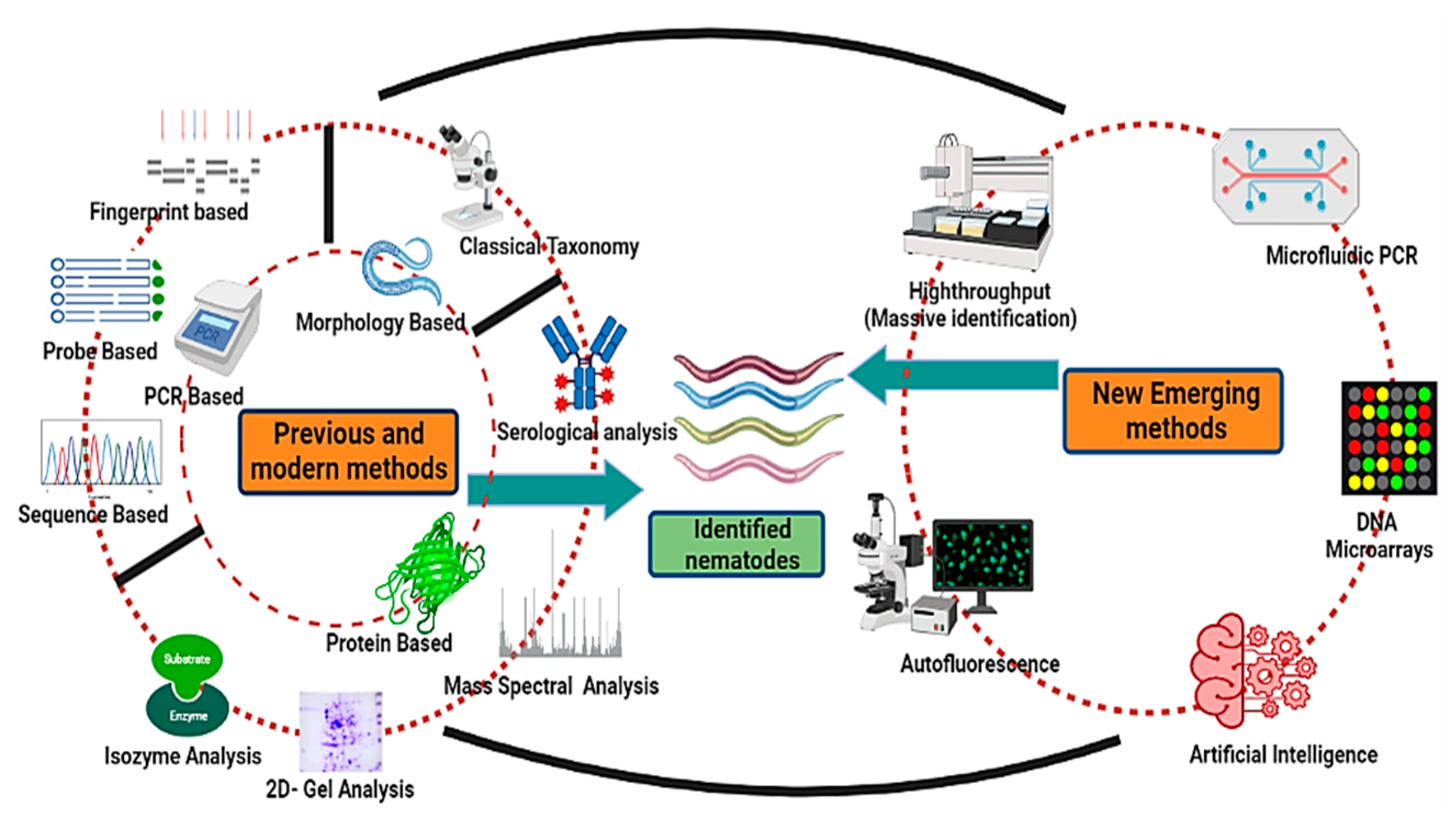
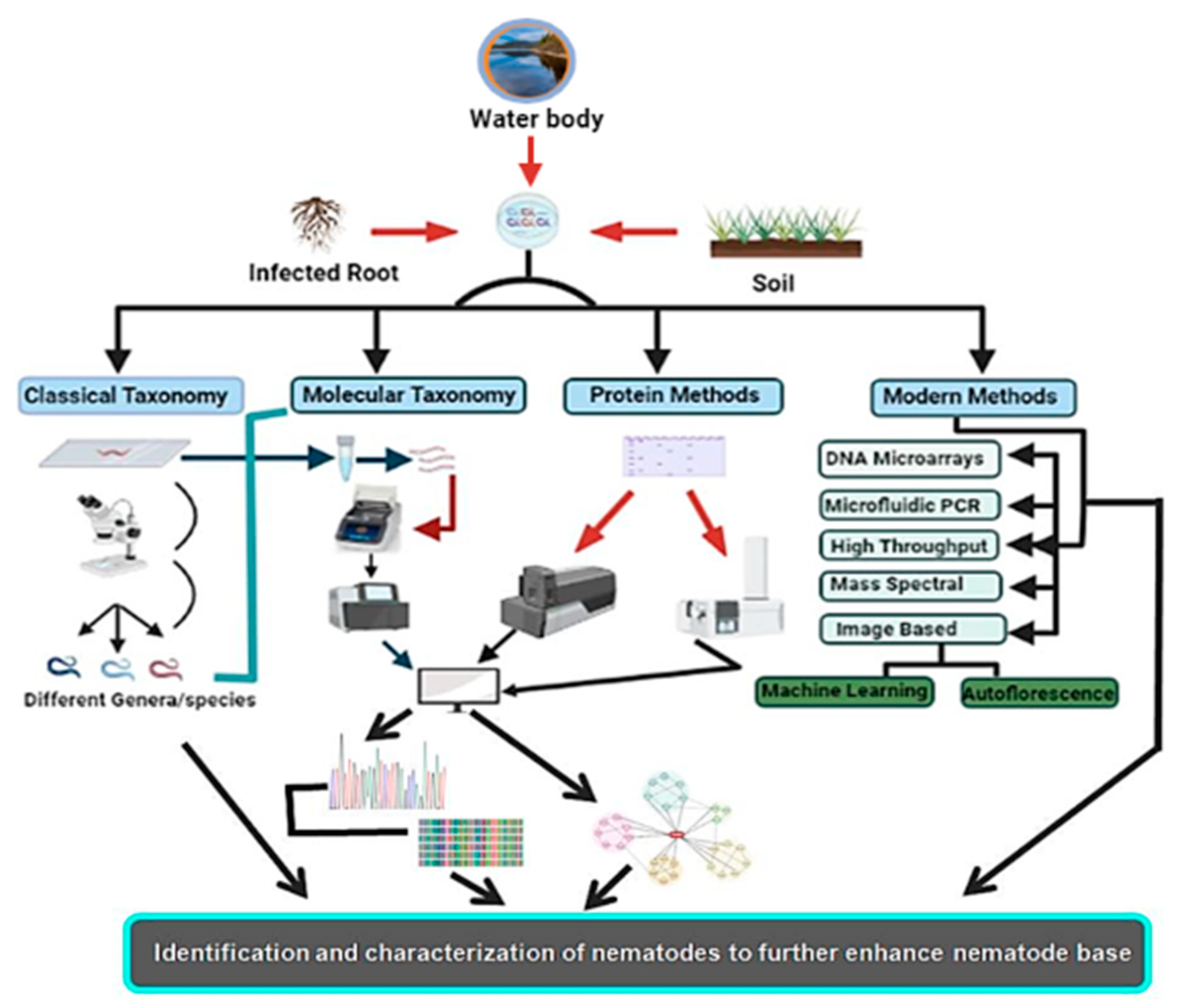
Nematodes are non-segmented roundworms evenly distributed with various habitats ranging to approximately every ecological extremity. These are the least studied organisms despite being the most diversified group. Nematodes are the most critical equilibrium-maintaining factors, having implications on the yield and health of plants as well as well-being of animals. As a result of the lack of precise taxonomic features, nematode taxonomy remains uncertain. Morphology-based identification has proved inefficacious in identifying and exploring the diversity of nematodes, as there are insufficient morphological variations. Different molecular and new evolving methodologies have been employed to augment morphology-based approaches and bypass these difficulties with varying effectiveness. These identification techniques vary from molecular-based targeting DNA or protein-based targeting amino acid sequences to methods for image processing. High-throughput approaches such as next-generation sequencing have also been added to this league. These alternative approaches have helped to classify nematodes and enhanced the base for increased diversity and phylogeny of nematodes, thus helping to formulate increasingly more nematode bases for use as model organisms to study different hot topics about human well-being.

emerging methods identification meta-barcoding morphology PCR nematodes

1. Introduction

Nematodes are non-segmented roundworms that include a widespread group of metazoans with various habitats ranging from land to water. Nematodes are reported to be the most prevalent and copious in these habitats, with species ranging up to one million [1]. Nematodes are the least studied organisms despite such a vast distribution. According to studies, less than 0.01% of these species have been reported [2]. There have been roughly 26,000 species reported so far, out of which 4100 are reported as plant parasites. At the same time, insect-pathogenic nematodes can be used as beneficial organisms in crop production for biological plant protection [3].
Most soil nematodes contribute significantly to nutrient cycling in the natural environment. Some nematodes are reported to be very important in medical and veterinary sciences [4]. Precise detection is, thus, critical for understanding nematode biodiversity and developing important management practices. Primary identification and characterization are mainly based on morphological features, including external body structure; total length; differences in sex organs and stoma; organization of lips, mouth, and tail; and other visible morphological characteristics. This often leads to the absence of precise categorization among strongly linked taxa due to specific observable characteristics and a lack of trained taxonomists and thus becomes insufficient, especially when a large sample size is implicated [5]. Molecular and biochemical identification methods have recently been utilized to overcome the lacuna of morphological identification of nematodes. Other studies have also reported using nematode ribosomal DNA (rDNA) sequencing to evaluate nematode characterization and evolutionary relationships [6].
Consequently, it is important to correctly identify the nematodes and enhance their evolutionary lineages to understand and combat their adverse effects on animal health and plant yield, in addition to fruitful use in nutrient cycling and other related phenomena. Caenorhabditis elegans (C. elegans), a free-living nematode, has proved a good model organism that is preferred by many researchers [7]. Because of its simple structure, good heritable traits, the existence of a comprehensive molecular toolset, and an entire genome sequence, it is appropriate for immediate and efficient regulation of gene expression function. The various techniques viz morphological, DNA-based, protein/biochemical-based, and new emerging methods used to identify and classify nematodes are summarized in Figure 1.
Figure 1. Different nematode identification techniques (previous, advanced, and emerging).

2. Conventional or Morphometric Method of Identification

Light microscopy has traditionally been used to diagnose and classify nematodes based on morphological and anatomical characteristics. It allows fast and reliable identification of specimens. It is cost-effective and allows a clear distinction between the function and morphological characteristics of the studied specimen [8].

2.1. Morphological Methods of Identification

Nematode identification methods vary from the detection of a single plant parasite in the soil or from the plants to the whole community observations [9]. Nowadays, different methods for defining and classifying nematodes are attainable, from basic morphological investigative techniques to much more complex high-performance sequencing methods [4][10][11]. Nathan Cobb (the father of nematology) has performed a lot of work identifying various free-living and parasitic nematodes, mostly based on their morphological characters [12]. The significant morphological identification parameters in nematodes include the body length, shape of the head, stylet length, shape of the stoma, number of annules, stylet shape and knob, lateral field structure, shape of the female tail terminus, shape and size of the spicule [13][14] and gubernaculum, and spermatheca shape [15]. Measuring such parameters and processes of nematodes requires trained taxonomists, which is on decline. Morphological parameters can also show variations due to different host plants, nutrition, geographical locations, and other environmental factors as is reported in few free-living and plant parasitic nematodes [16]. Therefore, it can become difficult for untrained individuals or researchers to identify and characterize a particular nematode species on morphology parameters alone [17] and thus, an integration of molecular analysis such as DNA sequences can be promising for accurate and proper identification [18].

3. Molecular Identification Methods

Molecular methods have advanced over classical or conventional taxonomic approaches for the characterization of nematodes [19]. Polymerase chain reaction (PCR) is widely established and efficiently employed to classify nematodes [20][21]. Good-quality research has been carried out using these molecular markers during the last two decades (Figure 2).
Figure 2. Publications on nematode identification as per PUBMED (Query: Molecular markers, DNA barcoding).

3.1. PCR-Based Methods

PCR-based markers are important and reliable sources, permitting the classification and characterization of new species in nematode taxa, such as Rhabditid, Meloidogyne, Pratylenchus, Globodera, and Heterodera [22][23][24]. DNA-based technologies such as real-time polymerase chain reaction and multiplexed tandem PCR are efficient in the preliminary screening of strongylid nematodes in farm animals, displacing conventional larval culture methodologies. Compared to traditional diagnostic processes, PCR-based detection technologies have revolutionized the field of worm diagnostics [25]. These are used due to their improved qualities of sensitivity, specificity, speed, relative ease of use, and cost-effectiveness. Using this technique, many copies of a specified DNA template in vitro can be generated. During the last decade of the 20th century, some experiments have suggested the incorporation of effective methods apart from classical methods to decipher the nematode taxonomy [26][27]. The data generated through molecular analysis enhances the understanding of nematode systematics and its biology by exhibiting a particular target DNA sequence that will be favorable for the identification of nematode species [28]. When used to supplement conventional descriptive information, PCR-based molecular methods illustrate the improvements made in this field, providing sensitivity, correctness, and time savings. A significant accomplishment demonstrated that a PCR-based approach effectively elevates polymorphism patches that separate closely related worm species [29]. Identifying a particular species within a diverse array of nematodes from a soil community is indeed a breakthrough, and was first established using confined ratios of pure DNA and later verified by identifying single individuals from a potent mixture of worms within soil [30]. In PCR, amplification is performed using nematode DNA or the worm itself as a template [31]. Several researchers have suggested revised versions of the nematode classification and identification based on the 18S rRNA sequence similarities, which were again focused on using the PCR method [32]. Based on molecular biology and the need for taxa to be identified, emerging methods such as PCR techniques and sequence-based (ITS and COX), probe-based (qRT-PCR and multiplex PCR), and fingerprint-based (RFLP, AFLP, and RAPD) methods (Figure 3) have been developed.
Figure 3. Different molecular markers for the identification of nematodes based on probe-, DNA-, and sequence-based methods.

3.2. Fingerprint-Based Methods

3.2.1. RFLP (Restriction Fragment Length Polymorphism)

This is one of the first molecular techniques that was used to differentiate different nematode species depending on the usage of other restriction enzymes that help in the digestion of whole genomic DNA or a particular amplified product. This results in the generation of banding patterns depending on the degree of divergence in sequences among different isolates. This method works on the principle of sequence polymorphism, forming definite and distinct cleaving sites for restriction enzymes that produce variable fragments of different sizes [33]. Many authors used PCR-RFLP to improve Anisakis identification [34]. This method is simple to use to evaluate the H1 gene and second intergenic spacer of the lungworm Metastrongylus, which provided resistance to Globodera rostochiensis, a parasite of the potato cyst nematode, and distinguished between three populations of the Meloidogyn earenaria race while using this technique [35]. An individual study was carried out on 15 nematode isolates representing 6 species of Trichostronglus. RFLP was used to investigate the diversity of filarial parasite populations that were morphologically indistinguishable. When Mbo I and Tag I, two restriction endonucleases, were used in a combinatorial pattern with two probes viz, rDNA from C. elegans and pBM103 yielded fragments that were used to discriminate six filial species [36]. ITS-RFLP is an efficient tool for the classification of different nematodes, as in the case of Bursaphelenchus, where these nematodes were identified up to the species level. ITS-RFLP is used to discriminate between pathogenic and non-pathogenic isolates of B. xylophilus. A study was carried out to clarify the phylogeny and molecular discrimination in cereal cyst nematodes (CCNs) using restriction polymorphism of rDNA in different Heterodera and Gotland strain species. After digestion, this experiment differentiated H. avenae, H. lapitons, H. filipjevi, and the Gotland strain with the restriction enzyme (TaqI) [34]. Consequently, these reports suggest that RFLP-based characterization is a versatile tool for the examination of nematodes to understand their lineage.

3.2.2. Random Amplified Polymorphic DNA (RAPD)

This technique randomly amplifies genomic DNA in different genome sites, followed by traditional electrophoretic or “lab chip” analysis on any sequence of short DNA fragments/oligonucleotide primers [14][37]. It had been used for successful characterization of Meloidogyne spp. (plant-parasitic nematodes). For instance, two nematode isolates from northern Indiana with the same virulence for resistant soybean lines were separated from the other two southern Indiana isolates with different virulence activity, which led to the development of marker probes using dot blots [38]. These experiments demonstrated a diagnosis test using RAPD for different isolates of nematodes with some common virulence factors against resistant varieties.
In contrast, the differentiation of two races of devastating plant parasitic nematode Globodera rostochiensis (Roj and Ro2/3) was carried out using RAPD with similar conditions [39]. RAPD markers are suitable candidates for differentiating Meloidogyne species using isolated genomic DNA from nematodes, acting as a template [40][41]. RAPD characterization of the single female of the Meloidogyne species led to an observation that amplification patterns from a single female root-knot nematode are stable over three successive generations, leading to the assumption of the mitotic parthenogenetic reproductive mode of this nematode [42]. A shift from RAPD analysis was imperative as low polymorphism, the dominant mode of inheritance, is less ideal for differentiation patterns, and low reliability is the system’s key drawbacks.

3.2.3. Amplified Fragment Length Polymorphism (AFLP)

AFLP is a strong DNA fingerprinting technique for any organism without prior sequence information. The process entails PCR amplification of restriction fragments from complete digestion of genomic DNA, commonly generated using a combination of two restriction enzymes. The AFLP technique was developed to create high-density linkage maps in positional gene cloning and molecular breeding. For instance, Höglund and co-workers employed this technology to uncover genetic differences in parasitic nematodes, including lungworms [43]. The principle of selective and accurate amplification underpins the AFLP approach, which was developed in response to challenges with endonuclease digestion of genomic DNA and adaptor ligation [40]. This method investigates gene expression to discover possible parasitic diseases, such as the potato cyst nematode (Globodera rostochiensis) [44]. It also identified the tobacco cyst nematode (TCN) complex [41]. Even though AFLP and RAPD techniques have several similarities, the results of AFLP appear to be much more reliable under strict experimental parameters. In contrast to RAPD-PCR, AFLP is focused on a small quantity of DNA without requiring prior sequence information.

3.3. Probe-Based Detection Methods

Multiplex PCR and quantitative polymerase chain reaction (qPCR) are the two permitted probe-based detection methods of nematode species found in fish populations, such as Anisakis, Pseudo terranova, Hysterothylacium, and Contracaicum [45][46]. Multiplex PCR is efficiently applied in various biological and medical studies as it allows simultaneous amplification of many DNA fragments within one reaction [47]. A maximum of seven specific forward primers were merged with universal reverse primers that anneal to all species of DNA for the ITS region. Because the amplified species-specific primers were of varying sizes, a potential co-infection by multiple species was established [46]. qPCR is a polymerase chain reaction that incorporates a fluorescent label into every other amplified DNA copy, enabling exact real-time quantitative estimation of the number of multiple crafted copies for every PCR cycle. To identify and quantify Anisakis spp. and Pseudo terranova spp. nematodes parasitizing fish-based products, a TaqMan-based qPCR targeting the ITS-1 and 18S rRNA genes was established [48]. Correspondingly, ITS-2 was used to recognize A. pegriffi in fish via qPCR. An investigation into the danger of tomato injury was conducted using Meloidogyne spp. during an experiment [49]. Li and Co-workers (2014) developed a method for determining Heterodera glycine concentrations in soil samples taken from agricultural fields [50]. Utilizing this technique, researchers developed a real-time PCR assay to identify M. hapla in the root galls and surrounding soil. It enabled the differentiation of M. hapla DNA among 14 other Meloidogyne spp. except for M. eminor. The researcher detected DNA extracted from M. hapla from about 250 mg of soil, containing about one-third of an egg. Similar observations were carried out in other nematode species using the favorable TaqMan qPCR for detection and quantifications [51].

3.4. Sequence-Based Detection Method

Sequence-based molecular approaches involve the analysis of nucleotide sequences from particular segment(s) of the nuclear DNA, mitochondrial DNA (mtDNA), or whole genome [52]. The ribosomal DNA (rDNA) and mitochondrial cytochrome C oxidase subunit I (COX1) genes are usually preferred by most studies for diagnostic purposes as these have variable regions that remain conserved. The identification and PCR amplification is easy as both genes have multiple copy numbers in the nematode genome [53]. The sequence information generated is thus used to decipher the taxa’s phylogeny [26]. The rDNA consists of conserved coding regions, such as the 28S, 18S, and 5.8S subunits with variable non-coding regions as an internal transcribed spacer (ITS) and external transcribed spacer (ETS) organized as tandem repeats, with intergenic spacers separating the repeating units [54]. ITS provides the sequence variability in the rDNA, interspersed by the 5.8S coding region in the rDNA cistron into ITS-1 and ITS-2, making the ITS applicable in molecular systematics for the discrimination of closely related species or sibling species [55]. For Caenorhabditis spp., diagnosis involves genetic crosses with unknown biological species, which has been characterized using ITS-2 markers alone [56]. ITS-1 and ITS-2 of nuclear rDNA have consistently been shown to be reliable genetic markers in parasitic nematodes of livestock and have helped to identify different strongylid nematodes, including species of Haemonchus, Teladorsagia, Ostertagia, Trichostrongylus, Cooperia, Nematodirus, and Bunostomum [57][58]. When ITS sequences from a variety of Strongylid nematodes were compared, it was shown that ITS-1 (364–522 bp) is usually larger than ITS-2 (215–484 bp). Because of an internal 204 bp region that is repeated twice, the ITS-1 region of Ostertagia ostertagi and O. lyrata (801 bp) is longer than that of other Trichostrongylids, including congeners [59]. The lengths of the ITS-2 sequences of Teladorsagia/Ostertagia species showed no significant differences. Furthermore, investigations suggest that the sequence variation in both ITS-1 and ITS-2 within a species is less (typically less than 1.5%) than the sequence differences among different species, laying the groundwork for strongylid identification and infection diagnosis. ITS-1 and ITS-2 are valuable genetic markers for the development of strongylid nematode diagnostic PCR-based techniques. They are brief (typically less than 800 bp), repetitive, homogenized, and species-specific [60].

4. Metabarcoding

Barcoding and metabarcoding are complementary approaches and comprehensive solutions to the development of biodiversity analysis. The two procedures are related because they always employ DNA-based accurate detection and have significant advantages due to their unique sequencers and specialized purposes. Metabarcoding has been touted as a ground-breaking tool, displacing standard DNA barcoding [61]. As DNA barcoding requires the sequencing of one well-curated individual at a time, the metabarcoding process has the potential to make use of the significant gain provided by second-generation sequencing technology, which can create millions of copies in a single run. Metabarcoding requires massive serial sequencing of intricate bulk materials for which exact recognition and curation are impractical. Metabarcoding has aided investigations of tiny eukaryotic cells, either whole populations or individual groups, with marine eukaryotes often being the focus [62]. It is used with morphometric evaluations, as evidenced in the research findings of estuarine plankton [63] and nematodes in aquatic habitats [64][65].
Meanwhile, molecular-based species identification is a successful approach, with various metabarcoding-based indices produced. Although nematodes have been extensively investigated at the molecular level [66], their suitability for a biomarker-based molecular approach has received little attention. Complex microscopic examination and metabarcoding studies on microorganisms in soil and marine resources are only carried out locally. Metabarcoding analysis of microbiological metazoan was concentrated on ribosomal RNA (rRNA) loci and the 18S rRNA gene [67]. The rRNA gene can be conveniently magnified from a wide variety of organisms using only a diverse range of universal PCR primers. Their chronological use in phylogenetic analysis has contributed to sizeable public sequence repositories containing curated benchmark datasets such as SILVA and PR2 [68]. Even though rRNA loci experience highly synchronized evolution inside higher eukaryotes, contrasting rRNA gene replicas can prevail inside individuals, populations, and species [69]. Polymorphic sequences repeatedly result in loads of molecular operational taxonomic units (MOTUs) being associated amongst each individual; intragenomic rRNA variants can affect alpha and beta diversity calculations in metabarcoding investigations. A new analysis discussed the diagnosis of gastrointestinal nematode parasites, and several PCR methods and DNA microarrays were investigated [70]. One major problem of eukaryotic metabarcoding studies is how to identify and circumvent the issue of intra-genomic rRNA variation. Bioinformatic tools for copy correction of rRNA metabarcoding datasets have been developed exclusively for prokaryotes since bacterial and archaeal species can also have multiple rRNA gene copies and exhibit some intra-genomic polymorphism [71].

5. Biochemical- and Protein-Based Methods of Identification

The detection of microbial species using proteome-based matrix-assisted laser desorption/ionization time-of-flight mass spectrometry (MALDI-TOF/MS) profiling has already reformed diagnostic microbiology. The molecular weight of proteins such as ribosomal and other abundant proteins is used to identify species. DNA-based approaches, mass-to-charge ratios, protein sequences, and immunological approaches highlight the utilization of different protein contents and shapes to characterize nematode species. Furthermore, protein structure and post-translational alterations extend the spectrum of nematode species that may be identified and make identification easier [72].

5.1. Analysis Based on Isozymes

Isozyme analysis is one of the early approaches employed to identify nematodes that did not rely on appearance. This approach involves the removal of soluble proteins from whole nematodes in buffer solutions, then running the extracts over a starch or polyacrylamide gel, and labeling certain enzymes. The electrophoretic technique, also called multi-locus enzyme electrophoresis, is based on isozyme migration patterns generated by the electrical load, molecular weight, and conformance due to changes in the amino acid composition. Esterases were the most often utilized enzymes, although additional enzymes such as malate dehydrogenase, superoxide dismutase, and glutamate-oxaloacetate transaminase were also used to various degrees. This strategy aided in improving and clarifying evolutionary relationships, notably among economically important Meloidogyne species. Despite this, the procedure was inefficient and time-consuming, with one of its shortcomings being the requirement to compare known samples [72]

5.2. Use of Two-Dimensional Gel (2-DGE) Analysis

Two-dimensional electrophoresis, also called 2-DGE, has been useful in nematode taxonomic studies. The approach allows for charge-based resolution of complex protein mixtures, followed by mass-based characterization in a dimension perpendicular to the first one. The resolution spectrum is then assessed between samples to check whether similarities or variations may be categorized as existing or absent for phylogenic or cladistic analysis of such a resultant data matrix. For instance, Navas and Co-workers (2002) employed 2-DGE to compare the proteomes of 18 root-knot nematodes from 4 strains [73]. According to the researchers, it was sometimes difficult to analyze spots. It was difficult to discern which reported alterations were fundamental due to gel anomalies and other issues. As a result, only 95 spots per nematode were scored, as revealed by the 2 replicates employed per nematode. The technique has advantages and downsides [73]; hence, 2-DGE allows for making evolutionary conclusions about the taxa.

5.3. Serological Analysis or Use of Antibodies

Immunoassays have great application in agriculture for the diagnosis of crop diseases, pesticides, and other naturally occurring compounds. Bird’s first reported generation of antibodies against nematodes was shown in 1964 [74]. After that, several researchers have demonstrated promising outcomes using poly and monoclonal antibodies (mAbs). Meloidogyne, Heterodera, and Globodera genera have been classified using this approach. Lee (1965) attempted serological discrimination of Meloidogyne species [75]. Bird reported the probability of developing antiserum against nematodes. Various researchers have investigated the use of polyclonal and monoclonal antibodies against nematodes since Bird initially reported on the possibility of generating antiserum against nematodes with variable results. Lee observed that anti-serum generated against M. incognita did not form a typical arc-shaped precipitation band when coupled with antigens from another species within the same genus, with M. hapla demonstrating a lack of cross-reactivity in the Ouchterlony double diffusion assay [76]. The apparent specialty could thus be attributed to a small number of nematode samples. The cyst nematode Heterodera and Globodera species have shown similarly mixed results [77]. The development of Kohler and Milstein’s hybridoma technique raised expectations within the nematology community that mAbs could be developed for medical diagnostics [78]. The method involved fusing matured B cells from animals immunized with nematode antigens with mouse lymphoid tumor cells in vitro to form indefinite hybridomas for a continuous antibody yield. Using the hybridoma technique, immunoglobulins against many significant agronomic nematodes, such as Meloidogyne incognita, Globodera rostochiensis, Heterodera glycines, and Globodera pallid, have indeed been formed. Schots and colleagues discovered that only specific mAbs distinguished between G. rostochiensis and G. pallid isolates [79]. The hybridoma technique becomes more challenging as the number of nematode samples increases. Because the restructure of antigen-site-binding sequences for comparative observations is feasible through the single B cell sequence receptor (ScBCR-Secq), this line of nematode detection algorithms can be reconfigured using next-generation advanced technologies [80].

5.4. Use of Mass Spectrometry Analysis

Pathogenic nematode protein profiles can help researchers to understand how pathogenic nematodes interact with their hosts. Millares and co-workers (2012) classified proteins linked with antibiotic resistance in Haemonchus contortus, utilizing mass spectrometry and MALDI-ToF [81]. Without having access to a nematode’s whole genome sequence, the MALDI-ToF technology can be used to classify them by investigating their proteomes. Although MALDI-ToF generates minor fragments, the ions generated are often single-charged and non-fragmented, meaning parental ion masses can be easily determined from mass spectra [82]. Figure 4 shows the basic setup required for nematode characterization and identification using proteins as markers, with some modern and day-to-day techniques followed for nematode identification. Anguina tritici, A. funesta, and M. funesta were separated using complete second-stage juveniles and/or proteins generated from these in a range of organic solvents [83]. These factors revealed that even a single M. incognita nematode (adult female or J2), cleaned or unclean, squashed or undamaged, may be utilized for MALDI-ToF-MS treatment investigations [84]. Juvenile and J2s had different protein profiles, with everyone possessing their diagnostic peak; larger mass and higher peaks were found when shattered and/or washed samples were employed. However, leading to a shortage of appropriate information in the database systems at the time, their efforts to categorize proteins using resemblance matches completely failed [85]. 2-DGE and MALDI-ToF-MS were used to examine the proteomes of two neuromorphic species, Paragordius tricuspidate and Spinochordodes tellinii.
Figure 4. Different methods with their associated experimental setup, with the main focus on protein and modern approaches to decipher nematode identity and characterization.
In contrast, the species of hairworm shared 36.2% of the total protein spots in the 2-DGE analyses, where 38.0% were unique to P. tricuspidate and 25.8% to S. tellinii; a hereditary distance of 0.47 created 2 different species, verifying the strained relationship originally recorded for such species [86]. They have been engaged in the recognition of MS fingerprints of proteins obtained from digested gel plugs by conducting preliminary database searches.
Protein coding sequences in nematodes differ depending on their genetic makeup and environment. Furthermore, various variables can impact the results, the protein extraction procedure, the effectiveness of the 2-DGE activity, and the instrument design. These studies demonstrated that 2-DGE in conjunction with MALDI-ToF-MS is a dependable roundworm taxonomic classification method.

6. Emerging Methods of Nematode Identification

With the growing demand and utility of nematodes (free-living nematodes) as a model for different studies, which include disease detection, aging, and other important physiological and developmental studies, the need to characterize nematodes with modern methods (Figure 4) has emerged, which could further enhance the already established base of nematode characterization. In 2006, Gasser and co-workers acknowledged that the genomic, genetic, and protein fields were recognized to define technological advances that provide several applications in veterinary parasitology research. Universal microarrays, nanotechnology systems, and MS frameworks have recently been established for about a decade. They are increasingly being employed in several studies, including parasitology and identification. These technologies could help develop or diagnose completely integrated methods [11][87][88]. These approaches could impact budding protozoan parasite research. However, by selecting or designing the latest technology, the biological consequence should be appropriately examined [88]. This technology is less sophisticated, complex, and cost-effective than HTS for a small number of samples. The Sanger worm classification or characterization method has sequenced PCR results for over 20 years. A practical method of reading DNA sequences was devised using molecular evolutionary biology and comprehensive analysis of a specific individual. Scientists utilized sequencing to distinguish among distinct harmonic populations and validate the development of a novel Phaphid ascaris species in marine fishes [89]. Ristau and colleagues used sequencing and phylogenetic analysis to describe the first strongly linked sister species of the freshwater nematode Tobrilus gracility [90]. Nowadays, this technique is precious since it enables the identification of new species or populations without prior information. The Sanger technique might be used to verify next-generation sequencing (NGS). It is easy to analyze because it requires essential bioinformatics and, therefore, can manage a short sequence. A Sanger sequence’s average duration is more significant than a second-generation sequence’s average duration. However, this technique suffers from a critical bottleneck in large-scale detections [91]. The count of nematode species with sequenced and annotated genomes is increasing. This will aid researchers in understanding worms. This will most likely add research into the nematode genome evolution, comparative nematode genomics, and the biology of different parasitic nematodes. It is also helpful in the creation of new molecular markers for these species and population identification and characterization.

References

  1. Abad, P.; Gouzy, J.; Aury, J.M.; Castagnone-Sereno, P.; Danchin, E.G.J.; Deleury, E.; Perfus-Barbeoch, L.; Anthouard, V.; Artiguenave, F.; Blok, V.C.; et al. Genome sequence of the metazoan plant-parasitic nematode Meloidogyne incognita. Nat. Biotechnol. 2008, 26, 909–915.
  2. Abebe, E.; Mekete, T.; Thomas, W.K. A critique of current methods in nematode taxonomy. Afr. J. Biotechnol. 2011, 10, 312–323.
  3. Sibert, J.R. Intertidal hyperbenthic populations in the Nanaimo Estuary. Mar. Biol. 1981, 64, 259–265.
  4. Roeber, F.; Jex, A.R.; Gasser, R.B. Next-generation molecular-diagnostic tools for gastrointestinal nematodes of livestock, with an emphasis on small ruminants: A turning point? Adv. Parasitol. 2013, 83, 267–333.
  5. De Oliveira, C.M.G.; Monteiro, A.R.; Blok, V.C. Morphological and molecular diagnostics for plant-parasitic nematodes: Working together to get the Identification done. Trop. Plant Pathol. 2011, 36, 65–73.
  6. Blaxter, M.L.; De Lay, P.; Garey, J.R.; Liu, L.X.; Scheldeman, P.; Vierstraete, A.; Vanfleteren, A.; Vanfleteren, J.R.; Mackey, L.Y.; Dorris, M.; et al. A molecular evolutionary framework for the phylum Nematoda. Nature 1998, 392, 71–75.
  7. Meneely, P.M.; Dahlberg, C.L.; Rose, J.K. Working with worms: Caenorhabditis elegans as a model organism. Curr. Protoc. Essent. Lab. Tech. 2019, 19, e35.
  8. Ferri, E.; Barbuto, M.; Bain, O.; Galimberti, A.; Uni, S.; Guerrero, R.; Ferté, H.; Bandi, C.; Martin, C.; Casiraghi, M. Integrated taxonomy: Traditional approach and DNA barcoding for the identification of filarioid worms and related parasites (Nematoda). Front. Zool. 2009, 6, 1.
  9. Hunt, D.; Zafar, H. Taxonomy, Identification, and principal species. Root-Knot Nematodes 2009, 1, 55–97.
  10. Roeber, F.; Kahn, L. The specific diagnosis of gastrointestinal nematode infections in livestock: Larval culture technique, its limitations, and alternative DNA-based approaches. Vet. Parasitol. 2014, 205, 619–628.
  11. Gasser, R.B. Molecular tools—Advances, opportunities, and prospects. Vet. Parasitol. 2006, 136, 69–89.
  12. Huette, R.N.; Golden, A.M. Nathan augustus COBB: The Father of Nematology in the United States. Annu. Rev. Phytopathol. 1991, 29, 15–26.
  13. Dayrat, B. Towards integrative taxonomy. Biol. J. Linn. Soc. 2005, 85, 407–415.
  14. Floyd, R.; Abebe, E.; Papert, A.; Blaxter, M. Molecular barcodes for soil nematode identification. Mol. Ecol. 2002, 11, 839–850.
  15. Hunt, D.J.; Palomares-Rius, J.; Manzanilla-López, R.H. Identification, Morphology and Biology of Plant Parasitic Nematodes. In Plant Parasitic Nematodes in Subtropical and Tropical Agriculture, 3rd ed.; Sikora, R.A., Coyne, D., Hallmann, J., Timper, P., Eds.; CABI: Boston, MA, USA, 2018; Volume 10, pp. 20–61.
  16. Poveda, J.; Abril-Urias, P.; Escobar, C. Biological Control of Plant-Parasitic Nematodes by Filamentous Fungi Inducers of Resistance: Trichoderma, Mycorrhizal and Endophytic Fungi. Front. Microbiol. 2020, 11, 992.
  17. Evangelina, G.L.; Sánchez-Puerta, M.V. Characterization of a Root-Knot Nematode Population of Meloidogyne arenaria from Tupungato (Mendoza, Argentina). J. Nematol. 2012, 3, 291–301.
  18. Mir, R.A.; Bhat, K.A.; Rashid, G.; Ebinezer, L.B.; Masi, A.; Rakwal, R.; Shah, A.A.; Zargar, S.M. DNA barcoding: A way forward to obtain deep insights about the realistic diversity of living organisms. Nucleus 2021, 2, 157–165.
  19. Ahmed, M.; Sapp, M.; Prior, T.; Karssen, G.; Back, M. Nematode taxonomy: From morphology to metabarcoding. Soil Discuss. 2015, 2, 1175–1220.
  20. Blok, V.C. Achievements in and future prospects for molecular diagnostics of plant-parasitic nematodes. Can. J. Plant Path. 2005, 2, 176–185.
  21. Reslova, N.; Skorpikova, L.; Kyrianova, I.A.; Vadlejch, J.; Höglund, J.; Skuce, P.; Kasny, M. The identification and semi-quantitative assessment of gastrointestinal nematodes in faecal samples using multiplex real-time PCR assays. Parasit. Vectors 2021, 9, 391.
  22. Ibrahim, I.; Handoo, Z.A.; Basyony, A.B. The cyst nematodes Heterodera and Globodera species in Egypt. Pak. J. Nematol. 2017, 2, 151–154.
  23. Madani, M.; Subbotin, S.A.; Moens, M. Quantitative detection of the potato cyst nematode, Globodera pallida, and the beet cyst nematode, Heterodera schachtii, using Real-Time PCR with SYBR green I dye. Mol. Cell Probes 2005, 2, 81–86.
  24. Shah, A.A.; Mir, R.A. Role of DNA-based markers in nematode taxonomy: A review. Int. J. Nematol. 2015, 2, 208–214.
  25. Ndao, M. Diagnosis of parasitic diseases: Old and new approaches. Interdiscip. Perspect. Infect. Dis. 2009, 2009, 278246.
  26. Handoo, Z.A.; Carta, L.K.; Skantar, A.M. Taxonomy, Morphology, and Phylogenetics of Coffee-Associated Root-Lesion Nematodes, Pratylenchus spp. In Plant-Parasitic Nematodes of Coffee; Souza, R.M., Ed.; Springer: Dordrecht, The Netherlands, 2008.
  27. Gasser, R.B.; Bott, N.J.; Chilton, N.B.; Hunt, P.; Beveridge, I. Toward practical, DNA based diagnostic methods for parasitic nematodes of livestock—Bionomic and biotechnological implications. Biotechnol. Adv. 2008, 26, 325–334.
  28. Mattiucci, S.; Nascetti, G. Chapter 2: Advances and Trends in the Molecular Systematics of Anisakid Nematodes, with Implications for their Evolutionary Ecology and Host-Parasite Co-Evolutionary Processes. Adv. Parasitol. 2008, 66, 47–148.
  29. Thevenoux, R.; Folcher, L.; Esquibet, M.; Fouville, D.; Montarry, J.; Grenier, E. The hidden diversity of the potato cyst nematode Globodera pallida in the south of Peru. Evol. Appl. 2020, 13, 727–737.
  30. Carneiro, R.M.D.G.; Correa, V.R.; Almeida, M.R.A.; Gomes, A.C.M.; Deimi, A.M.; Castagnone-Sereno, P.; Karssen, G. Meloidogyne lucin. sp. (Nematoda: Meloidogynidae), a root-knot nematode parasitizing different crops in Brazil, Chile, and Iran. Nematology 2004, 16, 289–301.
  31. Seesao, Y.; Audebert, C.; Verrez-Bagnis, V.; Merlin, S.; Jérôme, M.; Viscogliosi, E.; Dei-Cas, E.; Aliouat-Denis, C.M.; Gay, M. Monitoring of four DNA extraction methods upstream of high-throughput sequencing of Anisakidae nematodes. J. Microbiol. Methods 2014, 102, 69–72.
  32. Dawkins, H.J.S.; Spencer, T.L. The isolation of nucleic acid from nematodes requires an understanding of the parasite and its cuticular structure. Parasitol. Today 1989, 5, 73–76.
  33. Karanastasi, E.; Decraemer, W.; Zheng, J.; De Almeida, M.T.M.; Brown, D.J. Interspecific differences in the fine structure of the body cuticle of Trichodoridae Thorne, 1935 (Nematoda: Diphtherophorina) and review of anchoring structures of the epidermis. Nematology 2001, 3, 525–533.
  34. Castagnone-Sereno, P. Molecular tools for diagnosis. In Genomics and Molecular Genetics of Plant Nematode Interactions, 1st ed.; Jones, J., Gheisen, G., Fenoll, C., Eds.; Springer: New York, NY, USA, 2011; pp. 443–464.
  35. Anderson, R.C. Nematode Parasites of Vertebrates: Their Development and Transmission; C.A.B.I. U.S.D.A.: Wallingford, UK, 2000; Volume 8.
  36. Bogale, M.; Baniya, A.; Di Gennaro, P. Nematode Identification Techniques, and Recent Advances. Plants 2020, 24, 1260.
  37. Seesao, Y.; Gay, M.; Merlin, S.; Viscogliosi, E.; Aliouat-Denis, C.M.; Audebert, C.A. review of methods for nematode identification. J. Microbiol. Methods 2017, 138, 37–49.
  38. Holterman, M.M.; Oggenfuss, M.; Frey, J.E.; Kiewnik, S. Evaluation of high-resolution melting curve analysis as a new tool for root-knot nematode diagnostics. Phytopathology 2012, 160, 59–66.
  39. Blok, V.C.; Powers, T.O. Biochemical and Molecular Identification; Root-Knot Nematodes by R.N. Perry, M. Moens, and J. L. Starr; C.A.B.I. Flanders Research Institute for agriculture, Fisheries and Food: Ghent, Belgium, 2009; pp. 98–118.
  40. Subbotin, S.A.; Halford, P.D.; Warry, A.; Perry, R.N. Variations in ribosomal DNA sequences and phylogeny of Globodera parasitizing solanaceous plantas. Nematology 2000, 2, 591–604.
  41. Mullis, K.; Faloona, F.; Scharf, S.; Saiki, R.; Horn, G.; Erlich, H. Specific Enzymatic Amplification of DNA in vitro: The Polymerase Chain Reaction. Cold Spring Harb. Symp. Quant. Biol. 1986, 51, 263–273.
  42. Pontes, T.; D’Amelio, S.; Costa, G.; Paggi. Molecular characterization of larval anisakid nematodes from marine fishes of Madeira by a PCR-based approach, with evidence for a new species. J. Parasitol. 2005, 91, 1430–1434.
  43. Pineda, O.; Bonierbale, M.W.; Plaisted, R.L.; Brodie, B.B.; Tanksley, S.D. Identification of RFLP markers linked to the H1 gene conferring resistance to the potato cyst nematode Globodera rostochiensis. Genome 1993, 36, 152–156.
  44. Cameron, M.; Levy, P.; Nutman, T.; Vanamala, C.; Narayanan, P.; Rajan, T. Use of restriction fragment length polymorphisms (RFLPs) to distinguish between nematodes of pathogenic significance. Parasitology 1988, 96, 381–390.
  45. Sedlák, P.; Melounová, M.; Skupinová, S.; Vejl, P.; Domkářová, J. Study of European and Czech populations of potato cyst nematodes (Globodera rostochiensis and G. pallida) by RAPD method. Plant Soil Environ. 2004, 50, 10.
  46. Correa, V.R.; Mattos, V.S.; Almeida, M.R.A.; Santos, M.F.A.; Tigano, M.S.; Castagnone-Sereno, P.; Carneiro, R.M.D.G. Genetic diversity of the root-knot nematode Meloidogyne ethiopica and development of a species-specific SCAR marker for its diagnosis. Plant Pathol. 2014, 63, 476–483.
  47. Castagnone-Sereno, P.; Vanlerberghe-Masutti, F.; Leroy, F. Genetic polymorphism between and within Meloidogyne species detected with RAPD markers. Genome Natl. Res. Counc. Can. Génome Cons. Natl. Rech. Can. 1995, 37, 904–909.
  48. Randig, O.; Leroy, F.; Castagnone-Sereno, P.R.A.P.D. Characterization of Single Females of the Root-knot Nematodes, Meloidogyne spp. Eur. J. Plant Pathol. 2001, 107, 639–643.
  49. Höglund, J.; Engström, A.; Morrison, D.; Mattsson, J. Genetic diversity assessed by amplified fragment length polymorphism analysis of the parasitic nematode Dictyocaulus viviparus the lungworm of cattle. Int. J. Parasitol. 2004, 34, 475–484.
  50. Li, Y.; Lawrence, G.W.; Lu, S.; Balbalian, C.; Klink, V.P. Quantitative field testing Heterodera glycines from metagenomic DNA samples isolated directly from soil under agronomic production. PLoS ONE 2014, 9, e89887.
  51. Marché, L.; Valette, S.; Grenier, E.; Mugniéry, D. Intra-species DNA polymorphism in the tobacco cyst-nematode complex (Globodera tabacum) using A.F.L.P. Genome 2001, 44, 941–946.
  52. Fang, W.; Xu, S.; Zhang, S.; Wang, Y.; Chen, X.; Luo, D. Multiple primer PCR for the Identification of anisakid nematodes from Taiwan Strait. Exp. Parasitol. 2010, 124, 197–201.
  53. Umehara, A.; Kawakami, Y.; Araki, J.; Uchida, A. Multiplex PCR for the Identification of Anisakis simplex Sensu stricto, Anisakis pegreffii, and the other anisakid nematodes. Parasitol. Int. 2008, 57, 49–53.
  54. Sint, D.; Raso, L.; Traugott, M. Advances in multiplex PCR: Balancing primer efficiencies and improving detection success. Methods Ecol Evol. 2012, 3, 898–905.
  55. Mossali, C.; Palermo, S.; Capra, E.; Piccolo, G.; Botti, S.; Bandi, C.; D’Amelio, S.; Giuffra, E. Sensitive detection and quantification of anisakid parasite residues in food products. Foodborne Pathog. Dis. 2010, 7, 391–397.
  56. Fang, W.; Liu, F.; Zhang, S.; Lin, J.; Xu, S.; Luo, D. Anisakis pegreffii: A quantitative fluorescence PCR assay for detection in situ. Exp. Parasitol. 2011, 127, 587–592.
  57. Stirling, G.; Griffin, D.; Ophel-Keller, K.; McKay, A.; Hartley, D.; Currar, J.; Stirling, A.; Monsour, C.; Winch, J.; Hardie, B. Combining an initial risk assessment process with DNA assays to improve prediction of soilborne diseases caused by root-knot nematode (Meloidogyne spp.) and Fusarium oxysporum f. sp. lycopersici in the Queensland tomato industry. Australas. Plant Pathol. 2004, 33, 285–293.
  58. Amiri, S.; Subottin, S.A.; Moens, M. Identification of the beet cyst nematode Heterodera schachtii by PCR. Eur. J. Plant Pathol. 2002, 108, 497–506.
  59. Sapkota, R.; Skantar, A.M.; Nicolaisen, M. A TaqMan real-time PCR assay for detection of Meloidogyne hapla in root galls and soil. Nematology 2016, 18, 147–154.
  60. Huang, D.; Yan, G.; Gudmestad, N.; Skantar, A. Quantification of Paratrichodorus allius in DNA extracted from soil using TaqMan Probe and S.Y.B.R. Green real-time PCR assays. Nematology 2017, 19, 987–1001.
  61. Toumi, F.; Waeyenberge, L.; Viaene, N.; Dababat, A.; Nicol, J.M.; Ogbonnaya, F.; Moens, M. Development of two species-specific primer sets to detect the cereal cyst nematodes Heterodera avenae and Heterodera filipjevi. Eur. J. Plant Pathol. 2013, 136, 613–624.
  62. Van Megen, H.; Van Den Elsen, S.; Holterman, M.; Karssen, G.; Mooyman, P.; Bongers, T.; Holovachov, O.; Bakker, J.; Helder, J. A phylogenetic tree of nematodes based on about 1200 full-length small subunit ribosomal DNA sequences. Nematology 2009, 11, 927–950.
  63. Hadziavdic, K.; Lekang, K.; Lanzen, A.; Jonassen, I.; Thompson, E.M.; Troedsson, C. Characterization of the 18s rRNA gene for designing universal eukaryote specific primers. PLoS ONE 2014, 9, e87624.
  64. DeSalle, R.; Egan, M.G.; Siddall, M. The unholy trinity: Taxonomy, species delimitation, and DNA barcoding. Philos. Trans. R. Soc. B Biol. Sci. 2005, 360, 1905–1916.
  65. Bu, Y.; Niu, H.; Zhang, L. Phylogenetic analysis of the genus Cylicocyclus (Nematoda: Strongylidae) based on nuclear ribosomal sequence data. Acta Parasitol. 2013, 58, 167–173.
  66. Félix, M.A.; Braendle, C.; Cutter, A.D. A streamlined system for species diagnosis in Caenorhabditis (Nematoda: Rhabditidae) with name designations for 15 distinct biological species. PLoS ONE 2014, 9, e94723.
  67. Zajac, A.M. Gastrointestinal nematodes of small ruminants: Life cycle, anthelmintics, and diagnosis. Vet. Clin. N. Am. Food Anim. Pract. 2006, 22, 529–541.
  68. McLeod, R.S. Costs of major parasites to the Australian livestock industries. Int. J. Parasitol. 1995, 25, 1363–1367.
  69. Zarlenga, D.S.; Hoberg, E.P.; Stringfellow, F.; Lichtenfels, J.R. Comparisons of two polymorphic species of Ostertagia and phylogenetic relationships within the Ostertagiinae (Nematoda: Trichostrongyloidea) inferred from ribosomal DNA repeat and mitochondrial DNA sequences. J. Parasitol. 2001, 84, 806–812.
  70. Chilton, N.B.; Newton, L.A.; Beveridge, I.; Gasser, R.B. Evolutionary relationships of trichostrongyloid nematodes (Strongylida) inferred from ribosomal DNA sequence data. Mol. Phylogenet. Evol. 2001, 19, 367–386.
  71. DellAnno, A.; Carugati, L.; Corinaldesi, C.; Riccioni, G.; Danovaro, R. Unveiling the Biodiversity of Deep-Sea Nematodes through Metabarcoding: Are We Ready to Bypass the Classical Taxonomy? PLoS ONE 2015, 10, e0144928.
  72. Pedram, M.; Pourjam, E.; Atighi, M.R.; Panahandeh, Y. Further studies on soil nematode fauna in northwestern Iran with the description of one new species. J. Nematol. 2015, 47, 148.
  73. Holovachov, O.; Haenel, Q.; Bourlat, S.J.; Jondelius, U. Taxonomy assignment approach determines the efficiency of identification of O.T.U.s in marine nematodes. R. Soc. Open Sci. 2017, 4, 170315.
  74. Valentini, A.; Mattiucci, S.; Bondanelli, P. Genetic relationships among Anisakis species (Nematoda: Anisakidae) inferred from mitochondrial Cox2 sequences, and comparison with allozyme data. J. Parasitol. 2006, 92, 156–166.
  75. Navas, A.; López, J.A.; Espárrago, G.; Camafeita, E.; Albar, J.P. Protein variability in Meloidogyne spp. (Nematoda: Meloidogynidae) revealed by two-dimensional gel electrophoresis and mass spectrometry. J. Proteome Res. 2002, 1, 421–427.
  76. Millares, P.; LaCourse, E.J.; Perally, S.; Ward, D.A.; Prescott, M.C.; Hodgkinson, J.E.; Brophy, P.M.; Rees, H.H. Proteomic profiling and protein identification by MALDI-TOF mass spectrometry in unsequenced parasitic nematodes. PLoS ONE 2012, 7, e33590.
  77. Schots, A.; Hermsen, T.; Schouten, S.; Gommers, F.J.; Egberts, E. Serological differentiation of the potato-cyst nematodes Globdera pallida and G. rostochiensis: II. Preparation and characterization of species-specific monoclonal antibodies. Hybridoma 1989, 8, 401–413.
  78. Akintayo, A.; Tylka, G.L.; Singh, A.K.; Ganapathysubramanian, B.; Singh, A.; Sarkar, S. A deep learning framework to discern and count microscopic nematode eggs. Sci. Rep. 2018, 8, 9145.
  79. Hakim, A.; Mor, Y.; Toker, I.A.; Levine, A.; Neuhof, M.; Markovitz, Y.; Rechavi, O. WorMachine: Machine learning-based phenotypic analysis tool for worms. BMC Biol. 2018, 16, 8.
  80. Qazi, F.; Khalid, A.; Poddar, A.; Tetienne, J.P.; Nadarajah, A.; Aburto-Medina, A.; Shahsavari, E.; Shukla, R.; Prawer, S.; Ball, A.S.; et al. Real-time detection and Identification of nematode eggs genus and species through optical imaging. Sci. Rep. 2020, 10, 7219.
  81. Eves-van den Akker, S.; Lilley, C.J.; Reid, A.; Pickup, J.; Anderson, E.; Cock, P.J.; Blaxter, M.; Urwin, P.E.; Jones, J.T.; Blok, V.C. A metagenetic approach to determine the diversity and distribution of cyst nematodes at the level of the country, the field and the individual. Mol. Ecol. 2015, 24, 5842–5851.
  82. Golden, T.; Hubbard, A.; Melov, S. Microarray analysis of variation in individual aging C. elegans: Approaches and challenges. Exp. Gerontol. 2006, 41, 1040–1045.
  83. Esbenshade, P.R.; Triantaphyllou, A.C. Isozyme phenotypes for the Identification of Meloidogyne species. J. Nematol. 1990, 22, 10–15.
  84. Bird, A.F. Serological studies on the plant-parasitic nematode, Meloidogyne javanica. Exp. Parasitol. 1964, 15, 350–360.
  85. Lee, S.H. Attempts to use immunodiffusion for species identification of Meloidogyne (Abstr.). Nematologica 1965, 11, 41.
  86. Misaghi, I.; McClure, M.A. Antigenic Relationship of Meloidogyne incognita, M. javanica, and M. arenaria. Phytopathology 1974, 64, 698–701.
  87. Köhler, G.; Milstein, C. Continuous cultures of fused cells secreting antibody of predefined specificity. Nature 1975, 256, 495–497.
  88. Goldstein, L.D.; Chen, Y.J.J.; Wu, J.; Chaudhuri, S.; Hsiao, Y.C.; Schneider, K.; Hoi, K.H.; Lin, Z.; Guerrero, S.; Jaiswal, B.S.; et al. Massively parallel single-cell B-cell receptor sequencing enables rapid discovery of diverse antigen-reactive antibodies. Commun. Biol. 2019, 2, 304.
  89. Ahmad, F.; Babalola, O.O.; Tak, H.I. Potential of MALDI-ToF mass spectrometry as a rapid detection technique in plant pathology: Identification of plant-associated microorganisms. Anal. Bioanal. Chem. 2012, 404, 1247–1255.
  90. Perera, M.R.; Vanstone, V.A.; Jones, M.G.K. A novel approach to identify plant-parasitic nematodes using matrix-assisted laser desorption/ionization time-of-flight mass spectrometry. Rapid Commun. Mass Spectrom. 2005, 19, 1454–1460.
  91. Biron, D.G.; Joly, C.; Marché, L.; Galéotti, N.; Calcagno, V.; Schmidt-Rhaesa, A.; Renault, L.; Thomas, F. First analysis of the proteome in two nematomorph species, Paragordius tricuspidatus (Chordodidae) and Spinochordodes tellinii (Spinochordodidae). Infect. Genet. Evol. 2005, 5, 167–175.
More
Information
Contributors MDPI registered users' name will be linked to their SciProfiles pages. To register with us, please refer to https://encyclopedia.pub/register : , , , , , , , , , , , ,
View Times: 2.3K
Revisions: 2 times (View History)
Update Date: 12 Jul 2022
1000/1000
Video Production Service