Submitted Successfully!
To reward your contribution, here is a gift for you: A free trial for our video production service.
Thank you for your contribution! You can also upload a video entry or images related to this topic.
Version Summary Created by Modification Content Size Created at Operation
1 -- 2468 2022-07-11 06:15:14 |
2 update layout and references -20 word(s) 2448 2022-07-11 07:45:56 |

Video Upload Options

Do you have a full video?

Confirm

Are you sure to Delete?
Cite
If you have any further questions, please contact Encyclopedia Editorial Office.
Geng, M.;  Li, L.;  Ai, M.;  Jin, J.;  Hu, D.;  Song, K. Metal-Based Nanoparticle-Mediated Biological Effects in Arabidopsis thaliana. Encyclopedia. Available online: https://encyclopedia.pub/entry/24986 (accessed on 21 July 2024).
Geng M,  Li L,  Ai M,  Jin J,  Hu D,  Song K. Metal-Based Nanoparticle-Mediated Biological Effects in Arabidopsis thaliana. Encyclopedia. Available at: https://encyclopedia.pub/entry/24986. Accessed July 21, 2024.
Geng, Min, Linlin Li, Mingjun Ai, Jun Jin, Die Hu, Kai Song. "Metal-Based Nanoparticle-Mediated Biological Effects in Arabidopsis thaliana" Encyclopedia, https://encyclopedia.pub/entry/24986 (accessed July 21, 2024).
Geng, M.,  Li, L.,  Ai, M.,  Jin, J.,  Hu, D., & Song, K. (2022, July 11). Metal-Based Nanoparticle-Mediated Biological Effects in Arabidopsis thaliana. In Encyclopedia. https://encyclopedia.pub/entry/24986
Geng, Min, et al. "Metal-Based Nanoparticle-Mediated Biological Effects in Arabidopsis thaliana." Encyclopedia. Web. 11 July, 2022.
Metal-Based Nanoparticle-Mediated Biological Effects in Arabidopsis thaliana
Edit

The widespread application of metal-based nanoparticles (MNPs) has prompted great interest in nano-biosafety. Consequently, as more and more MNPs are released into the environment and eventually sink into the soil, plants, as an essential component of the ecosystem, are at greater risk of exposure and response to these MNPs. Therefore, to understand the potential impact of nanoparticles on the environment, their effects should be thoroughly investigated. Arabidopsis (Arabidopsis thaliana L.) is an ideal model plant for studying the impact of environmental stress on plants’ growth and development because the ways in which Arabidopsis adapt to these stresses resemble those of many plants, and therefore, conclusions obtained from these scientific studies have often been used as the universal reference for other plants.

metal-based nanoparticles Arabidopsis thaliana

1. Introduction

Metal nanoparticles (MNPs) are widely used in biosensors, medical imaging, diagnostic and therapeutic materials, antimicrobial agents and drugs, chemical catalysis, optoelectronics and other areas due to their unique physical and excellent chemical properties [1][2][3][4][5][6]. However, during their production and recycling process, MNPs are inevitably leaked into the environment and may become exogenous stimulation for plants.
MNPs have been extensively studied for their effects on plant growth and development. For instance, TiO2 NPs were found to promote seed germination in tomato, onion and radish, Cu NPs concentration-dependently inhibit seedling growth and root growth in both mung bean and wheat, and Al2O3 NPs were observed to have no pharmacological effect on the root elongation of Arabidopsis, radish, rape, ryegrass, lettuce or cucumber [7][8][9].
Soil has gradually become a significant reservoir for MNP deposits in the environment. Several previous studies have shown that the number of residual nanoparticles in the soil of certain regions could reach up to 1.9–865 mg/kg [10]. These MNPs were absorbed by plant roots and leaves and transported to other tissues via the plant’s vascular system [11][12][13]. However, most studies found that MNPs have a low rate of internalization [14][15][16]. For example, the translocation of Ce from the roots to the stems was shown to be around 1.44% and 1.79% [17]. Nevertheless, these internalized MNPs still pose a risk due to their ability to translocate, accumulate and even transform within the plants to interact with biomolecules, leading to changes in the morphological characteristics, physiological responses and growth of plants at a molecular level. Therefore, it is necessary to thoroughly evaluate the interactions between nanoparticles and plants.
Due to the limited number of plant species and technical means used in the existing literature, observations of the combination of different plants and MNPs have reached no unified conclusions. Previous works have shown that Arabidopsis thaliana is an ideal plant that can be used to model the impact of environmental factors on plants because the ways in which they develop, reproduce and respond to adapt to these factors are representative of those of other plants.

2. Absorption and Transport of MNPs in Arabidopsis thaliana

Based on studies performed to assess the ecological safety of nanomaterials, it is generally believed that nanoparticles exposed to the surface of plants can be attached to tissue surfaces and hinder the transmission of water, nutrients and ion exchange. Some hydrophilic nanoparticles have been found to cross plants’ cell walls and accumulate between cell walls and cell membranes or between cell walls of adjacent cells, indicating a potential plasmatic exosomal transport mode for nanoparticles in plant tissues. Despite limited evidence showing that intact plant roots can absorb and translocate nanoparticles [18][19][20][21], there are still controversies surrounding this issue [22][23].

2.1. Absorption and Transport of Monometallic Nanoparticles in Arabidopsis thaliana

Geisler-Lee et al. tested different sizes of Ag NPs (20, 40, 80 nm) in a hydroponic growth media using different microscopy methods to study the effects of Ag NPs’ toxicity in Arabidopsis root tips. They found that Ag NPs were absorbed and gradually accumulated in the root tips, from the marginal cells to the root cap, epidermis and columella, and then penetrated the initial part of the root meristem (Figure 1a). At low concentrations, smaller Ag NPs accumulated more than larger ones, while at high concentrations, the opposite occurred [24][25]. Ag NPs were first absorbed by underground tissues (primary root and lateral roots) and then transferred to aboveground parts (stems, leaves, flowers, etc.) where they tended to influence the growth and development of Arabidopsis thaliana. They appeared to accumulate in the plastid exosomes of root tissues while only a tiny fraction was transported to aboveground tissues. In the places they accumulated, i.e., on the surface of bare plant roots and leaves, they demonstrated a low internalization rate. It was found that the particle size of Ag NPs in plant tissues was larger than their initial diameter, suggesting that the internalized Ag NPs no longer existed as intact individual particles but rather appeared to aggregate and biotransform in the plants [26]. In contrast to observations made for Ag NPs, Au NPs (60 nm) tended to remain unaggregated after being absorbed by Arabidopsis roots. Yeonjong Koo et al. compared leaf acoustic signal distributions from Arabidopsis leaves exposed to media with high (2.4 × 10¹⁰ NP mL−2) or low (4.8 × 10⁸ NP mL−2) GNP concentrations. The high GNP concentration increased the percentage of the leaf surface area, but regardless of concentration, nearly all the signals remained in the 90–200 mV amplitude range. A lack of high-amplitude signals suggests that GNPs did not aggregate in plants (Figure 1b) [27]. Thus, it seems that in addition to the changes in morphology and concentrations of monometallic nanomaterials that occur in Arabidopsis, other factors also affect the state of MNPs in plants.
Figure 1. (a) Localization of 40 nm silver nanoparticles (Ag NPs) in Arabidopsis roots. (a1) Two-week-old control root tip demonstrating no Ag NP signal. (a2) 267.36 mg/L of Ag NPs, 1 week. Ag NPs are shown in the columella cells as an illuminating white crown. (a3) A surface overview of a brown root tip. (b) Statistical analysis of acoustic signals detected from GNPs in Arabidopsis leaves. (b1,b2) The frequency of leaf signal amplitudes is compared between (b1) high- and low-GNP-concentration exposure to detached leaf petioles and (b2) high- and low-GNP-concentration exposure to whole plants for two different durations. Signal amplitudes below 200 mV and above 200 mV are indicated on upper side of each graph. (b3) Percentage of leaf surface that emitted detectable signal (% surface with signal, x axis) and acoustic signal amplitude (average signal amplitude over 90 mV—average signal amplitude below 90 mV, y axis) from (b1) and (b2) are plotted. Detached leaf data are shown in green; whole-plant exposure data are shown in orange.
The surface charge of nanoparticles is generally assumed to be a key factor affecting their uptake and translocation. Using DF-HSI and nano-CT, Astrid et al. observed that negatively charged nanoparticles were transported along plastid exosome in Arabidopsis while positively charged nanoparticles uptake occurred to a small extent, possibly through other processes, such as clathrin-mediated endocytosis, in the phytoplankton (Figure 2) [29]. However, Milewska-Hendel et al. modified the surface of AuNPs using polyethylene glycol (PEG) and branched polyethyleneimine (BPEI) and citrate to achieve neutral, positive and negative charges, as demonstrated by HRTEM analysis, which demonstrated that, regardless of the surface charge of Au NPs, they did not traverse the cell wall barrier of Arabidopsis root bark cells or root cap cells but were internalized by the protoplasm [30]. Although there seems to be some strong co-localization of Au NPs in root tips, it has not yet been possible to determine whether Au NPs are adsorbed on or accumulated in the roots.
Figure 2. Spectral libraries used for the nanomaterial mapping of (a) (−) Au-NPs and (b) (+) Au-NPs. (a1a3) Dark-field microscopy images of Arabidopsis thaliana roots exposed to 10 mg/L of (−) Au-NPs (left) and (+) Au-NPs (right). Red pixels: (−/+) Au-NPs mapped using the spectral angular mapping algorithm (SAM; 0.085 rad). Images of different root compartments in the top root. (a1) Root cap with border-like cells and mucilage. (a2) Detaching border-like cells. (a3) Lateral root cap and epidermis. (The orange arrows points to where the Au-NPs are distributed.)

2.2. Absorption and Transport of Metal Oxide Nanoparticles in Arabidopsis thaliana

The use of zinc oxide nanoparticles (ZnO NPs) as Zn fertilizer has been shown to be effective for correcting Zn deficiency in soils [31]. However, it has also been shown that ZnO NPs may dissolve rapidly once they are released into the soil, releasing Zn ions, and may lead to a far higher concentration of Zn than expected [32]. In plants, Zn homeostasis is mediated through transporter proteins involved in the intracellular acquisition of Zn, mobilization and sequestration [33]. The Arabidopsis transporter proteins AtZIP4, AtZIP9 and AtZIP12 are involved in the acquisition of Zn from roots and subsequent mobilization to aerial tissues, while AtHMA3 and AtHMA4 mediate root-to-crown Zn transport [34][35]. Prakash et al. observed Arabidopsis seedlings after treatment with ZnO NPs under fluorescent labeling. They detected an intense green fluorescence in the primordial root tip region, primordial lateral root junctions and aboveground root junctions, but ZnO NPs treatment resulted in Zn accumulation only in the root apex and root–shoot junctions, whereas Zn ion treatment caused a root-to-shoot uptake and translocation of the element (Figure 3) [36].
Figure 3. Accumulation of zinc in roots of A. thaliana seedlings evidenced by Zynpyr-1fluorescence after exposure to various concentrations of zinc and ZnO NPs. (a) Control and seedlings grown in the presence of 20, 50, 100 and 200 mg/L of (be) Zn and (fi) ZnO NPs.
In experiments where Arabidopsis was exposed to 5–40 mg/L of CuO NPs, the Cu content in Arabidopsis roots was significantly increased compared to the Cu content in Arabidopsis stems and leaves. Additionally, while the transfer rate of CuO NPs from root to shoot was found to be low (1.1–2.8%), under the same conditions, that of Cu2+ occurred at a higher rate (10.8%), indicating a weak transport capacity of CuO NPs (Figure 4) [37]. Wang et al. exposed Arabidopsis to 50 mg/L of CuO NPs and found that the Cu contents in the roots were significantly higher than those in leaves, flowers and harvested seeds in the investigated ecotypes of Arabidopsis. In all the tissues tested, the Cu contents were significantly higher after exposure to 50 mg/L of CuO NPs than exposure to 0.15 mg/L of Cu2+, indicating that a large number of CuO NPs were transformed and transported as Cu2+ in Arabidopsis [38]. Thus, based on metal oxide nanoparticles’ solubility, comparing the effect of the nanoparticles themselves with that of a single metal ion is important to determine the extent of their internalization in plants.
Figure 4. Effect of CuO NPs and Cu2+ on copper uptake and transfer. (A,B) Effect of CuO NPs (0–40 mg/L) and Cu2+ (1.4 mg/L) on copper accumulation in roots and shoots. (C) Effect of CuO NPs (0–40 mg/L) and Cu2+ (1.4 mg/L) on copper transfer in roots and shoots. Lowercase ‘a to f’ indicated the significant different p < 0.05 in histogram.
Unlike highly soluble MNPs, TiO2 NPs are difficult for plant roots to absorb due to their low solubility. In addition, titanium also plays a key role in plants as it stimulates the production of more carbohydrates and helps in encouraging growth and the rate of photosynthesis. Ti/TiO2, widely used in the agricultural sector, exhibited both phytotoxic and positive effects on the size, concentration and plant species tested [39].
Although Ti elements are non-essential elements for Arabidopsis thaliana because their cell membranes lack corresponding transport receptors, Kurepa et al. found that TiO2 NPs (<5 nm) could be absorbed, translocated and distributed among the tissues and cells of Arabidopsis seedlings [12]. Via morphological and histological assessment of ultrasmall TiO2 NPs, García-Sánchez et al. observed that TiO2 NPs could enter Arabidopsis cells, accumulate in subcellular (including vesicular) locations such as the cytosol and root cell nuclei and further disrupt Arabidopsis microtubule dynamics [12][40][41]. This suggests that there are still other unknown ways and pathways for MNPs to enter Arabidopsis, and it would be helpful to further assess TiO2 NPs using traceable signals.
CeO2 NPs are a class of MNPs that tend to aggregate and precipitate in aqueous solutions due to their size and surface properties. In a study by Yang et al., the investigators introduced an agar curing medium to prevent the aggregation of CeO2 NPs, allowing them to be uniformly dispersed. It was found that the transport of Ce compounds by Arabidopsis grown in the agar medium behaved similarly to internalized CuO NPs in plants [42]. Ma et al. digested and analyzed Arabidopsis exposed at 0–1000 ppm CeO2 NPs by ICP-MS and observed measurable amounts of the elements in the root and stem tissues of Arabidopsis. However, the underlying mechanism of this transport is yet to be uncovered. Despite these observations, the accumulation and translocation of CeO2 NPs in plants seem to vary depending on the plant species. Birbaum et al. found that CeO2 NPs did not undergo translocation in maize, while Ce elements were found to be accumulated in plants such as alfalfa, cucumber and tomato [43][44][45][46].

2.3. Absorption and Transport of Other Metal-Based Nanoparticles in Arabidopsis thaliana

Given their promising water solubility and small size, quantum dots (QDs) were believed to be easily absorbed by plants; this was also confirmed in the recent study of pumpkin’s physiological responses to zinc oxide quantum dots and nanoparticles [47]. The experimental results for water-dispersible CdSe/ZnSe QDs showed no significant results [48]. Using confocal fluorescence microscopy, Navarro et al. found that water-soluble CdSe/ZnS QDs with carboxyl groups were strongly adsorbed to polar/charged root surfaces but could not enter the roots. Moreover, despite a 7-day exposure period, the plant cells remained impermeable to QDs, and therefore, QDs could neither be endocytosed nor passively or actively transported through the plant root system (Figure 5), suggesting the significant effect of the surface charge of nanoparticles on their uptake by Arabidopsis. In addition to the barrier created by the plant’s cell wall, when QDs are electrostatically adsorbed on the root surface, they form bulky agglomerates, which further impedes their entry as endocytosis cannot occur [49].
Figure 5. Superposition of fluorescence and light microscopy images of plants’ roots from exposure to QD suspensions in Hoagland’s solution (HS) for (a) 1 day and (b) 7 days, and HS + humic acids (HAs) for (d) 1 day and (e) 7 days. Images of unexposed plants in (c) HS and (f) HS + HA are also provided for comparison. QD emission is shown in pink. Endogenous emission is shown in blue-green. Reprinted with permission from Ref. [49].
Taken together, the current body of literature suggests that although the uptake of most MNPs is associated with ion transporters on Arabidopsis root cell membranes, they have a low rate of internalization [50]. Small numbers of MNPs that are ingested or able to enter root cells via other routes are biotransformed into an ionic state and transported to other parts of Arabidopsis. Besides this, the importance of nanomaterials’ entry through stomata has also been extensively studied [51][52]. Moreover, the size, charge and growth media of nanoparticles affect the extent to which they are absorbed and transported.

References

  1. Rocha-Santos, T.A.P. Sensors and biosensors based on magnetic nanoparticles. TrAC Trends Anal. Chem. 2014, 62, 28–36.
  2. Prashant, K.J.; Huang, X.; Ivan, H.E.-S.; Mostafa, A.E.-S. Noble metals on the nanoscale: Optical and photothermal properties and some applications in imaging, sensing, biology, and medicine. Acc. Chem. Res. 2007, 41, 1578–1586.
  3. Patra, J.K.; Das, G.; Fraceto, L.F.; Campos, E.V.R.; Rodriguez-Torres, M.D.P.; Acosta-Torres, L.S.; Diaz-Torres, L.A.; Grillo, R.; Swamy, M.K.; Sharma, S.; et al. Nano based drug delivery systems: Recent developments and future prospects. J. Nanobiotechnol. 2018, 16, 71.
  4. Huh, A.J.; Kwon, Y.J. “Nanoantibiotics”: A new paradigm for treating infectious diseases using nanomaterials in the antibiotics resistant era. J. Control. Release 2011, 156, 128–145.
  5. Wu, J.; Wang, X.; Wang, Q.; Lou, Z.; Li, S.; Zhu, Y.; Qin, L.; Wei, H. Nanomaterials with enzyme-like characteristics (nanozymes): Next-generation artificial enzymes (II). Chem. Soc. Rev. 2019, 48, 1004–1076.
  6. Tan, C.; Cao, X.; Wu, X.-J.; He, Q.; Yang, J.; Zhang, X.; Chen, J.; Zhao, W.; Han, S.; Nam, G.-H.; et al. Recent advances in ultrathin two-dimensional nanomaterials. Chem. Rev. 2017, 117, 6225–6331.
  7. Haghighi, M.; Teixeira da Silva, J.A. The effect of N-TiO2 on tomato, onion, and radish seed germination. J. Crop Sci. Biotechnol. 2014, 17, 221–227.
  8. Zhang, Z.; Ke, M.; Qu, Q.; Peijnenburg, W.; Lu, T.; Zhang, Q.; Ye, Y.; Xu, P.; Du, B.; Sun, L.; et al. Impact of copper nanoparticles and ionic copper exposure on wheat (Triticum aestivum L.) root morphology and antioxidant response. Environ. Pollut. 2018, 239, 689–697.
  9. Lee, C.W.; Mahendra, S.; Zodrow, K.; Li, D.; Tsai, Y.-C.; Braam, J.; Alvarez, P.J. Alvarez. developmental phytotoxicity of metal oxide nanoparticles to Arabidopsis thaliana. Environ. Toxicol. Chem. 2010, 29, 669–675.
  10. Chen, Y.; Chen, H.; Zheng, X.; Mu, H. The impacts of silver nanoparticles and silver ions on wastewater biological phosphorous removal and the mechanisms. J. Hazard. Mater. 2012, 239–240, 88–94.
  11. Zhu, H.; Han, J.; Xiao, J.Q.; Jin, Y. Uptake, translocation, and accumulation of manufactured iron oxide nanoparticles by pumpkin plants. J. Environ. Monit. 2008, 10, 713–717.
  12. Kurepa, J.; Paunesku, T.; Vogt, S.; Arora, H.; Rabatic, B.M.; Lu, J.; Wanzer, M.B.; Woloschak, G.E.; Smalle, J.A. Uptake and distribution of ultrasmall anatase TiO2 Alizarin red S nanoconjugates in Arabidopsis thaliana. Nano Lett. 2010, 10, 2296–2302.
  13. Corredor, E.; Testillano, P.S.; Coronado, M.J.; Gonzalez-Melendi, P.; Fernandez-Pacheco, R.; Marquina, C.; Ibarra, M.R.; de la Fuente, J.M.; Rubiales, D.; Perez-de-Luque, A.; et al. Nanoparticle penetration and transport in living pumpkin plants: In situ subcellular identification. BMC Plant Biol. 2009, 9, 45.
  14. Shams, G.; Ranjbar, M.; Amiri, A. Effect of silver nanoparticles on concentration of silver heavy element and growth indexes in cucumber (Cucumis sativus L. negeen). J. Nanopart. Res. 2013, 15, 1630.
  15. Daohui Lin, B.X. Root Uptake and phytotoxicity of ZnO nanoparticles. Environ. Sci. Technol. 2008, 42, 5580–5585.
  16. Song, U.; Jun, H.; Waldman, B.; Roh, J.; Kim, Y.; Yi, J.; Lee, E.J. Functional analyses of nanoparticle toxicity: A comparative study of the effects of TiO2 and Ag on tomatoes (Lycopersicon esculentum). Ecotoxicol. Environ. Saf. 2013, 93, 60–67.
  17. Zhao, L.; Sun, Y.; Hernandez-Viezcas, J.A.; Servin, A.D.; Hong, J.; Niu, G.; Peralta-Videa, J.R.; Duarte-Gardea, M.; Gardea-Torresdey, J.L. Influence of CeO2 and ZnO nanoparticles on cucumber physiological markers and bioaccumulation of Ce and Zn: A life cycle study. J. Agric. Food Chem. 2013, 61, 11945–11951.
  18. Matthed, D.; Whiteside, K.K.T.; Peter, R.A. The brighter side of soils: Quantum dots track organic nitrogen through fungi and plants. Ecology 2009, 90, 100–108.
  19. Ruttkay-Nedecky, B.; Krystofova, O.; Nejdl, L.; Adam, V. Nanoparticles based on essential metals and their phytotoxicity. J. Nanobiotechnol. 2017, 15, 33.
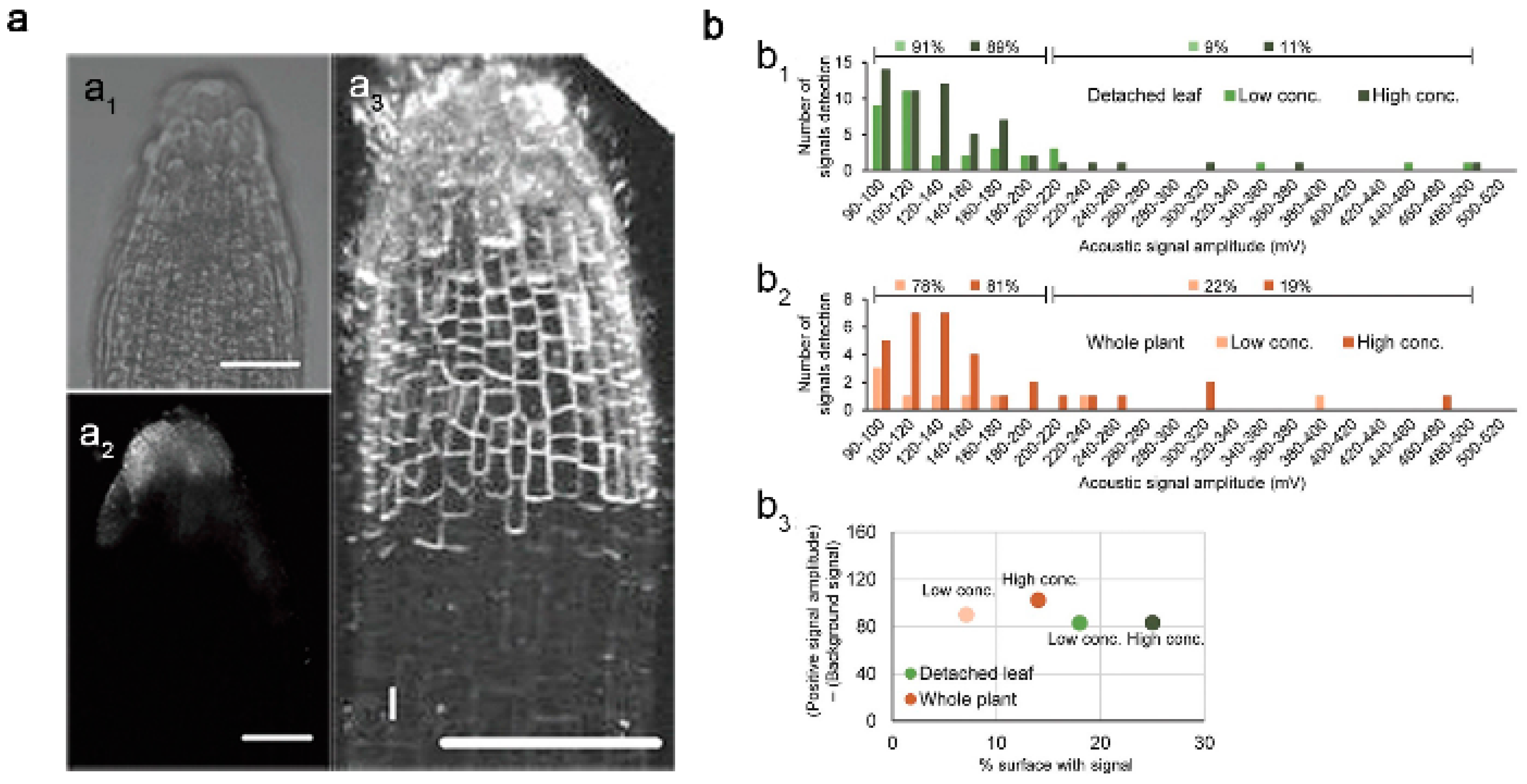
  20. Khodakovskaya, M.V.; de Silva, K.; Nedosekin, D.A.; Dervishi, E.; Biris, A.S.; Shashkov, E.V.; Galanzha, E.I.; Zharov, V.P. Complex genetic, photothermal, and photoacoustic analysis of nanoparticle-plant interactions. Proc. Natl. Acad. Sci. USA 2011, 108, 1028–1033.
  21. Zhai, G.; Walters, K.S.; Peate, D.W.; Alvarez, P.J.; Schnoor, J.L. Transport of gold nanoparticles through plasmodesmata and precipitation of gold ions in woody poplar. Environ. Sci. Technol. Lett. 2014, 1, 146–151.
  22. Taylor, A.F.; Rylott, E.L.; Anderson, C.W.; Bruce, N.C. Investigating the toxicity, uptake, nanoparticle formation and genetic response of plants to gold. PLoS ONE 2014, 9, e93793.
  23. Rico, C.M.; Majumdar, S.; Duarte-Gardea, M.; Peralta-Videa, J.R.; Gardea-Torresdey, J.L. Interaction of nanoparticles with edible plants and their possible implications in the food chain. J. Agric. Food Chem. 2011, 59, 3485–3498.
  24. Kohan-Baghkheirati, E.; Geisler-Lee, J. Gene expression, protein function and pathways of Arabidopsis thaliana responding to silver nanoparticles in comparison to silver ions, cold, salt, drought, and heat. Nanomaterials 2015, 5, 436–467.
  25. Katsumiti, A.; Gilliland, D.; Arostegui, I.; Cajaraville, M.P. Mechanisms of toxicity of Ag nanoparticles in comparison to bulk and ionic Ag on mussel hemocytes and gill cells. PLoS ONE 2015, 10, e0129039.
  26. Scherer, M.D.; Sposito, J.C.V.; Falco, W.F.; Grisolia, A.B.; Andrade, L.H.C.; Lima, S.M.; Machado, G.; Nascimento, V.A.; Goncalves, D.A.; Wender, H.; et al. Cytotoxic and genotoxic effects of silver nanoparticles on meristematic cells of Allium cepa roots: A close analysis of particle size dependence. Sci. Total Environ. 2019, 660, 459–467.
  27. Koo, Y.; Lukianova-Hleb, E.Y.; Pan, J.; Thompson, S.M.; Lapotko, D.O.; Braam, J. In planta response of arabidopsis to photothermal impact mediated by gold nanoparticles. Small 2016, 12, 623–630.
  28. Geisler-Lee, J.; Wang, Q.; Yao, Y.; Zhang, W.; Geisler, M.; Li, K.; Huang, Y.; Chen, Y.; Kolmakov, A.; Ma, X. Phytotoxicity, accumulation and transport of silver nanoparticles by Arabidopsis thaliana. Nanotoxicology 2013, 7, 323–337.
  29. Avellan, A.; Schwab, F.; Masion, A.; Chaurand, P.; Borschneck, D.; Vidal, V.; Rose, J.; Santaella, C.; Levard, C. Nanoparticle uptake in plants: Gold nanomaterial localized in roots of Arabidopsis thaliana by X-ray computed nanotomography and hyperspectral imaging. Environ. Sci. Technol. 2017, 51, 8682–8691.
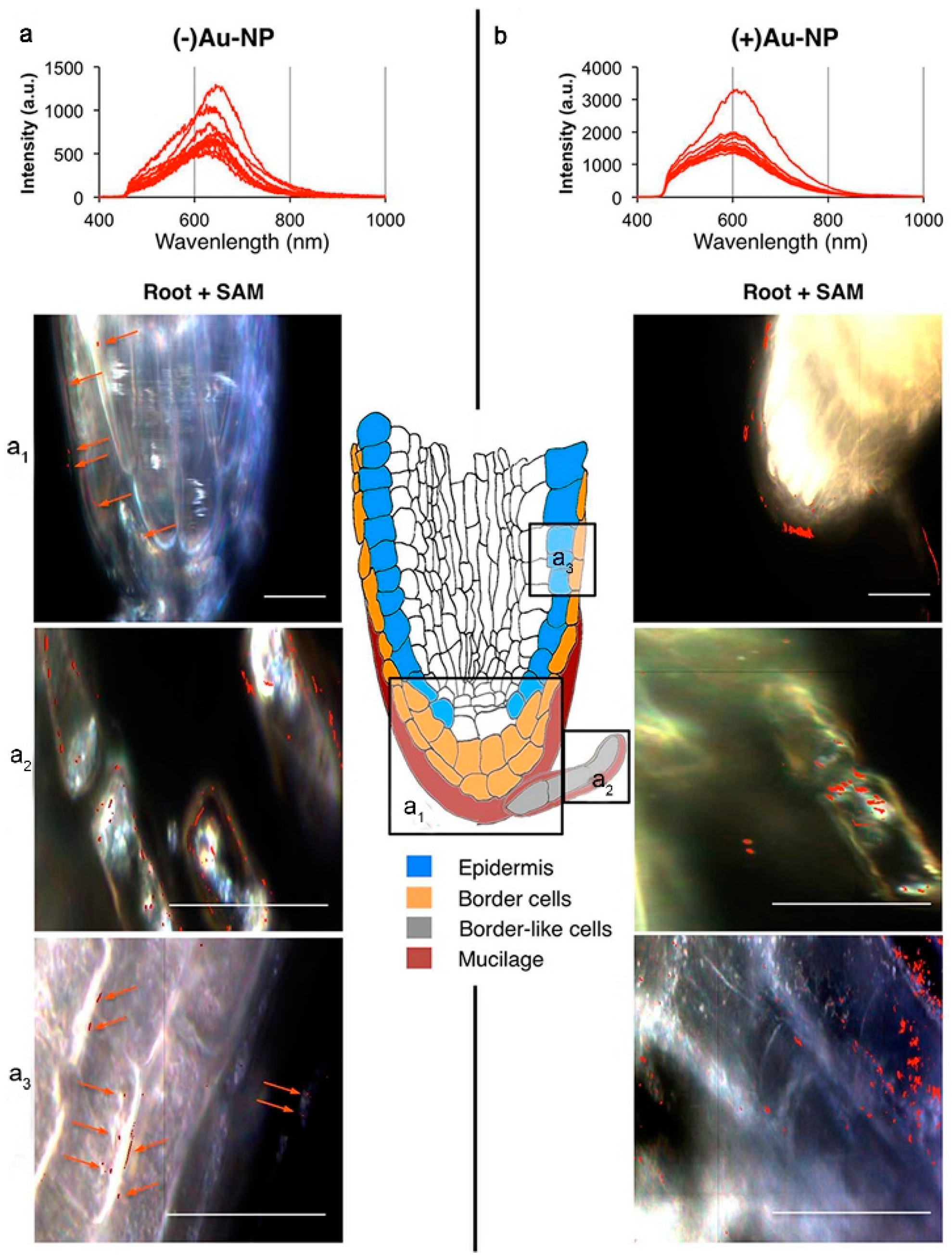
  30. Milewska-Hendel, A.; Zubko, M.; Stroz, D.; Kurczynska, E.U. Effect of nanoparticles surface charge on the Arabidopsis thaliana (L.) roots development and their movement into the root cells and protoplasts. Int. J. Mol. Sci. 2019, 20, 1650.
  31. Milani, N.; McLaughlin, M.J.; Stacey, S.P.; Kirby, J.K.; Hettiarachchi, G.M.; Beak, D.G.; Cornelis, G. Dissolution kinetics of macronutrient fertilizers coated with manufactured zinc oxide nanoparticles. J. Agric. Food Chem. 2012, 60, 3991–3998.
  32. Mir, A.H.; Qamar, A.; Qadir, I.; Naqvi, A.H.; Begum, R. Accumulation and trafficking of zinc oxide nanoparticles in an invertebrate model, Bombyx mori, with insights on their effects on immuno-competent cells. Sci. Rep. 2020, 10, 1617.
  33. Bafaro, E.; Liu, Y.; Xu, Y.; Dempski, R.E. The emerging role of zinc transporters in cellular homeostasis and cancer. Signal Transduct. Target. Ther. 2017, 2, 17029.
  34. Jain, A.; Sinilal, B.; Dhandapani, G.; Meagher, R.B.; Sahi, S.V. Effects of deficiency and excess of zinc on morphophysiological traits and spatiotemporal regulation of zinc-responsive genes reveal incidence of cross talk between micro- and macronutrients. Environ. Sci. Technol. 2013, 47, 5327–5335.
  35. Hussain, D.; Haydon, M.J.; Wang, Y.; Wong, E.; Sherson, S.M.; Young, J.; Camakaris, J.; Harper, J.F.; Cobbett, C.S. P-type ATPase heavy metal transporters with roles in essential zinc homeostasis in Arabidopsis. Plant Cell 2004, 16, 1327–1339.
  36. Nair, P.M.G.; Chung, I.M. Regulation of morphological, molecular and nutrient status in Arabidopsis thaliana seedlings in response to ZnO nanoparticles and Zn ion exposure. Sci. Total Environ. 2017, 575, 187–198.
  37. Jia, H.; Chen, S.; Wang, X.; Shi, C.; Liu, K.; Zhang, S.; Li, J. Copper oxide nanoparticles alter cellular morphology via disturbing the actin cytoskeleton dynamics in Arabidopsis roots. Nanotoxicology 2020, 14, 127–144.
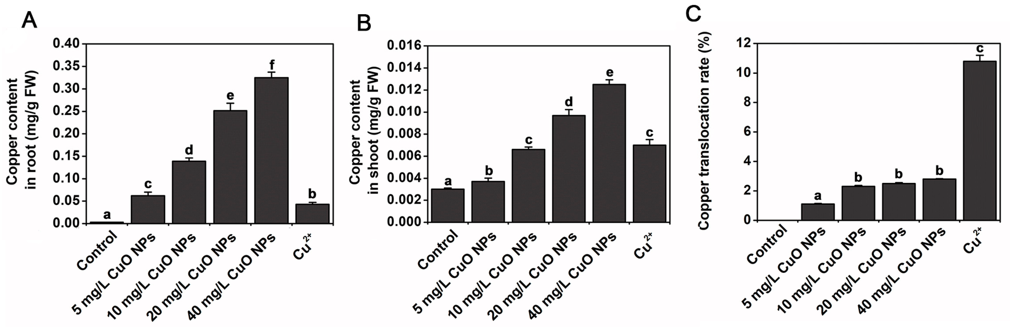
  38. Wang, Z.; Xu, L.; Zhao, J.; Wang, X.; White, J.C.; Xing, B. CuO Nanoparticle interaction with Arabidopsis thaliana: Toxicity, parent-progeny transfer, and gene expression. Environ. Sci. Technol. 2016, 50, 6008–6016.
  39. Hayat, S.; Pichtel, J.; Faizan, M. Sustainable agriculture reviews 41 nanotechnology for plant growth and development. In Nanotechnology for Plant Growth and Development; Springer: Berlin/Heidelberg, Germany, 2020.
  40. Garcia-Sanchez, S.; Bernales, I.; Cristobal, S. Early response to nanoparticles in the Arabidopsis transcriptome compromises plant defence and root-hair development through salicylic acid signalling. BMC Genom. 2015, 16, 341.
  41. Wang, S.; Kurepa, J.; Smalle, J.A. Ultra-small TiO2 nanoparticles disrupt microtubular networks in Arabidopsis thaliana. Plant Cell Environ. 2011, 34, 811–820.
  42. Ma, C.; Chhikara, S.; Xing, B.; Musante, C.; White, J.C.; Dhankher, O.P. Physiological and molecular response of Arabidopsis thaliana (L.) to nanoparticle cerium and indium oxide exposure. ACS Sustain. Chem. Eng. 2013, 1, 768–778.
  43. Birbaum, K.; Brogioli, R.; Schellenberg, M.; Martinoia, E.; Wendelin, E.; Stark, J.W.; Detlef, G.; Ludwig, K.L. No evidence for cerium dioxide nanoparticle translocation in maize plants. Environ. Sci. Technol. 2010, 44, 8718–8723.
  44. Martha, L.L.-M.; Rose, G.D.L.; Jose, A.H.-V.; Castillo-Michel, H.; Botez, C.E.; Jose, R.P.-V.; Jorge, L.G.-T. Evidence of the differential biotransformation and genotoxicity of ZnO and CeO2 nanoparticles on soybean (Glycine max) plants. Environ. Sci. Technol. 2010, 44, 7315–7320.
  45. Priester, J.H.; Ge, Y.; Mielke, R.E.; Horst, A.M.; Moritz, S.C.; Espinosa, K.; Gelb, J.; Walker, S.L.; Nisbet, R.M.; An, Y.J.; et al. Soybean susceptibility to manufactured nanomaterials with evidence for food quality and soil fertility interruption. Proc. Natl. Acad. Sci. USA 2012, 109, E2451–E2456.
  46. Zhang, P.; Ma, Y.; Zhang, Z.; He, X.; Zhang, J.; Guo, Z.; Tai, R.; Zhao, Y.; Chai, Z. Biotransformation of ceria nanoparticles in cucumber plants. ACS Nano 2012, 6, 9943–9950.
  47. Xu, X.; Zhao, C.; Qian, K.; Sun, M.; Hao, Y.; Han, L.; Wang, C.; Ma, C.; White, J.C.; Xing, B. Physiological responses of pumpkin to zinc oxide quantum dots and nanoparticles. Environ. Pollut. 2022, 296, 118723.
  48. Kolackova, M.; Moulick, A.; Kopel, P.; Dvorak, M.; Adam, V.; Klejdus, B.; Huska, D. Antioxidant, gene expression and metabolomics fingerprint analysis of Arabidopsis thaliana treated by foliar spraying of ZnSe quantum dots and their growth inhibition of Agrobacterium tumefaciens. J. Hazard. Mater. 2019, 365, 932–941.
  49. Navarro, D.A.; Bisson, M.A.; Aga, D.S. Investigating uptake of water-dispersible CdSe/ZnS quantum dot nanoparticles by Arabidopsis thaliana plants. J. Hazard. Mater. 2012, 211–212, 427–435.
  50. Laaveri, T.; Sterne, J.; Rombo, L.; Kantele, A. Systematic review of loperamide: No proof of antibiotics being superior to loperamide in treatment of mild/moderate travellers’ diarrhoea. Travel Med. Infect. Dis. 2016, 14, 299–312.
  51. Shen, Y.; Borgatta, J.; Ma, C.; Singh, G.; Tamez, C.; Schultes, N.P.; Zhang, Z.; Dhankher, O.P.; Elmer, W.H.; He, L.; et al. Role of foliar biointerface properties and nanomaterial chemistry in controlling Cu transfer into wild-type and mutant Arabidopsis thaliana leaf tissue. J. Agric. Food Chem. 2022, 70, 4267–4278.
  52. He, J.; Zhang, L.; He, S.Y.; Ryser, E.T.; Li, H.; Zhang, W. Stomata facilitate foliar sorption of silver nanoparticles by Arabidopsis thaliana. Environ. Pollut. 2022, 292, 118448.
More
Information
Subjects: Biophysics
Contributors MDPI registered users' name will be linked to their SciProfiles pages. To register with us, please refer to https://encyclopedia.pub/register : , , , , ,
View Times: 404
Entry Collection: Environmental Sciences
Revisions: 2 times (View History)
Update Date: 11 Jul 2022
1000/1000
Video Production Service