Submitted Successfully!
To reward your contribution, here is a gift for you: A free trial for our video production service.
Thank you for your contribution! You can also upload a video entry or images related to this topic.
Version Summary Created by Modification Content Size Created at Operation
1 -- 4384 2022-06-30 06:45:35 |
2 layout + 3 word(s) 4387 2022-07-01 06:16:14 | |
3 layout Meta information modification 4387 2022-07-01 06:20:15 | |
4 rollback to version 1 -3 word(s) 4384 2022-07-01 06:21:23 | |
5 layout + 40 word(s) 4424 2022-07-01 07:41:40 | |
6 layout Meta information modification 4424 2022-07-01 07:57:22 | |
7 layout -2 word(s) 4422 2022-07-01 08:06:58 |

Video Upload Options

Do you have a full video?

Confirm

Are you sure to Delete?
Cite
If you have any further questions, please contact Encyclopedia Editorial Office.
Ng, M.F.;  Simmons, J.L.;  Boyle, G.M. Intertumoural and Intratumoural Heterogeneity in Melanoma. Encyclopedia. Available online: https://encyclopedia.pub/entry/24717 (accessed on 20 May 2024).
Ng MF,  Simmons JL,  Boyle GM. Intertumoural and Intratumoural Heterogeneity in Melanoma. Encyclopedia. Available at: https://encyclopedia.pub/entry/24717. Accessed May 20, 2024.
Ng, Mei Fong, Jacinta L. Simmons, Glen M. Boyle. "Intertumoural and Intratumoural Heterogeneity in Melanoma" Encyclopedia, https://encyclopedia.pub/entry/24717 (accessed May 20, 2024).
Ng, M.F.,  Simmons, J.L., & Boyle, G.M. (2022, July 01). Intertumoural and Intratumoural Heterogeneity in Melanoma. In Encyclopedia. https://encyclopedia.pub/entry/24717
Ng, Mei Fong, et al. "Intertumoural and Intratumoural Heterogeneity in Melanoma." Encyclopedia. Web. 01 July, 2022.
Intertumoural and Intratumoural Heterogeneity in Melanoma
Edit

Tumour heterogeneity is a phenomenon where the cancer cells evolve diversely over the course of the disease. As a result of the evolution, the cancer cells can be found to be genetically, epigenetically and/or phenotypically different in order to survive in the human body. The tumour microenvironment also plays a crucial role during the evolution.

melanoma tumour heterogeneity intertumoural heterogeneity

1. Introduction

There were an estimated 325,000 new cases of melanoma diagnosed worldwide in 2020, causing an economic burden and placing enormous pressure on healthcare systems [1]. Cutaneous melanoma arises when melanocytes, the cells that produce melanin and provide ultraviolet (UV) protection to the skin [2][3], undergo malignant transformation. The transformation of melanocytes to melanoma has been extensively described [4]. Melanoma may arise from single melanocytes or clusters of melanocytes known as naevi, with or without BRAF or NRAS mutation [5][6][7][8]. Vast evidence has suggested that exposure to UV radiation results in mutations in DNA, in particular UV-induced C > T transitions that are implicated in melanoma development [9][10]. Both UVA and UVB radiation can induce DNA damage responses through production of pyrimidine dimers and generation of oxidative responses, although at different rates via different mechanisms [11]. Various genetic factors are correlated with the progression of melanoma. In general, melanoma can be classified into four subtypes based on the mutation carried in the tumour, i.e., BRAF, NRAS, NF1 or triple wild type [4]. Approximately 50% of all cutaneous melanoma cases exhibit BRAFV600E mutations, while NRAS mutations are found in up to 20% of the cases [12][13]. TERT promoter mutations have been detected in intermediate lesions, while NF1 mutation is observed in melanoma in situ [14][15]. In the advanced stages of melanoma, mutations in CDKN2A, PTEN and TP53 were observed [15]. Melanoma has the highest mutation frequency among human cancers [16][17][18][19]. The increasing mutational burden as the disease progresses is thought to contribute to increased heterogeneity. Heterogeneity in all cancers, including haematological and solid tumours, is now a well-recognised barrier, particularly in looking for the best treatment for cancer patients [20]. In general, heterogeneity can be categorised into interpatient heterogeneity, intertumoural heterogeneity and intratumoural heterogeneity (Figure 1A,B). While interpatient heterogeneity describes the differences in tumours between patients, intertumoural heterogeneity refers to differences between lesions in the same patient. It is now known that populations of cancer cells within a given tumour may also be different from each other, described as intratumoural heterogeneity. Melanoma tumour heterogeneity has been reviewed previously [21][22].

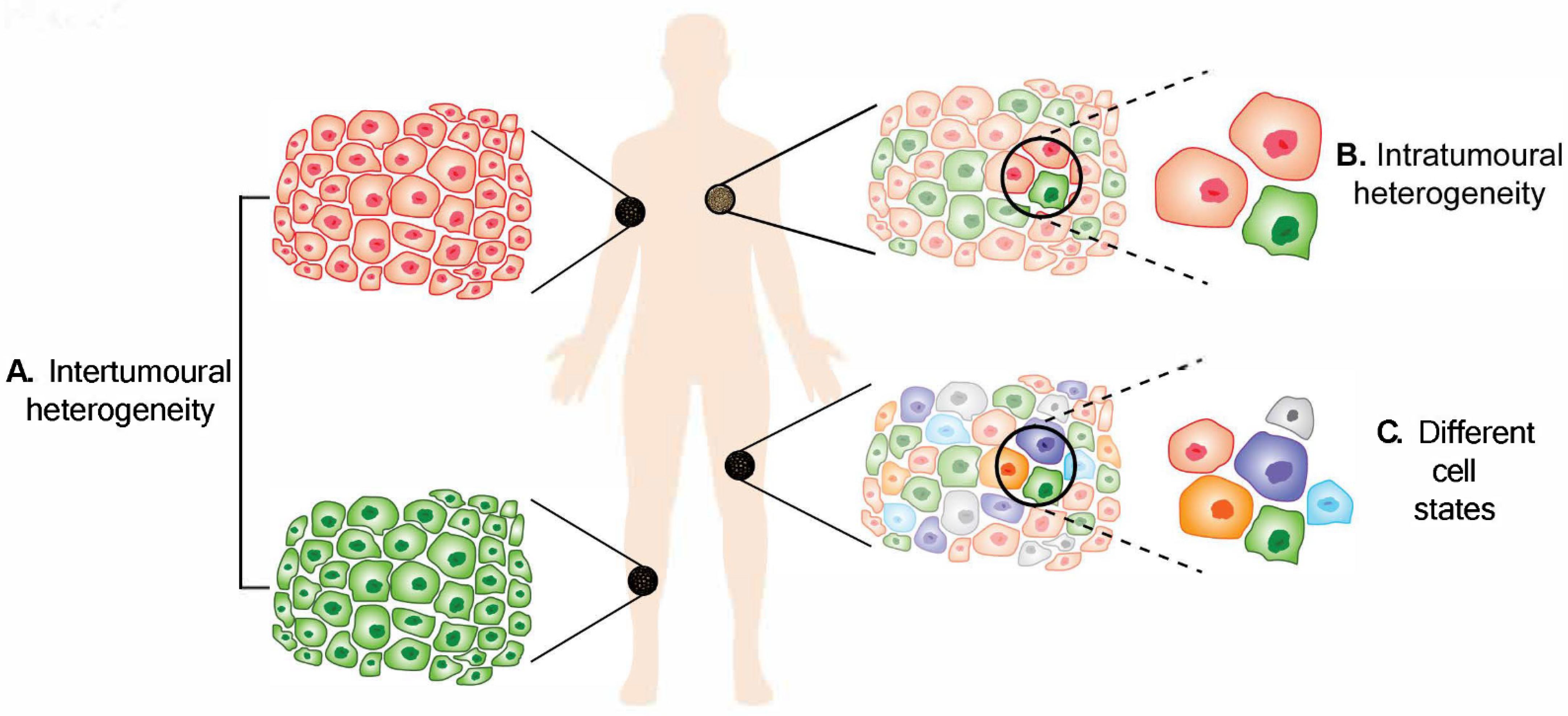
Figure 1. Melanoma heterogeneity. (A) Intertumoural heterogeneity refers to differences between lesions in a patient, while (B) intratumoural heterogeneity describes the differences between cells or subpopulations of cells within a tumour. Advancement in technology has revealed (C) different cell states in melanoma tumours.

2. Intertumoural Heterogeneity

Advancements in molecular biology and sequencing techniques have made detection of intertumoural heterogeneity easier. Sakaizawa and colleagues investigated mutational diversification between circulating tumour cells in patients where paired primary and metastatic lesions were available. Their data revealed discrepancies in the BRAF mutation status in 33% (3/9) of the paired samples and KIT mutations in 75% (3/4) [23]. Similarly, another study showed that mismatched BRAF/NRAS mutations were found in 15% (15/99) of the paired primary and metastatic lesions [24]. This discordant BRAF mutation status has similarly been identified in multiple studies and poses a significant problem for treatment selection and response [25][26][27][28][29][30]. Moreover, differential methylation was detected between three paired primary and metastatic cell lines, where at least 7220 differential methylation fragments were identified [31]. Subsequent data revealed that EBF3 promoter methylation causes elevated EBF3 expression, which leads to metastatic characteristics in melanoma cells. Aberrant expression of methyltransferases in melanoma has also been proposed as a mechanism of malignant progression [32]. More detailed studies have subsequently been conducted to understand the phylogenetic relationship between the primary tumour and metastases. The work demonstrated that metastases are genetically diverse in comparison to the primary tumour or other metastases from the same patient. Whole-genome sequencing (WGS) analysis indicated that metastases can emerge from either identical or distinct cell populations from the primary tumour [33][34].
Overall, it is conceivable that the metastases are subpopulations disseminated from a primary tumour or tumour cells that have gone through further adaptations and/or selection to become genetically or phenotypically distinct from the primary tumour or a regressed primary tumour. It is clear that mutations accumulate throughout the development of metastases and metastases within a single patient do not share the same mutations. Cancer cells gain mutations, resulting in additional mutational clusters, hyperploidy and whole-genome doubling, whereas cancer cells also lose mutations, causing a loss in heterozygosity. This intertumoural divergence, or better described as intertumoural evolution, likely enhances survival and promotes metastasis.

3. Intratumoural Heterogeneity

Intratumoural heterogeneity describes the differences between cells or subpopulations of cells within a tumour. Heterogeneous cells within a tumour can display remarkable variability in phenotypic traits. A cross-sectional analysis that screened 1165 tumour exomes from 12 different cancers revealed that intratumoural heterogeneity was found in all cancer types, with 86% of the samples consisting of at least two subpopulations, with melanoma being the most heterogeneous [18]. Furthermore, a study has shown that 6/9 (67%) melanoma tumours exhibited intratumoural heterogeneity [25].

3.1. Genetic Contribution to Intratumoural Heterogeneity

Genetic heterogeneity in melanoma can be explored by examining allelic variations using DNA microsatellite loci analysis [35][36]. Alternatively, analyses, such as sequencing, high-resolution melting analysis, immunohistochemistry (IHC) and mutation-specific PCR, have also been utilised to reveal the presence of cells with either wild type or mutant copies of BRAF in the same lesion/metastasis [25][37][38]. Busam and colleagues found heterogeneous BRAFV600E staining as determined by IHC analysis with a mutation-specific antibody in 27% (6/22) of the genetically confirmed BRAFV600E-positive cases [39], suggesting either heterogeneity of the BRAF mutation in the lesions or a lack of protein expression. In a case study, subpopulations carrying KITWT and its mutant KITL576P were identified in circulating DNA of a metastatic vaginal mucosal melanoma patient [40]. While it is common to find multiple mutations within a tumour, mutations that are mutually exclusive in different tumour cell populations within a single tumour have also been identified. Sensi and colleagues discovered that 75% (75/100) of the clones of a short-term melanoma cell line expressed BRAFV600E/NRASWT, while the remaining expressed BRAFWT/NRASQ61R [41]. Subsequent investigations revealed that the two subpopulations of cells exhibit different phenotypes. For example, while the BRAFV600E/NRASWT cells remained viable under an ultra-low attachment condition, the BRAFWT/NRASQ61R cells were more proliferative and tumourigenic. Further, subpopulations NRASWT and NRASG13R were found in the same BRAFi-resistant tumour in a melanoma patient [42]. In a sophisticated study, a metastatic lesion from a patient that had undergone multiple treatment regimens was divided into several regions in which deep targeted DNA sequencing was performed [43]. The data showed that only 28% (15/53) of the somatic mutations identified were prevalent in all 41 regions sequenced, while 30% of the mutations were confined to a single region. Further, copy number alteration analysis not only revealed modifications, such as gains of chromosome 6p and 20q and losses of chromosome 6q and 9p, that happened across all 41 regions but also alterations that only occurred in certain regions, such as chromosome 7 gain in four regions, whole-chromosome 10 loss in five regions, 10p loss in one region and chromosome 13 gain in four regions. Taken together, there are large amounts of genetic heterogeneity in melanoma, resulting in subpopulations within a tumour that can possess certain survival advantages and, therefore, possibly affect the treatment outcome.

3.2. Intratumoural Heterogeneity from Non-Genetic Sources

3.2.1. Epigenetic Heterogeneity

Epigenetic mechanisms can contribute to melanoma development via DNA methylation, chromatin remodelling, such as histone modification, and non-coding RNA regulation [44]. Changes in methylation status between subpopulations of cells in melanoma tumours have been observed across multiple studies. For instance, heterogeneous hypermethylation of at least one of the promoters of various tumour-related genes (RASSF1A, CDKN2A, DAPK, MGMT and RB1) was observed in 70% (16/23) of the patient samples [45]. Of note, expression of RASSF1A was observed at the periphery but not in the core of the tumours, indicating that varied degrees of hypermethylation can occur in different areas of a tumour. Differential methylation was observed in clones derived from a melanoma lesion, which then resulted in heterogeneous expressions of MAGE-A3, a previously identified immunotherapeutic target in melanoma [46]. This could dampen the therapeutic efficacy in melanoma patients. In addition, H3K27 hypermethylation in the core of solid tumours caused by low glutamine can lead to cell dedifferentiation and reduced sensitivity to BRAF inhibitor treatment [47]. JARID1B, a member of the jumonji/ARID1 (JARID1) histone 3 K4 demethylase family, was expressed in a slow-cycling population and identified as being essential for tumour maintenance and metastatic progression [48]. Roesch and colleagues also found that melanoma cells maintain heterogeneous expressions of JARID1B. Pure JARID1B+ or JARID1B− cell populations were generated by cell sorting; however, the populations became heterogeneous for JARID1B after just 72 h in culture, showing that expression of JARID1B is dynamically regulated [49]. Epigenetic alterations are also often associated with drug resistance. Shaffer and colleagues found more than 10,000 differentially accessible sites from the assay for transposase-accessible chromatin with sequencing (ATAC-seq) by comparing pre-resistant cells to drug-induced resistant cells [50], indicating that drug treatment promotes epigenetic alterations, which lead to treatment resistance and cancer cell survival.

3.2.2. Phenotypic Heterogeneity

It is possible for a tumour to display dynamic expression of certain markers in different regions in response to microenvironmental cues, disease development, therapeutic treatment or progression without going through genetic alterations. This has been described as phenotypic heterogeneity. For instance, a single human melanoma cell can grow into a heterogeneous tumour in an immunocompromised mouse [51]. In addition, heterogeneous expression of various markers (such as ABCB5, NGFR, CD54, KIT, etc.) has been identified in tumour samples, although there are no significant differences in the tumourigenicity or metastatic properties of the subpopulations with differing expression [51][52]. Hoek and colleagues initially proposed a reversible proliferative and invasive model based on their observations [53], which coincides with the occurrence of heterogeneity in melanoma. Many studies have identified gene signatures associated with reversible proliferative or invasive states in melanoma, where genes that were normally expressed in the proliferative state were often inversely correlated in cells in the invasive state and vice versa [54][55][56]. Remarkably, cells in a proliferative state have been shown to behave similarly to invasive cells when the two populations were co-cultured together [57][58]. Subsequent findings revealed that heterogeneous populations of proliferative and invasive cells are more metastatic and invasive compared to single proliferative populations [57][58], suggesting cooperation between heterogeneous populations in driving metastasis.
Heterogeneous expression of microphthalmia-associated transcription factor (MITF) has been observed extensively in melanoma development and progression. Importantly, MITFhigh and MITFlow subpopulations co-exist in different regions of patient tumour samples [59][60][61][62]. While MITFhigh expression is known to promote differentiation and proliferation, MITFlow expression is associated with an invasive and stem-cell-like phenotype [54][63]. The interaction between MITFhigh and MITFlow subpopulations was elegantly shown in a zebrafish melanoma xenograft model. When invasive MITFlow (WM266-4) cells and less invasive MITFhigh (501mel) cells were co-injected into zebrafish embryos, the MITFhigh cells phenocopied the MITFlow cells without changing the MITF expression and became invasive [57]. When the mixed populations of MITFlow (75%) and MITFhigh (25%) were co-injected into mice, the proliferation rate of the tumour was higher compared to single populations and the MITFhigh population increased from 25% to 50% [64]. Subsequent RNA sequencing results revealed that even the heterogeneous tumours expressed higher MITF as compared to the MITFlow tumours; the tumours were enriched for markers of epithelial mesenchymal transition (EMT) and more invasive, less proliferative phenotypes, showing that there is phenotype adaptation that happens due to cooperativity between MITFhigh and MITFlow cells in enhancing metastasis. Notably, a MITFlow zebrafish model has been established to address a subgroup of melanoma patients with low MITF having a poor prognosis. The researchers first incorporated a BRAFV600 mutation into the MITFlow zebrafish and found that the zebrafish developed nevi and melanoma [65]. Subsequently, a TP53 mutation was introduced into the model and the zebrafish first developed black pigmented lesions that then progressed into superficial and nodular melanoma [66]. As the disease progressed, the expression of MITF shifted from low levels in superficial melanoma to high levels in nodular melanoma. Interestingly, by integrating BRAFV600 and/or TP53 mutation(s) in the MITFlow zebrafish model, melanoma onset is triggered and, over time, the disease progresses by alternating MITF expression. Collectively, this evidence has shown that MITF is heterogeneously expressed in tumours, and the role of MITF in promoting melanoma heterogeneity cannot be disregarded.
The co-existence of the MITFhigh subpopulation with other subpopulations can result in melanoma progression. BRN2, a transcription factor encoded by the gene POU3F2, was found heterogeneously expressed in mouse xenografts [67]. BRN2high cells were migratory and less pigmented, suggesting they potentially expressed low levels of MITF. Intriguingly, MITF and BRN2 were found mutually exclusively expressed in two distinct subpopulations of cells [68][69][70]. Mechanistically, BRN2 repression is mediated by miR-211, which itself is under transcriptional control of MITF [71]. Conversely, BRN2 can promote melanoma invasion via repressing MITF expression by binding to the MITF promoter or through activation of Notch signalling, followed by activation of miR-222/221 promoter [68][69][72]. Heterogeneous expression of PAX3 in melanoma also contributes to MITF regulation by increasing binding of BRN2 to the MITF promoter [73]. When cells are BRN2high/PAX3low, the MITF expression is mainly regulated by BRN2, while, when BRN2low/PAX3high in cells, the MITF expression is driven by PAX3. Additionally, MITF and BRN2-expressing cells act jointly in melanoma progression by first stimulating tumour growth with MITFhigh cells and then activating tumour invasion with BRN2high cells [70]. Moreover, expression of both MITF and BRN2 proved indispensable for melanoma to efficiently metastasise [74]. Overall, the complex interactions between MITF and BRN2 are essential in regulating each other, creating a heterogeneous tumour and in driving melanoma progression.
The expression level of MITF may be associated with melanoma cell behaviour in conjunction with other factors. Of note, MITFhigh/NF-κBlow cells can transition to MITFlow/NF-κBhigh/AXLhigh cells during acquisition of resistance, suggesting the importance of low MITF expression in treatment resistance [75][76]. Heterogeneous expression of AXL is commonly observed in melanomas [77]. It has been suggested that AXL has a pertinent role in the development of acquired resistance to BRAFi and MEKi. Similarly, a shift from ZEB2high/SNAI2high/ZEB1low/TWIST1low primary melanoma to ZEB2low/SNAI2low/ZEB1high/TWIST1high metastatic disease was observed in patient samples [78]. Follow-up experiments suggested that ZEB2 and SNAI2 act as tumour suppressors but ZEB1 and TWIST1 facilitate BRAF signalling in melanoma transformation by downregulating MITF. Thus, a loss of MITF not only correlated with treatment resistance but also melanoma metastasis.
A subpopulation of melanoma cells has been described as slow-cycling due to a slower proliferation rate than other cells under the same conditions. The population, however, could be much more aggressive or invasive compared to other cells due to expression of ABCB5, a transmembrane glycoprotein from the ATP-binding cassette (ABC) superfamily [79][80]. In the study, the ABCB5+ subpopulation exhibited self-renewing and differentiating phenotypes in addition to playing significant roles in melanoma growth, tumourigenicity and metastasis [79][80]. The data also showed that the ABCB5+ subpopulation included slow-cycling cells and was potentially involved in drug resistance mechanisms [81]. Furthermore, a stem cell marker that is frequently found co-expressed with ABCB5, CD133, was also expressed in tumourigenic, tumour-initiating cells [82][83]. CD133+ cells were also more proliferative, able to survive ultra-low attachment conditions and have an immunosuppressive phenotype compared to the CD133− subpopulation in a mouse xenograft model [83][84]. Moreover, CD133+ cells are invasive and metastatic when tested in both in vitro and in vivo (zebrafish and athymic mice) systems [85]. Evidence has shown that ABCB5- and CD133-expressing cells are co-localised in the perivascular niche where vasculogenic mimicry takes place in melanoma xenografts. This suggests the involvement of ABCB5+ and CD133+ cells in tumour growth and progression by promoting vasculogenic mimicry and morphogenesis of tumour microcirculation [86]. A NGFR+ subpopulation has also been identified as cancer initiating cells due to their capability of forming tumours and metastasising in mice compared to NGFR− subpopulations [87][88]. Interestingly, only NGFR+ xenografts formed in nude and NOD/SCID mice, not NSG mice, were able to phenocopy parental tumours. This was contrary to earlier work demonstrating no change in the tumourigenic potential of cells on the basis of ABCB5, CD133 or NGFR expression [51][52]. However, changes in the expression of these proteins during tumour formation in mice is unknown, and, therefore, it is possible that heterogeneous populations are regenerated as tumours develop. Nonetheless, Su and colleagues observed a trajectory of phenotypic changes in patient-derived cell lines upon BRAFi treatment from day 0 until a stably drug-resistant population is established [89]. The cells initially expressed MART-1, a melanocytic marker, then transitioned into a slow-cycling (MART-1neg/NGFRhigh) phenotype after 2 weeks, followed by a mesenchymal (MART-1neg/NGFRneg) phenotype at day 62. This has showcased the phenotypic changes in a process of developing resistance to BRAFi treatment. Increased expression of SerpinE2, JARID1B or PKH26 was associated with a slow-cycling phenotype [90]. Further, ALDH+ melanoma cells are more tumourigenic compared to ALDH−cells and differentiation capabilities [91]. In recent studies, NME1 demonstrated interesting characteristics depending on its expression level in melanoma cells [92][93]. High expression of NME1 promoted cell proliferation, growth of melanoma spheres and stem-cell-like features, whereas NME1low cells were more aggressive and metastatic while exhibiting neural crest-like features.
Peroxisome proliferator-activated receptor gamma coactivator 1-alpha (PGC1α), a transcriptional coactivator responsible for mitochondrial biogenesis and respiration [94], was heterogeneously expressed in melanoma [95][96]. PGC1αhigh populations showed a proliferative phenotype, while PGC1αlow populations displayed a more metastatic phenotype [96]. More recently, a study showed that melanoma cells with higher metastatic potential have elevated levels of the MCT1 transporter, which takes in lactate. Both MCT1high and MCT1low cells were able to form tumours in NSG mice; however, MCT1high cells formed more metastases compared to MCT1low cells [97]. Emerging studies indicate that metabolic changes in melanoma cells are playing a part in melanoma metastasis. However, more studies need to be conducted to obtain enough information in determining how metabolic changes affect melanoma metastasis.

3.2.3. Intratumoral Heterogeneity from Other Perspectives

Tumour Microenvironment

Bidirectional interactions between tumour cells and their microenvironment can also cultivate intratumoural heterogeneity. The tumour microenvironment consists of an extracellular matrix (ECM), stromal cells, fibroblasts and endothelial cells, amongst others, which play vital roles in tumour progression and metastasis. Tirosh and colleagues found that cancer associated fibroblasts (CAFs) expressed high levels of AXL in melanoma tumours that also expressed high AXL but apparent low levels of MITF, both markers of therapeutic resistance. In addition, genes expressed by CAFs were strongly correlated to infiltration of T cells into the tumour [61], which may impact patient response to immune checkpoint inhibitor therapy.

Immune Heterogeneity

Heterogeneous expression of melanoma differentiation antigens and immune infiltrates (CD4+ and CD8+ IHC staining) was observed in 11 melanoma samples [98]. Tissue microarray analysis was conducted on 21 samples (2–5 regions from each sample), where 11 samples showed a diverse presence of cytotoxic lymphocytes [99], indicating that there is no preferential localisation of immune cells within the tumour. Similarly, a diversified distribution of immune cells was observed in a tumour sample excised from a patient who was resistant to anti-PD-1 therapy [43]. Differential expression in immune profile (such as neoantigens, CD4, CD8 and Treg) was detected in a study of 33 synchronous metastases from 15 patients [100]. Of note, less than 8% of the total T-cell clones were shared between synchronous tumours, indicating significant immune heterogeneity observed intratumourally. It was found that highly heterogeneous tumours correlated with reduced anti-tumour immune cells, such as CD8+ T cells, follicular helper T cells and pro-inflammatory M1-like macrophages, and more immune suppressing cells, such as alternatively activated M2-macrophages [101]. Exposing B2905 murine melanoma cells to UVB irradiation, the main etiological cause of melanoma, considerably increased mutational loads from 2745 to 5479 exonic mutations, as well as increased the number of C > T transversions, an increase in the ultraviolet radiation mutation signature and immune heterogeneity [102]. The irradiated cells were more tumorigenic, but single-cell clones isolated from irradiated cells were less tumourigenic and more immunogenic. This could suggest that a reduction in the tumour mutational burden could reduce tumour aggressiveness and improve the treatment response.

4. Impact of Heterogeneity on Treatment Responses

The overall survival rate for metastatic melanoma patients has significantly improved owing to the development and advancement in targeted therapies and immunotherapies [103]. However, there is a large proportion of patients who do not benefit from these new treatments due to differential responses resulting from tumour heterogeneity. Resistance to BRAFi treatment is often associated with reactivation of the MAPK pathway through mechanisms such as RAS mutation, BRAF amplification and alternative splicing [104]. For example, a patient developed resistance to BRAFi treatment after subpopulations in the tumour switched from NRASWT/BRAFV600E to NRASG13R/BRAFV600E, thereby reactivating the MAPK pathway [42]. In a rare case of transplacental transmission of metastatic melanoma with BRAFV600E mutation, the mother was refractory to BRAFi treatment, whereas the baby was responsive to therapy [105]. Genomic analyses showed that there were at least two related but distinct subpopulations causing resistance, which later led to relapse. Notably, single-cell analysis of a panel of 16 melanoma cell lines and a primary cell line (epidermal melanocyte) found the status of markers, such as MITF, AXL and NGFR, impacted the BRAF/MEKi treatment [106]. AXLhigh cells were more resistant to various treatments in comparison to MITFhigh or NGFRhigh cells. Resistant AXLhigh cells exhibited partial MAPK pathway inhibition with high Ki67 expression, while both MITFhigh and NGFRhigh cells demonstrated considerable inhibition of the MAPK pathway, suggesting that the MAPK pathway is crucial for AXLhigh cells but not MITFhigh and NGFRhigh cells. Co-treatment of BRAF/MEKi with different epigenetic modulators in the cells revealed selective sensitivities presented by different populations of cells [106]. Several slow-cycling subpopulations, such as MITFlow/NF-κBhigh/AXLhigh, are also correlated to reactivation of the MAPK pathway [75][76]. Importantly, elevated expression of AXL was observed following BRAFi and MEKi (either single or combination) treatments in patient-derived xenografts (PDXs) [77]. To better understand the mechanisms behind resistance, Shaffer and colleagues compared the expression of resistance markers in pre-resistant cells and BRAFi-treated cells [50]. Interestingly, the number of resistance markers expressed increased from 72 in the pre-resistant cells to 600 (of the 1456 genes tested) in the post-drug-treatment cells by transcriptome analysis. Only 33 differentially accessible sites were discovered by comparing the non-resistant and pre-resistant cells versus more than 10,000 differentially accessible sites between pre- and post-drug-treatment using ATAC-seq, suggesting minute variations between the non-resistant and pre-resistant cells, and that the treatment induced phenotype switching resistance. Yet, whether the 33 differentially accessible sites between non-resistant and pre-resistant cells are sufficient to provoke a remarkable effect in drug resistance is arguable. In another study, resistance-conferring genetic alterations were evidenced in PDXs established from treatment-naïve patient samples, suggesting the existence of intrinsic resistant populations in melanoma patients [107]. Nevertheless, acquired resistance was observed in some PDXs too, indicating variabilities in causations of resistance to treatments. Meanwhile, other slow-cycling populations, such as JARID1Bhigh cells, CD133+, ABCB5+ and NGFR+ cells, have also been implicated in resistance to BRAFi and other anti-cancer drugs [49][83][85][108][109]. Furthermore, the PIK3CAE545K mutation was found to confer resistance to combined MEKi + CDK4i in a melanoma patient [110]. Nonetheless, aberrant expression of methyltransferases in melanomas could affect the sensitivity of melanoma cells to chemotherapy [111].
In addition, not only did pre-existing NGFRhigh melanoma cells show reduced sensitivity to the combination of BRAFi and MEKi but the cells were also resistant to antigen-specific cytotoxic CD8+ T cells [112]. Consistent with this finding, patients whose tumours are NGFRhigh were not responsive to anti-PD-1 and/or a combination of anti-PD-1 and anti-CTLA-4 therapy [112]. Furthermore, acquired JAK1 and JAK2 loss-of-function mutations were found in tumours after patients developed resistance to anti-PD-1 therapy [113]. The mutations were discovered by comparing the whole-exome sequencing data of tumour samples before treatment and after relapse. The cells with JAK1 mutations were resistant to interferon α, β and γ-induced growth arrest, while the cells with JAK2 mutations were insensitive to interferon γ-induced growth arrest. The same study also revealed the mutation of beta-2-microglobulin (B2M) in another patient’s baseline and matched relapse samples, where the loss of B2M reduced the immune-cell recognition of cancer cells. Nonetheless, there are ambiguous responses to immunotherapy due to tumour heterogeneity. For instance, data from a mouse study suggested that tumours with a low level of intratumoural heterogeneity may be more immunogenic and, hence, have an improved overall survival rate [102]. Lin and colleagues found that tumours with high heterogeneity were associated with decreased immune cell infiltration, immune response activation and overall patient survival [101]. In contrast, a positive correlation was observed between high mutational load and enhanced T cell infiltration in patient samples [100], indicating that tumours with a high mutational load may be more immunogenic. Despite the inconsistencies observed, it is possible that the identity of the genes mutated is potentially more pertinent in determining the treatment response regardless of the amount of mutational load [114].
Altogether, studies have shown that drug treatment could trigger secondary mutations in tumour cells, which lead to resistance. It is also evident that some tumour cells harbour pre-existing resistance, conferring genetic properties that reduce sensitivities to treatments and later lead to treatment resistance. The heterogeneous tumour environment similarly contributes to immunotherapy treatment resistance. The development of immunotherapy has greatly transformed the treatment for advanced melanoma and dramatically increased the overall survival rate for melanoma patients. However, there are still nearly 40% of advanced melanoma patients that do not respond initially to immunotherapies, with adverse side effects observed in 60% of the patients [115]. The reason why some patients respond well while others do not respond at all is still unclear, although it is clear that heterogeneity at the tumour and immune levels plays an important role.

5. Conclusions and Implications

Heterogeneity within melanoma was initially described by Norris, an attentive general practitioner, in 1820 [116]. Since that time, the researchers have gained significantly more discerning information regarding melanoma development and tumour heterogeneity, particularly at the molecular level. However, many aspects of the development and role of heterogeneity remain unclear. For example, the true identity of each of the subpopulations within the patient tumour remains to be clarified. Further, the specific roles of communication between different subpopulations within the tumour and the various biological components within the tumour microenvironment (ECM and immune cells) are not well characterised. As more subpopulations develop in the tumour, the more challenges patients must face, such as tumour growth, metastasis, efficacy of the therapeutic treatments and, finally, resistance to treatment. These open questions must serve as a reminder that much more needs to be done in order to fill in the missing pieces and truly understand the role of heterogeneity in melanoma metastasis and treatment resistance in order to effectively tackle the disease. Effectively targeting the tumour heterogeneity in melanoma could lead to better therapeutic options and better outcomes for patients.

References

  1. Sung, H.; Ferlay, J.; Siegel, R.L.; Laversanne, M.; Soerjomataram, I.; Jemal, A.; Bray, F. Global Cancer Statistics 2020: GLOBOCAN Estimates of Incidence and Mortality Worldwide for 36 Cancers in 185 Countries. CA Cancer J. Clin. 2021, 71, 209–249.
  2. Wood, J.M.; Jimbow, K.; Boissy, R.E.; Slominski, A.; Plonka, P.M.; Slawinski, J.; Wortsman, J.; Tosk, J. What’s the use of generating melanin? Exp. Dermatol. 1999, 8, 153–164.
  3. Lin, J.Y.; Fisher, D.E. Melanocyte biology and skin pigmentation. Nature 2007, 445, 843–850.
  4. Shain, A.H.; Bastian, B.C. From melanocytes to melanomas. Nat. Rev. Cancer 2016, 16, 345–358.
  5. Weatherhead, S.C.; Haniffa, M.; Lawrence, C.M. Melanomas arising from naevi and de novo melanomas—Does origin matter? Br. J. Dermatol. 2007, 156, 72–76.
  6. Kiuru, M.; Tartar, D.M.; Qi, L.; Chen, D.; Yu, L.; Konia, T.; McPherson, J.D.; Murphy, W.J.; Fung, M.A. Improving classification of melanocytic nevi: Association of BRAF V600E expression with distinct histomorphologic features. J. Am. Acad. Dermatol. 2018, 79, 221–229.
  7. Bauer, J.; Curtin, J.; Pinkel, D.; Bastian, B.C. Congenital Melanocytic Nevi Frequently Harbor NRAS Mutations but no BRAF Mutations. J. Investig. Dermatol. 2007, 127, 179–182.
  8. Tschandl, P.; Berghoff, A.S.; Preusser, M.; Burgstaller-Muehlbacher, S.; Pehamberger, H.; Okamoto, I.; Kittler, H. NRAS and BRAF Mutations in Melanoma-Associated Nevi and Uninvolved Nevi. PLoS ONE 2013, 8, e69639.
  9. Alexandrov, L.B.; Nik-Zainal, S.; Wedge, D.C.; Aparicio, S.A.; Behjati, S.; Biankin, A.V.; Bignell, G.R.; Bolli, N.; Borg, A.; Børresen-Dale, A.-L.; et al. Signatures of mutational processes in human cancer. Nature 2013, 500, 415–421.
  10. Hayward, N.K.; Wilmott, J.S.; Waddell, N.; Johansson, P.A.; Field, M.A.; Nones, K.; Patch, A.-M.; Kakavand, H.; Alexandrov, L.B.; Burke, H.; et al. Whole-genome landscapes of major melanoma subtypes. Nature 2017, 545, 175–180.
  11. Cadet, J.; Douki, T.; Ravanat, J.-L. Oxidatively Generated Damage to Cellular DNA by UVB and UVA Radiation. Photochem. Photobiol. 2015, 91, 140–155.
  12. Sanchez-Vega, F.; Mina, M.; Armenia, J.; Chatila, W.K.; Luna, A.; La, K.C.; Dimitriadoy, S.; Liu, D.L.; Kantheti, H.S.; Saghafinia, S.; et al. Oncogenic Signaling Pathways in The Cancer Genome Atlas. Cell 2018, 173, 321–337.e310.
  13. Muñoz-Couselo, E.; Adelantado, E.Z.; Ortiz, C.; Soberino-García, J.; Perez-Garcia, J. NRAS-mutant melanoma: Current challenges and future prospect. OncoTargets Ther. 2017, 10, 3941–3947.
  14. Osella-Abate, S.; Bertero, L.; Senetta, R.; Mariani, S.; Lisa, F.; Coppola, V.; Metovic, J.; Pasini, B.; Puig, S.S.; Fierro, M.T.; et al. TERT Promoter Mutations are Associated with Visceral Spreading in Melanoma of the Trunk. Cancers 2019, 11, 452.
  15. Shain, A.H.; Yeh, I.; Kovalyshyn, I.; Sriharan, A.; Talevich, E.; Gagnon, A.; Dummer, R.; North, J.; Pincus, L.; Ruben, B.; et al. The Genetic Evolution of Melanoma from Precursor Lesions. New Engl. J. Med. 2015, 373, 1926–1936.
  16. Hodis, E.; Watson, I.R.; Kryukov, G.V.; Arold, S.T.; Imielinski, M.; Theurillat, J.-P.; Nickerson, E.; Auclair, D.; Li, L.; Place, C.; et al. A Landscape of Driver Mutations in Melanoma. Cell 2012, 150, 251–263.
  17. Lawrence, M.S.; Stojanov, P.; Polak, P.; Kryukov, G.V.; Cibulskis, K.; Sivachenko, A.; Carter, S.L.; Stewart, C.; Mermel, C.H.; Roberts, S.A.; et al. Mutational heterogeneity in cancer and the search for new cancer-associated genes. Nature 2013, 499, 214–218.
  18. Andor, N.; Graham, T.A.; Jansen, M.; Xia, L.C.; Aktipis, C.A.; Petritsch, C.; Ji, H.P.; Maley, C.C. Pan-cancer analysis of the extent and consequences of intratumor heterogeneity. Nat. Med. 2016, 22, 105–113.
  19. Ding, L.; Kim, M.; Kanchi, K.L.; Dees, N.D.; Lu, C.; Griffith, M.; Fenstermacher, D.; Sung, H.; Miller, C.A.; Goetz, B.; et al. Clonal Architectures and Driver Mutations in Metastatic Melanomas. PLoS ONE 2014, 9, e111153.
  20. Janku, F. Tumor heterogeneity in the clinic: Is it a real problem? Ther. Adv. Med. Oncol. 2014, 6, 43–51.
  21. Grzywa, T.M.; Paskal, W.; Włodarski, P.K. Intratumor and Intertumor Heterogeneity in Melanoma. Transl. Oncol. 2017, 10, 956–975.
  22. Ito, T.; Tanaka, Y.; Murata, M.; Kaku-Ito, Y.; Furue, K.; Furue, M. BRAF Heterogeneity in Melanoma. Curr. Treat. Options Oncol. 2021, 22, 20.
  23. Sakaizawa, K.; Goto, Y.; Kiniwa, Y.; Uchiyama, A.; Harada, K.; Shimada, S.; Saida, T.; Ferrone, S.; Takata, M.; Uhara, H.; et al. Mutation analysis of BRAF and KIT in circulating melanoma cells at the single cell level. Br. J. Cancer 2012, 106, 939–946.
  24. Colombino, M.; Capone, M.; Lissia, A.; Cossu, A.; Rubino, C.; De Giorgi, V.; Massi, D.; Fonsatti, E.; Staibano, S.; Nappi, O.; et al. BRAF/NRAS Mutation Frequencies Among Primary Tumors and Metastases in Patients with Melanoma. J. Clin. Oncol. 2012, 30, 2522–2529.
  25. Yancovitz, M.; Litterman, A.; Yoon, J.; Ng, E.; Shapiro, R.L.; Berman, R.S.; Pavlick, A.C.; Darvishian, F.; Christos, P.; Mazumdar, M.; et al. Intra- and inter-tumor heterogeneity of BRAF(V600E)) mutations in primary and metastatic melanoma. PLoS ONE 2012, 7, e29336.
  26. Heinzerling, L.; Baiter, M.; Kühnapfel, S.; Schuler, G.; Keikavoussi, P.; Agaimy, A.; Kiesewetter, F.; Hartmann, A.; Schneider-Stock, R. Mutation landscape in melanoma patients clinical implications of heterogeneity of BRAF mutations. Br. J. Cancer 2013, 109, 2833–2841.
  27. Saint-Jean, M.; Quéreux, G.; Nguyen, J.M.; Peuvrel, L.; Brocard, A.; Vallée, A.; Knol, A.C.; Khammari, A.; Denis, M.G.; Dréno, B. Is a single BRAF wild-type test sufficient to exclude melanoma patients from vemurafenib therapy? J. Investig. Dermatol. 2014, 134, 1468–1470.
  28. Bradish, J.R.; Richey, J.D.; Post, K.M.; Meehan, K.; Sen, J.D.; Malek, A.J.; Katona, T.M.; Warren, S.; Logan, T.F.; Fecher, L.A.; et al. Discordancy in BRAF mutations among primary and metastatic melanoma lesions: Clinical implications for targeted therapy. Mod. Pathol. 2015, 28, 480–486.
  29. Yaman, B.; Kandiloglu, G.; Akalin, T. BRAF-V600 Mutation Heterogeneity in Primary and Metastatic Melanoma: A Study with Pyrosequencing and Immunohistochemistry. Am. J. Dermatopathol. 2016, 38, 113–120.
  30. Eriksson, H.; Zebary, A.; Vassilaki, I.; Omholt, K.; Ghaderi, M.; Hansson, J. BRAFV600E protein expression in primary cutaneous malignant melanomas and paired metastases. JAMA Dermatol. 2015, 151, 410–416.
  31. Chatterjee, A.; Stockwell, P.A.; Ahn, A.; Rodger, E.J.; Leichter, A.L.; Eccles, M.R. Genome-wide methylation sequencing of paired primary and metastatic cell lines identifies common DNA methylation changes and a role for EBF3 as a candidate epigenetic driver of melanoma metastasis. Oncotarget 2016, 8, 6085–6101.
  32. Campagna, R.; Pozzi, V.; Sartini, D.; Salvolini, E.; Brisigotti, V.; Molinelli, E.; Campanati, A.; Offidani, A.; Emanuelli, M. Beyond Nicotinamide Metabolism: Potential Role of Nicotinamide N-Methyltransferase as a Biomarker in Skin Cancers. Cancers 2021, 13, 4943.
  33. Sanborn, J.Z.; Chung, J.; Purdom, E.; Wang, N.J.; Kakavand, H.; Wilmott, J.S.; Butler, T.; Thompson, J.F.; Mann, G.J.; Haydu, L.E.; et al. Phylogenetic analyses of melanoma reveal complex patterns of metastatic dissemination. Proc. Natl. Acad. Sci. USA 2015, 112, 10995–11000.
  34. Rabbie, R.; Ansari-Pour, N.; Cast, O.; Lau, D.; Scott, F.; Welsh, S.J.; Parkinson, C.; Khoja, L.; Moore, L.; Tullett, M.; et al. Multi-site clonality analysis uncovers pervasive heterogeneity across melanoma metastases. Nat. Commun. 2020, 11, 4306.
  35. Bogdan, I.; Xin, H.; Burg, G.; Böni, R. Heterogeneity of allelic deletions within melanoma metastases. Melanoma Res. 2001, 11, 349–354.
  36. Harbst, K.; Lauss, M.; Cirenajwis, H.; Winter, C.; Howlin, J.; Törngren, T.; Kvist, A.; Nodin, B.; Olsson, E.; Häkkinen, J.; et al. Molecular and genetic diversity in the metastatic process of melanoma. J. Pathol. 2014, 233, 39–50.
  37. Lin, J.; Goto, Y.; Murata, H.; Sakaizawa, K.; Uchiyama, A.; Saida, T.; Takata, M. Polyclonality of BRAF mutations in primary melanoma and the selection of mutant alleles during progression. Br. J. Cancer 2011, 104, 464–468.
  38. Manfredi, L.; Meyer, N.; Tournier, E.; Grand, D.; Uro-Coste, E.; Rochaix, P.; Brousset, P.; Lamant, L. Highly Concordant Results Between Immunohistochemistry and Molecular Testing of Mutated V600E BRAF in Primary and Metastatic Melanoma. Acta Derm. Venereol. 2016, 96, 630–634.
  39. Busam, K.J.; Hedvat, C.; Pulitzer, M.; von Deimling, A.; Jungbluth, A.A. Immunohistochemical analysis of BRAF(V600E) expression of primary and metastatic melanoma and comparison with mutation status and melanocyte differentiation antigens of metastatic lesions. Am. J. Surg. Pathol. 2013, 37, 413–420.
  40. Gremel, G.; Lee, R.J.; Girotti, M.R.; Mandal, A.K.; Valpione, S.; Garner, G.; Ayub, M.; Wood, S.; Rothwell, D.; Fusi, A.; et al. Distinct subclonal tumour responses to therapy revealed by circulating cell-free DNA. Ann. Oncol. 2016, 27, 1959–1965.
  41. Sensi, M.; Nicolini, G.; Petti, C.; Bersani, I.; Lozupone, F.; Molla, A.; Vegetti, C.; Nonaka, D.; Mortarini, R.; Parmiani, G.; et al. Mutually exclusive NRASQ61R and BRAFV600E mutations at the single-cell level in the same human melanoma. Oncogene 2006, 25, 3357–3364.
  42. Wilmott, J.S.; Tembe, V.; Howle, J.R.; Sharma, R.; Thompson, J.F.; Rizos, H.; Lo, R.S.; Kefford, R.F.; Scolyer, R.A.; Long, G.V. Intratumoral Molecular Heterogeneity in a BRAF-Mutant, BRAF Inhibitor-Resistant Melanoma: A Case Illustrating the Challenges for Personalized Medicine. Mol. Cancer Ther. 2012, 11, 2704–2708.
  43. Mitra, A.; Andrews, M.C.; Roh, W.; De Macedo, M.P.; Hudgens, C.W.; Carapeto, F.; Singh, S.; Reuben, A.; Wang, F.; Mao, X.; et al. Spatially resolved analyses link genomic and immune diversity and reveal unfavorable neutrophil activation in melanoma. Nat. Commun. 2020, 11, 1839.
  44. Giunta, E.F.; Arrichiello, G.; Curvietto, M.; Pappalardo, A.; Bosso, D.; Rosanova, M.; Diana, A.; Giordano, P.; Petrillo, A.; Federico, P.; et al. Epigenetic Regulation in Melanoma: Facts and Hopes. Cells 2021, 10, 2048.
  45. Rastetter, M.; Schagdarsurengin, U.; Lahtz, C.; Fiedler, E.; Marsch, W.C.; Dammann, R.; Helmbold, P. Frequent intra-tumoural heterogeneity of promoter hypermethylation in malignant melanoma. Histol. Histopathol. 2007, 22, 1005–1015.
  46. Sigalotti, L.; Fratta, E.; Coral, S.; Tanzarella, S.; Danielli, R.; Colizzi, F.; Fonsatti, E.; Traversari, C.; Altomonte, M.; Maio, M. Intratumor Heterogeneity of Cancer/Testis Antigens Expression in Human Cutaneous Melanoma Is Methylation-Regulated and Functionally Reverted by 5-Aza-2′-deoxycytidine. Cancer Res. 2004, 64, 9167–9171.
  47. Pan, M.; Reid, M.; Lowman, X.H.; Kulkarni, R.P.; Tran, T.Q.; Liu, X.; Yang, Y.; Hernandez-Davies, J.E.; Rosales, K.K.; Li, H.; et al. Regional glutamine deficiency in tumours promotes dedifferentiation through inhibition of histone demethylation. Nat. Cell Biol. 2016, 18, 1090–1101.
  48. Roesch, A.; Fukunaga-Kalabis, M.; Schmidt, E.C.; Zabierowski, S.E.; Brafford, P.A.; Vultur, A.; Basu, D.; Gimotty, P.; Vogt, T.; Herlyn, M. A Temporarily Distinct Subpopulation of Slow-Cycling Melanoma Cells Is Required for Continuous Tumor Growth. Cell 2010, 141, 583–594.
  49. Roesch, A.; Vultur, A.; Bogeski, I.; Wang, H.; Zimmermann, K.M.; Speicher, D.; Körbel, C.; Laschke, M.W.; Gimotty, P.A.; Philipp, S.E.; et al. Overcoming intrinsic multidrug resistance in melanoma by blocking the mitochondrial respiratory chain of slow-cycling JARID1B(high) cells. Cancer Cell 2013, 23, 811–825.
  50. Shaffer, S.M.; Dunagin, M.C.; Torborg, S.R.; Torre, E.A.; Emert, B.; Krepler, C.; Beqiri, M.; Sproesser, K.; Brafford, P.A.; Xiao, M.; et al. Rare cell variability and drug-induced reprogramming as a mode of cancer drug resistance. Nature 2017, 546, 431–435.
  51. Quintana, E.; Shackleton, M.; Sabel, M.S.; Fullen, D.R.; Johnson, T.M.; Morrison, S.J. Efficient tumour formation by single human melanoma cells. Nature 2008, 456, 593–598.
  52. Quintana, E.; Shackleton, M.; Foster, H.R.; Fullen, D.R.; Sabel, M.S.; Johnson, T.M.; Morrison, S.J. Phenotypic Heterogeneity among Tumorigenic Melanoma Cells from Patients that Is Reversible and Not Hierarchically Organized. Cancer Cell 2010, 18, 510–523.
  53. Hoek, K.S.; Eichhoff, O.M.; Schlegel, N.C.; Döbbeling, U.; Kobert, N.; Schaerer, L.; Hemmi, S.; Dummer, R. In vivo switching of human melanoma cells between proliferative and invasive states. Cancer Res. 2008, 68, 650–656.
  54. Eichhoff, O.M.; Zipser, M.C.; Xu, M.; Weeraratna, A.T.; Mihic, D.; Dummer, R.; Hoek, K.S. The immunohistochemistry of invasive and proliferative phenotype switching in melanoma: A case report. Melanoma Res. 2010, 20, 349–955.
  55. Widmer, D.S.; Cheng, P.F.; Eichhoff, O.M.; Belloni, B.C.; Zipser, M.C.; Schlegel, N.C.; Javelaud, D.; Mauviel, A.; Dummer, R.; Hoek, K.S. Systematic classification of melanoma cells by phenotype-specific gene expression mapping. Pigment. Cell Melanoma Res. 2012, 25, 343–353.
  56. Verfaillie, A.; Imrichova, H.; Atak, Z.K.; Dewaele, M.; Rambow, F.; Hulselmans, G.; Christiaens, V.; Svetlichnyy, D.; Luciani, F.; Van den Mooter, L.; et al. Decoding the regulatory landscape of melanoma reveals TEADS as regulators of the invasive cell state. Nat. Commun. 2015, 6, 6683.
  57. Chapman, A.; del Ama, L.F.; Ferguson, J.; Kamarashev, J.; Wellbrock, C.; Hurlstone, A. Heterogeneous Tumor Subpopulations Cooperate to Drive Invasion. Cell Rep. 2014, 8, 688–695.
  58. Campbell, N.R.; Rao, A.; Hunter, M.V.; Sznurkowska, M.K.; Briker, L.; Zhang, M.; Baron, M.; Heilmann, S.; Deforet, M.; Kenny, C.; et al. Cooperation between melanoma cell states promotes metastasis through heterotypic cluster formation. Dev. Cell 2021, 56, 2808–2825.e10.
  59. Salti, G.I.; Manougian, T.; Farolan, M.; Shilkaitis, A.; Majumdar, D.; Das Gupta, T.K. Micropthalmia transcription factor: A new prognostic marker in intermediate-thickness cutaneous malignant melanoma. Cancer Res. 2000, 60, 5012–5016.
  60. Ennen, M.; Keime, C.; Gambi, G.; Kieny, A.; Coassolo, S.; Thibault-Carpentier, C.; Margerin-Schaller, F.; Davidson, G.; Vagne, C.; Lipsker, D.; et al. MITF-High and MITF-Low Cells and a Novel Subpopulation Expressing Genes of Both Cell States Contribute to Intra- and Intertumoral Heterogeneity of Primary Melanoma. Clin. Cancer Res. 2017, 23, 7097–7107.
  61. Tirosh, I.; Izar, B.; Prakadan, S.M.; Wadsworth, M.H., II; Treacy, D.; Trombetta, J.J.; Rotem, A.; Rodman, C.; Lian, C.; Murphy, G.; et al. Dissecting the multicellular ecosystem of metastatic melanoma by single-cell RNA-seq. Science 2016, 352, 189–196.
  62. Carreira, S.; Goodall, J.; Denat, L.; Rodriguez, M.; Nuciforo, P.; Hoek, K.S.; Testori, A.; LaRue, L.; Goding, C.R. Mitf regulation of Dia1 controls melanoma proliferation and invasiveness. Genes Dev. 2006, 20, 3426–3439.
  63. Goding, C.R. A picture of Mitf in melanoma immortality. Oncogene 2011, 30, 2304–2306.
  64. Rowling, E.J.; Miskolczi, Z.; Nagaraju, R.; Wilcock, D.; Wang, P.; Telfer, B.; Li, Y.; Lasheras-Otero, I.; Redondo-Muñoz, M.; Sharrocks, A.D.; et al. Cooperative behaviour and phenotype plasticity evolve during melanoma progression. Pigment Cell Melanoma Res. 2020, 33, 695–708.
  65. Lister, J.A.; Capper, A.; Zeng, Z.; Mathers, M.E.; Richardson, J.; Paranthaman, K.; Jackson, I.J.; Patton, E.E. A Conditional Zebrafish MITF Mutation Reveals MITF Levels Are Critical for Melanoma Promotion vs. Regression In Vivo. J. Investig. Dermatol. 2014, 134, 133–140.
  66. Travnickova, J.; Wojciechowska, S.; Khamseh, A.; Gautier, P.; Brown, D.V.; Lefevre, T.; Brombin, A.; Ewing, A.; Capper, A.; Spitzer, M.; et al. Zebrafish MITF-Low Melanoma Subtype Models Reveal Transcriptional Subclusters and MITF-Independent Residual Disease. Cancer Res. 2019, 79, 5769–5784.
  67. Pinner, S.; Jordan, P.; Sharrock, K.; Bazley, L.; Collinson, L.; Marais, R.; Bonvin, E.; Goding, C.; Sahai, E. Intravital Imaging Reveals Transient Changes in Pigment Production and Brn2 Expression during Metastatic Melanoma Dissemination. Cancer Res. 2009, 69, 7969–7977.
  68. Goodall, J.; Carreira, S.; Denat, L.; Kobi, D.; Davidson, I.; Nuciforo, P.; Sturm, R.A.; Larue, L.; Goding, C.R. Brn-2 Represses Microphthalmia-Associated Transcription Factor Expression and Marks a Distinct Subpopulation of Microphthalmia-Associated Transcription Factor–Negative Melanoma Cells. Cancer Res. 2008, 68, 7788–7794.
  69. Thurber, A.E.; Douglas, G.; Sturm, E.C.; Zabierowski, S.E.; Smit, D.J.; Ramakrishnan, S.N.; Hacker, E.E.; Leonard, J.H.; Herlyn, M.; Sturm, R.A. Inverse expression states of the BRN2 and MITF transcription factors in melanoma spheres and tumour xenografts regulate the NOTCH pathway. Oncogene 2011, 30, 3036–3048.
  70. Fane, M.; Chhabra, Y.; Hollingsworth, D.E.; Simmons, J.; Spoerri, L.; Oh, T.G.; Chauhan, J.; Chin, T.; Harris, L.; Harvey, T.J.; et al. NFIB Mediates BRN2 Driven Melanoma Cell Migration and Invasion Through Regulation of EZH2 and MITF. eBioMedicine 2017, 16, 63–75.
  71. Boyle, G.M.; Woods, S.L.; Bonazzi, V.F.; Stark, M.S.; Hacker, E.; Aoude, L.G.; Dutton-Regester, K.; Cook, A.L.; Sturm, R.A.; Hayward, N.K. Melanoma cell invasiveness is regulated by miR-211 suppression of the BRN2 transcription factor. Pigment Cell Melanoma Res. 2011, 24, 525–537.
  72. Golan, T.; Messer, A.R.; Amitai-Lange, A.; Melamed, Z.; Ohana, R.; Bell, R.E.; Kapitansky, O.; Lerman, G.; Greenberger, S.; Khaled, M.; et al. Interactions of Melanoma Cells with Distal Keratinocytes Trigger Metastasis via Notch Signaling Inhibition of MITF. Mol. Cell 2015, 59, 664–676.
  73. Smith, M.P.; Rana, S.; Ferguson, J.; Rowling, E.J.; Flaherty, K.T.; Wargo, J.A.; Marais, R.; Wellbrock, C. A PAX3/BRN2 rheostat controls the dynamics of BRAF mediated MITF regulation in MITF(high)/AXL(low) melanoma. Pigment Cell Melanoma Res. 2019, 32, 280–291.
  74. Simmons, J.; Pierce, C.J.; Al-Ejeh, F.; Boyle, G.M. MITF and BRN2 contribute to metastatic growth after dissemination of melanoma. Sci. Rep. 2017, 7, 1–12.
  75. Konieczkowski, D.J.; Johannessen, C.M.; Abudayyeh, O.; Kim, J.W.; Cooper, Z.A.; Piris, A.; Frederick, D.T.; Barzily-Rokni, M.; Straussman, R.; Haq, R.; et al. A Melanoma Cell State Distinction Influences Sensitivity to MAPK Pathway Inhibitors. Cancer Discov. 2014, 4, 816–827.
  76. Müller, J.; Krijgsman, O.; Tsoi, J.; Robert, L.; Hugo, W.; Song, C.; Kong, X.; Possik, P.A.; Cornelissen-Steijger, P.D.; Foppen, M.H.G.; et al. Low MITF/AXL ratio predicts early resistance to multiple targeted drugs in melanoma. Nat. Commun. 2014, 5, 5712.
  77. Boshuizen, J.; Koopman, L.A.; Krijgsman, O.; Shahrabi, A.; van den Heuvel, E.G.; Ligtenberg, M.A.; Vredevoogd, D.W.; Kemper, K.; Kuilman, T.; Song, J.-Y.; et al. Cooperative targeting of melanoma heterogeneity with an AXL antibody-drug conjugate and BRAF/MEK inhibitors. Nat. Med. 2018, 24, 203–212.
  78. Caramel, J.; Papadogeorgakis, E.; Hill, L.; Browne, G.J.; Richard, G.; Wierinckx, A.; Saldanha, G.; Osborne, J.; Hutchinson, P.; Tse, G.; et al. A Switch in the Expression of Embryonic EMT-Inducers Drives the Development of Malignant Melanoma. Cancer Cell 2013, 24, 466–480.
  79. Schatton, T.; Murphy, G.F.; Frank, N.Y.; Yamaura, K.; Waaga-Gasser, A.M.; Gasser, M.; Zhan, Q.; Jordan, S.; Duncan, L.M.; Weishaupt, C.; et al. Identification of cells initiating human melanomas. Nature 2008, 451, 345–349.
  80. Wang, S.; Tang, L.; Lin, J.; Shen, Z.; Yao, Y.; Wang, W.; Tao, S.; Gu, C.; Ma, J.; Xie, Y.; et al. ABCB5 promotes melanoma metastasis through enhancing NF-kappaB p65 protein stability. Biochem. Biophys. Res. Commun. 2017, 492.
  81. Wilson, B.J.; Saab, K.R.; Ma, J.; Schatton, T.; Pütz, P.; Zhan, Q.; Murphy, G.F.; Gasser, M.; Waaga-Gasser, A.M.; Frank, N.Y.; et al. ABCB5 Maintains Melanoma-Initiating Cells through a Proinflammatory Cytokine Signaling Circuit. Cancer Res. 2014, 74, 4196–4207.
  82. Monzani, E.; Facchetti, F.; Galmozzi, E.; Corsini, E.; Benetti, A.; Cavazzin, C.; Gritti, A.; Piccinini, A.; Porro, D.; Santinami, M.; et al. Melanoma contains CD133 and ABCG2 positive cells with enhanced tumourigenic potential. Eur. J. Cancer 2007, 43, 935–946.
  83. Jamal, S.M.E.; Alamodi, A.; Wahl, R.U.; Grada, Z.; Shareef, M.A.; Hassan, S.Y.; Murad, F.; Hassan, S.L.; Santourlidis, S.; Gomez, C.R.; et al. Melanoma stem cell maintenance and chemo-resistance are mediated by CD133 signal to PI3K-dependent pathways. Oncogene 2020, 39, 5468–5478.
  84. Zimmerer, R.M.; Korn, P.; Demougin, P.; Kampmann, A.; Kokemüller, H.; Eckardt, A.M.; Gellrich, N.-C.; Tavassol, F. Functional features of cancer stem cells in melanoma cell lines. Cancer Cell Int. 2013, 13, 78.
  85. Simbulan-Rosenthal, C.M.; Dougherty, R.; Vakili, S.; Ferraro, A.M.; Kuo, L.-W.; Alobaidi, R.; Aljehane, L.; Gaur, A.; Sykora, P.; Glasgow, E.; et al. CRISPR-Cas9 Knockdown and Induced Expression of CD133 Reveal Essential Roles in Melanoma Invasion and Metastasis. Cancers 2019, 11, 1490.
  86. Lai, C.-Y.; Schwartz, B.E.; Hsu, M.-Y. CD133+ Melanoma Subpopulations Contribute to Perivascular Niche Morphogenesis and Tumorigenicity through Vasculogenic Mimicry. Cancer Res. 2012, 72, 5111–5118.
  87. Boiko, A.D.; Razorenova, O.V.; van de Rijn, M.; Swetter, S.M.; Johnson, D.L.; Ly, D.P.; Butler, P.D.; Yang, G.P.; Joshua, B.; Kaplan, M.J.; et al. Human melanoma-initiating cells express neural crest nerve growth factor receptor CD271. Nat. Cell Biol. 2010, 466, 133–137.
  88. Civenni, G.; Walter, A.; Kobert, N.; Mihic-Probst, D.; Zipser, M.; Belloni, B.; Seifert, B.; Moch, H.; Dummer, R.; van den Broek, M.; et al. Human CD271-positive melanoma stem cells associated with metastasis establish tumor heterogeneity and long-term growth. Cancer Res. 2011, 71, 3098–3109.
  89. Su, Y.; Bintz, M.; Yang, Y.; Robert, L.; Ng, A.H.C.; Liu, V.; Ribas, A.; Heath, J.R.; Wei, W. Phenotypic heterogeneity and evolution of melanoma cells associated with targeted therapy resistance. PLoS Comput. Biol. 2019, 15, e1007034.
  90. Perego, M.; Maurer, M.; Wang, J.X.; Shaffer, S.; Müller, A.C.; Parapatics, K.; Li, L.; Hristova, D.; Shin, S.; Keeney, F.; et al. A slow-cycling subpopulation of melanoma cells with highly invasive properties. Oncogene 2018, 37, 302–312.
  91. Luo, Y.; Dallaglio, K.; Chen, Y.; Robinson, W.A.; Robinson, S.E.; McCarter, M.D.; Wang, J.; Gonzalez, R.; Thompson, D.C.; Norris, D.A.; et al. ALDH1A Isozymes are Markers of Human Melanoma Stem Cells and Potential Therapeutic Targets. Stem Cells 2012, 30, 2100–2113.
  92. Wang, Y.; Leonard, M.K.; Snyder, D.E.; Fisher, M.L.; Eckert, R.L.; Kaetzel, D.M. NME1 Drives Expansion of Melanoma Cells with Enhanced Tumor Growth and Metastatic Properties. Mol. Cancer Res. 2019, 17, 1665–1674.
  93. Snyder, D.; Wang, Y.; Kaetzel, D.M. A rare subpopulation of melanoma cells with low expression of metastasis suppressor NME1 is highly metastatic in vivo. Sci. Rep. 2020, 10, 1971.
  94. Scarpulla, R.C. Metabolic control of mitochondrial biogenesis through the PGC-1 family regulatory network. Biochim. Biophys. Acta 2011, 1813, 1269–1278.
  95. Vazquez, F.; Lim, J.-H.; Chim, H.; Bhalla, K.; Girnun, G.; Pierce, K.; Clish, C.B.; Granter, S.R.; Widlund, H.R.; Spiegelman, B.M.; et al. PGC1α expression defines a subset of human melanoma tumors with increased mitochondrial capacity and resistance to oxidative stress. Cancer Cell 2013, 23, 287–301.
  96. Luo, C.; Lim, J.-H.; Lee, Y.; Granter, S.R.; Thomas, A.; Vazquez, F.; Widlund, H.; Puigserver, P. A PGC1α-mediated transcriptional axis suppresses melanoma metastasis. Nature 2016, 537, 422–426.
  97. Tasdogan, A.; Faubert, B.; Ramesh, V.; Ubellacker, J.M.; Shen, B.; Solmonson, A.; Murphy, M.M.; Gu, Z.; Gu, W.; Martin, M.; et al. Metabolic heterogeneity confers differences in melanoma metastatic potential. Nature 2020, 577, 115–120.
  98. Bernsen, M.R.; Diepstra, J.H.S.; van Mil, P.; Punt, C.J.; Figdor, C.G.; van Muijen, G.N.; Adema, G.J.; Ruiter, D.J. Presence and localization of T-cell subsets in relation to melanocyte differentiation antigen expression and tumour regression as assessed by immunohistochemistry and molecular analysis of microdissected T cells. J. Pathol. 2003, 202, 70–79.
  99. Bosisio, F.M.; Antoranz, A.; Van Herck, Y.; Bolognesi, M.M.; Marcelis, L.; Chinello, C.; Wouters, J.; Magni, F.; Alexopoulos, L.G.; Stas, M.; et al. Functional heterogeneity of lymphocytic patterns in primary melanoma dissected through single-cell multiplexing. eLife 2020, 9, e53008.
  100. Reuben, A.; Spencer, C.N.; Prieto, P.A.; Gopalakrishnan, V.; Reddy, S.; Miller, J.P.; Mao, X.; De Macedo, M.P.; Chen, J.; Song, X.; et al. Genomic and immune heterogeneity are associated with differential responses to therapy in melanoma. NPJ Genom. Med. 2017, 2, 10.
  101. Lin, Z.; Meng, X.; Wen, J.; Corral, J.M.; Andreev, D.; Kachler, K.; Schett, G.; Chen, X.; Bozec, A. Intratumor Heterogeneity Correlates with Reduced Immune Activity and Worse Survival in Melanoma Patients. Front. Oncol. 2020, 10, 596493.
  102. Wolf, Y.; Bartok, O.; Patkar, S.; Eli, G.B.; Cohen, S.; Litchfield, K.; Levy, R.; Jiménez-Sánchez, A.; Trabish, S.; Lee, J.S.; et al. UVB-Induced Tumor Heterogeneity Diminishes Immune Response in Melanoma. Cell 2019, 179, 219–235.e21.
  103. Siegel, R.L.; Miller, K.D.; Jemal, A. Cancer statistics, 2020. CA Cancer J. Clin. 2020, 70, 7–30.
  104. Kakadia, S.; Yarlagadda, N.; Awad, R.; Kundranda, M.; Niu, J.; Naraev, B.; Mina, L.; Dragovich, T.; Gimbel, M.; Mahmoud, F. Mechanisms of resistance to BRAF and MEK inhibitors and clinical update of US Food and Drug Administration-approved targeted therapy in advanced melanoma. OncoTargets Ther. 2018, 11, 7095–7107.
  105. Sekulic, A.; Hingorani, P.; Lenkiewicz, E.; Holley, T.; Barrett, M.; Zismann, V.; Trent, J. Clonal evolution underlying trans-placental transferand vemurafenib resistance in melanoma. Society for Melanoma Research 2012 Congress. Pigment Cell Melanoma Res. 2012, 25, 836–903.
  106. Khaliq, M.; Manikkam, M.; Martinez, E.D.; Fallahi-Sichani, M. Epigenetic modulation reveals differentiation state specificity of oncogene addiction. Nat. Commun. 2021, 12, 1536.
  107. Marin-Bejar, O.; Rogiers, A.; Dewaele, M.; Femel, J.; Karras, P.; Pozniak, J.; Bervoets, G.; Van Raemdonck, N.; Pedri, D.; Swings, T.; et al. Evolutionary predictability of genetic versus nongenetic resistance to anticancer drugs in melanoma. Cancer Cell 2021, 39, 1135–1149.e8.
  108. El-Khattouti, A.; Sheehan, N.T.; Monico, J.; Drummond, H.A.; Haikel, Y.; Brodell, R.T.; Megahed, M.; Hassan, M. CD133 + melanoma subpopulation acquired resistance to caffeic acid phenethyl ester-induced apoptosis is attributed to the elevated expression of ABCB5: Significance for melanoma treatment. Cancer Lett. 2015, 357, 83–104.
  109. Ravindran Menon, D.; Das, S.; Krepler, C.; Vultur, A.; Rinner, B.; Schauer, S.; Kashofer, K.; Wagner, K.; Zhang, G.; Bonyadi Rad, E.; et al. A stress-induced early innate response causes multidrug tolerance in melanoma. Oncogene 2015, 34, 4448–4459.
  110. Romano, G.; Chen, P.L.; Song, P.; McQuade, J.L.; Liang, R.J.; Liu, M.; Roh, W.; Duose, D.Y.; Carapeto, F.C.L.; Li, J.; et al. A Preexisting Rare PIK3CA(E545K) Subpopulation Confers Clinical Resistance to MEK plus CDK4/6 Inhibition in NRAS Melanoma and Is Dependent on S6K1 Signaling. Cancer Discov. 2018, 8, 556–567.
  111. Campagna, R.; Salvolini, E.; Pompei, V.; Pozzi, V.; Salvucci, A.; Molinelli, E.; Brisigotti, V.; Sartini, D.; Campanati, A.; Offidani, A.; et al. Nicotinamide N-methyltransferase gene silencing enhances chemosensitivity of melanoma cell lines. Pigment Cell Melanoma Res. 2021, 34, 1039–1048.
  112. Boshuizen, J.; Vredevoogd, D.W.; Krijgsman, O.; Ligtenberg, M.A.; Blankenstein, S.; De Bruijn, B.; Frederick, D.T.; Kenski, J.C.N.; Parren, M.; Brüggemann, M.; et al. Reversal of pre-existing NGFR-driven tumor and immune therapy resistance. Nat. Commun. 2020, 11, 3946.
  113. Zaretsky, J.M.; Garcia-Diaz, A.; Shin, D.S.; Escuin-Ordinas, H.; Hugo, W.; Hu-Lieskovan, S.; Torrejon, D.Y.; Abril-Rodriguez, G.; Sandoval, S.; Barthly, L.; et al. Mutations Associated with Acquired Resistance to PD-1 Blockade in Melanoma. N. Engl. J. Med. 2016, 375, 819–829.
  114. Strickler, J.H.; Hanks, B.A.; Khasraw, M. Tumor Mutational Burden as a Predictor of Immunotherapy Response: Is More Always Better? Clin. Cancer Res. 2021, 27, 1236–1241.
  115. Asher, N.; Ben-Betzalel, G.; Lev-Ari, S.; Shapira-Frommer, R.; Steinberg-Silman, Y.; Gochman, N.; Schachter, J.; Meirson, T.; Markel, G. Real World Outcomes of Ipilimumab and Nivolumab in Patients with Metastatic Melanoma. Cancers 2020, 12, 2329.
  116. Rebecca, V.W.; Sondak, V.K.; Smalley, K.S. A brief history of melanoma: From mummies to mutations. Melanoma Res. 2012, 22, 114–122.
More
Information
Subjects: Oncology; Cell Biology
Contributors MDPI registered users' name will be linked to their SciProfiles pages. To register with us, please refer to https://encyclopedia.pub/register : , ,
View Times: 316
Revisions: 7 times (View History)
Update Date: 01 Jul 2022
1000/1000
Video Production Service