 +1 credit
| Version | Summary | Created by | Modification | Content Size | Created at | Operation |
|---|---|---|---|---|---|---|
| 1 | Karma Yeshi | -- | 4635 | 2022-07-01 01:57:21 | | | |
| 2 | Peter Tang | Meta information modification | 4635 | 2022-07-01 03:17:53 | | |
Video Upload Options
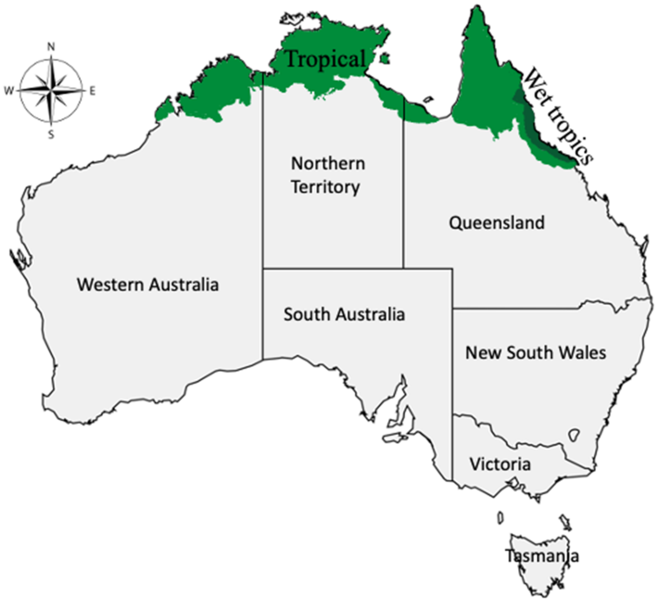
Australian tropical plants have been a rich source of food (bush food) and medicine to the first Australians (Aboriginal people), who are believed to have lived for more than 50,000 years. Plants such as spreading sneezeweed (Centipeda minima), goat’s foot (Ipomoea pes-caprae), and hop bush (Dodonaea viscosa and D. polyandra) are a few popular Aboriginal medicinal plants.
1. Introduction
2. Ethnomedical Uses of Selected Medicinal Plants
Out of 78 tropical medicinal plants used by the Aboriginal people of Australia for treating inflammation and inflammatory-related diseases, 45 species were selected (Table 1).
Table 1. Ethnomedical uses and the compounds isolated from Aboriginal tropical medicinal plants of Australia.
|
Species and Family |
Ethnomedical Uses |
Countries from Where the Plant Has Been Collected for Chemical Studies |
Parts Used for Chemical Isolation |
Isolated Compounds |
|---|---|---|---|---|
|
Acalypha wilkesiana Müll.Arg. (Euphorbiaceae) |
Pulped shoots (i.e., collected when leaves are still red) are applied to cuts and open sores [21]. |
Nigeria |
Leaves; stem and root barks |
Gallic acid, Corilagin, Geraniin, Rutin, Kaempferol 3-O-rutinoside [22]. |
|
Ageratum conyzoides (L.) L. (Asteraceae) |
Meshed whole plant applied to wounds to enhance healing [21][23]. |
Brazil, India |
Whole plant |
5,6,7,8,3′,4′,5′-Heptamethoxyflavone, Coumarin [24]; Ageconyflavones A-C, Linderoflavone B, Eupalestin, Nobiletin, 5,6,7,5′-Tetramethoxy-3′,4′-methylenedioxyflavone, Sinensetin, 5,6,7,3′,4′,5′-Hexamethoxyflavone, 5,6,7,8,3′-Pentamethoxy-4′-hydroxyflavone, 5,6,7,8,3′,5′-Hexamethoxy-4′-hydroxyflavone [24][25]. |
|
Alphitonia excelsa (Fenzl) Reissek ex Benth. (Rhamnaceae) |
Leaves are applied to sore eyes; warm aqueous leaves infusion is used as a bath to ease headaches; decoction from bark, wood, and roots is applied externally to relieve body pains; bark and wood decoction are used as a mouth wash to relieve toothache [21][26]. |
Philippines |
Twigs |
Betulinic acid [27]. |
|
Alphitonia petriei Braid & C.T.White (Rhamnaceae) |
A decoction made from the bark is applied externally to relieve body pain [26]. |
Australia |
Leaves; stems |
Embolic acid, Alphitolic acid, trans- and cis-Coumaroyl esters of alphitolic acid, Betulinic acid [28]. |
|
Angophora costata (Gaertn.) Hochr. ex Britten (Myrtaceae) |
An aqueous solution of reddish exudate from the trunk is taken orally against diarrhoea [8][29]. |
Australia |
Leaves |
Costatamins A-C [30]. |
|
Antidesma bunius (L.) Spreng. (Phyllanthaceae) |
Indicated for headaches, colds, and fevers [23]. |
Vietnam |
Leaves; fruits |
Antidesoside, Podocarpusflavone A, Amentoflavone, Byzantionoside B, Roseoside [31]. |
|
Barringtonia racemosa (L.) Spreng. (Lecythidaceae) |
Pulverized roots are applied to skin sores [21]. |
Bangladesh, China, India, Taiwan, and Vietnam |
Stem bark; seeds; roots; leaves |
Olean-18-en-3β-O-E-coumaroyl ester, Olean-18-en-3β-O-Z-coumaroyl ester, Germanicol, Germanicone, Betulinic acid, Lupeol, Taraxerol [32]; 3,3’-Dimethoxy ellagic acid, Dihydromyticetin, Gallic acid, Bartogenic acid, Stigmasterol [33][34]; Rutin [35][36]; Nasimalun A and B [37]; Barringtin D1-D3, and M1, Casuarictin, Tellimagrandin I, Valoneic acid dilactone, Schimawalin A [38]; Isoracemosol A, Racemosaceramide A, Racemosol A and E [34][39]; Barringtogenol C [34]; 3β-p-E-Coumaroymaslinic acid, cis-Careaborin, Careaborin, Maslinic acid, 2α,3β,19α-Trihydroxyolean-12-ene-24,28-dioic acid, 3β-p-Z-coumaroylcorosolic acid, Corosolic acid, 1α,2α,3β,19α-Tetrahydroxyurs-12-en-28-oic acid, 19α-Hydroxyl ursolic acid, 3α,19α-Dihydroxyurs-12-en-24,28-dioic acid, Tormentic acid, 3-Hydroxy-7,22-dien-ergosterol [40]; Barringosides G-I [41]. |
|
Brasenia schreberi J.F.Gmel. (Combretaceae) |
Canada |
Quercetin-7-O-glucoside, Gallic acid [43]. |
||
|
Brucea javanica (L.) Merr. (Simaroubaceae) |
Roots and leaves are used as analgesics [23]. |
China and Thailand |
Aerial; seeds; roots |
Brusatol [44]; Demethyl-dehydrobrusatol, Deacetyl-yadanzioside I, Javanicoside G, Yadanziolide C and E, Bruceine A-D and H, Bruceoside A-E, Yadanzioside C and I, Yadanzioside K and L, Dehydrobruceine B, Dehydro-bruceantinol, Deacetylated isobrucein B [45]; brujavanol A and B, bruceine, 11-dehydroklaineanone, 15β-hydroxyklaineanone, 14,15β-dihydroxyklaineanone, 15β-O-acetyl-14hydroxyklaieanone [46] |
|
Calophyllum inophyllum L. (Calophyllaceae) |
Nut kernel ground with red pigment is mixed with water and rubbed to ease body pain [21]. |
China, France, Fiji, French Polynesia, India, Indonesia, Malaysia, Thailand, Taiwan, and Vietnam |
Leaves; seeds; twigs; stems; roots |
Inophinnin, Inophinone [47][48]; Inophyllin A, Friedelin, Stigmasterol [48][49][50]; Macluraxanthone, Pyranojacareubin, 4-Hydroxyxanthone, Betulinic acid, Inophyxanthone A, Pancixanthone A, Gerontoxanthone B, Jacareubin [48][51][52][53]; Inocalophyllin A and B [54]; Caloxanthone O and P [55]; Tamanolide, Tamanolide D, E1, E2, and P [56][57]; Calophyllolide [58][59]; 3β,23-epoxy-Friedelan-28-oic acid, Epifriedelanol, Canophyllal, Canophyllol, Canophyllic acid, 3-oxo-Friedelan-28-oic acid, Oleanolic acid, 3,4-Secofriedelan-3,28-dioic acid, 27-Hydroxyacetate canophyllic acid, 3-oxo-27-Hydroxyacetate friedelan-28-oic acid [50][60][61]; Caloxanthone Q, 2-Deprenylrheediaxanthone B, 6-Deoxyjacareubin [52][62]; 1,3,6,7-Tetrahydroxy-5-methoxy-4-(1′,1′-dimethyl-2′-propenyl)-8-(3″,3″-dimethyl-2″-propenyl)-xanthone, (2′S)-7-Hydroxycaloxanthone B, Caloxanthone A-C, 7-Prenyljacareubin, Daphnifolin, Tovopyrifolin C, 1,3,5-Trihydroxyxanthone, 2-Hydroxyxanthone [53]; Inophyllums G-1, G-2, and P [63]; Isocalophyllic acid, Amentoflavone [61][64]; 27-[(E)-p-Coumaroyloxy]canophyllic acid, 27-[(Z)-p-coumaroyloxy]canophyllic acid, Methyl shikimate, (3S,5R,6R,7E,9R)-3,5,6-Trihydroxy-β-ionyl-3-O-β-d-glucopyranoside, Benzyl-O-α-l-rhanmopyranosyl (1→6)-β-d-glucopyranoside, Hexylrutinoside, Kaempferol-3-O-α-l-rhamnoside, 27-[(Z)-p-Coumaroyloxy]friedelin-28-carboxylic acid, (22E,24R)-24-Methyl-5α-cholesta-7,22-diene-3β,5,6β-triol, 3-oxo-Friedelan-28-oicacid [64]; trans-2-[2-(Trifluoromethyl)phenyl]-10b,10c-dimethyl-10b,10c-dihydropyrene, anti-4-aza-B-Homo-5α-cholestane-3-one [65]. |
|
Centella asiatica (L.) Urb. (Apiaceae) |
Juice derived from the plant is taken orally or applied locally for non-specific ulcerations. Powered leaves mixed with lime are applied to sores on babies, and the plant is also indicated for skin diseases [21][23][42][66]. |
China, Japan, India, Madagascar, USA, and Vietnam |
Whole plant |
Asiaticoside, Asiaticoside C, F, G-I, 23-O-Acetyl madecassoside, Asiatic acid, Madecassic acid, Madecassoside, 23-O-Acetylasiaticoside B, Stigmasterol 3-O-β-glucoside, Quercetin 3-O-glucuronide [67][68][69][70][71][72]; Inositol, Centellose [69]; 4′-Hydroxyl-7-methoxyl-6-prenyl-3-O-trans-p-Coumaroyl-flavonol, (2R,3R,2′′S)-3-Furanoyl-brosimacutin E, Epigallocatechin 3-O-p-coumaroate, Pinobanksin-3-propanoate, Kaempferol, Pachypodol, Coryaurone A [71][73]; Asiaticoside B [70][74]; Isomadecassoside [75]; Quadranoside IV, Quercetin, Astragalin, Isoquercetrin [71]; Centelloside E-G, 11-oxo-Asiaticoside B, 11-oxo-Madecassoside, 11(β)-Methoxy asiaticoside B, 11(β)-Methoxy madecassoside, Centellasaponin A, Isoasiaticoside, Scheffoleoside A [70]; 2α,3β,20,23-Tetrahydroxyurs-28-oic acid [76]; Ursolic acid lactone, Ursolic acid, Pomolic acid, Epi-maslinic acid, Corosolic acid, Rosmarinic acid [72]. |
|
Centipeda minima (L.) A.Braun & Asch. (Asteraceae) |
Infusion and decoction from the whole plant, along with other two species (C. cunninghamii and C. thespidioides) is used to wash eye inflammation due to conjunctiva and purulent ophthalmia [21][77]. |
China, Japan, Nepal, South Korea, and Thailand |
Whole plant |
Brevilin A [78][79]; Apigenin, Quercetin-3-Me-ether, Quercetin-3,3′-diMe-ether, Quercetin-3,7,3′-trimethyl-ether, Quercetin-3,7,3′,4′-tetramethyl-ether, Isobutyroylplenolin, Senecioylplenolin, Aurantiamide acetate, Tetrahydrohelenalin, α-Cyperone [80]; 6-O-Methylacrylylplenolin, 6-O-Isobutyroylplenolin, 6-O-Angeloylplenolin [81]; 2β-(Isobutyryloxy)florilenalin [82]; 2R,3R)-(+)-7,4′-di-O-Methyldihydrokaempferol, Iristectorin A, 4′,5,8-Trihydroxy-7-methoxyisoflavone, 3-Trimethoxyquercetin, 3-O-Caffeoyl-α-glueopyranose, 3-O-Caffeoyl-β-glucopyranose, Quercetin, Epipinoresinol, Hispidulin [83]; Minimaoside A and B [84]; Minimolides G and H [85]; Minimolide A-F, J-L, Cenminolide A, B, Centiplide A, (1S,2S,4R,5S,7R,8S,10R)-2α-Tigloyloxy-4α-angeloyloxyguaia- 11(13)-en-8α,12-olide, Centiplide C-I [79][86][87]; 8,10-Dihydroxy-9(2)-methylbutyryloxythymol, 10-Hydroxy-8,9-dioxyisopropylidene-thymol, 8,9,10-Trihydroxythymol, Thymol-β-glucopyranoside, 9-Hydroxythymol, 8,10-Dihydroxy-9-isobutyryloxythymol, 8-Hydroxy-9,10-diisobutyryloxythymol [88]; 4,5β-Dihydroxy-2β-(isobutyryloxy)-10βH-guai-11(13)-en-12,8β-olide, 4-Hydroxyguaia-9,11(13)-dien-12,8β-olide, 2β-(Isobutyryloxy) florilenalin, Pulchellin-2α-O-tiglate, Florilenalin-2α-O-tiglate [89]; Microhelenalin B and C, Arnicolides B-D, Helenalin-angelate, Helenalin-isovalerate, Helenalin-isobutyrate, Helenalin-3-methyl-2-butanoate, Minimolide E, Minimolide B, 2α-Methoxy-6α-angeloyl-2,3-helenalin [79]; Caloinophyllin A, Nobiletin, Quercetin pentamethyl ether, 3′,4′5,7-Tetramethoxyflavone, 4′,5,7-Trimethoxyflavone, 1,5-Dihydroxyxanthone, 1,8-Dimethoxy-2-hydroxyxanthone, 1,6-Dihydroxy-7-methoxyxanthone, 4-Methoxycaffeic acid [90]. |
|
Cleome viscosa L. (Cleomaceae) |
The whole meshed plant is applied externally to relieve rheumatism, swellings, headaches, colds, ulcers, and open-sores; seeds are eaten to relieve fever and diarrhoea [8][21]. |
India, USA, Nigeria, and Vietnam |
Seeds; aerial; leaves |
Quercetin 3-O-(2″-acetyl)-glucoside [91]; Malabaric acid, Stigmast-4-en-3-one, Stigmast-4-ene-3,6-dione [92]; Cleomaldeic acid [93]; Lupeol [94]; Astragalin, Visconoside A-C, Vincetoxicoside A and B, Kaempferitrin, Kaempferide 3-O-β-d-glucopyranoside 7-O-α-l-rhamnopyranoside, Kaempferol 3-O-β-d-glucopyranoside 7-O-α-l-rhamnopyranoside, Isorhamnetin 3-O-β-d-glucopyranoside [95][96]; Lactam nonanoic acid [97]. |
|
Clerodendrum inerme (L.) Gaertn. (Heliotropiaceae) |
China, Egypt, India, Taiwan, Thailand, and Vietnam |
Aerial; flowers; roots; leaves |
3-Hydroxy-3′,4′-dimethoxychalcone, 3,2′-Dihydroxy-3′,4′-dimethoxychalcone, 5-Hydroxy-7,8-dimethoxyflavone, Eucalyptin [98]; 2-(3-Methoxy-4-hydroxylphenyl) ethyl-O-2”,3”-diacetyl-α-l-rhamnopyranosyl-(1→3)-4-O-(E)-feruloyl-β-d-glucopyranoside, monomelittoside, Melittoside, Inerminoside A1, Acteoside, Isoacteoside, Campneoside I [99][100][101]; 4α-Methyl-24β-ethyl-5α-cholesta-14,25-dien-3β-ol; 24β-Ethylcholesta-5,9(11),22E-trien-3β-ol; 11-Pentacosanone; 6-Nonacosanone, Clerodermic acid [102]; Inerminoside A-D [103][104]; Sammangaosides A-C, Leucosceptoside A, Decaffeoyl-acteoside, Darendoside B, Monomelittoside, Melittoside, (7S,8R)-Dehydrodiconiferyl alcohol 9-O-β-glucopyranoside, (7S,8R)-Dehydrodiconiferyl alcohol 4-O-β-glucopyranoside, β-Glucopyranoside, β-(2′-O-β-Xylopyranosyl) glucopyranoside, Salidroside, (Z)-3-Hexenyl-β-glucopyranoside, 2,6-Dimethoxy-p-hydroquinone 1-O-β-glucopyranoside, Seguinoside K [101]; Lup-1,5,20(29)-trien-3-O-β-d-glucopyranoside [100]; Octacosane, Friedelin, β-Amyrin [105]; Crolerodendrum A and B, Uncinatone, Harwickiic acid, Acacetin, Kaempferol 3,7,4′-trimethyl ether, 5α,8α-Epidioxyergosta-6,22-diene-3β-ol [106][107]; Inermes A and B, 14,15-Dihydro-15β-methoxy-3-epicaryoptin [108]; Hispidulin, Diosmetin [107]. |
|
|
Corymbia terminalis (F.Muell.) K.D.Hill & L.A.S.Johnson (Myrtaceae) |
The plant is used for dysentery [109]. |
Australia |
Gum |
Cianidanol, Taxifolin, Aromadendrin, Farrerol [110]. |
|
Crinum pedunculatum R.Br. (Amaryllidaceae) |
Crushed whole plant-rubbed on body parts stung by marine organism [21][23]. |
NA |
NA |
NA |
|
Dodonaea polyandra Merr. & L.M.Perry (Sapindaceae) |
The plant is used for toothache, mouth inflammation, cuts, and open wounds [23]. |
Australia |
Leaves; stems; leaf resins |
Polyandric acid A [111]; 13,17-Epoxy-13-methyl-15-oxo-labda-7-ene, 17-Hydroxy-13-methyl-labda-7,13Z-diene-15-oic acid, 13-Methyl-17-oxo-labda-7,13Z-diene-15-oic acid, Labdane [112]; 15,16-Epoxy-8α-(benzoyloxy)methylcleroda-3,13(16),14-trien-18-oic acid, 15,16-Epoxy-8α-(benzoyloxy)methyl-2α-hydroxycleroda-3,13(16),14-trien-18-oic acid, 15,16-Epoxy-8α-(benzoyloxy)methyl-2-oxocleroda-3,13(16),14-trien-18-oic acid, 15,16-Epoxy-2α-benzoyloxycleroda-3,13(16),14-trien-18-oic acid [113]; 5,7,4′-Trihydroxy-3′(3-methylbut-2-enyl)-3-methoxy flavone, 5,7-Dihydroxy-3′(3-methylbut-2-enyl)-3,4′-dimethoxy flavone, 5,7,4′-Trihydroxy-3′,5′(3-methylbut-2-enyl)-3-methoxy flavone, 5,7,4′-Trihydroxy-3′,5′(3-methylbut-2-enyl)-3,6-dimethoxy flavone, Viscosol, 5,4′-Dihydroxy-3,7-dimethoxyflavone [114]. |
|
Dodonaea viscosa (L.) Jacq. (Sapindaceae) |
Leaves are chewed to relieve toothache; root juice is used as a mouthwash; leaf juice is used to heal stonefish and stingray wounds; root decoction is applied to wounds [21][26]. |
Cameroon, China, and Mexico |
Stems; bark |
Dodovisins A-F, Dodovisnoid E, (+)-hardwickiic acid, ent-15,16-Epoxy-1,3,13(16),14-clerodatetraen-18-oic acid, Hautriwaic lactone, Dodovisnoid G, Methyl-dodovisate B, 5α-Hydroxy-1,2-dehydro-5,10-dihydroprintziasaure-methylester, Strictic acid, Dodonolide [115]; Hautriwaic acid [116]; 2,18-Dihydroxylabda-7,13(E)-dien-15-oic acid, 5,7-Dihydroxy-3,6,4′-trimethoxy-3′-(4-hydroxy-3-methyl-but-2-enyl)flavone, 2,17-Dihydroxylabda-7,13(E)-dien-15-oic acid, 2-Hydroxylabda-7,13(E)-dien-15-oic acid, 3,6-Dimethoxy-5,7,4′-trihydroxyflavone, Penduletin, Santin [117]. |
|
Eleocharis dulcis (Burm.f.) Trin. ex Hensch. (Cyperaceae) |
Whole plant infusion in saltwater (preferred for those growing in or near saltwater) is applied to wounds and sealed with a hollow stem of the same plant [118]. |
China |
Whole plant; peel |
6′-(4″-Hydroxy-3″-methoxy-phenylpropenyl)-1-(10-methoxy-phenylacetone)-1′-O-β-d-glucopyranoside, Susaroyside A, Clausenaglycoside A-D, Emarginone A and B, Thoreliin B, 4-O-(1′,3′-Dihydroxypropan-2′-yl)-dihydroconiferyl alcohol 9-O-β-d-glucopyranoside, 2-[4-(3-Methoxy-1-propenyl)-2-methoxy-phenoxy]-propane-1,3-diol, 6′-O-(E-Cinnamoyl)-coniferin, Methyl 3-(2-O-β-d-glucopyranosyl-3,4,5,6-tetramethoxyphenyl) propanoate, 9-O-(E-Cinnamoyl)-coniferin, 6′-O-(E-Cinnamoyl)-syringin, 2′-O-(E-Cinnamoyl)-syringin [119]. |
|
Eucalyptus camaldulensis Dehnh. (Myrtaceae) |
Gum (or kino) mixed with water is taken orally (recommended not more than 1.3 g of kino) against diarrhoea; infusion made from aerial parts is used for washing head to heal colds and fevers [21][120][121]. |
NA |
NA |
|
|
Euphorbia hirta L. (Euphorbiaceae) |
A decoction from dried herb (whole plant) is used for deworming, dysentery, bowel problems, and colic warts [21][42]. |
India |
Whole plant |
Kaempferol, Rutin, Quercetin [122]. |
|
Euphorbia tirucalli L. (Euphorbiaceae) |
The plant is known for healing skin cancer [23]. |
China |
Aerial; latex |
12-O-(2E,4E,6E,8E-Tetradecatetraenoyl)-13-O-isobutyroyl-4β-deoxyphorbol, 13-O-acetyl-12-O-(2Z,4E-Octadienoyl)-4β-deoxyphorbol, Pedilstatin, 4β-Deoxy-phorbol-13-acetate, 4α-deoxy-phorbol-13-acetate, 3-O-(2,4,68-Tetradecatetraenoyl) ingenol [123]. |
|
Excoecaria agallocha L. (Euphorbiaceae) |
Toxic juice from this plant is applied externally to relieve painful punctures caused by marine organisms, such as the sharp spines of some fish. Infusion from the bark is rubbed against body pain [21][23]. |
Australia, China, India, Japan, and Vietnam |
Leaves; stems; resinous wood; roots; twigs; bark |
12-Deoxyphorbol 13-(3E,5E-decadienoate) [124]; Excoecarins R1 and R2 [125]; 3α,11β-Dihydroxy-ent-isopimara-8(14),15-dien-2-one, 16β-Hydroxy-ent-atisan-3-one, Ribenone, ent-labda-8(17),13E-diene-3β,15-diol, ent-3β-Hydroxybeyer-15-ene-2,12-dione [126]; Excoecarins S, T1-T2, ent-12-oxo-2,3-Secobeyer-15-ene-2,3-dioic acid, ent-15-epoxy-Beyerane-3α-ol, Agallochin H [127]; Excoecarins V1—V3, 3,5,7,3′,5′-Pentahydroxy-2R,3R-flavanonol 3-O-α-l-rhamnopyranoside, ent-Atisane-16α-ol, ent-2,3-Secobeyer-15-ene-2,3-dioic acid, ent-15,18-Dihydroxybezoate, 3,4,5-Trimethoxyphenol 1-O-β-d-(6-galloyl)-glucopyranoside [128]; 3β-[(2E,4E)-5-oxo-Decadienoyloxy]-olean-12-ene, β-Amyrin acetate, Taraxerone, 3-Epitaraxerol, Epilupeol, Taraxerol, Taraxerone, 3β-[(2E,4E)-6-oxo-Decadienoyloxy]-olean-12-ene, Acetyl aleuritolic acid, Cycloart-22-ene-3β,25-diol, β-Sitostenone, (24R)-24-Ethylcholesta-4,22-dien-3-one, β-Sitosterol [129][130]; Excoagallochaols A–E [131]; Agallochins A-E [132][133]; Excoecarins D, E, and K [134]; Agallochins J-L [133][135]; Agallochins F-I, 2-Acetoxy-1,15-beyeradiene-3,12-dione, 2-Hydroxy-1,15-beyeradiene-3,12-dione, ent-kauran-16β-ol-3-one [127][133][136]; Excoecariphenols A-D [137]; Agallochaols K–P, Agallochaol Q, ent-17-Hydroxykaur-15-en-3-one, ent-Kaur-15-en-3β,17-diol, 7-Deoxogeayine, ent-15-Hydroxylabd-8(17),13E-dien-3-one, ent-15,18-Dihydroxylabd-8(17),13E-diene, ent-3β,11α-Dihydroxyisopimara-8(14),15-dien-2-one, ent-3β-Hydroxybeyer-15-en-2,12-dione [138]; ent-16α-Hydroxy-atisane-3,4-lactone, ent-16α-Hydroxy-atisane-3-one, ent-Atisane-3β,16α-diol, ent-3,4-seco-16α-Hydroxyatis-4(19)-en-3-oic acid [139]; Triacontane [140]; Agallochins M-P [138][141][142]; Excagallonoid A, ent-(3α,5β,8α,9β,10α,12α)-3-Hydroxyatis-16-en-14-one, Atis-16-ene-3,14-dione, 2-Hydroxy-atis-1,16-diene-3,14-dione, 12-Hydroxy-13-methylpodocarpa-8,11,13-trien-3-one [143]; Excolides A-B [144]; Afzelin, Quercitrin, Rutin, Kaempferol-3-O-(2-O-acetyl)-α-l-rhamnopyranoside, Kaempferide 3-O-α-l-rhamnopyranoside, Kaempferol 3-O-α-l-arabinofuranoside [145]; Agallolides A-M [146] |
|
Flueggea virosa (Roxb. ex Willd.) Royle (Phyllanthaceae) |
An aqueous leaf infusion is taken orally to heal internal pains, such as toothache; the liquid is applied to skin sores [21][147]. |
China and Taiwan |
Aerial; roots |
Flueggether A, Virosinine A [148]; Flueggenines A, B, and D, Norsecurinine [149][150][151]; Flueggines A and B [152]; Fluevirosines A-C [153]; Virosaines A and B [150][154]; 3β,12-Dihydroxy-13-methylpodocarpa-6,8,11,13-tetraene, 3β,12-Dihydroxy-13-methylpodocarpa-8,11,13-triene, Spruceanol, ent-3β,12α-Dihydroxypimara-8(14),15-diene, 3α-Hydroxy-12-methoxy-13-methyl-entpodocarp-6,8,11,13-tetraene, 3α-Hydroxy-13-hydroxymethyl-12-methoxy-ent-podocarp-6,8,11,13-tetraene, 3β-Hydroxy-13-hydroxymethyl-12-methoxy-ent-podocarp-6,8,11,13-tetraene, 12-Hydroxy-13-methylent-podocarp-6,8,11,13-tetraen-3-one, 12-Methoxy-13-methyl-ent-podocarp-6,8,11,13-tetraen-3-one, 6β,12-Dihydroxy-13-methyl-ent-podocarp-8,11,13-trien-3-one, 7α,20-Epoxy-3α-hydroxy-12-methoxy-13-methyl-ent-podocarp-8,11,13-triene, 3α,20-Epoxy-3β-hydroxy-12-methoxy-13-methyl-ent-podocarp-8,11,13-triene [155][156]; Fluvirosaones A and B, Virosecurinine [151][157]; 9(10→20)-Abeo-ent-podocarpane; 3,10-Dihydroxy-12-methoxy-13-methyl-9(10→20)-abeo-ent-podocarpa-6,8,11,13-tetraene; 4E-Dehydrochebulic acid trimethyl ester; 12-Hydroxy-20(10→5)-abeo-4,5-seco-podocarpa-5(10),6,8,11,13-pentaen-3-one; Betulinic acid 3β-calfeate, (+)-Ampelosin E [156]; Flueggrenes A and B [158]; Flueggenoids A–E, 6,12-Dihydroxy-13-methyl-7-oxo-ent-podocarpa−5,8,11,13-tetraeno-20,3α-lactone; 10α,12-Dihydroxy-13-methyl-9(10→20)-abeo-ent-podocarpa−6,8,11,13-tetraen-3-one; 12-Hydroxy-20(10→5)-abeo-4,5-seco-podocarpa-5(10),6,8,11,13-pentaen-3-one; Securinine, Bergenin, Norbergenin [150]; Fluevirines E and F, Viroallosecurinine [151]; Flueindolines A–C, Donaxanine, Methyltryptamine, N,N-Dimethyltryptamine, 1-Acetyl-β-carboline, 1-Hydroxymethyl-β-carboline, N-Methyl-1,2,3,4-tetrahydro-β-carboline, Strychnocarpine, Racemate, Hydromethyl-2-methyl-tetrahydro-β-carboline [159]. |
|
Heliotropium ovalifolium Forss (Heliotropiaceae) |
Herb extract is used to relieve fevers [160]. |
India, Egypt, and Zimbabwe |
Aerial |
Heliophenanthrone [161]; Retronecine, Helifoline [162]; Supinine, 7-Angelyl-heliotridine [163]; 4,7,8-Trimethoxy-naphthalene-2-carboxylic acid, 6-Hydroxy-5,7-dimethoxy-naphthalene-2-carbaldehyde [164]; Heliotropamide [165]. |
|
Hibiscus tiliaceus L. (Malvaceae) |
Infusions from bark and sapwood (with salt or freshwater) are applied to wounds and covered with the bark of the same plant [21][118]. |
China, Japan, and Taiwan |
Stem; wood; bark |
Hibiscusin, Hibiscusamide, Vanillic acid, 4-Hydroxybenzoic acid, Syringic acid, 4-Hydroxybenzaldehyde, Scopoletin, N-trans-Feruloyltyramine, N-cis-Feruloyltyramine [166]; 27-oic-3-oxo-28-Friedelanoic acid, 3α-Hydroxyfriedelane-2-one, 4α-Hydroxyfriedelane-3-one, Friedelin, Epifriedelanol, Pachysandiol A, 3β-O-(p-Hydroxy-Z-cinnamoyl)oleanolic acid, 3β-O-(p-hydroxy-E-cinnamoyl)oleanolic acid, oleanolic acid [167]; Hibiscusterpene I-V, Hibiscone B and C, Isohemigossypol-1-methyl ether, Virginicin, Parvifloral A, Syriacusin A [168]. |
|
Ipomoea brasiliensis (L.) Sweet (I. pes-caprae (L.) R. Br.) (Combretaceae) |
Leaves decoction is applied externally for sores; the heated leaves are used to discharge boils [21][23]. |
China, India, Mexico, and Thailand |
Whole plant |
Pescapreins X-XVII [169]; β-Damascenone, Phytol [170]; Pescaproside A and B, Pescapreins I-IX, Stoloniferin III [171]; Ipomeolides A and B, Presqualene alcohol, Icosyl (E)-3-(4-hydroxyphenyl)acrylate, β-Sitosterol-3-O-β-d-glucopyranoside, Stigmasterol, Lupeol [172]. |
|
Litsea glutinosa (Lour.) C.B.Rob. (Heliotropiaceae) |
Leaves and bark decoctions are applied to sores and to relieve body pain; sometimes, chewed leaves are applied to cuts and sores [21][23][26]. |
China and India |
Leaves; twigs; heartwood |
Glutin, β-sitosterol, Stigmasterol, (−)-Epicatechin, Sitosterol-β-d-glucopyranoside [173]; (3R,4S,5S)-2-Hexadecyl-3-hydroxy-4-methylbutanolide, Litsealactone C, D, and G, Eusmoside C [174]. |
|
Macaranga tanarius (L.) Müll.Arg. (Euphorbiaceae) |
The plant is known for wound healing [175]. |
Japan, Taiwan, Thailand, and Vietnam |
Bark; leaves; fruits; glandular trichomes |
(2β,5β,10α,13α)-2-Hydroxypimara-9(11),15-dien-12-one, Methyl 2α-hydroxy-3β-[(4-hydroxybenzoyl)oxy]taraxer-14-en-28-oate, 2α-Acetoxy-3β-[(4-hydroxybenzoyl)oxy]-taraxer-14-en-28-oic acid, β-Sitosterol, Friedelin, Friedelin-3β-ol, β-Amyline, Macarangonol, 3β-Acetoxytaraxer-14-en-28-oic acid, 2α-Hydroxy-3β-[(4-hydroxybenzoyl)oxy]taraxer-14-en-28-oic acid [176]; (+)-Pinoresinol 4-O-[6″-O-galloyl]-β-d-glucopyranoside, Roseoside, Icariside B5, (6R,9R)-3-oxo-α-ionol β-d-glucoside, (6R,9S)-3-oxo-α-Ionol β-d-glucoside, (2S,3R)-Dihydrodehydrodiconiferyl alcohol β-d-glucoside, (+)-Pinoresinol 4-O-β-d-glucopyranoside, Scopoline, Rutin, Quercetin 3-O-galactopyranoside, Quercetin 3-O-arabinopyranoside, Isovitexin, Methyl gallate, Hexenyl β-d-glucoside, (E)-2-Hexenyl β-d-glucoside, Malloapeltine [177]; Macarangiosides A-F, Mallophenol B, Lauroside E [178]; Tanariflavanones A-D [177][179][180]; Macaflavanones A-G, Kolavenol [181]; 3′-Geranyl-naringenin [182]; Nymphaeol A-C, Isonymphaeol B, 3′-Geranyl naringenin [179][180][181][182][183]; Macatanarin D, Schweinfurthins E-H, and K-Q,5-((E)-3,5-Dihydroxystyryl)-3-((E)-3,7-dimethylocta-2,6-dien-1-yl)benzene-1,2-diol [184]; Tanarifuranonol, Vomifoliol, Blumenol B, vedelianin, mappain, methyl-mappain [180][185]. |
|
Manihot esculenta Crantz (Euphorbiaceae) |
The plant is known to be effective against belly aches and diarrhoea [175]. |
NA |
NA |
NA |
|
Melaleuca leucadendra (L.) L. (Myrtaceae) |
The plant is known to be effective against headache, sinusitis, cough and colds, and skin sores [21][23]. |
Egypt |
Essential oil |
Stachyurin (or casuarinin), Ellagitannin [186]. |
|
Merremia tridentata (L.) Hallier f. (Combretaceae) |
The whole plant is chewed or soaked in the water before applying it to the sores [109]. |
Vietnam |
Stem bark |
Apigenin, Cynaroside, Luteolin, Cosmosiin, Quercitrin [187]. |
|
Morinda citrifolia L. (Rubiaceae) |
French Polynesia and Japan |
Fruits |
(+)-3,4,3′,4′-Tetrahydroxy-9,7′α-epoxylignano-7α,9′-lactone, (+)-3,3′-Bisdemethyltanegool, (−)-Pinoresinol, (−)-3,3″-Bisdemethylpinoresinol, Quercetin, Kaempferol, Scopoletin, Isoscopoletin, Vanillin [189]; 1,5,15-Tri-O-methylmorindol, 2-O-(β-d-glucopyranosyl)-1-O-hexanoyl-β-d-gluropyranose, 2-O-(β-d-glucopyranosyl)-1-O-octanoyl-β-d-gluropyranose, 5,15-Di-O-methylmorindol, 1,3-Dihydroxy-2-methoxyanthracene-9,10-dione, 6-O-(β-d-Glucopyranosyl)-1-O-hexanoyl-β-d-glucopyranose, 6-O-(β-d-glucopyranosyl)-1-O-octanoyl-β-d-glucopyranose, 2,6-Di-O-(β-d-Glucopyranosyl)-1-O-hexanoyl-β-d-glucopyranose, 3-Methylbut-3-enyl-β-d-glucopyranose, 3-Methylbut-3-enyl-6-O-β-d-glucopyranosyl-β-d-glucopyranose, Asperulosidic acid, Rutin [190][191]; Nonioside A, (2E,4E,7Z)-deca-2,4,7-trienoate-2-O-β-d-glucopyranosyl-β-d-glucopyranoside, Tricetin [191]. |
|
|
Nauclea orientalis (L.) L. (Rubiaceae) |
Aqueous bark infusion is used for sore belly; it is also applied externally to relieve rheumatic pains; the wood infusion is used for relieving fevers [23][77]. |
China, Japan, Laos, Papua New Guinea, Thailand, and Vietnam |
Heartwood; bark; leaves; stems; roots; |
Noreugenin, Naucleoside [192]; Angustine, 18,19-Dihydroangustine, 10-Hydroxyangustine, 3,14,18,19-Tetrahydroangustine, Parvine, Angustoline [193]; Nauclealines A and B, Naucleosides A and B, Strictosamide, Vincosamide, Pumiloside, Kelampayoside A, β-Sitosterol, Sitosteryl β-d-glucoside [194][195]; Naucleaorals A and B [196]; 10-Hydroxystrictosamide, 6′-O-Acetylstrictosamide [195]; α-Pinene, Loganetin, Loganin, Sweroside, Grandifloroside, Methyl 3,4-dihydroxybenzoate, 4-Hydroxycinnamic acid, 3-(2,4-Dihydroxylphenyl)propanoic acid, Methyl 3-(2,4-dihydroxylphenyl)propanoate, Skimmin, Adicardin, Aloe emodin, Pinoresinol [197]; Naucleaorine, Epimethoxynaucleaorine, Strictosidine lactam, 3,4,5-Trimethoxyphenol, 3α-Hydroxyurs-12-en-28-oic acid methyl ester, 3α,23-Dihydroxyurs-12-en-28-oic acid, 3α,19α,23-Trihydroxyurs-12-en-28-oic acid methyl ester, Oleanolic acid [198]; Nauclorienine, Antirhine, Iso-antirhine, Alangine, Naucline, Neonaucline, Angustidine, Subditine [199]. |
|
Nelumbo nucifera Gaertn. (Nelumbonaceae) |
Milky juice from leaves is used against diarrhoea [42]. |
China, India, and Japan |
Flowers; rhizome; leaves; seed embryo |
2α,24-Diacetoxy-3β-hydroxyolean-12-en-28-oic acid, Hyptatic acid A, Maslinic acid, Botulin, Lupeol [200]; (R)-Coclaurine, (S)-norcoclaurine, Quercetin 3-O-β-d-glucuronide [201]; Neferine [202][203]; Liensinine, Isoliensinine [204]; Betulinic acid [205]. |
|
Ochrosia elliptica Labill. (Apocynaceae) |
Bark is known to be good for dysentery [188]. |
China and Egypt |
Stems and leaves |
10-Methoxyconolidine, Apparicine, Vallesamine, Yunnanensine A, Angustilodine, Isositsirikine, (−)-Echitainine, Pseudo akuammigine [206]; Ursolic acid [207][208]; Ellipticine, elliptinine, methoxyellipticine, reserpiline (elliptine) [209]. |
|
Ocimum tenuiflorum L. (Heliotropiaceae) |
The plant is used to relieve fevers [210]. |
NA |
NA |
NA |
|
Phyllanthus urinaria L. (Phyllanthaceae) |
China and Taiwan |
Whole plant |
Phyllanthin, Phyltetralin, Trimethyl-3,4-dehydrochebulate, Methylgallate, Rhamnocitrin, Methyl brevifolincarboxylate, β-Sitosterol-3-O-β-d-glucopyranoside, Quercitrin, Rutin [211]; Geraniin [212]; Corilagin, Ellagic acid [213]. |
|
|
Phragmites australis (Cav.) Trin. ex Steud. (Plantaginaceae) |
China |
Roots |
N-p-Coumaroyl serotonin, N-p-Coumaroyl-trypamine, phranisines A-B [216]. |
|
|
Sarcostemma viminale (L.) R. Br (Apocynaceae) |
The plant is indicated for skin sores and eye complaints [217]. |
NA |
NA |
NA |
|
Scaevola taccada (Gaertn.) Roxb. (Euphorbiaceae) |
Leaves decoction is applied externally to skin sores [8][23]. |
Thailand |
Fruits |
Scataccanol, ent-ammirin, Nodachenetin, Marmesin, Xanthyletin, Umbelliferone, 4-Formylsyringol, 6-Hydroxy-7-methyl-1-oxo-4-carbomethoxy octahydrocyclopenta[c]pyran, Loganetin, Matairesinol, 2-(4-Hydroxyphenyl) 3-(3,4-dihydroxyphenyl)-2-propenoate [218]. |
|
Scoparia dulcis L. (Plantaginaceae) |
Leaves infusion is taken orally to heal stomach pain; the pulped whole plant is used for covering sores and cuts to enhance healing [23]. |
Bangladesh and Brazil |
Whole plant |
Glutinol [219]; Scoparinol [220]; iso-dulcinol, 4-epi-scopadulcic acid B, dulcidiol, scopanolal, dulcinol, and scopadiol [221]. |
|
Terminalia catappa L. (Combretaceae) |
The plant is indicated for sore throat [175]. |
China and New Caledonia |
Leaves; bark |
Ursolic acid, 2,3,23-Trihydroxyurs-12-en-28-oic acid [222]; 3,4,5-Trimethoxyphenyl-1-O-(4-sulfo)-β-d-glucopyranoside, Chebuloside II, Arjunoglucoside II, Arjunolic acid, Betulinic acid, β-Sitosterol-3-O-β-d-glucopyranoside [223]. |
|
Terminalia muelleri Benth. (Combretaceae) |
The plant is indicated for skin sores [175]. |
Egypt |
Leaves |
Apigenin-8-C-(2″-O-galloyl) glucoside 1, Luteolin-8-C-(2″-O-galloyl) glucoside 2, 1-O-Galloyl-2,3,4,6-dihexahydroxydiphenoyl-β-d-glucopyranoside, 1,4,6-Tri-O-galloyl-2,3-hexahydroxydiphenoyl-β-d-glucopyranoside, 1,2-Di-O-galloyl-4,6-hexahydroxydiphenoyl-β-d-glucopyranoside, Isostrictinin, 1-O-Galloyl-β-d-glucopyranoside, Combretum caffrum, Ellagic acid, Gallic acid [224][225]; Isoorientin, Vitexin, Chebulinic acid [225]. |
|
Verbena officinalis L. (Verbenaceae) |
A decoction made from the whole plant is applied externally to overcome fever and rheumatic pain [21][42][226]. |
China and India |
Aerial |
3,4-Dihydroverbenalin, Daucosterol [227]; Ursolic acid [228]; Verbenalin, Hastatoside, Acteoside, β-sitosterol-d-glucoside [229]. |
Abbreviation: NA, not available.
3. Overview of the Anti-Inflammatory Mechanism of Action/Pathways
4. Phytochemistry and Pharmacology of Medicinal Plants
4.1. Anti-Inflammatory Crude Extracts
4.2. Anti-Inflammatory Compounds
References
- Schippmann, U.; Leaman, D.J.; Cunningham, A.B. Impact of culitvation and gathering of medicinal plants on biodiversity: Global trends and issues (Case Study No 7). In Proceedings of the Biodiversity and the Ecosystem Approach in Agriculture, Forestry and Fisheries, Rome, Italy, 12–13 October 2002; pp. 140–167.
- Verpoorte, R. Pharmacognosy in the New Millennium: Leadfinding and Biotechnology. J. Pharm. Pharmacol. 2000, 52, 253–262.
- Calixto, J.B. Twenty-five years of research on medicinal plants in Latin America: A personal view. J Ethnopharmacol 2005, 100, 131–134.
- Wangchuk, P.; Yeshi, K.; Vennos, C.; Mandal, S.C.; Kloos, S.; Nugraha, A.S.; Tashi; Samten. Three medicinal Corydalis species of the Himalayas: Their ethnobotany, pharmacognosy, phytochemistry and pharmacology. J. Herbal Med. 2020, 23, 100384.
- Australian Bureau of Statistics. Cultural Identification. National Aboriginal and Torres Strait Islander Health Survey Table Builder. 2019. Available online: https://www.abs.gov.au/statistics/people/aboriginal-and-torres-strait-islander-peoples/national-aboriginal-and-torres-strait-islander-health-survey/latest-release (accessed on 21 January 2022).
- Australian Bureau of Statistics. Estimates and projections, Aboriginal and Torres Strait Islander Australians, 2006 to 2031; ABS cat. no. 3238.0; ABS: Canberra, Australia, 2019.
- McConvell, P.; Thieberger, N. Keeping track of Indigenous language endangerment in Australia. In Language Diversity in the Pacific: Endangerment and Survival; Cunningham, D., Ingram, D.E., Sumbuk, K., Eds.; Multilingual Matters: Clevedon, Austrailia, 2006; pp. 54–84.
- Lassak, E.V.; McCarthy, T. Australian Medicinal Plants; New Holland Publishers: Wahroonga, Australia, 2006.
- Barr, A. Aboriginal communities of the Northern Territory of Australia. In Traditional Bush Medicines. An Aboriginal Pharmacopoeia; Greenhouse Publications: Darwin, Australia, 1988.
- Locher, C.; Semple, S.J.; Simpson, B.S. Traditional Australian Aboriginal medicinal plants: An untapped resource for novel therapeutic compounds? Future. Med. Chem. 2013, 5, 733–736.
- Packer, J.; Turpin, G.; Ens, E.; Venkataya, B.; Mbabaram, C.; Yirralka, R.; Hunter, J. Building partnerships for linking biomedical science with traditional knowledge of customary medicines: A case study with two Australian Indigenous communities. J. Ethnobiol. Ethnomed. 2019, 15, 69.
- Barr, A. Traditional Bush Medicines: An Aboriginal Pharmacopoeia; Greenhouse Publications: Richmond, VIC, Australia, 1988.
- Guo, Y.; Sakulnarmrat, K.; Konczak, I. Anti-inflammatory potential of native Australian herbs polyphenols. Toxicol. Rep. 2014, 1, 385–390.
- Yeshi, K.; Crayn, D.; Ritmejeryte, E.; Wangchuk, P. Plant Secondary Metabolites Produced in Response to Abiotic Stresses Has Potential Application in Pharmaceutical Product Development. Molecules 2022, 27, 313.
- Zubair, M.; Rizwan, K.; Rashid, U.; Saeed, R.; Saeed, A.A.; Rasool, N.; Riaz, M. GC/MS profiling, in vitro antioxidant, antimicrobial and haemolytic activities of Smilax macrophylla leaves. Arab. J. Chem. 2017, 10, S1460–S1468.
- Ashraf, I.; Zubair, M.; Rizwan, K.; Rasool, N.; Jamil, M.; Khan, S.A.; Tareen, R.B.; Ahmad, V.U.; Mahmood, A.; Riaz, M.; et al. Chemical composition, antioxidant and antimicrobial potential of essential oils from different parts of Daphne mucronata Royle. Chem. Cent. J. 2018, 12, 135.
- Khalid, A.; Shahid, S.; Khan, S.A.; Kanwal, S.; Yaqoob, A.; Rasool, Z.G.; Rizwan, K. Antioxidant activity and hepatoprotective effect of Cichorium intybus (Kasni) seed extract against carbon tetrachloride-induced liver toxicity in rats. Trop. J. Pharm. Res. 2018, 17.
- Adegboye, O.; Field, M.A.; Kupz, A.; Pai, S.; Sharma, D.; Smout, M.J.; Wangchuk, P.; Wong, Y.; Loiseau, C. Natural-product-based solutions for tropical infectious diseases. Clin. Microbiol. Rev. 2021, 34, e0034820.
- Cock, I.E. Medicinal and aromatic plants—Australia. In Ethnopharmacology Section, Biological, Physiological and Health Sciences, Encyclopedia of Life Support Systems (EOLSS), Developed under the Auspices of the UNESCO; EOLSS Publishers: Oxford, UK, 2011; Available online: http://www.eolss.net (accessed on 30 April 2022).
- Bureau of Meteorology. Map of climate zones of Australia: Government of Australia. 2001. Available online: http://www.bom.gov.au/climate/how/newproducts/images/zones.shtml (accessed on 5 May 2022).
- Lassak, E.V.; McCarthy, T. Australian Medicinal Plants; Methuen: Sydney, Australia, 1983; ISBN 9780454004380.
- Adesina, S.K.; Idowu, O.; Ogundaini, A.O.; Oladimeji, H.; Olugbade, T.A.; Onawunmi, G.O.; Pais, M. Antimicrobial constituents of the leaves of Acalypha wilkesiana and Aacalypha hispida. Phytother Res. 2000, 14, 371–374.
- Webb, L.J. Some New Records of Medicinal Plants Used by the Aborigines of Tropical Queensland and New Guinea; Royal Society of Queensland: Brisbane, Australia, 1959; Volume 71.
- Moreira, M.D.; Picanco, M.C.; Barbosa, L.C.; Guedes, R.N.; Barros, E.C.; Campos, M.R. Compounds from Ageratum conyzoides: Isolation, structural elucidation and insecticidal activity. Pest Manag. Sci. 2007, 63, 615–621.
- Vyas, A.V.; Mulchandani, N.B. Polyoxygenated flavones from Ageratum conyzoides. Phytochemistry 1986, 25, 2625–2627.
- Webb, L.J. The use of plant medicines and poisons by Australian aborigines. Mankind 1969, 7, 137.
- Fuentes, R.G.; Valenciano, A.L.; Cassera, M.B.; Kingston, D.G.I. Antiproliferative and antiplasmodial investigation of Alphitonia excelsa and Arcangelesia flava. Philipp. J. Sci. 2020, 149, 115–120.
- Raju, R.; Gunawardena, D.; Ahktar, M.A.; Low, M.; Reddell, P.; Munch, G. Anti-Inflammatory Chemical Profiling of the Australian Rainforest Tree Alphitonia petriei (Rhamnaceae). Molecules 2016, 21, 1521.
- Lauterer, J. Chemical and physiological notes on native and acclimatised mydriatic plants of Queensland. Australas. Med. Gaz. 1895, 14, 457–460.
- Raju, R.; Singh, A.; Bodkin, F.; Munch, G. Costatamins A-C, new 4-phenylcoumarins with anti-inflammatory activity from the Australian woodland tree Angophora costata (Myrtaceae). Fitoterapia 2019, 133, 171–174.
- Trang, D.T.; Huyen, L.T.; Nhiem, N.X.; Quang, T.H.; Hang, D.T.T.; Yen, P.H.; Tai, B.H.; Anha, H.L.T.; Binh, N.Q.; Van Minha, C.; et al. Tirucallane glycoside from the leaves of Antidesma bunius and inhibitory NO production in BV2 cells and RAW264.7 macrophages. Nat. Prod. Commun. 2016, 11, 935–937.
- Yang, Y.; Deng, Z.; Proksch, P.; Lin, W. Two new 18-en-oleane derivatives from marine mangrove plant, Barringtonia racemosa. Pharmazie 2006, 61, 365–366.
- Sun, H.Y.; Long, L.J.; Wu, J. Chemical constituents of mangrove plant Barringtonia racemosa. Zhong Yao Cai. 2006, 29, 671–672. (In Chinese)
- Gowri, P.M.; Radhakrishnan, S.V.; Basha, S.J.; Sarma, A.V.; Rao, J.M. Oleanane-type isomeric triterpenoids from Barringtonia racemosa. J. Nat. Prod. 2009, 72, 791–795.
- Samanta, S.K.; Bhattacharya, K.; Mandal, C.; Pal, B.C. Identification and quantification of the active component quercetin 3-O-rutinoside from Barringtonia racemosa, targets mitochondrial apoptotic pathway in acute lymphoblastic leukemia. J. Asian Nat. Prod. Res. 2010, 12, 639–648.
- Patil, K.R.; Patil, C.R.; Jadhav, R.B.; Mahajan, V.K.; Patil, P.R.; Gaikwad, P.S. Anti-arthritic activity of bartogenic acid isolated from fruits of Barringtonia racemosa Roxb. (Lecythidaceae). Evid. Based Complement. Alternat. Med. 2011, 2011, 785245.
- Hasan, C.M.; Khan, S.; Jabbar, A.; Rashid, M.A. Nasimaluns A and B: Neo-clerodane diterpenoids from Barringtonia racemosa. J. Nat. Prod. 2000, 63, 410–411.
- Yoshikawa, S.; Chen, L.G.; Yoshimura, M.; Amakura, Y.; Hatano, T.; Taniguchi, S. Barricyclin D1-a dimeric ellagitannin with a macrocyclic structure-and accompanying tannins from Barringtonia racemosa. Biosci. Biotechnol. Biochem. 2021, 85, 1609–1620.
- Ponnapalli, M.G.; Dangeti, N.; Sura, M.B.; Kothapalli, H.; Akella, V.S.; Shaik, J.B. Self gelating isoracemosol A, new racemosaceramide A, and racemosol E from Barringtonia racemosa. Nat. Prod. Res. 2017, 31, 63–69.
- Xia, H.; Zhang, X.L.; Wang, G.H.; Tong, Y.C.; He, L.; Wang, H.F.; Pei, Y.H.; Chen, Y.J.; Sun, Y. Chemical constituents from Barringtonia racemosa. Zhongguo Zhong Yao Za Zhi. 2016, 41, 2460–2465. (In Chinese)
- Van, Q.T.T.; Vien, L.T.; Hanh, T.T.H.; Huong, P.T.T.; Cuong, N.T.; Thao, N.P.; Thuan, N.H.; Dang, N.H.; Thanh, N.V.; Cuong, N.X.; et al. Acylated flavonoid glycosides from Barringtonia racemosa. Nat. Prod. Res. 2020, 34, 1276–1281.
- Maiden, J.H. The useful native plants of Australia; Turner and Henderson: Sydney, Australia, 1889.
- Legault, J.; Perron, T.; Mshvildadze, V.; Girard-Lalancette, K.; Perron, S.; Laprise, C.; Sirois, P.; Pichette, A. Antioxidant and anti-inflammatory activities of quercetin 7-O-beta-D-glucopyranoside from the leaves of Brasenia schreberi. J. Med. Food 2011, 14, 1127–1134.
- Zhou, J.; Tan, L.; Xie, J.; Lai, Z.; Huang, Y.; Qu, C.; Luo, D.; Lin, Z.; Huang, P.; Su, Z.; et al. Characterization of brusatol self-microemulsifying drug delivery system and its therapeutic effect against dextran sodium sulfate-induced ulcerative colitis in mice. Drug Deliv. 2017, 24, 1667–1679.
- He, X.; Wu, J.; Tan, T.; Guo, W.; Xiong, Z.; Yang, S.; Feng, Y.; Wen, Q. Quassinoids from Brucea javanica and attenuates lipopolysaccharide-induced acute lung injury by inhibiting PI3K/Akt/NF-kappaB pathways. Fitoterapia 2021, 153, 104980.
- Chumkaew, P.; Srisawat, T. Antimalarial and cytotoxic quassinoids from the roots of Brucea javanica. J. Asian Nat. Prod. Res. 2017, 19, 247–253.
- Mah, S.H.; Lian Ee, G.C.; Teh, S.S.; Sukari, M.A. Antiproliferative xanthone derivatives from Calophyllum inophyllum and Calophyllum soulattri. Pak. J. Pharm. Sci. 2015, 28, 425–429.
- Lian Ee, G.C.; Mah, S.H.; Rahmani, M.; Taufiq-Yap, Y.H.; Teh, S.S.; Lim, Y.M. A new furanoxanthone from the stem bark of Calophyllum inophyllum. J. Asian Nat. Prod. Res. 2011, 13, 956–960.
- Lian Ee, G.C.; Kua, A.S.; Lim, C.K.; Jong, V.; Lee, H.L. Inophyllin A, a new pyranoxanthone from Calophyllum inophyllum (Guttiferae). Nat. Prod. Res. 2006, 20, 485–491.
- Li, Y.Z.; Li, Z.L.; Yin, S.L.; Shi, G.; Liu, M.S.; Jing, Y.K.; Hua, H.M. Triterpenoids from Calophyllum inophyllum and their growth inhibitory effects on human leukemia HL-60 cells. Fitoterapia 2010, 81, 586–589.
- Li, Y.; Li, Z.L.; Liu, M.S.; Li, D.Y.; Zhang, H.; Hua, H.M. Xanthones from leaves of Calophyllum inophyllum Linn. Yao Xue Xue Bao 2009, 44, 154–157. (In Chinese)
- Wei, D.J.; Mei, W.L.; Zhong, H.M.; Zeng, Y.B.; Wu, X.D.; Dai, H.F. A new prenylated xanthone from the branches of Calophyllum inophyllum. J. Asian Nat. Prod. Res. 2011, 13, 265–269.
- Haerani, S.N.; Raksat, A.; Pudhom, K. Two new xanthones from the root of Thai Calophyllum inophyllum and their toxicity against colon and liver cancer cells. J. Nat. Med. 2021, 75, 670–674.
- Shen, Y.C.; Hung, M.C.; Wang, L.T.; Chen, C.Y. Inocalophyllins A, B and their methyl esters from the seeds of Calophyllum inophyllum. Chem. Pharm. Bull. 2003, 51, 802–806.
- Dai, H.F.; Zeng, Y.B.; Xiao, Q.; Han, Z.; Zhao, Y.X.; Mei, W.L. Caloxanthones O and P: Two new prenylated xanthones from Calophyllum inophyllum. Molecules 2010, 15, 606–612.
- Leu, T.; Raharivelomanana, P.; Soulet, S.; Bianchini, J.P.; Herbette, G.; Faure, R. New tricyclic and tetracyclic pyranocoumarins with an unprecedented C-4 substituent. Structure elucidation of tamanolide, tamanolide D and tamanolide P from Calophyllum inophyllum of French Polynesia. Magn. Reson. Chem. 2009, 47, 989–993.
- Ginigini, J.; Lecellier, G.J.; Nicolas, M.; Nour, M.; Hnawia, E.; Lebouvier, N.; Herbette, G.; Lockhart, P.; Raharivelomanana, P. Chemodiversity of Calophyllum inophyllum L. oil bioactive components related to their specific geographical distribution in the South Pacific region. PeerJ 2019, 7, e6896.
- Kalyanaraman, L.; Mohan Kumar, R.; Vishweshwar, P.; Pichai, R.; Narasimhan, S. 5-Meth-oxy-2,2-dimethyl-6--10-phenyl-2H,8H-pyranochromen-8-one (calophyllolide). Acta Crystallogr. Sect. E Struct. Rep. Online 2010, 66, o1115.
- Nguyen, V.L.; Truong, C.T.; Nguyen, B.C.Q.; Vo, T.V.; Dao, T.T.; Nguyen, V.D.; Trinh, D.T.; Huynh, H.K.; Bui, C.B. Anti-inflammatory and wound healing activities of calophyllolide isolated from Calophyllum inophyllum Linn. PLoS ONE 2017, 12, e0185674.
- Laure, F.; Herbette, G.; Faure, R.; Bianchini, J.P.; Raharivelomanana, P.; Fogliani, B. Structures of new secofriedelane and friedelane acids from Calophyllum inophyllum of French Polynesia. Magn. Reson. Chem. 2005, 43, 65–68.
- Prasad, J.; Shrivastava, A.; Khanna, A.K.; Bhatia, G.; Awasthi, S.K.; Narender, T. Antidyslipidemic and antioxidant activity of the constituents isolated from the leaves of Calophyllum inophyllum. Phytomedicine 2012, 19, 1245–1249.
- Li, Z.L.; Liu, D.; Li, D.Y.; Hua, H.M. A novel prenylated xanthone from the stems and leaves of Calophyllum inophyllum. Nat. Prod. Res. 2011, 25, 905–908.
- Patil, A.D.; Freyer, A.J.; Eggleston, D.S.; Haltiwanger, R.C.; Bean, M.F.; Taylor, P.B.; Caranfa, M.J.; Breen, A.L.; Bartus, H.R.; Johnson, R.K.; et al. The inophyllums, novel inhibitors of HIV-1 reverse transcriptase isolated from the Malaysian tree, Calophyllum inophyllum Linn. J. Med. Chem. 1993, 36, 4131–4138.
- Van Thanh, N.; Jang, H.J.; Vinh, L.B.; Linh, K.T.P.; Huong, P.T.T.; Cuong, N.X.; Nam, N.H.; Van Minh, C.; Kim, Y.H.; Yang, S.Y. Chemical constituents from Vietnamese mangrove Calophyllum inophyllum and their anti-inflammatory effects. Bioorg. Chem. 2019, 88, 102921.
- Susanto, D.F.; Aparamarta, H.W.; Widjaja, A.; Jadid, N.; Gunawan, S. Isolation and identification of cholestane and dihydropyrene from Calophyllum inophyllum. Heliyon 2019, 5, e02893.
- Hurst, E. The Poison Plants of NSW; Snelling Printing Works Pty Ltd.: Sydney, Australia, 1942.
- Ren, B.; Luo, W.; Xie, M.-J.; Zhang, M. Two new triterpenoid saponins from Centella asiatica. Phytochem. Lett. 2021, 44, 102–105.
- Rumalla, C.S.; Ali, Z.; Weerasooriya, A.D.; Smillie, T.J.; Khan, I.A. Two new triterpene glycosides from Centella asiatica. Planta Med. 2010, 76, 1018–1021.
- Singh, B.; Rastogi, R.P. A reinvestigation of the triterpenes of Centenella asiatica. Phytochem 1968, 8, 917–921.
- Wu, Z.-W.; Li, W.-B.; Zhou, J.; Liu, X.; Wang, L.; Chen, B.; Wang, M.-K.; Ji, L.; Hu, W.-C.; Li, F. Oleanane- and ursane-type triterpene saponins from Centella asiatica exhibit neuroprotective effects. J. Agric. Food Chem. 2020, 68, 6977–6986.
- Nhiem, N.X.; Tai, B.H.; Quang, T.H.; Kiem, P.V.; Minh, C.V.; Nam, N.H.; Kim, J.H.; Im, L.R.; Lee, Y.M.; Kim, Y.H. A new ursane-type triterpenoid glycoside from Centella asiatica leaves modulates the production of nitric oxide and secretion of TNF-alpha in activated RAW 264.7 cells. Bioorg. Med. Chem. Lett. 2011, 21, 1777–1781.
- Yoshida, M.; Fuchigami, M.; Nagao, T.; Okabe, H.; Matsunaga, K.; Takata, J.; Karube, Y.; Tsuchihashi, R.; Kinjo, J.; Mihashi, K.; et al. Antiproliferative Constituents from Umbelliferae Plants VII.1) Active Triterpenes and Rosmarinic Acid from Centella asiatica. Biol. Pharm. Bull. 2005, 28, 173–175.
- Zheng, J.; Zhou, Q.; Cao, X.; Meng, Y.; Jiang, G.; Xu, P. Two new flavonol derivatives from the whole plants of Centella asiatica and their cytotoxic activities. Phytochem. Lett. 2022, 47, 34–37.
- Sahu, N.P.; Roy, S.K.; Mahato, S.B. Spectroscopic determination of structures of triterpenoid trisaccharides from Centella asiatica. Phytochemistry 1989, 28, 2852–2854.
- Chianese, G.; Masi, F.; Cicia, D.; Ciceri, D.; Arpini, S.; Falzoni, M.; Pagano, E.; Taglialatela-Scafati, O. Isomadecassoside, a New Ursane-Type Triterpene Glycoside from Centella asiatica Leaves, Reduces Nitrite Levels in LPS-Stimulated Macrophages. Biomolecules 2021, 11, 494.
- Yu, Q.-L.; Duan, H.-Q.; Takaishi, Y.; Gao, W.-Y. A novel triterpene from Centella asiatica. Molecules 2006, 11, 661–665.
- Reid, E.; Betts, T.J. The records of Western Australian plants used by Aboriginals as medicinal agents. Planta Med. 1979, 36, 164–173.
- Oh, H.M.; Kwon, B.-M.; Baek, N.I.; Kim, S.H.; Lee, J.H.; Eun, J.S.; Yang, J.H.; Kim, D.K. Inhibitory activity of 6-O-angeloylprenolin from Centipeda minima on farnesyl protein transferase. Arch Pharm Res. 2006, 29, 64–66.
- Xue, P.H.; Zhang, N.; Liu, D.; Zhang, Q.R.; Duan, J.S.; Yu, Y.Q.; Li, J.Y.; Cao, S.J.; Zhao, F.; Kang, N.; et al. Cytotoxic and anti-inflammatory sesquiterpenes from the whole plants of Centipeda minima. J. Nat. Prod. 2021, 84, 247–258.
- Wu, J.-B.; Chun, Y.-T.; Ebizuka, Y.; Sankawa, U. Biologically active constituents of Centipeda minima: Sesquiterpenes of potential anti-allergy activity. Chem. Pharm. Bull. 1991, 39, 3272–3275.
- Taylor, R.S.; Towers, G.H. Antibacterial constituents of the Nepalese medicinal herb, Centipeda minima. Phytochemistry 1998, 47, 631–634.
- Su, M.; Li, Y.; Chung, H.Y.; Ye, W. 2beta-(Isobutyryloxy)florilenalin, a sesquiterpene lactone isolated from the medicinal plant Centipeda minima, induces apoptosis in human nasopharyngeal carcinoma CNE cells. Molecules 2009, 14, 2135–2146.
- Cao, J.; Li, G. Chemical constituents of Centipeda minima. China J. Chin. Mater. Med. 2012, 37, 2301–2303.
- Ding, L.F.; Liu, Y.; Liang, H.X.; Liu, D.P.; Zhou, G.B.; Cheng, Y.X. Two new terpene glucosides and antitumor agents from Centipeda minima. J. Asian Nat. Prod. Res. 2009, 11, 732–736.
- Wu, P.; Li, X.G.; Liang, N.; Wang, G.C.; Ye, W.C.; Zhou, G.X.; Li, Y.L. Two new sesquiterpene lactones from the supercritical fluid extract of Centipeda minima. J. Asian Nat. Prod. Res. 2012, 14, 515–520.
- Wu, P.; Su, M.X.; Wang, Y.; Wang, G.C.; Ye, W.C.; Chung, H.Y.; Li, J.; Jiang, R.W.; Li, Y.L. Supercritical fluid extraction assisted isolation of sesquiterpene lactones with antiproliferative effects from Centipeda minima. Phytochemistry 2012, 76, 133–140.
- Li, Y.; Zeng, Y.; Huang, Q.; Wen, S.; Wei, Y.; Chen, Y.; Zhang, X.; Bai, F.; Lu, Z.; Wei, J.; et al. Helenalin from Centipeda minima ameliorates acute hepatic injury by protecting mitochondria function, activating Nrf2 pathway and inhibiting NF-kappaB activation. Biomed Pharmacother. 2019, 119, 109435.
- Liang, H.; Bao, F.; Dong, X.; Tan, R.; Zhang, C.; Lu, Q.; Cheng, Y. Antibacterial thymol derivatives isolated from Centipeda minima. Molecules 2007, 12, 1606–1613.
- Liang, H.-X.; Bao, F.-K.; Dong, X.-P.; Zhu, H.-J.; Lu, X.-J.; Shi, M.; Lu, Q.; Cheng, Y.-X. Two New Antibacterial Sesquiterpenoids from Centipeda minima. Chem 2007, 4, 2810–2816.
- Ponguschariyagul, S.; Sichaem, J.; Khumkratok, S.; Siripong, P.; Lugsanangarm, K.; Tip-Pyang, S. Caloinophyllin A, a new chromanone derivative from Calophyllum inophyllum roots. Nat. Prod. Res. 2018, 32, 2535–2541.
- Senthamilselvi, M.M.; Kesavan, D.; Sulochana, N. An anti-inflammatory and anti-microbial flavone glycoside from flowers of Cleome viscosa. Org. Med. Chem. Lett. 2012, 2, 1–5.
- Dissanayake, A.A.; Georges, K.; Nair, M.G. Cyclooxygenase enzyme and lipid peroxidation inhibitory terpenoids and steroidal compounds as major constituents in Cleome viscosa leaves. Planta Med. 2021, 88.
- Jente, R.; Jakupovic, J.; Olatunji, G.A. A cembranoid diterpene from Cleome viscosa. Phytochemistry 1990, 29, 666–667.
- Singh, H.; Ali, S.S.; Khan, N.A.; Mishra, A.; Mishra, A.K. Wound healing potential of Cleome viscosa Linn. seeds extract and isolation of active constituent. S. Afr. J. Bot. 2017, 112, 460–465.
- Phan, N.M.; Nguyen, T.P.; Le, T.D.; Mai, T.C.; Phong, M.T.; Mai, D.T. Two new flavonol glycosides from the leaves of Cleome viscosa L. Phytochem. Lett. 2016, 18, 10–13.
- Nguyen, T.P.; Tran, C.L.; Vuong, C.H.; Do, T.H.T.; Le, T.D.; Mai, D.T.; Phan, N.M. Flavonoids with hepatoprotective activity from the leaves of Cleome viscosa L. Nat. Prod. Res. 2017, 31, 2587–2592.
- Jana, A.; Biswas, S.M. Lactam nonanic acid, a new substance from Cleome viscosa with allelopathic and antimicrobial properties. J. Biosci. 2011, 36, 27–35.
- Shahabuddin, S.K.; Munikishore, R.; Trimurtulu, G.; Gunasekar, D.; Devillee, A.; Bodo, B. Two new chalcones from the flowers of Clerodendrum inerme. Nat. Prod. Commun. 2013, 8, 459–460.
- Nan, H.; Wu, J.; Zhang, S. A new phenylethanoid glycoside from Clerodendrum inerme. Pharmazie 2005, 60, 798–799.
- Fauvel, M.T.; Gleye, J.; Andary, C. Verbascoside: A constituent of Clerodendrum inerme. Planta Med. 1989, 55, 577.
- Kanchanapoom, T.; Kasai, R.; Chumsri, P.; Hiraga, Y.; Yamasaki, K. Megastigmane and iridoid glucosides from Clerodendrum inerme. Phytochemistry 2001, 58, 333–336.
- Pandey, R.; Verma, R.K.; Singh, S.C.; Gupta, M.M. 4α-Methyl-24β-ethyl-5α-cholesta-14,25-dien-3β-ol and 24β-ethylcholesta-5, 9(11), 22E-trien-3β-ol, sterols from Clerodendrum inerme. Phytochemistry 2003, 63, 415–420.
- Caliş, I.; Hosny, M.; Yürüker, A.; Wright, A.D.; Sticher, O. Inerminosides A and B, two novel complex iridoid glycosides from Clerodendrum inerme. J. Nat. Prod. 1994, 57, 494–500.
- Caliş, I.; Hosny, M.; Yürüker, A. Inerminosides A1, C and D, three iridoid glycosides from Clerodendrum inerme. Phytochemistry 1994, 37, 1083–1085.
- Parveen, M.; Khanam, Z.; Ali, M.; Rahman, S.Z. A novel lupene-type triterpenic glucoside from the leaves of Clerodendrum inerme. Nat. Prod. Res. 2010, 24, 167–176.
- Ba Vinh, L.; Thi Minh Nguyet, N.; Young Yang, S.; Hoon Kim, J.; Thi Vien, L.; Thi Thanh Huong, P.; Van Thanh, N.; Xuan Cuong, N.; Hoai Nam, N.; Van Minh, C.; et al. A new rearranged abietane diterpene from Clerodendrum inerme with antioxidant and cytotoxic activities. Nat. Prod. Res. 2018, 32, 2001–2007.
- Huang, W.J.; Lee, H.J.; Chen, H.L.; Fan, P.C.; Ku, Y.L.; Chiou, L.C. Hispidulin, a constituent of Clerodendrum inerme that remitted motor tics, alleviated methamphetamine-induced hyperlocomotion without motor impairment in mice. J. Ethnopharmacol. 2015, 166, 18–22.
- Pandey, R.; Verma, R.K.; Gupta, M.M. Neo-clerodane diterpenoids from Clerodendrum inerme. Phytochem. 2005, 66, 643–648.
- Roth, W.E. Superstition, Magic and Medicine; North Queensland Ethnography Bulletin Number 5; Government Printer: Brisbane, Australia, 1903.
- Marzieh, N. The Medicinal Effects of Two Australian Native Plants. Ph.D. Thesis, Queensland University of Technology, Brisbane, Austrailia, 2020.
- Simpson, B.S.; Luo, X.; Costabile, M.; Caughey, G.E.; Wang, J.; Claudie, D.J.; McKinnon, R.A.; Semple, S.J. Polyandric acid A, a clerodane diterpenoid from the Australian medicinal plant Dodonaea polyandra, attenuates pro-inflammatory cytokine secretion in vitro and in vivo. J. Nat. Prod. 2014, 77, 85–91.
- Simpson, B.S.; Claudie, D.J.; Smith, N.M.; McKinnon, R.A.; Semple, S.J. Rare, seven-membered cyclic ether labdane diterpenoid from Dodonaea polyandra. Phytochemistry 2012, 84, 141–146.
- Simpson, B.S.; Claudie, D.J.; Gerber, J.P.; Pyke, S.M.; Wang, J.; McKinnon, R.A.; Semple, S.J. In vivo activity of benzoyl ester clerodane diterpenoid derivatives from Dodonaea polyandra. J. Nat. Prod. 2011, 74, 650–657.
- Simpson, B.S.; Claudie, D.J.; Smith, N.M.; Gerber, J.P.; McKinnon, R.A.; Semple, S.J. Flavonoids from the leaves and stems of Dodonaea polyandra: A Northern Kaanju medicinal plant. Phytochemistry 2011, 72, 1883–1888.
- Lei, C.; Wang, X.H.; Liu, Y.N.; Zhao, T.; Hu, Z.; Li, J.Y.; Hou, A.J. Clerodane diterpenoids from Dodonaea viscosa and their inhibitory effects on ATP citrate lyase. Phytochemistry 2021, 183, 112614.
- Salinas-Sanchez, D.O.; Herrera-Ruiz, M.; Perez, S.; Jimenez-Ferrer, E.; Zamilpa, A. Anti-inflammatory activity of hautriwaic acid isolated from Dodonaea viscosa leaves. Molecules 2012, 17, 4292–4299.
- Wabo, H.K.; Chabert, P.; Tane, P.; Note, O.; Tala, M.F.; Peluso, J.; Muller, C.; Kikuchi, H.; Oshima, Y.; Lobstein, A. Labdane-type diterpenes and flavones from Dodonaea viscosa. Fitoterapia 2012, 83, 859–863.
- Levitt, D. Unwritten pharmacopoeia. Hemisphere 1979, 23, 244–249.
- Wei, R.R.; Ma, Q.G.; Sang, Z.P.; Dong, J.H. Studies on phenylpropanoids from Eleocharis dulcis and their hepatoprotective activities. Zhongguo Zhong Yao Za Zhi. 2021, 46, 1430–1437.
- Campbell, A. Pharmacy of Victorian Aborigines. Aust. J. Pharm. 1973, 54, 894–900.
- Maiden, J.H. The Forest Flora of New South Wales; Government Printer: Sydney, Australia, 1922; Volume 7.
- Gupta, S.S.; Azmi, L.; Mohapatra, P.K.; Rao, C.V. Flavonoids from whole plant of Euphorbia hirta and their evaluation against experimentally induced gastroesophageal reflux disease in rats. Pharmacogn. Mag. 2017, 13, S127–S134.
- Weng, H.Z.; Tian, Y.; Zhang, J.S.; Huang, J.L.; Tang, G.H.; Yin, S. A new tigliane-type diterpenoid from Euphorbia tirucalli. Nat. Prod. Res. 2021, 35, 1–7.
- Erickson, K.L.; Beutler, J.A.; Cardellina, J.H.; McMahon, J.B.; Newman, D.J.; Boyd, M.R. A novel phorbol ester from Excoecaria agallocha. J. Nat. Prod. 1995, 58, 769–772.
- Konishi, T.; Yamazoe, K.; Konoshima, T.; Maoka, T.; Fujiwara, Y.; Miyahara, K. New bis-secolabdane diterpenoids from Excoecaria agallocha. J. Nat. Prod. 2003, 66, 108–111.
- Kang, J.; Chen, R.Y.; Yu, D.Q. A new isopimarane-type diterpene and a new natural atisane-type diterpene from Excoecaria agallocha. J. Asian Nat. Prod. Res. 2005, 7, 729–734.
- Konishi, T.; Yamazoe, K.; Konoshima, T.; Fujiwara, Y. Seco-labdane type diterpenes from Excoecaria agallocha. Phytochemistry 2003, 64, 835–840.
- Konishi, T.; Yamazoe, K.; Kanzato, M.; Konoshima, T.; Fujiwara, Y. Three diterpenoids (excoecarins V1-V3) and a flavanone glycoside from the fresh stem of Excoecaria agallocha. Chem. Pharm. Bull. 2003, 51, 1142–1146.
- Zou, J.H.; Dai, J.; Chen, X.; Yuan, J.Q. Pentacyclic triterpenoids from leaves of Excoecaria agallocha. Chem. Pharm. Bull. 2006, 54, 920–921.
- Tian, M.Q.; Bao, G.M.; Ji, N.Y.; Li, X.M.; Wang, B.G. Triterpenoids and steroids from Excoecaria agallocha. Zhongguo Zhong Yao Za Zhi 2008, 33, 405–408.
- Wang, J.D.; Zhang, W.; Li, Z.Y.; Xiang, W.S.; Guo, Y.W.; Krohn, K. Elucidation of excogallochaols A-D, four unusual diterpenoids from the Chinese mangrove Excoecaria agallocha. Phytochemistry 2007, 68, 2426–2431.
- Anjaneyulu, A.S.; Rao, V.L. Five diterpenoids (agallochins A-E) from the mangrove plant Excoecaria agallocha Linn. Phytochemistry 2000, 55, 891–901.
- Jiang, Z.P.; Zou, B.H.; Li, X.J.; Liu, J.J.; Shen, L.; Wu, J. Ent-kauranes from the Chinese Excoecaria agallocha L. and NF-kappaB inhibitory activity. Fitoterapia 2019, 133, 159–170.
- Konishi, T.; Konoshima, T.; Fujiwara, Y.; Kiyosawa, S. Excoecarins D, E, and K, from Excoecaria agallocha. J. Nat. Prod. 2000, 63, 344–346.
- Anjaneyulu, A.S.; Rao, V.L.; Sreedhar, K. Agallochins J-L, new isopimarane diterpenoids from Excoecaria agallocha L. Nat. Prod. Res. 2003, 17, 27–32.
- Anjaneyulu, A.S.; Rao, V.L.; Sreedhar, K. ent-Kaurane and beyerane diterpenoids from Excoecaria agallocha. J. Nat. Prod. 2002, 65, 382–385.
- Li, Y.; Yu, S.; Liu, D.; Proksch, P.; Lin, W. Inhibitory effects of polyphenols toward HCV from the mangrove plant Excoecaria agallocha L. Bioorg. Med. Chem. Lett. 2012, 22, 1099–1102.
- Li, Y.; Liu, J.; Yu, S.; Proksch, P.; Gu, J.; Lin, W. TNF-alpha inhibitory diterpenoids from the Chinese mangrove plant Excoecaria agallocha L. Phytochemistry 2010, 71, 2124–2131.
- Wang, Z.C.; Lin, Y.M.; Feng, D.Q.; Ke, C.H.; Lin, P.; Yan, C.L.; Chen, J.D. A new atisane-type diterpene from the bark of the mangrove plant Excoecaria agallocha. Molecules 2009, 14, 414–422.
- Satyan, R.S.; Sakthivadivel, M.; Shankar, S.; Dinesh, M.G. Mosquito larvicidal activity of linear alkane hydrocarbons from Excoecaria agallocha L. against Culex quinquefasciatus Say. Nat. Prod. Res. 2012, 26, 2232–2234.
- Anjaneyulu, A.R.S.; Rao, V.L. Seco diterpenoids from Excoecaria agallocha L. Phytochemistry 2003, 62, 585–589.
- Huyen, T.L.; Nguyen, V.T.; Nguyen, H.M.; Nguyen, T.A.; Tran, T.Q.; Tran, T.H.; Tran, T.H. Agallochin P, a new diterpene from Vietnamese mangrove Excoecaria agallocha L. Nat. Prod. Res. 2021, 35, 1–6.
- Liu, G.; Zhang, Z.; Wang, Y.; Li, X. Highly oxygenated ent-atisane and podocarpane diterpenoids from Excoecaria agallocha. Nat. Prod. Res. 2021, 34, 1–7.
- Rao Annam, S.C.V.; Ankireddy, M.; Sura, M.B.; Ponnapalli, M.G.; Sarma, A.V.S.; Basha, S.J. Epimeric excolides from the stems of Excoecaria agallocha and structural revision of rhizophorin A. Org. Lett. 2015, 17, 2840–2843.
- Rifai, Y.; Arai, M.A.; Sadhu, S.K.; Ahmed, F.; Ishibashi, M. New Hedgehog/GLI signaling inhibitors from Excoecaria agallocha. Bioorg. Med. Chem. Lett. 2011, 21, 718–722.
- Jiang, Z.-P.; Yu, Y.; Shen, L. Agallolides A-M, including two rearranged ent-atisanes featuring a bicyclooctane motif, from the Chinese Excoecaria agallocha. Bioorg. Chem. 2020, 104, 104206.
- Fletcher, T.G. Thanakupi’s Guide to Language & Culture: A Thaynakwith Dictionary; Jennifer Isaacs Arts & Publishing: North Sydney, NSW, USA, 2007.
- Zhang, H.; Zhu, K.K.; Han, Y.S.; Luo, C.; Wainberg, M.A.; Yue, J.M. Flueggether A and Virosinine A, Anti-HIV Alkaloids from Flueggea virosa. Org. Lett. 2015, 17, 6274–6277.
- Gan, L.S.; Fan, C.Q.; Yang, S.P.; Wu, Y.; Lin, L.P.; Ding, J.; Yue, J.M. Flueggenines A and B, two novel C,C-linked dimeric indolizidine alkaloids from Flueggea virosa. Org. Lett. 2006, 8, 2285–2288.
- Wang, X.F.; Liu, F.F.; Zhu, Z.; Fang, Q.Q.; Qu, S.J.; Zhu, W.; Yang, L.; Zuo, J.P.; Tan, C.H. Flueggenoids A-E, new dinorditerpenoids from Flueggea virosa. Fitoterapia 2019, 133, 96–101.
- Yang, X.; Liu, J.; Huo, Z.; Yuwen, H.; Li, Y.; Zhang, Y. Fluevirines E and F, two new alkaloids from Flueggea virosa. Nat. Prod. Res. 2020, 34, 2001–2006.
- Zhao, B.X.; Wang, Y.; Zhang, D.M.; Jiang, R.W.; Wang, G.C.; Shi, J.M.; Huang, X.J.; Chen, W.M.; Che, C.T.; Ye, W.C. Flueggines A and B, two new dimeric indolizidine alkaloids from Flueggea virosa. Org. Lett. 2011, 13, 3888–3891.
- Zhang, H.; Zhang, C.R.; Zhu, K.K.; Gao, A.H.; Luo, C.; Li, J.; Yue, J.M. Fluevirosines A-C: A biogenesis inspired example in the discovery of new bioactive scaffolds from Flueggea virosa. Org. Lett. 2013, 15, 120–123.
- Zhao, B.X.; Wang, Y.; Zhang, D.M.; Huang, X.J.; Bai, L.L.; Yan, Y.; Chen, J.M.; Lu, T.B.; Wang, Y.T.; Zhang, Q.W.; et al. Virosaines A and B, two new birdcage-shaped Securinega alkaloids with an unprecedented skeleton from Flueggea virosa. Org. Lett. 2012, 14, 3096–3099.
- Chao, C.H.; Cheng, J.C.; Shen, D.Y.; Wu, T.S. Anti-hepatitis C virus dinorditerpenes from the roots of Flueggea virosa. J. Nat. Prod. 2014, 77, 22–28.
- Chao, C.H.; Lin, Y.J.; Cheng, J.C.; Huang, H.C.; Yeh, Y.J.; Wu, T.S.; Hwang, S.Y.; Wu, Y.C. Chemical constituents from Flueggea virosa and the structural revision of dehydrochebulic acid trimethyl ester. Molecules 2016, 21, 1239.
- Luo, X.K.; Cai, J.; Yin, Z.Y.; Luo, P.; Li, C.J.; Ma, H.; Seeram, N.P.; Gu, Q.; Xu, J. Fluvirosaones A and B, Two indolizidine alkaloids with a pentacyclic skeleton from Flueggea virosa. Org. Lett. 2018, 20, 991–994.
- Chao, C.H.; Cheng, J.C.; Hwang, T.L.; Shen, D.Y.; Wu, T.S. Trinorditerpenes from the roots of Flueggea virosa. Bioorg. Med. Chem. Lett 2014, 24, 447–449.
- Xie, Q.-J.; Zhang, W.-Y.; Wu, Z.-L.; Xu, M.-T.; He, Q.-F.; Huang, X.-J.; Che, C.-T.; Wang, Y.; Ye, W.-C. Alkaloid constituents from the fruits of Flueggea virosa. Chin. J. Nat. Med. 2020, 18, 385–392.
- Palmer, E. On plants used by the natives of North Queensland, Flinders and Mitchell Rivers for food, medicine and clothing. In Journal and Proceedings of the Royal Society of New South Wales; Acting Government Printer: Sydney, Australia, 1883; Volume 17, pp. 93–113.
- Guilet, D.; Guntern, A.; Loset, J.R.; Queiroz, E.F.; Ndjoko, K.; Foggin, C.M.; Hostettmann, K. Absolute configuration of a tetrahydrophenanthrene from Heliotropium ovalifolium by LC-NMR of its Mosher esters. J. Nat. Prod. 2003, 66, 17–20.
- Mohanraj, S.; Kulanthaivel, P.; Subramanian, P.S.; Herz, W. Helifoline, a pyrrolizidine alkaloid from Heliotropium ovalifolium. Phytochemistry 1981, 20, 1991–1995.
- Rizk, A.M.; Hammouda, F.M.; Hassan, N.M. Pyrrolizidine alkaloids from Heliotropium arbainense and Heliotropium ovalifolium. Qatar Univ. Sci. Bull. 1991, 11, 113–119.
- Kulkarni-Almeida, A.; Suthar, A.; Goswami, H.; Vishwakarma, R.; Chauhan, V.S.; Balakrishnan, A.; Sharma, S. Novel leads from Heliotropium ovalifolium, 4,7,8-trimethoxy-naphthalene-2-carboxylic acid and 6-hydroxy-5,7-dimethoxy-naphthalene-2-carbaldehyde show specific IL-6 inhibitory activity in THP-1 cells and primary human monocytes. Phytomedicine 2008, 15, 1079–1086.
- Guntern, A.; Ioset, J.R.; Queiroz, E.F.; Sándor, P.; Foggin, C.M.; Hostettmann, K. Heliotropamide, a novel oxopyrrolidine-3-carboxamide from Heliotropium ovalifolium. J. Nat. Prod. 2003, 66, 1550–1553.
- Chen, J.J.; Huang, S.Y.; Duh, C.Y.; Chen, I.S.; Wang, T.C.; Fang, H.Y. A new cytotoxic amide from the stem wood of Hibiscus tiliaceus. Planta Med. 2006, 72, 935–938.
- Li, L.; Huang, X.; Sattler, I.; Fu, H.; Grabley, S.; Lin, W. Structure elucidation of a new friedelane triterpene from the mangrove plant Hibiscus tiliaceus. Magn. Reson. Chem. 2006, 44, 624–628.
- Muatsumoto, T.; Imahori, D.; Achiwa, K.; Saito, Y.; Ohta, T.; Yoshida, T.; Kojima, N.; Yamashita, M.; Nakayama, Y.; Watanabe, T. Chemical structures and cytotoxic activities of the constituents isolated from Hibiscus tiliaceus. Fitoterapia 2020, 142, 104524.
- Tao, H.; Hao, X.; Liu, J.; Ding, J.; Fang, Y.; Gu, Q.; Zhu, W. Resin glycoside constituents of Ipomoea pes-caprae (beach morning glory). J. Nat. Prod. 2008, 71, 1998–2003.
- Pongprayoon, U.; Baeckström, P.; Jacobsson, U.; Lindström, M.; Bohlin, L. Antispasmodic activity of beta-damascenone and E-phytol isolated from Ipomoea pes-caprae. Planta Med. 1992, 58, 19–21.
- Escobedo-Martínez, C.; Pereda-Miranda, R. Resin glycosides from Ipomoea pes-caprae. J. Nat. Prod. 2007, 70, 974–978.
- Sura, M.B.; Ponnapalli, M.G.; Annam, S.C.V.A.; Bobbili, V.V.P. Ipomeolides A and B, resin glycosides from Ipomoea pes-caprae and combination therapy of ipomeolide A with doxorubicin. J. Nat. Prod. 2019, 82, 1292–1300.
- Wang, Y.S.; Huang, R.; Lu, H.; Li, F.Y.; Yang, J.H. A new 2′-oxygenated flavone glycoside from Litsea glutinosa (Lour.) C. B. Rob. Biosci Biotechnol Biochem. 2010, 74, 652–654.
- Agrawal, N.; Pareek, D.; Dobhal, S.; Sharma, M.C.; Joshi, Y.C.; Dobhal, M.P. Butanolides from methanolic extract of Litsea glutinosa. Chem Biodivers. 2013, 10, 394–400.
- Nai Agama Aboriginal Corporation. Bush Medicine of the Northern Peninsula Area of Cape York; Nai Agama Aboriginal Corporation: Bamaga, Australia, 1995.
- Wada, S.; Tanaka, R. Isolation, DNA topoisomerase-II inhibition, and cytotoxicity of three new terpenoids from the bark of Macaranga tanarius. Chem Biodivers. 2006, 3, 473–479.
- Matsunami, K.; Otsuka, H.; Kondo, K.; Shinzato, T.; Kawahata, M.; Yamaguchi, K.; Takeda, Y. Absolute configuration of (+)-pinoresinol 4-O--beta-D-glucopyranoside, macarangiosides E, and F isolated from the leaves of Macaranga tanarius. Phytochemistry 2009, 70, 1277–1285.
- Matsunami, K.; Takamori, I.; Shinzato, T.; Aramoto, M.; Kondo, K.; Otsuka, H.; Takeda, Y. Radical-scavenging activities of new megastigmane glucosides from Macaranga tanarius (L.) MULL.-ARG. Chem. Pharm. Bull. 2006, 54, 1403–1407.
- Tseng, M.H.; Chou, C.H.; Chen, Y.M.; Kuo, Y.H. Allelopathic prenylflavanones from the fallen leaves of Macaranga tanarius. J. Nat. Prod. 2001, 64, 827–828.
- Phommart, S.; Sutthivaiyakit, P.; Chimnoi, N.; Ruchirawat, S.; Sutthivaiyakit, S. Constituents of the leaves of Macaranga tanarius. J. Nat. Prod. 2005, 68, 927–930.
- Kawakami, S.; Harinantenaina, L.; Matsunami, K.; Otsuka, H.; Shinzato, T.; Takeda, Y. Macaflavanones A-G, prenylated flavanones from the leaves of Macaranga tanarius. J. Nat. Prod. 2008, 71, 1872–1876.
- Nam, S.H.; Yamano, A.; Kim, J.A.; Lim, J.; Baek, S.H.; Kim, J.E.; Kwon, T.G.; Saito, Y.; Teruya, T.; Choi, S.Y.; et al. Prenylflavonoids isolated from Macaranga tanarius stimulate odontoblast differentiation of human dental pulp stem cells and tooth root formation via the mitogen-activated protein kinase and protein kinase B pathways. Int. Endod. J. 2021, 54, 1142–1154.
- Natsume, N.; Yonezawa, T.; Saito, Y.; Woo, J.T.; Teruya, T. Prenylflavonoids from fruit of Macaranga tanarius promote glucose uptake via AMPK activation in L6 myotubes. J. Nat. Med. 2021, 75, 813–823.
- Doan, T.M.H.; Nguyen, T.L.; Trinh, T.T.V.; Vu, V.N.; Phi, T.D.; Litaudon, M.; Roussi, F.; Chau, V.M.; Pham, V.C. Cytotoxic Phenolic Compounds from Fruit Glandular Trichomes of Macaranga tanarius. J. Anal. Methods Chem. 2019, 2019, 2917032.
- Peresse, T.; Jezequel, G.; Allard, P.M.; Pham, V.C.; Huong, D.T.M.; Blanchard, F.; Bignon, J.; Levaique, H.; Wolfender, J.L.; Litaudon, M.; et al. Cytotoxic prenylated stilbenes isolated from Macaranga tanarius. J. Nat. Prod. 2017, 80, 2684–2691.
- Al-Sayed, E.; Michel, H.E.; Khattab, M.A.; El-Shazly, M.; Singab, A.N. Protective role of casuarinin from Melaleuca leucadendra against ethanol-induced gastric ulcer in rats. Planta Med. 2020, 86, 32–44.
- Vo Van, L.; Pham, E.C.; Nguyen, C.V.; Duong, N.T.N.; Vi Le Thi, T.; Truong, T.N. In vitro and in vivo antidiabetic activity, isolation of flavonoids, and in silico molecular docking of stem extract of Merremia tridentata (L.). Biomed. Pharmacother. 2022, 146.
- Webb, L.J. Guide to the Medicinal and Poisonous Plants of Queensland; CSIRO Bulletin Number 232; Government Printer: Melbourne, Australia, 1948.
- Deng, S.; Palu, K.; West, B.J.; Su, C.X.; Zhou, B.N.; Jensen, J.C. Lipoxygenase inhibitory constituents of the fruits of noni (Morinda citrifolia) collected in Tahiti. J. Nat. Prod. 2007, 70, 859–862.
- Akihisa, T.; Matsumoto, K.; Tokuda, H.; Yasukawa, K.; Seino, K.; Nakamoto, K.; Kuninaga, H.; Suzuki, T.; Kimura, Y. Anti-inflammatory and potential cancer chemopreventive constituents of the fruits of Morinda citrifolia (Noni). J. Nat. Prod. 2007, 70, 754–757.
- Lee, D.; Yu, J.S.; Huang, P.; Qader, M.; Manavalan, A.; Wu, X.; Kim, J.C.; Pang, C.; Cao, S.; Kang, K.S.; et al. Identification of Anti-Inflammatory Compounds from Hawaiian Noni (Morinda citrifolia L.) Fruit Juice. Molecules 2020, 25, 4968.
- Fujita, E.; Fujita, T.; Suzuki, T. On the constituents of Nauclea orientalis L. I. Noreugenin and naucleoside, a new glycoside. (Terpenoids V). Chem. Pharm. Bull. 1967, 15, 1682–1686.
- Erdelmeier, C.A.; Regenass, U.; Rali, T.; Sticher, O. Indole alkaloids with in vitro antiproliferative activity from the ammoniacal extract of Nauclea orientalis. Planta Med. 1992, 58, 43–48.
- Zhang, Z.; ElSohly, H.N.; Jacob, M.R.; Pasco, D.S.; Walker, L.A.; Clark, A.M. New Indole Alkaloids from the Bark of Nauclea orientalis. J. Nat. Prod. 2001, 64, 1001–1005.
- Erdelmeier, C.A.J.; Wright, A.D.; Orjala, J.; Baumgartner, B.; Ralt, T.; Sticher, O. New Indole Alkaloid Glycosides from Nauclea orientalis. Planta Med. 1991, 57, 149–152.
- Sichaem, J.; Surapinit, S.; Siripong, P.; Khumkratok, S.; Jong-aramruang, J.; Tip-pyang, S. Two new cytotoxic isomeric indole alkaloids from the roots of Nauclea orientalis. Fitoterapia 2010, 81, 830–833.
- Dao, P.T.; Quan, T.L.; Mai, N.T. Constituents of the stem of Nauclea orientalis. Nat. Prod. Commun. 2015, 10, 1901–1903.
- He, Z.D.; Ma, C.Y.; Zhang, H.J.; Tan, G.T.; Tamez, P.; Sydara, K.; Bouamanivong, S.; Southavong, B.; Soejarto, D.D.; Pezzuto, J.M.; et al. Antimalarial constituents from Nauclea orientalis (L.) L. Chem Biodivers. 2005, 2, 1378–1386.
- Liu, Y.P.; Ju, P.K.; Long, J.T.; Lai, L.; Zhao, W.H.; Zhang, C.; Zhang, Z.J.; Fu, Y.H. Cytotoxic indole alkaloids from Nauclea orientalis. Nat. Prod. Res. 2018, 32, 2922–2927.
- Chaudhuri, P.K.; Singh, D. A new triterpenoid from the rhizomes of Nelumbo nucifera. Nat. Prod. Res. 2013, 27, 532–536.
- Kashiwada, Y.; Aoshima, A.; Ikeshiro, Y.; Chen, Y.P.; Furukawa, H.; Itoigawa, M.; Fujioka, T.; Mihashi, K.; Cosentino, L.M.; Morris-Natschke, S.L.; et al. Anti-HIV benzylisoquinoline alkaloids and flavonoids from the leaves of Nelumbo nucifera, and structure-activity correlations with related alkaloids. Bioorg. Med. Chem. 2005, 13, 443–448.
- Tomita, M.; Furukawa, H.; Yang, T.H.; Liu, T.J. Hiroshi Furukawa: On the alkaloids of Nelumbo nucifera Gaertn, IX. alkaloids of Loti embryo. (2). Structure of Neferine, a new Biscoclaurine alkaloid. Chem. Pharm. Bull. 1965, 13, 39.
- Itoh, A.; Saitoh, T.; Tani, K.; Uchigaki, M.; Sugimoto, Y.; Yamada, J.; Nakajima, H.; Oshiro, H.; Sun, S.; Tanahashi, T. Bisbenzylisoquinoline Alkaloids from Nelumbo nucifera. Chem. Pharm. Bull. 2011, 59, 947–951.
- Tomita, M.; Furukawa, H.; Yang, T.-H.; Lin, T.-J. On the Alkaloids of Nelumbo nucifera Gaertn. VIII. Studies on the alkaloids of Loti embryo. (1). Structure of Isoliensinine a New Biscoclaurine Type Alkaloid. Chem. Pharm. Bull. 1965, 13, 39–43.
- Mukherjee, P.K.; Saha, K.; Das, J.; Pal, M.; Saha, B.P. Studies on the anti-inflammatory activity of rhizomes of Nelumbo nucifera. Planta Med. 1997, 63, 367–369.
- Chen, A.-H.; Liu, Y.-P.; Wang, Z.-X.; Ma, Y.-L.; Jiang, Z.-H.; Lai, L.; Guo, R.-R.; Long, J.-T.; Lin, S.-X.; Xu, W.; et al. Structurally diverse indole alkaloids from Ochrosia elliptica. Heterocycles 2017, 94, 743–749.
- Labib, R.M.; Ebada, S.S.; Youssef, F.S.; Ashour, M.L.; Ross, S.A. Ursolic acid, a natural pentacylcic triterpene from Ochrosia elliptica and its role in the management of certain neglected tropical diseases. Pharmacogn. Mag. 2016, 12, 319–325.
- Elshamy, A.I.; Farrag, A.-R.H.; Mohamed, S.H.; Ali, N.A.; Mohamed, T.A.; Menshawy, M.M.; Zaglool, A.W.; Efferth, T.; Hegazy, M.-E.F. Gastroprotective effects of ursolic acid isolated from Ochrosia elliptica on ethanol-induced gastric ulcer in rats. Med. Chem. Res. 2019, 29, 113–125.
- Goodwin, S.; Smith, A.F.; Horning, E.C. Alkaloids of Ochrosia elliptica Labill. J. Am. Chem. Soc. 1959, 81, 1903–1908.
- Bailey, F.M. Medicinal plants of Queensland. Proc. Linn. Soc. New South Wales 1880, 5, 1–29.
- Fang, S.H.; Rao, Y.K.; Tzeng, Y.M. Anti-oxidant and inflammatory mediator’s growth inhibitory effects of compounds isolated from Phyllanthus urinaria. J. Ethnopharmacol. 2008, 116, 333–340.
- Lin, S.Y.; Wang, C.C.; Lu, Y.L.; Wu, W.C.; Hou, W.C. Antioxidant, anti-semicarbazide-sensitive amine oxidase, and anti-hypertensive activities of geraniin isolated from Phyllanthus urinaria. Food Chem. Toxicol. 2008, 46, 2485–2492.
- Jikai, L.; Yue, H.; Henkel, T.; Weber, K. One step purification of corilagin and ellagic acid from Phyllanthus urinaria using high-speed countercurrent chromatography. Phytochem. Anal. 2002, 13, 1–3.
- Gott, B. SAUSE Database, South Australian Plants Used by Aborigines; Department of Ecology and Evolutionary Biology, Monash University: Melbourne, Australia, 1992.
- Clarke, P.A. Aboriginal uses of plants as medicines, narcotics and poisons in southern South Australia. J. Anthropol. Soc. South Aust. 1987, 25, 3–23.
- Chen, Y.; Li, L.; Jiang, L.R.; Tan, J.Y.; Guo, L.N.; Wang, X.L.; Dong, W.; Wang, W.B.; Sun, J.K.; Song, B. Alkaloids constituents from the roots of Phragmites australis (Cav.) Trin. ex Steud. with their cytotoxic activities. Nat. Prod. Res. 2022, 36, 1454–1459.
- Forster, P.I. A taxonomic revision of Sarcostemma R.Br. subgenus Sarcostemma (Asclepiadaceae: Asclepiadeae) in Australia. Austral. Syst. Bot. 1992, 5, 53–70.
- Suthiwong, J.; Thongsri, Y.; Yenjai, C. A new furanocoumarin from the fruits of Scaevola taccada and antifungal activity against Pythium insidiosum. Nat. Prod. Res. 2017, 31, 453–459.
- Freire, S.M.; Torres, L.M.; Roque, N.F.; Souccar, C.; Lapa, A.J. Analgesic activity of a triterpene isolated from Scoparia dulcis L. (Vassourinha). Mem. Inst. Oswaldo Cruz. 1991, 86, 149–151.
- Ahmed, M.; Shikha, H.A.; Sadhu, S.K.; Rahman, M.T.; Datta, B.K. Analgesic, diuretic, and anti-inflammatory principle from Scoparia dulcis. Pharmazie. 2001, 56, 657–660.
- Ahsan, M.; Islam, S.K.N.G.; Gray, A.I.; Stimson, W.H. Cytotoxic Diterpenes from Scoparia dulcis. J. Nat. Prod. 2003, 66, 958–961.
- Fan, Y.M.; Xu, L.Z.; Gao, J.; Wang, Y.; Tang, X.H.; Zhao, X.N.; Zhang, Z.X. Phytochemical and antiinflammatory studies on Terminalia catappa. Fitoterapia 2004, 75, 253–260.
- Pertuita, D.; Mitaine-Offera, A.-C.; Miyamotob, T.; Tanakab, C.; Delemasurec, S.; Dutartrec, P.; Lacaille-Duboisa, M.-A. A new aromatic compound from the stem bark of Terminalia catappa. Nat. Prod. Commun. 2015, 10, 1005–1007.
- El-Kashak, W.A.; Osman, S.M.; Gaara, A.H.; El-Toumy, S.A.; Mohamed, T.K.; Brouard, I. Phenolic metabolites, biological activities, and isolated compounds of Terminalia muelleri extract. Pharm Biol 2017, 55, 2277–2284.
- Fahmy, N.M.; Al-Sayed, E.; Abdel-Daim, M.M.; Singab, A.N. Anti-inflammatory and analgesic activities of Terminalia muelleri Benth. (Combretaceae). Drug Dev. Res. 2017, 78, 146–154.
- Woolls, W. A Contribution to the Flora of Australia; F. White: Sydney, Australia, 1867.
- Zhang, T.; Ruan, J.L.; Lu, Z.M. Studies on chemical constituents of aerial parts of Verbena officinalis L. Zhongguo Zhong Yao Za Zhi. 2000, 25, 676–678. (In Chinese)
- Liu, C.H.; Liu, Y. Determination of ursolic acid in herba of Verbena officinalis by HPLC. Zhongguo Zhong Yao Za Zhi. 2002, 27, 916–918. (In Chinese)
- Deepak, M.; Handa, S.S. Antiinflammatory activity and chemical composition of extracts of Verbena officinalis. Phytother. Res. 2000, 14, 463–465.
- Wang, N.; Liang, H.; Zen, K. Molecular mechanisms that influence the macrophage m1-m2 polarization balance. Front Immunol. 2014, 5, 614.
- Liu, T.; Zhang, L.; Joo, D.; Sun, S.C. NF-kappaB signaling in inflammation. Signal Transduct Target 2017, 2.
- Newton, K.; Dixit, V.M. Signaling in innate immunity and inflammation. Cold Spring Harb. Perspect. Biol. 2012, 4, a006049.
- Hayden, M.S.; Ghosh, S. NF-kappaB in immunobiology. Cell Res. 2011, 21, 223–244.
- Lu, Y.C.; Yeh, W.C.; Ohashi, P.S. LPS/TLR4 signal transduction pathway. Cytokine 2008, 42, 145–151.
- Knowles, R.G.; Moncada, S. Nitric oxide synthases in mammals. Biochem J. 1994, 298, 249–258.
- Solà, C.; Barrón, S.; Tusell, J.M.; Serratosa, J. The Ca2+/calmodulin system in neuronal hyperexcitability. Int. J. Biochem. Cell Biol. 2001, 33, 439–455.
- Wink, D.A.; Mitchell, J.B. Chemical biology of nitric oxide: Insights into regulatory, cytotoxic, and cytoprotective mechanisms of nitric oxide. Free Radic. Biol. Med. 1998, 25, 434–456.
- Huang, S.S.; Chiu, C.S.; Lin, T.H.; Lee, M.M.; Lee, C.Y.; Chang, S.J.; Hou, W.C.; Huang, G.J.; Deng, J.S. Antioxidant and anti-inflammatory activities of aqueous extract of Centipeda minima. J. Ethnopharmacol. 2013, 147, 395–405.
- Chan, B.D.; Wong, W.Y.; Lee, M.M.; Leung, T.W.; Shum, T.Y.; Cho, W.C.; Chen, S.; Tai, W.C. Centipeda minima extract attenuates dextran sodium sulfate-induced acute colitis in mice by inhibiting macrophage activation and monocyte chemotaxis. Front Pharmacol. 2021, 12, 738139.
- Li, S.Y.; Zhou, Y.L.; He, D.H.; Liu, W.; Fan, X.Z.; Wang, Q.; Pan, H.F.; Cheng, Y.X.; Liu, Y.Q. Centipeda minima extract exerts antineuroinflammatory effects via the inhibition of NF-kappaB signaling pathway. Phytomedicine 2020, 67, 153164.
- Huang, Y.F.; Zhou, J.T.; Qu, C.; Dou, Y.X.; Huang, Q.H.; Lin, Z.X.; Xian, Y.F.; Xie, J.H.; Xie, Y.L.; Lai, X.P.; et al. Anti-inflammatory effects of Brucea javanica oil emulsion by suppressing NF-kappaB activation on dextran sulfate sodium-induced ulcerative colitis in mice. J. Ethnopharmacol. 2017, 198, 389–398.
- Daram, P.; Jitta, S.R.; Shreedhara, C.S.; Misra, C.S.; Gourishetti, K.; Lobo, R. Investigation of anti-inflammatory and anti-arthritic potentials of Terminalia catappa bark using in vitro assays and carrageenan-induced inflammation, complete Freund’s adjuvant induced arthritis model in rats. S. Afr. J. Bot. 2021, 141, 313–321.

