Submitted Successfully!
To reward your contribution, here is a gift for you: A free trial for our video production service.
Thank you for your contribution! You can also upload a video entry or images related to this topic.
Version Summary Created by Modification Content Size Created at Operation
1 -- 2365 2022-06-28 19:09:02 |
2 Reference format revised. -4 word(s) 2361 2022-06-29 05:18:24 |

Video Upload Options

Do you have a full video?

Confirm

Are you sure to Delete?
Cite
If you have any further questions, please contact Encyclopedia Editorial Office.
Roy, S.C. History of X-ray Research in Colonial India. Encyclopedia. Available online: https://encyclopedia.pub/entry/24594 (accessed on 07 May 2024).
Roy SC. History of X-ray Research in Colonial India. Encyclopedia. Available at: https://encyclopedia.pub/entry/24594. Accessed May 07, 2024.
Roy, Suprakash C.. "History of X-ray Research in Colonial India" Encyclopedia, https://encyclopedia.pub/entry/24594 (accessed May 07, 2024).
Roy, S.C. (2022, June 28). History of X-ray Research in Colonial India. In Encyclopedia. https://encyclopedia.pub/entry/24594
Roy, Suprakash C.. "History of X-ray Research in Colonial India." Encyclopedia. Web. 28 June, 2022.
History of X-ray Research in Colonial India
Edit

This is the stroy of the beginning of X-ray research in colonial India and its progress to understand the India's respectable position globally in X-ray researh in modern times.The history of X-ray research in colonial India started with the procurement of X-ray tube by Dr. Mahendralal Sircar within six months of the discovery of X-rays at the Indian Association for the Cultivation of Science (IACS). The first application of X-rays for clinical purposes was done, in private capacity, independently by Dr. Sircar and Sir J.C. Bose. After his return from England in 1921, C.V. Raman initiated serious X-ray scattering studies in the structure of matter at the IACS which proliferated to all parts of India and laid the foundation stone of modern X-ray research in India.

X-ray colonial India history

1. Introduction

India entered the international mainstream of scientific research in the late nineteenth and early twentieth century. It was Sir Jagadish Chandra Bose (1858–1937) who first showed the world that it is possible to do science at par with Western standards in colonial India when he demonstrated his millimetre wave experiment with equipment made in India with limited resources before the Royal Society of London during his first scientific mission to Europe (1896–1897). Serious research began in universities in the 1920s and 1930s in Allahabad, Dhaka (now in Bangladesh), Punjab, Delhi, Mysore, Andhra Pradesh, Banaras, etc., pursuing the idea of teaching-cum-research institutions in contrast to the purely teaching universities set up by the British [1].
So far as X-ray research in India is concerned, it started immediately after the discovery of X-rays first by excitation to reproduce X-ray photographs indigenously and then to pursue research more seriously [2][3]. C.V. Raman started his scientific research as an amateur at Indian Association in Calcutta and later turned into a serious researcher cum teacher and earned the Nobel Prize in physics in 1930. After his return from England in 1921, C.V. Raman started serious scientific research using X-rays with a host of talented students which paved the way for building X-ray crystallography research centres across India.

2. X-ray Discovery and Its Impact in India

It is not known exactly when and how the news of X-ray discovery reached Calcutta (now Kolkata), but it is fascinating to note that it had produced huge interest among the science lovers of colonial India. Within a few months of the discovery of X-rays, Dr. Mahendralal Sircar (MLS) ordered a Roentgen tube from Ducretet Company in Europe and received it in June 1896. In his diary dated 11 June 1896, “The cases (3 in number) from Ducretet containing apparatus for experimenting with Roentgen Rays were brought from the Customs House to the Association today. When Amrita (son of Mahendralal Sircar) opened them, he found that they have omitted to send the Flourescent Screen. The cathode disc was slightly bent. The cells were too big” [4].
The first experiment using X-rays in India was carried out by Mahendralal Sircar (MLS) on 20 June 1896 by taking the photograph of a hand using the procured Roentgen’s apparatus and it was noted in his diary that he did not obtain a good picture in his first attempt, probably due to over exposure. As noted in his diary, he repeated his experiment to obtain a better photograph and was successful on 23 June. Therefore, according to his diary, the first successful X-ray photograph was produced in India on 23 June 1896 in Calcutta by Mahendralal Sircar [5].
Experiments with X-rays were continued at the IACS by Amrita Lal Sircar under the guidance of MLS with blocks of wood and books of different thickness, with sheets of iron, tin foil and zinc foil as has been recorded in the diary dated 13 December 1899 [6]. This was the beginning of X-ray research in India after the discovery of X-rays and thus IACS has the distinction of introducing X-ray research in India.
A spurt of activities continued in India when people were trained in X-ray photography from England and there is evidence of obtaining an excellent photograph of the right hand of Earl of Elgin, the then Viceroy of India, wearing rings [5].

3. J. C. Bose and His X-ray Apparatus

Acharya Jagadish Chandra Bose (1858–1937) (Figure 1) is known for his discovery of microwaves [7] and his epoch-making work on plant physiology demonstrating that plants respond to external stimuli in a way very similar to animals [8][9][10]. However, what is less known is that he was the first person in India who built an improved Roentgen’s apparatus on his return from England in 1897. Acharya Bose visited Europe in 1896 at a time when the excitement of the discovery of Roentgen rays in Europe was at its peak. It is possible that Jagadish Chandra, being a physicist and an exceptional experimentalist, was attracted to this discovery and studied in detail about the production of Roentgen rays.
Figure 1. Jagadish Chandra Bose (1858–1937).
Jagadish Chandra Bose returned to India in April 1897 and started building his apparatus [3]. D M Bose, his nephew, and former Director of Bose Institute, remarked in one of his articles that after ‘reading a newspaper account of Roentgen’s discovery’ he built an X-ray apparatus in Presidency College, Calcutta [11].
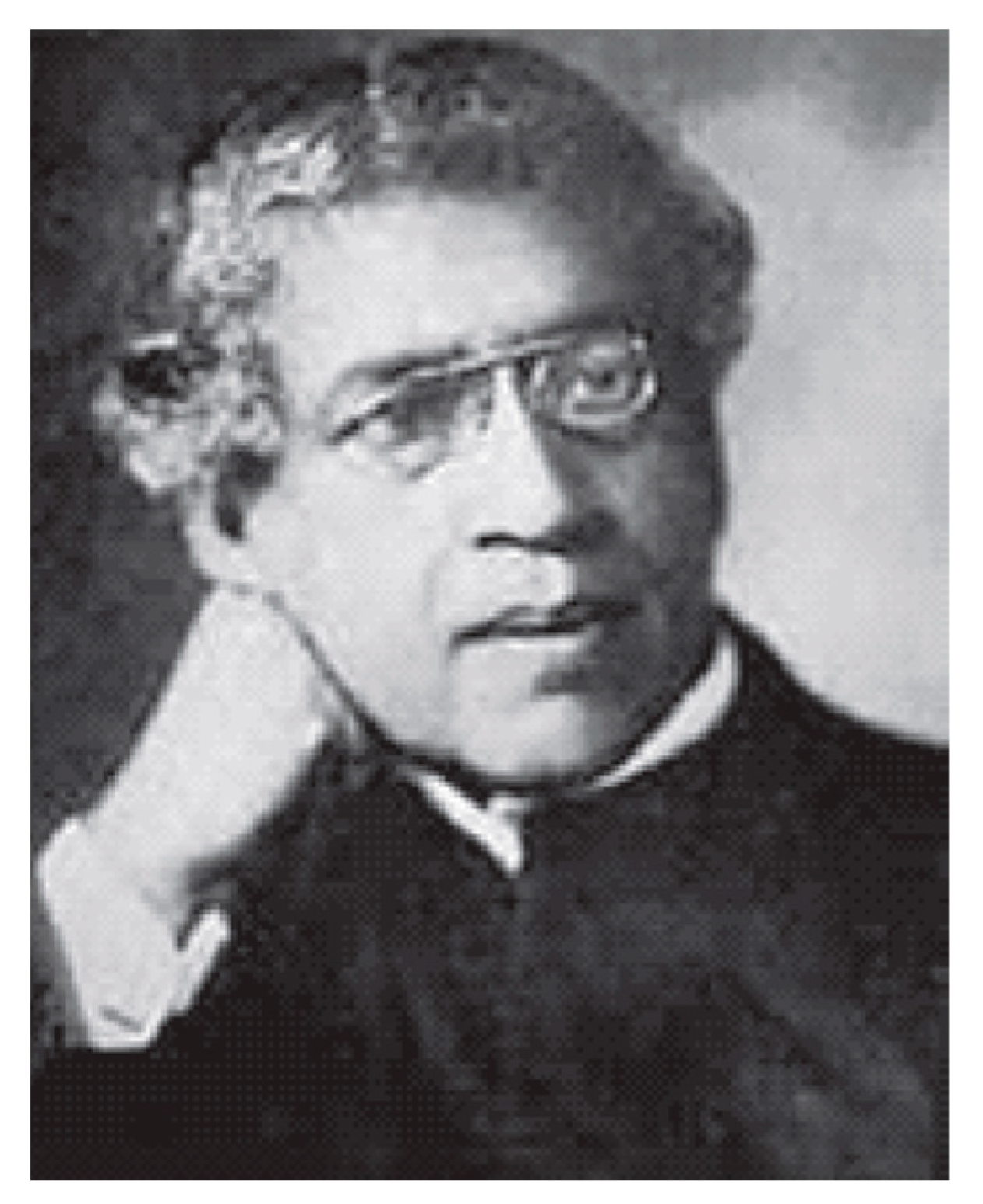
The exact date of his building the apparatus is not known but it appears to have been built sometime around 1897–1898. It found the first mention of his X-ray apparatus in a letter written to Rabindranath Tagore, the first Asian to receive the Nobel Prize in literature, in February 1898 [12]. Unfortunately, neither the apparatus nor any sketch of his apparatus is available either at Presidency College, Calcutta, the place where he built the apparatus or at Bose Institute, Calcutta, the institute that he built later, to understand the mechanism of the apparatus. Fortunately, scholars have been able to find a Press Report published on the 5th of May 1898 edition of the Amrita Bazar Patrika, an erstwhile reputed English daily of India, under the title ‘Professor Bose and the New Light’ which describes the X-ray apparatus built by him and its demonstration.
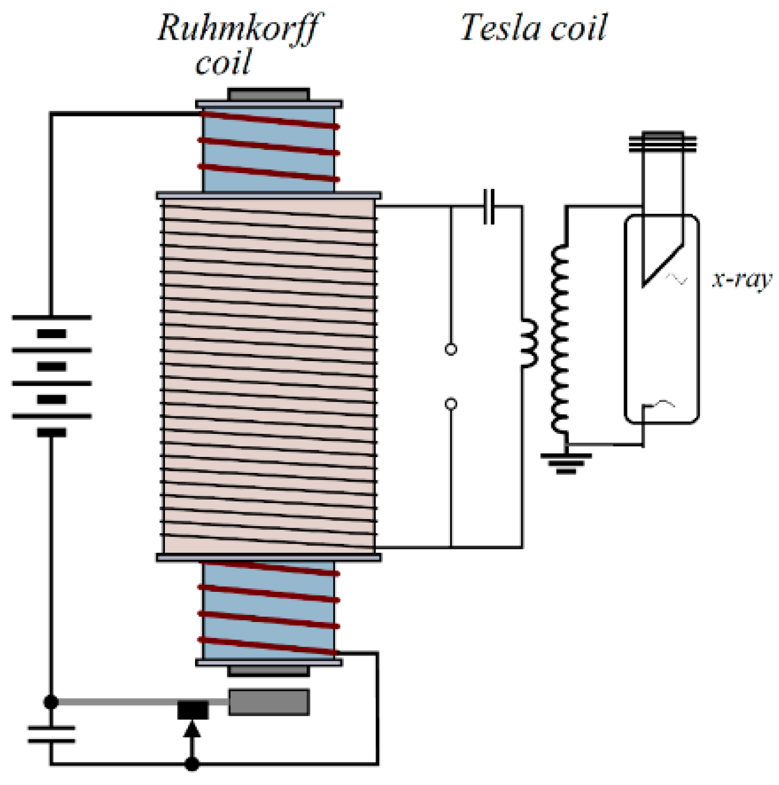
Roentgen used Ruhmkorff’s coil as a source of transient high voltage in discharging the gas in Crooke’s tube. Ruhmkorff’s coil produces high voltage pulse in the secondary coil by electromagnetic induction every time when a dc supply in the primary is interrupted by mechanical means. The secondary is connected with the discharge tube (X-ray tube) and the high voltage pulse was enough to produce X-rays. By coupling a Tesla coil with the secondary of Ruhmkorff’s coil (which then acts as the primary of the tesla transformer).
Jagadish Chandra had been able to produce a higher voltage many times more than that can be produced using a single Ruhmkorff’s coil. On the basis of the published information, scholars have presented a schematic diagram (Figure 2) to represent the apparatus he had probably used. The newspaper reported “We were shown a photograph of human palm taken by the Professor with the new light, and the ghastly sight will long be vividly imprinted in our memory, for there, in the photograph, instead of the ordinary fleshy palm is seen depicted a long range of bones presenting a skeleton-like appearance.” Using Barium Platinocyanide screen prepared by himself with his assistants in the Presidency College, he took X-ray photographs of different objects.
Figure 2. Schematic diagram of the high voltage source J. C. Bose probably used.
Although use of X-rays in India was started within a few months of X-ray discovery by importing X-ray tube from abroad, commercial production of indigenous machine made in India started only a few years before India’s independence in 1947 [3].

4. Medical Uses of X-rays

As per available literature, the evidence of first clinical uses of X-rays in India, at least in an informal setting, was started by Jagadish Chandra Bose in 1898. In a letter (Sen, 1994) written possibly in February 1898 to Rabindranath Tagore, he expressed his inability to meet Rabindranath at a time when he will be busy examining a patient with a broken back using Roentgen kal (machine in Bengali) [12]. The history of application of X-rays by professionals for clinical purposes in India is not very clearly known. As reported [13], Dr. Kartick Bose (1873–1955), an eminent doctor, medical scholar and industrialist was “the first clinician in India to start clinical and X-ray laboratories”.
Installation of the first X-ray machine in hospitals for public use is full of claims, counter-claims and conflicting reports so much so that it is very difficult to arrive at a definite conclusion [5]. It was reported by K P Mody in one of his editorials published in the Indian Journal of Radiology & Imaging that “the first X-ray machine was imported by a chemist in 1902 into India; that was only 7 years after the discovery” [14]. Madras Medical College website (www.mmc.ac.in (accessed on 17 June 2021)) claimed that the “first X-ray outfit was obtained for the general hospital in the year 1900 almost five years after the discovery of X-rays, the first in South East Asia”. This might be true considering that it is a very old medical institution started first as a Government General Hospital on 16 November 1664 to treat sick soldiers of the East India Company.
Use of X-rays for therapeutical purposes was also carried out at the beginning of twentieth century. Successful treatment of leukaemia using X-rays was reported in Calcutta Medical Journal [15] within five years of the first ever treatment of leukaemia, as reported in a paper published in the Journal of the American Medical Association in 1902.
Excitement of using X-rays in medical science waned with time and was replaced by Xray research in physical sciences.

5. C.V. Raman and Initiation of X-ray Research in India

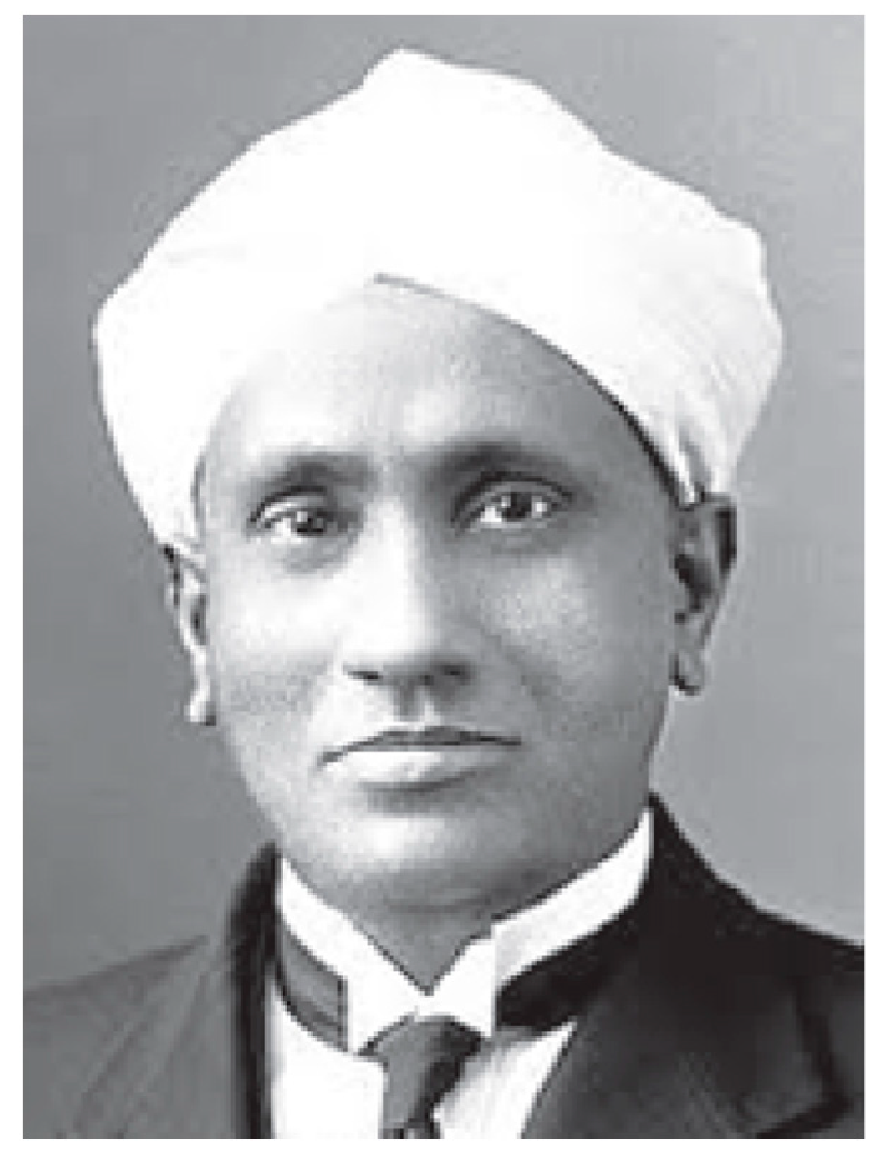
In July 1921, C.V. Raman (Figure 3) on his voyage to England as a delegate of Calcutta and Banaras University to attend University Congress, was attracted to the “mystery of the blue colour of the sea”. On his return to India, he undertook a comprehensive programme of research on molecular scattering of light by solid, liquid and gases which led to the discovery of a new light effect. The new light effect was later known as ‘Raman effect’ and earned him the Nobel Prize in physics in 1930. During this time Raman and his students were interested in the study of optical anisotropy of molecules from scattering of light. Raman and Ramanathan [16] extended the optical theory of scattering to X-ray diffraction by liquids. Experiments were also carried out to compare the theory with experimental results. Sogani [17] recorded the X-ray diffraction patterns of 22 aliphatic and aromatic liquids and estimated the average molecular distance from the position of the maxima of the diffraction haloes. It became clear from these experiments that the shape of the molecules and the intermolecular forces had profound influence on the diffraction patterns. Krishnamurti [18], another student of Raman, initiated small angle X-ray scattering which enabled people to understand the size and distribution of the particles in a sample. This is reportedly the first small angle X-ray scattering experiment performed in India [19].
Figure 3. C.V. Raman.
Raman had an encounter with William Bragg and he was of the opinion that Bragg’s first structure of naphthalene was not consistent with the birefringence, while the second one was. In order to confirm this, he and his students started extensive studies on the optical and magnetic anisotropy of organic crystals to understand the arrangements of molecules in crystalline state. The work on crystal magnetism was initiated by one of his successful students K.S. Krishnan. In many cases the orientation of the molecules in the unit cell could be calculated with a high degree of precision from purely magnetic data, which helped in refining X-ray analysis of the crystal structure [20][21].
Bidhubhusan Ray, another student of Raman, made significant contribution to X-ray spectroscopy, although he remained unknown to most physicists until a book was published by Rajinder Singh 
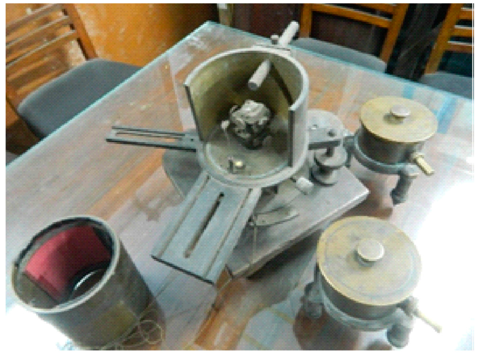
[22]. Bidhubhushan Ray (B. B. Ray) worked with Manne Siegbahn, a Nobel Laureate in Physics, at Uppsala and on his return to India established X-ray laboratory at Calcutta University. Satyendranath Bose who visited Europe during 1924–1926 met de Broglie brothers and was attracted to X-ray crystal structure. His experience was utilized both at Dhaka University and later at the Calcutta University where he took charge of the X-ray laboratory after the premature death of B. B. Ray and built an X-ray diffraction apparatus in the laboratory (Figure 4).
Figure 4. X-ray Diffraction Apparatus built by SN Bose. (Courtesy: Prof. Alok Mukherjee).
Historically the first crystallographic activity in India started with the determination of crystal structures of napthaelene and anthracene by Kedareswar Banerjee (KB) at IACS [23][24]. In 1930, in response to KB’s article in “Nature” on the structure of naphthalene, J.M. Robertson of Michigan University, Ann Arbor, U.S.A., wrote: “I believe Dr. Banerjee’s structure to be essentially correct. It has been clear to me for some time that the last two sections of my paper to which Dr. Banerjee refers must be amended as regards the distribution of the scattering centres in the a and b directions”. His further research on crystallography paved the way for a ‘complete crystallography structure analysis’ and for understanding statistical relationships in the amplitudes of diffracted waves. Further details of his life and work are available in a recently published book [25].
With Raman having good contact with the giants in the field of X-ray crystallography, he was able to send many of his students abroad to let them work with leading scientists of the field to gain contemporary knowledge of the subject. This made IACS the nucleus of X-ray research in India.
After independence, there has been a great surge of scientific research in India through establishment of laboratories by the Govt. of India. X-ray crystallography also flourished and expanded into areas of biological sciences. Contributions of Kedareswar Banerjee were followed by equally profound works by other researchers in India such as G. N. Ramachandran (University of Madras and Indian Institute of Science, Bangalore), A. R. Verma (Banaras Hindu University), S. Ramashesan (National Aeronautical Laboratory, Bangalore), VR. Chidambaram (Bhabha Atomic Research Centre, Mumbai) and others [26].
G.N. Ramachandran, a student of C.V. Raman, and most distinguished crystallographer of independent India worked on fibrous proteins and was recognized for the advancement of triple helix model of the structure of collagen [27] and the Ramachandran plot for validation of protein structure.

References

  1. International Union of Crystallography Newsletter; 2007; Volume 15, No. 4. Available online: https://www.iucr.org/news/newsletter/volume-15/number-4 (accessed on 17 June 2021).
  2. Roy, S.C. Early years of X-ray research in India. Sci. Cult. 2015, 81, 72.
  3. Roy, S.C. Discovery of X-rays and its impact in India. Indian J. Hist. Sci. 2017, 52, 66.
  4. Biswas, A.K. Gleanings of the Past and the Science Movement. In The Diaries of Drs. Mahendralal Sircar and Amritlal Sircar; The Asiatic Society: Kolkata, India, 2000; pp. 295–296.
  5. Roy, S.C. History of X-ray research in colonial India. Indian J. Hist. Sci. 2018, 53, T-123.
  6. Grigg, E.R.N. The Trail of the Invisible Light; Charles C Thomas Publisher: Springfield, IL, USA, 1965.
  7. Bose, J.C. Electromagnetic Radiation and Polarisation of Electric Rays; Collected Physical Papers; Longmans, Green & Co.: London, UK, 1927; pp. 77–101.
  8. Bose, J.C. Responses in the Living and Non-Living; Longmans, Green & Co.: London, UK, 1902.
  9. Bose, J.C. Plant Responses as a Means of Physiological Investigations; Longmans, Green & Co.: London, UK, 1906.
  10. Bose, J.C. Comparative Electrophysiology; Longmans, Green & Co.: London, UK, 1907.
  11. Bose, D.M. The scientific activities of Acharya Jagadish Chandra Bose. Sci. Cult. 1949, 14, 366.
  12. Patrabali. Collection of Letters of Acharya Jagadis Chandra Bose to Rabindranath Tagore; Sen, D., Ed.; (Letter No. 1); Bose Institute: Kolkata, India, 1994; pp. 1–2.
  13. Bose, K. Kartick Chandra Bose. Curr. Sci. 2010, 98, 133.
  14. Kumar, P.N.; Sharma, S. Study on Cost-Benefit Analysis of Computerised Tomography (CT) Scan. Int. J. Health Sci. Res. 2014, 4, 206.
  15. Chatterjee, G.C. A case of Leukaemia successfully treated with X-rays. Calcutta Med. J. 1907, 1, 269.
  16. Raman, C.V.; Ramanathan, K.R. The diffraction of X-rays in liquids, liquid mixtures, solutions, fluid crystals amorphous solids. Proc. Indian Assoc. Cultiv. Sci. 1923, 8, 127.
  17. Sogani, C.M. X-ray diffraction in liquids. Indian J. Phys. 1927, 1, 357.
  18. Krishnamurti, P. Studies in X-ray diffraction: The structures of Amorphous Carbon. Indian J. Phys. 1930, 5, 473.
  19. Chu, B.; Hsiao, S. Small-angle X-ray scattering by polymers. Chem. Rev. 2001, 101, 1727.
  20. Krishnan, K.S.; Banerjee, S. Investigations on magne-crystallic action IV. Magnetic behaviour of paramagnetic ions in the S-state in crystals. Phil. Trans. Roy. Soc. 1936, A235, 343.
  21. Krishnan, K.S.; Mookherji, A. Investigations on magne-crystallic action V. Paramagnetic salts of the rare earth and the ion groups. Phil. Trans. Roy. Soc. 1938, A237, 135.
  22. Singh, R. Bidhu Bhushan Ray-A Pioneer of X-ray Spectroscopy; Shaker Publishers: Maastricht, Germany, 2017.
  23. Banerjee, K. Structure of naphthalene and anthracene. Nature 1931, 125, 456.
  24. Banerjee, K. Determination of the signs of the Fourier terms in complete crystal structure. Proc. R. Soc. 1933, 141, 188.
  25. Rajinder Singh, C.V. Raman’s Student Kedareswar Banerjee: His Scientific Work in Indian Context; Shaker: Maastricht, Germany, 2021.
  26. Mande, S. Early Developments in crystallography. Resonance 2014, 19, 1077.
  27. Venkataraman, V. Quartz India. 15 May 2017. Available online: https://qz.com/india/983439/madras-triple-helix-the-world-has-nearly-forgotten-the-indian-scientist-who-cracked-the-structure-of-collagen/ (accessed on 20 March 2022).
More
Information
Contributor MDPI registered users' name will be linked to their SciProfiles pages. To register with us, please refer to https://encyclopedia.pub/register :
View Times: 2.3K
Revisions: 2 times (View History)
Update Date: 29 Jun 2022
1000/1000