Submitted Successfully!
To reward your contribution, here is a gift for you: A free trial for our video production service.
Thank you for your contribution! You can also upload a video entry or images related to this topic.
Version Summary Created by Modification Content Size Created at Operation
1 -- 2737 2022-06-23 02:14:05 |
2 Reference format revised. Meta information modification 2737 2022-06-23 11:00:20 |

Video Upload Options

Do you have a full video?

Confirm

Are you sure to Delete?
Cite
If you have any further questions, please contact Encyclopedia Editorial Office.
Nayl, A.A.;  Abd-Elhamid, A.I.;  Awwad, N.S.;  Abdelgawad, M.A.;  Wu, J.;  Mo, X.;  Gomha, S.M.;  Aly, A.A.;  Bräse, S. Electrospun Nanofibers in Regeneration of Tissues and Organs. Encyclopedia. Available online: https://encyclopedia.pub/entry/24366 (accessed on 26 April 2024).
Nayl AA,  Abd-Elhamid AI,  Awwad NS,  Abdelgawad MA,  Wu J,  Mo X, et al. Electrospun Nanofibers in Regeneration of Tissues and Organs. Encyclopedia. Available at: https://encyclopedia.pub/entry/24366. Accessed April 26, 2024.
Nayl, Abdelaziz A., Ahmed I. Abd-Elhamid, Nasser S. Awwad, Mohamed A. Abdelgawad, Jinglei Wu, Xiumei Mo, Sobhi M. Gomha, Ashraf A. Aly, Stefan Bräse. "Electrospun Nanofibers in Regeneration of Tissues and Organs" Encyclopedia, https://encyclopedia.pub/entry/24366 (accessed April 26, 2024).
Nayl, A.A.,  Abd-Elhamid, A.I.,  Awwad, N.S.,  Abdelgawad, M.A.,  Wu, J.,  Mo, X.,  Gomha, S.M.,  Aly, A.A., & Bräse, S. (2022, June 23). Electrospun Nanofibers in Regeneration of Tissues and Organs. In Encyclopedia. https://encyclopedia.pub/entry/24366
Nayl, Abdelaziz A., et al. "Electrospun Nanofibers in Regeneration of Tissues and Organs." Encyclopedia. Web. 23 June, 2022.
Electrospun Nanofibers in Regeneration of Tissues and Organs
Edit

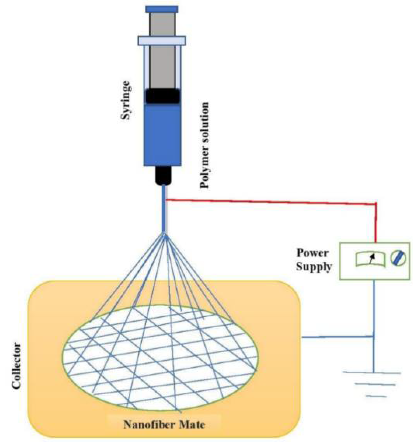
Electrospun techniques are promising and flexible technologies to fabricate ultrafine fiber/nanofiber materials from diverse materials with unique characteristics under optimum conditions. These fabricated fibers/nanofibers via electrospinning can be easily assembled into several shapes of three-dimensional (3D) structures and can be combined with other nanomaterials. Therefore, electrospun nanofibers, with their structural and functional advantages, have gained considerable attention from scientific communities as suitable candidates in biomedical fields, such as the regeneration of tissues and organs, where they can mimic the network structure of collagen fiber in its natural extracellular matrix(es). Due to these special features, electrospinning has been revolutionized as a successful technique to fabricate such nanomaterials from polymer media.

electrospinning nanofibers polymers biomedical applications regeneration of tissues

1. Introduction

During the last century, the utilization of polymers has rapidly increased, leading to the development of techniques used to produce polymer fibers to satisfy the high-performance requirements of different industries and modern applications [1]. Nanofibrous materials are thin and long substances and they are 1D materials, with diameters ranging from 50.0 to 500.0 nm in length, having diameter ratios >1.0:200.0. These nanofibrous materials can be synthesized from polymer solutions or melts. Nanofiber structures have gained extensive attention and show large potential for various applications, due to their favorable and unique characteristics [2][3][4]. The unique properties of electrospun nanofiber materials, such as large surface area and porosity, in addition to adjustability of pore sizes, due to their similar morphology and extracellular matrix, gives nanofibers the edge, when compared with bulk materials, in the regeneration of tissues and organs. Nanofibers can resemble or mimic the human anatomy’s tissues and organs [4].
Tissue engineering is focused on fabricating scaffolds that can temporarily substitute for the native extracellular matrix (ECM), to guide and regenerate specific tissue functions. Tissue engineering technologies encompass rapidly advancing techniques, which combine features and advantages of biochemicals/biomaterials sciences and transplantation of cells to generate bioartificial tissues or organs to cure damage to skin [5], cartilage [6], bone [7], nerve [8], and vascular [9] tissues. Over the last three decades, much investigative work has been done to fabricate vascular grafts in small diameter for clinical applications, but most of the attempts failed or were not satisfactory for applications. Therefore, new cell-based approaches have been investigated to fabricate tissue-engineered scaffolds similar to native ECM [10]. Electrospinning technology provides several interesting characteristics to the design and manufacture of scaffolds, with topography in the micro- and nano-scales, as scaffolding biomaterials for tissue repair and regeneration [11]. Electrospun nanofiber scaffolds (ENFSs) also provide internal porous network structures similar to the natural extracellular matrix (ECM), which supports the growth of cells, tissues, and organs. To fulfill these features, ENFSs must be biocompatible, have a high surface area, high porosity, gas permeability, possess appropriate mechanical properties, and transfer bioactive molecules that support cell growth, spreading, proliferation, migration, infiltration, and attachment. These promising and unique characteristics of electrospun nanofibrous structures promote their stabilities and versatilities in surface functionality, leading to their being perceived as significant biomaterials. They have gained great attention in regards to bone cell proliferation, nerve regeneration, vascular tissue and skin tissue engineering applications [11][12][13]. Furthermore, a tissue engineering scaffold should be biodegradable, have low immunogenicity, be economical, easy to process, and commercially available. Moreover, nanofibers possess elasticity in the functionalization of their surfaces, where they can be modified by applying pretreatment or post-treatment strategies. In the pretreatment approach, the most widely utilized modifiers are directly blended with spinning materials, and the modifier will already be present in the nanofiber. On the other hand, in post-treatment, the nanofiber is first prepared and then treated with the modifying agent, where only the surface is functionalized [14].
Nanoparticles suffer from aggregations in liquid during use forming a slurry, and these powders are difficult to remove after treatment operation, which reduces their applicable viability. In this respect, nanofibers introduce a valuable substrate for the nano-powder, whether the nanoparticles are embedded into [15] or decorate [16] the nanofiber. This strategy keeps the nanoparticles separated, increases the nanoparticles’ surface area exposure, and increases the materials’ efficiency. Moreover, using nanofibers avoids a high cost and painful removal process. Previous strategies in modification of nanofibers, by adding function groups or adding nanoparticles, were seen to be effective in enhancing various properties (wettability, water contact angle, mechanical properties, cell adhesion and proliferation, adsorptive capacities, filtration, photocatalysis, catalytic degradation, etc.) of the nanofibers towards different applications.

2. Fabrication of Nanofibers via Electrospinning

Electrospinning is a simple, cost-effective, and highly versatile technique used to fabricate a layer of nanofibers with diameters of 3.0–>5.0 nm, in the presence of a high-voltage electric field used on injected polymer solutions to stretch the droplets [17][18][19][20][21][22][23]. The high-voltage induces the interaction of charged polymer precursors and external electrical fields to form polymer nanofibers (PmNFs) [18], as in Figure 1. Where the high-voltage power source provides a high voltage of various tens of kilovolts [19] the fabricated nanofibers have unique characteristics, like higher surface areas and inter-/intra- fibrous porosity [4]. Therefore, it is used on a large scale to fabricate 1D continuous polymeric nanomaterials and 1D nanocomposites/inorganic nanomaterials [19].
Figure 1. Schematic diagram for a conventional electrospinning process.
Consequently, this technology has attracted great attention in fabricating different polymer ultrafine fibers [22]. Recently, the ability to fabricate new electrospun nanofibers and nanomaterials quickly by means of electrospinning technology led to the emergence of new applications in different fields [20].
According to the chemical composition of the fabricated nanofibers, they can be classified into four major types: 1—Inorganic nanofibers, 2—Carbon nanofibers, 3—Polymer-based nanofibers, 4—Composite nanofibers [24]. All of these types have potential application and functional performance in tissue engineering technologies, as discussed in the following sections. On the other hand, various types of nanofibers were recently fabricated from different organic and inorganic precursors, and the synthesis of nanofibers with distinguished morphological properties and satisfying yields is still challenging.

3. Applications of Nanofibers

3.1. Biomedical Applications

3.1.1. Bone Cell Proliferation

Bone cell proliferation processes are complicated and have many defects where self-regeneration is restricted. Therefore, scaffolds are widely utilized in replacement and regeneration processes for damaged bone tissues. During the last decade, various types of bionanomaterials have been used to design porous scaffolds that mimic the structure of the original ECM [25][26][27].
To culture the bone marrow endothelial progenitor cells (BEPCs), two varied hybrid scaffolds, composed of collagen/polycaprolactone (PCL) (70:30%) and gelatin (Gel)/PCL (70:30%), were prepared by electrospinning [25]. BEPCs were separately seeded on the two scaffolds and on glass slides as a control group. BEPCs spread well and adhered strongly to the collagen/PCL and gel/PCL scaffolds, compared with the control group (glass slide).
Recently, gelatin nanofibers have acquired considerable attention due to their promising characteristics as eco-friendly materials and they have considerable influences on cell adhesion, proliferation, and differentiation of diverse tissues [27]. Glucose cross-linked gel/zein scaffolds were evaluated to regenerate bone in vivo and in vitro. The nanofiber scaffolds presented rapid mineralization in the concentrated mimicked body fluid with precipitated octacalcium phosphate, and dicalcium phosphate dehydrate. Cytotoxic effect on MC3T3e1 cells in a CCK-8 test of the nanofibrous scaffolds was negligible. Osteogenesis characterizations were tested with Alizarin Red staining and showed enhanced calcium precipitation on the cross-linked scaffolds, while the alkaline phosphatase (ALP) exhibited no variation.
Structures of many natural materials, such as chitosan and its bioactive polymers, mimic exceedingly well to glycosaminoglycan (GAG). GAG is one of the components of bone extracellular matrix (ECM), which plays a vital function in cell-cell adhesion by interaction with collagen fibers [28][29][30][31]. These biomaterials have good biodegradability, biocompatibility, suitable mechanical properties, easy processing ability, water solubility, and can chelate with Ca or other active components during biomineralization processes [29][30]. Such biomaterials have attracted great attention during recent years in biomedical applications. Therefore, as a solvent, chitosan (Chi) nanofibers were electrospun from aqueous chitosan solutions via concentrated solutions of acetic acid (CH3COOH). Polyethylene oxide (PEO), of different weights (10–60 wt%), was blended with Chi solutions which acted as a plasticizer to enable spinnability of the Chi solutions that formed. MTT-assay and ALP expression analysis shows that it reached eleven days of cell seed. The fabricated electrospun Chi scaffolds developed MG 63 cell proliferation and differentiation into mature osteoblasts [28]. Also, metformin-loaded polycaprolactone/chitosan nanofibrous membranes were prepared using an electrospinning strategy for bone repairing membranes with better osteoinductive properties, subsequently espoused to glutaraldehyde crosslinking to improve the stability of chitosan in aqueous media. Furthermore, rats’ bone mesenchymal stem cells were seeded on membranes for the evaluation of the effect of metformin-loaded polycaprolactone/chitosan nanofibrous membranes on cell morphology, alkaline phosphate activity, and osteogenic mineralization in vitro. Moreover, in vitro experiments supposed that the crosslinked-polycaprolactone/chitosan/metformin membranes provide a preferable environment for cell attachment, proliferation, and osteogenic differentiation of bone mesenchymal stem cells [29]. Also, electrospun PCL/carboxymethyl chitosan (PCL/CMChi) nanofibers cured by helium cold atmospheric plasma (CAP) and incubated with bone morphogenic protein-2 (BMP-2) acted as scaffolds for the osteodifferentiation of stem cells. For the in vitro test, human bone marrow-derived mesenchymal stem cells (hMSCs) were seeded on the fabricated scaffolds, and their activities were followed. The findings exhibited that scaffolds supported the proliferation of hMSCs and enhanced their osteodifferentiation without using additional osteogenic differential agents. Moreover, the RT-PCR and ICC results indicated that CAP treatment and BMP-2-fmodifications exhibit synergic improvement on the ossification of hMSCs [30].

3.1.2. Nerve Regeneration

Peripheral nerve injuries (PNI), caused by traffic accidents, natural disasters, and unsatisfactory treatments [32], are intractable clinical problems that create heavy burdens for patients. Therefore, various electrospinning nanofiber materials play a considerable role in nerve regeneration. So, an aligned fibrin/functionalized self-assembling peptide (AFG/fSAP) interpenetrating hydrogel was fabricated by electrospinning and molecular self-assembly, where the prepared hydrogels show synergistic topographical and biochemical cues [32]. The scaffolds with aligned structures illustrated considerable impact on the regeneration of peripheral nerves [32]. Also, they upregulated regeneration-associated gene expression and activated PI3K/Akt and MAPK signaling pathways in regenerated nerves.
Aligned chitosan nanofiber hydrogel (AChiG) incorporated with a bioactive peptide mixture composed of Ac-RGIDKRHWNSQGG (RGI) and Ac-KLTWQELYQLKYKGIGG (KLT), named AChiG-RGI/KLT, was applied as nerve conduit filler to remodel sciatic nerve defects in rats. AChiG-RGI/KLT oriented the Schwann cells, and supported the proliferation and excretion of neurotrophic factors by Schwann cells. At an early infection stage, AChiG-RGI/KLT promoted nerve regeneration and enhanced vascular infiltration. After twelve weeks, AChiG-RGI/KLT facilitated nerve repair and functional restoration in rats [33].

3.1.3. Vascular Tissue

Recently, electrospinning has been utilized to synthesize nanofiber-based scaffolds. Several works have investigated and described the applications of nanofiber-based scaffolds for vascular scaffolds of small diameter and vascular grafts [34][35][36][37][38][39][40]. Though the vascular scaffolds fabricated by present electrospinning techniques can mimic the compositions of human blood vessels, these technologies have many difficulties in fabrication of vascular scaffolds of small-diameters (<1.5 mm) [34]. Therefore, a biodegradable polyl-lactide-co-caprolactone (PLCL) with biomimetic mechanical characteristics was employed to fabricate small diameter <1.5 mm PLCL/tussah silk fibroin (TSF) nanofiber vascular scaffolds for grafting. The biological behaviors of PLCL/TSF nanofiber vascular scaffolds were tested by in vitro culture of vascular endothelial cells (ECs). The scaffolds efficiently allowed vascular endothelial cell adhesion and proliferation [34]. Also, to mimic and fabricate the specific structures of natural blood vessels, a new approach has been developed [35]. In this process dual-oriented/bilayered small-diameter tubular nanofiber scaffolds were prepared by a mixture of PCL, poly d,l-lactide-co-glycolide (PLGA) and gelatin. The two bilayered nanofibers were orientated perpendicular to each other, aiming at guiding cell-specific orientation of smooth muscle cells (SMCs) and endothelial cells (ECs) in vitro, respectively. The findings revealed that the presence of gelatin highly induced the hydrophilicity of the scaffold as well as its mechanical property. The in vitro degradation indicated that by blending of three biodegradable polymers, the degradation rate of the scaffold accelerated. Moreover, electrospun scaffolds could enhance proliferation of both SMCs and ECs. Furthermore, topographic cues signed by oriented nanofibers could direct the growth and orientation of smooth muscle cells and endothelial cells [35].

3.1.4. Skin Tissue Engineering

Skin is the outermost and the most important barrier organ in the body and plays a vital role in protection of internal tissues from external damage [41][42][43]. Therefore, the protection of this barrier against damage is vital to prevent microorganisms from penetrating and forming infections in wounds and other dangerous side effects. Also, the protection and healing of wounds are very urgent factors in keeping patients safe and healthy [42][43]. Recently, the development of novel nanomaterials for wound dressing and antibiotic agents is one of the more important challenges facing current medical technological innovations [44]. Thus, many researchers have been motivated to fabricate an ideal wound dressing and antibiotics with appropriate properties using nanomaterials, such as different types of fabricated electrospun nanofibers to enhance wound healing. Electrospun nanofibers have opened new ways to synthesizing and fabricating of novel materials to be used in skin tissue engineering through nano/microscale polymeric fibers, inorganic/organic compositions, biomaterials, elastomers, and other types of materials.
Polymeric nanofibers can be incorporated with nanofillers, such as nanoparticles and nanotubes, to improve their superior properties and enhance applications in skin tissue engineering. Electrospun nanofibers of glucose (G)-reduced graphene oxide (rGO) (0–1.0 wt%) blended with PVA to form PVA/GrGO scaffolds, referred as (PG) scaffolds, and crosslinked chemically with acidic glutaraldehyde (GA) in acetone medium to mimic the (ECM) to apply in skin tissue engineering. Further increase in concentrations of G-rGO in PG scaffolds cause a decrease in tensile strengths and elongations, increasing the thermal properties. The biological properties of PG scaffolds were examined using in vitro hemolysis, using CCD-986Sk (a human skin fibroblast cell line). Results indicated G-rGO incorporation in PVA nanofibers induced a small shift from hydrophilic to hydrophobic. Moreover, the PVA/G-rGO scaffolds did not possess hemolysis of red blood cells, even at a G-rGO immobilizing of 1.0 wt%, and PG-1.0 scaffold (with a GRGO loading of 1.0 wt%) there was good compatibility with fibroblasts and highly increased metabolic activity after seed for twenty-one days, as compared with PG-0 controls [41].
Another nanofiber scaffold was synthesized from PU and cellulose acetate via electrospinning. RGO/Ag nanocomposite and/or curcumin was incorporated with the materials attributed to the antibacterial activity of rGO/Ag-nanocomposite. The scaffolds could prevent both of Gram-negative and Gram-positive bacteria via direct contact with them. In vivo histopathological experiments demonstrated that the scaffolds of the blended rGO/Ag nanocomposites and curcumin showed excellent wound healing and could promote the healing rate of artificial wounds, which provides considerable biomedical potential for nanomaterials in wound healing [42].

4. Conclusions

Nowadays, electrospun nanofibers with superior performance have illustrated considerable and promising results for biomedical applications. Their unique characteristics makes them convenient to apply in various fields. However, fabrication of nanofibers with distinct morphological and superior properties and considerable yield is still challenging. Such fabricated nanofibers can be functionalized through various techniques to widespread application in different strategic fields. Hence, considerable developments have been achieved even though there are still various challenges. Much effort has been put into improving desired performance of fabricated electrospun nanofiber and functionalized nanofiber application in specific areas. However, further work still needs to be done to obtain more promising results and eventual practical electrospun nanofibrous materials by advanced electrospinning techniques and scaling up the fabrication of nanofibers from the laboratory to commercial scales, especially for biomedical applications. The development of nanomaterials/biomaterials from biodegradable, biobased electrospun nanofibers is imperative to improve the applications of electrospun nanofibers in diverse fields, such as biomedical applications.
Also, electrospun nanofiber materials can play a vital role to overcome many challenges facing tissue engineering technologies in regeneration of tissues and organs where the scaffolds fabricated by electrospun nanofibers materials can mimic 3D with other physiological properties of tissues and organs in vitro to then transplant in vivo. Also, they are promising materials to regenerate injured nerve through tissue engineering technologies. Another serious challenge facing tissue engineering technologies to mimic and fabricate nerve grafts is losing the structural integrity, or biological functionality, of some natural polymer nanofibers used in a media simulating that in the human body. Therefore, further intensive research is required to explore and fabricate new materials to overcome such challenges. Using nanomaterials has many disadvantages, such as aggregation, uncontrollable release, costs, and potential cellular toxicity. Therefore, further research is needed to avoid such disadvantages.
On the other hand, current utilization of electrospun nanofibers in biomedical applications, such as bone cell proliferation, nerve regeneration, vascular tissue, and skin tissue engineering has been demonstrated. Many of these results were obtained for small rodent models which may be accompanied by restrictions regarding their immunological responses compared with humans. Therefore, these studies must be confirmed with suitable, and larger, animal models.

References

  1. Anstey, A.; Chang, E.; Kim, E.S.; Rizvi, A.; Kakroodi, A.C.; Park, B.; Lee, P.C. Nanofibrillated polymer systems: Design, application, and current state of the art. Prog. Polym. Sci. 2021, 113, 101346.
  2. Guerrero-Pérez, M.O. Research Progress on the Applications of Electrospun Nanofibers in Catalysis. Catalysts 2022, 12, 9.
  3. Phan, D.-N.; Khan, M.Q.; Nguyen, N.-T.; Phan, T.-T.; Ullah, A.; Khatri, M.; Kien, N.N.; Kim, I.-S. A review on the fabrication of several carbohydrate polymers into nanofibrous structures using electrospinning for removal of metal ions and dyes. Carbohydr. Polym. 2021, 252, 117175.
  4. Reddy, V.S.; Tian, Y.; Zhang, C.; Ye, Z.; Roy, K.; Chinnappan, A.; Ramakrishna, S.; Liu, W.; Ghosh, R. A Review on Electrospun Nanofibers Based Advanced Applications: From Health Care to Energy Devices. Polymers 2021, 13, 3746.
  5. Yu, R.; Zhang, H.; Guo, B. Conductive Biomaterials as Bioactive Wound Dressing for Wound Healing and Skin Tissue Engineering. Nano-Micro Lett. 2022, 14, 1.
  6. Yu, Y.S.; Ahn, C.B.; Son, K.H.; Lee, J.W. Motility Improvement of Biomimetic Trachea Scaffold via Hybrid 3D-Bioprinting Technology. Polymers 2021, 13, 971.
  7. Samadian, H.; Khastar, H.; Ehterami, A.; Salehi, M. Bioengineered 3D nanocomposite based on gold nanoparticles and gelatin nanofibers for bone regeneration: In vitro and in vivo study. Sci. Rep. 2021, 11, 13877.
  8. Ikegami, Y.; Ijima, H. Development of heparin-conjugated nanofibers and a novel biological signal by immobilized growth factors for peripheral nerve regeneration. J. Biosci. Bioeng. 2020, 129, 354–362.
  9. Rahmati, M.; Mills, D.K.; Urbanska, A.M.; Saeb, M.R.; Venugopal, J.R.; Ramakrishna, S.; Mozafari, M. Electrospinning for tissue engineering applications. Prog. Mater. Sci. 2021, 117, 100721.
  10. Lee, K.S.; Kayumov, M.; Emechebe, G.A.; Kim, D.-W.; Cho, H.-J.; Jeong, Y.-J.; Lee, D.-W.; Park, J.-K.; Park, C.-H.; Kim, C.-S.; et al. A Comparative Study of an Anti-Thrombotic Small-Diameter Vascular Graft with Commercially Available e-PTFE Graft in a Porcine Carotid Model. Tissue Eng. Regen. Med. 2022.
  11. Jiang, J.; Chen, S.; Wanga, H.; Carlson, M.A.; Gombart, A.F.; Xie, J. CO2-expanded nanofiber scaffolds maintain activity of encapsulated bioactive materials and promote cellular infiltration and positive host response. Acta Biomater. 2018, 68, 237–248.
  12. Cojocaru, E.; Ghitman, J.; Biru, E.I.; Pircalabioru, G.G.; Vasile, E.; Iovu, H. Synthesis and Characterization of Electrospun Composite Scaffolds Based on Chitosan-Carboxylated Graphene Oxide with Potential Biomedical Applications. Materials 2021, 14, 2535.
  13. El-Ghazali, S.; Kobayashi, H.; Khatri, M.; Phan, D.-N.; Khatri, Z.; Mahar, S.K.; Kobayashi, S.; Kim, I.-S. Preparation of a Cage-Type Polyglycolic Acid/Collagen Nanofiber Blend with Improved Surface Wettability and Handling Properties for Potential Biomedical Applications. Polymers 2021, 13, 3458.
  14. Mehrani, Z.; Ebrahimzadeh, H.; Moradi, E.; Yamini, Y. Using three-dimensional poly (vinyl alcohol)/sodium hexametaphosphate nanofiber as a non-toxic and efficient nanosorbent for extraction and recovery of Lanthanide ions from aqueous solutions. J. Mol. Liq. 2020, 307, 112925.
  15. Kumar, P.S.; Venkatesh, K.; Gui, E.L.; Sundaramurthy, J.; Singh, G.; Arthanareeswaran, G. Electrospun carbon nanofibers/TiO2-PAN hybrid membranes for effective removal of metal ions and cationic dye, Environmental Nanotechnology. Monit. Manag. 2018, 10, 366–376.
  16. Yin, J.; Zhan, F.; Jiaoa, T.; Deng, H.; Zou, G.; Bai, Z.; Zhang, Q.; Peng, Q. Highly efficient catalytic performances of nitro compounds via hierarchical PdNPs-loaded MXene/polymer nanocomposites synthesized through electrospinning strategy for wastewater treatment. Chin. Chem. Lett. 2020, 31, 992–995.
  17. Zhang, Z.; Wu, X.; Kou, Z.; Song, N.; Nie, G.; Wang, C.; Verpoort, F.; Mu, S. Rational design of electrospun nanofiber-typed electrocatalysts for water splitting: A review. Chem. Eng. J. 2022, 428, 131133.
  18. Zhou, X.; Wang, Y.; Gong, C.; Liu, B.; Weia, G. Production, structural design, functional control, and broad applications of carbon nanofiber-based nanomaterials: A comprehensive review. Chem. Eng. J. 2020, 402, 126189.
  19. Lu, X.; Wang, C.; Favier, F.; Pinna, N. Electrospun nanomaterials for supercapacitor electrodes: Designed architectures and electrochemical performance. Adv. Energy Mater. 2017, 7, 1601301.
  20. Xue, J.; Wu, T.; Dai, Y.; Xia, Y. Electrospinning and electrospun nanofibers: Methods, materials, and applications. Chem. Rev. 2019, 119, 5298–5415.
  21. Joshi, M.K.; Pant, H.R.; Tiwari, A.P.; Kim, H.J.; Park, C.H.; Kim, C.S. Multi-layered macroporous three-dimensional nanofibrous scaffold via a novel gas foaming technique. Chem. Eng. J. 2015, 275, 79–88.
  22. Wang, X.-X.; Yu, G.-F.; Zhang, J.; Yu, M.; Ramakrishna, S.; Long, Y.-Z. Conductive polymer ultrafine fibers via electrospinning: Preparation, physical properties and applications. Prog. Mater. Sci. 2021, 115, 100704.
  23. Shikhi-Abadi, P.G.; Irani, M. A review on the applications of electrospun chitosan nanofibers for the cancer treatment. Int. J. Biol. Macromol. 2021, 183, 790–810.
  24. El-Aswar, E.I.; Ramadan, H.; Elkik, H.; Taha, A.G. A comprehensive review on preparation, functionalization and recent applications of nanofiber membranes in wastewater treatment. J. Environ. Manag. 2022, 301, 113908.
  25. Hu, Y.; Feng, B.; Zhang, W.; Yan, C.; Yao, Q.; Shao, C.; Yu, F.; Li, F.; Fu, Y. Electrospun gelatin/PCL and collagen/PCL scaffolds for modulating responses of bone marrow endothelial progenitor cells. Exp. Ther. Med. 2019, 17, 3717–3726.
  26. Ali, M.G.; Mousa, H.M.; Blaudez, F.; Abd El-sadek, M.S.; Mohamed, M.A.; Abdel-Jaber, G.T.; Abdal-hay, A.; Ivanovski, S. Dual nanofiber scaffolds composed of polyurethane-gelatin/nylon 6-gelatin for bone tissue engineering. Colloids Surf. A 2020, 597, 124817.
  27. Deng, L.; Li, Y.; Zhang, H. In vitro and in vivo assessment of glucose cross-linked gelatin/zein nanofibrous scaffolds for cranial bone defects regeneration. J. Biomed. Mater. Res. 2020, 108B, 1505–1517.
  28. Singh, Y.P.; Dasgupta, S.; Nayar, S.; Bhaskar, R. Optimization of electrospinning process & parameters for producing defect-free chitosan/polyethylene oxide nanofibers for bone tissue engineering. J. Biomater. Sci. Polym. Ed. 2020, 31, 781–803.
  29. Zhu, J.; Ye, H.; Deng, D.; Li, J.; Wu, Y. Electrospun metformin-loaded polycaprolactone/chitosan nanofibrous membranes as promoting guided bone regeneration membranes: Preparation and characterization of fibers, drug release, and osteogenic activity in vitro. J. Biomater. Appl. 2020, 34, 1282–1293.
  30. Sharifi, F.; Atyabi, S.M.; Irani, S.; Bakhshi, H. Bone morphogenic protein-2 immobilization by cold atmospheric plasma to enhance the osteoinductivity of carboxymethyl chitosan-based nanofibers. Carbohydr. Polym. 2020, 231, 115681.
  31. Guo, S.; He, L.; Yang, R.; Chen, B.; Xie, X.; Jiang, B.; Weidong, T.; Ding, Y. Enhanced effects of electrospun collagen-chitosan nanofiber membranes on guided bone regeneration. J. Biomater. Sci. Polym. Ed. 2020, 31, 155–168.
  32. Yang, S.; Zhu, J.; Lu, C.; Chai, Y.; Cao, Z.; Lu, J.; Zhang, Z.; Zhao, H.; Huang, Y.; Yao, S.; et al. Aligned fibrin/functionalized self-assembling peptide interpenetrating nanofiber hydrogel presenting multi-cues promotes peripheral nerve functional recovery. Bioact. Mater. 2022, 8, 529–544.
  33. Rao, F.; Wang, Y.; Zhang, D.; Lu, C.; Cao, Z.; Sui, J.; Wu, M.; Zhang, Y.; Pi, W.; Wang, B.; et al. Aligned chitosan nanofiber hydrogel grafted with peptides mimicking bioactive brain-derived neurotrophic factor and vascular endothelial growth factor repair long-distance sciatic nerve defects in rats. Theranostics 2020, 10, 1590–1603.
  34. Liu, F.; Liao, X.; Liu, C.; Li, M.; Chen, Y.; Shao, W.; Weng, K.; Li, F.; Ou, K.; He, J. Poly(L-lactide-co-caprolactone)/tussah silk fibroin nanofiber vascular scaffolds with small diameter fabricated by core-spun electrospinning technology. J. Mater. Sci. 2020, 55, 7106–7119.
  35. Li, X.; Huang, L.; Li, L.; Tang, Y.; Liu, Q.; Xie, H.; Tian, J.; Zhou, S.; Tang, G. Biomimetic dual-oriented/bilayered electrospun scaffold for vascular tissue engineering. J. Biomater. Sci. Polym. Ed. 2020, 31, 439–455.
  36. Chen, M.; Li, L.; Xia, L.; Zhang, F.; Jiang, S.; Hu, H.; Li, X.; Wang, H. Temperature Responsive Shape-Memory Scaffolds with Circumferentially Aligned Nanofibers for Guiding Smooth Muscle Cell Behavior. Masthead Macromol. Biosci. 2020, 20, 1900312.
  37. Liua, G.; Fu, M.; Li, F.; Fu, W.; Zhao, Z.; Xia, H.; Niu, Y. Tissue-engineered PLLA/gelatine nanofibrous scaffold promoting the phenotypic expression of epithelial and smooth muscle cells for urethral reconstruction. Mater. Sci. Eng. C 2020, 111, 110810.
  38. Liu, Y.; Huang, L.; Yuan, W.; Zhang, D.; Gu, Y.; Huang, J.; Murphy, S.; Ali, M.; Zhang, Y.; Song, L. Sustained release of stromal cell–derived factor-1 alpha from silk fibroin microfiber promotes urethral reconstruction in rabbits. J. Biomed. Mater. Res. 2020, 108, 1591–1786.
  39. Jia, W.; Li, M.; Weng, H.; Gu, G.; Chen, Z. Design and comprehensive assessment of a biomimetic tri-layer tubular scaffold via biodegradable polymers for vascular tissue engineering applications. Mater. Sci. Eng. C 2020, 110, 110717.
  40. Eilenberg, M.; Enayati, M.; Ehebruster, D.; Grasl, C.; Walter, I.; Messner, B.; Baudis, S.; Potzmann, P.; Kaun, C.; Podesser, B.K.; et al. Long Term Evaluation of Nanofibrous, Bioabsorbable Polycarbonate Urethane Grafts for Small Diameter Vessel Replacement in Rodents. Eur. J. Vasc. Endovasc. Surg. 2020, 59, 643–652.
  41. Narayanana, K.B.; Park, G.T.; Han, S.S. Electrospun poly(vinyl alcohol)/reduced graphene oxide nanofibrous scaffolds for skin tissue engineering. Colloids Surf. B Biointerfaces 2020, 191, 110994.
  42. Esmaeili, E.; Eslami-Arshaghi, T.; Hosseinzadeh, S.; Elahirad, E.; Jamalpoor, Z.; Hatamie, S.; Soleimani, M. The biomedical potential of cellulose acetate/polyurethane nanofibrous mats containing reduced graphene oxide/silver nanocomposites and curcumin: Antimicrobial performance and cutaneous wound healing. Int. J. Biol. Macromol. 2020, 152, 418–427.
  43. Asadi, H.; Ghaee, A.; Nourmohammadi, J.; Mashak, A. Electrospun zein/graphene oxide nanosheet composite nanofibers with controlled drug release as antibacterial wound dressing. Int. J. Polym. Mater. Polym. Biomater. 2020, 69, 173–185.
  44. Bakhsheshi-Rad, H.R.; Ismail, A.F.; Aziz, M.; Akbari, M.; Hadisi, Z.; Omidi, M.; Chen, X. Development of the PVA/CS nanofibers containing silk protein sericin as a wound dressing: In vitro and in vivo assessment. Int. J. Biol. Macromol. 2020, 149, 513–521.
More
Information
Contributors MDPI registered users' name will be linked to their SciProfiles pages. To register with us, please refer to https://encyclopedia.pub/register : , , , , , , , ,
View Times: 422
Revisions: 2 times (View History)
Update Date: 28 Jun 2022
1000/1000