Submitted Successfully!
To reward your contribution, here is a gift for you: A free trial for our video production service.
Thank you for your contribution! You can also upload a video entry or images related to this topic.
Version Summary Created by Modification Content Size Created at Operation
1 -- 2326 2022-06-15 18:24:46 |
2 format change Meta information modification 2326 2022-06-16 04:52:01 |

Video Upload Options

Do you have a full video?

Confirm

Are you sure to Delete?
Cite
If you have any further questions, please contact Encyclopedia Editorial Office.
Gonzalez-Gronow, M.; Pizzo, S. GRP78 Autoantibodies in Immune-Mediated Neurological Diseases. Encyclopedia. Available online: https://encyclopedia.pub/entry/24078 (accessed on 26 April 2024).
Gonzalez-Gronow M, Pizzo S. GRP78 Autoantibodies in Immune-Mediated Neurological Diseases. Encyclopedia. Available at: https://encyclopedia.pub/entry/24078. Accessed April 26, 2024.
Gonzalez-Gronow, Mario, Salvatore Pizzo. "GRP78 Autoantibodies in Immune-Mediated Neurological Diseases" Encyclopedia, https://encyclopedia.pub/entry/24078 (accessed April 26, 2024).
Gonzalez-Gronow, M., & Pizzo, S. (2022, June 15). GRP78 Autoantibodies in Immune-Mediated Neurological Diseases. In Encyclopedia. https://encyclopedia.pub/entry/24078
Gonzalez-Gronow, Mario and Salvatore Pizzo. "GRP78 Autoantibodies in Immune-Mediated Neurological Diseases." Encyclopedia. Web. 15 June, 2022.
GRP78 Autoantibodies in Immune-Mediated Neurological Diseases
Edit

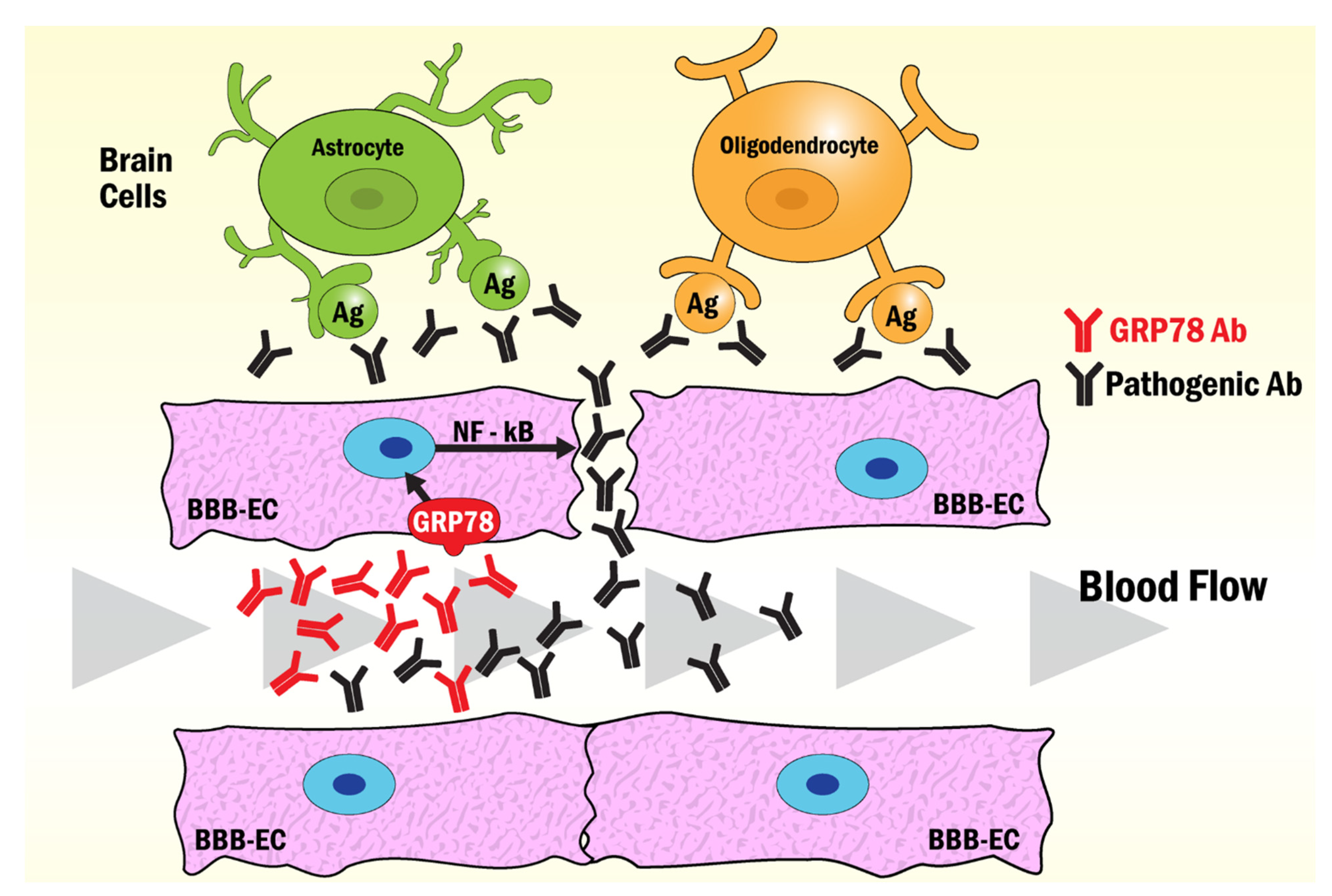
The 78 kDa glucose-regulated protein (GRP78), a member of the 70 kDa heat-shock family of molecular chaperones (HSP70), is essential for the regulation of the unfolded protein response (UPR) resulting from cellular endoplasmic reticulum (ER) stress. During ER stress, GRP78 evades retention mechanisms and is translocated to the cell surface (csGRP78) where it functions as an autoantigen. Autoantibodies to GRP78 appear in prostate, ovarian, gastric, malignant melanoma, and colorectal cancers. They are also found in autoimmune pathologies such as rheumatoid arthritis (RA), neuromyelitis optica (NMO), anti-myelin oligodendrocyte glycoprotein antibody-associated disorder (AMOGAD), Lambert-Eaton myasthenic syndrome (LEMS), multiple sclerosis (MS), neuropsychiatric systemic lupus erythematosus (NPSLE) and type 1 diabetes (T1D). 

Cancer Autoimmune disease ER Dysfunctions GRP78 autoantigenicity

1. Introduction

GRP78, a member of the 70 kDa heat-shock family of molecular chaperones (HSP70), is a resident of the ER [1]. The ubiquitous localization of GRP78 within the ER is indispensable for the regulation of the UPR resulting from ER stress [2]. During ER stress, GRP78 evades ER retention mechanisms and is translocated to the cell surface, where it functions as a receptor for multiple ligands and also behaves as an autoantigen for autoantibodies that contribute either positively or negatively to several human pathologies such as cancer and autoimmune diseases [3]. Autoantibodies to GRP78 have been identified in patients with prostate, ovarian, gastric, malignant melanoma, and colorectal cancers [4][5]. They are also present in serum from patients with autoimmune pathologies such as RA [6], NMO [7], LEMS [8] and T1D [9]. GRP78 autoantibodies found in the peripheral circulation of MS and NPSLE patients are able to disrupt and move across the BBB reaching the brain [10].
The lumen of the ER is an ideal environment for the proper synthesis and folding of proteins destined for secretion or display on the cell surface [11]. Homeostasis in the ER is maintained via coordination of the UPR and ER-associated degradation; however, a variety of disturbances can increase protein misfolding leading to ER stress, where GRP78 initiates signaling cascades that regulate the UPR [12]. Although csGRP78 behaves as an autoantigen in several pathologies, there are major differences in the specificity of its autoantibodies that may be traced down to ER dysfunctions specifically associated with the pathologies of cancer [13] or inflammatory diseases [14].

2. Molecular Mechanisms and ER Stress Conditions That Promote GRP78 Autoimmunity in Cancer

GRP78 binds hydrophobic surfaces on newly synthesized polypeptides and is first in line for protein folding, a function that is enhanced when misfolded polypeptides accumulate within the ER as a consequence of cellular stress [2][15]. GRP78 binds to unfolded proteins in its ATP-bound form and mediates their folding at the expense of ATP [16]. When GRP78 functions as a chaperone, it dissociates from the ER transmembrane stress sensor proteins inositol requiring enzyme 1 (Ire1), protein kinase RNA-like ER kinase (PERK) and activating transcription factor 6 (ATF6), which trigger the UPR [17]. Once activated, the UPR signaling promotes the transcription of numerous chaperones to protect the cell from accumulated proteins, and GRP78 itself is a transcriptional target of the UPR via ER stress-responsive elements that can bind to ATF6 [18]. The UPR leads to elevated expression of GRP78 in tumor tissue and controls not only its aberrant localization in the cytosol and the mitochondria but also in the plasma membrane [19]. The over-expression of GRP78 leads to the saturation of the KDEL receptor retrieval mechanism [20], permitting evasion and migration of GRP78 from the ER to these aberrant sites, a phenomenon common to several types of cancer [21]. In prostate cancer and melanoma, csGRP78 induces the production of GRP78 autoantibodies [22][23]. The interaction of these autoantibodies with csGRP78 triggers phosphoinositide 3-kinase (PI3K), protein kinase B (Akt) and mitogen-activated protein kinase (MAPK) signaling pathways, increasing cell survival during tumor growth and metastasis [24][25]. GRP78 is also functionally linked to the immune system via binding to the major histocompatibility complex class I (MHC-I), regulating peptide presentation by MHC-I in the plasma membrane [26]. Overexpression of csGRP78 under ER stress decreases MHC-I expression on the surface [27], and this mechanism is used by tumor cells to evade immune surveillance [28][29].
In prostate cancer, the GRP78 autoantibody recognizes a tertiary structure motif with the amino acid sequence CNVKSDKC [30], contained within the GRP78 primary amino acid sequence L98IGRTWNDPSVQQDIKFL115 (L98-L115) [22]. This csGRP78 N-terminal region site is also a second receptor for activated α2-macroglobulin (α2M*) [31] which initiates pro-proliferative signaling pathways upon the binding of either α2M* or GRP78 autoantibodies to prostate [22] or melanoma cancer cells [23]. Structural analyses of anti-GRP78 antibodies from a group of patients at different stages of malignant melanoma show that the IgG Fab is asymmetrically glycosylated, while the Fc regions are aberrantly glycosylated [23] in a manner similar to that observed in ovarian cancer [32]. Functionally, the asymmetric glycosylation matters, as reflected by the capacity of these IgG to activate the Akt signaling pathway while promoting the growth of prostate, lung and gastric cancers [33][34]. Asymmetric antibodies have two paratopes, one of high affinity, with Kd similar to that of symmetric antibodies and the other one with an affinity for the antigen significantly lower [35]. As a consequence, these IgG function as univalent and fail to trigger some of the major biological reactions of the immune response such as complement fixation, phagocytic activity and antigen clearance, thereby potentially exerting protective immunomodulatory effects on the host tissues [36]. The aberrant glycosylation of the Fc region in the GRP78 Abs in malignant melanoma [23] is the result of a lack of processing of D-mannosyl residues, which leads to a change in the size of the IgG-Fc fragment (27–33 kDa) [37]. These changes decrease their affinity for the Fc receptor (FcR), inducing changes in immune modulation that affect FcR activation [38]. These observations suggest that anti-GRP78 antibodies produced in cancer patients are functionally synthesized to protect the target tissue from immune responses, providing the tumor with defensive mechanisms that facilitate cell survival and proliferation. Figure 1 shows a model summarizing some of the properties and functions that characterize the anti-GRP78 IgG and its associated N-glycan structures identified in cancer.
Figure 1. Schematic representation of the anti-GRP78 IgG molecular structure and its associated N-glycan structures identified in cancer patients. It consists of two interconnected heavy (H) and two light chains (L) with two domains that infer different properties, the fragment antigen-binding (Fab) and fragment crystallizable (Fc) domains. The Fab domain is responsible for recognizing and binding the antigen. The Fc domain contains two glycans attached to Asn297 in conserved regions of the CH2 domain. The Fc domain binds to the FcR on natural killers and other inflammatory cells. In cancer, the Fab region is asymmetrically glycosylated with one additional N-glycan that converts the anti-GRP78 IgG into a univalent molecule that binds the antigen with a decreased affinity, preventing it from triggering some IgG-linked functions of the immune response such as complement fixation, phagocytic activity and antigen clearance. Moreover, the N-glycans of the Fc region are aberrantly glycosylated, mainly at the levels of D-mannosylation and galactosylation with a reduction in the affinity of the antibody for the FcR and its activation, thus inhibiting the inflammatory effects of the IgG.

3. Mechanisms and ER Stress Conditions That Promote GRP78 Autoimmunity in RA

RA is a systemic autoimmune disease characterized by chronic inflammation of the joints resulting in hypertrophy of the synovium called pannus, which ultimately leads to joint destruction [39]. The ER response has been implicated in chronic autoimmune inflammatory diseases [12] and ER stressors such as hypoxia, low glucose, and the pro-inflammatory cytokine milieu are responsible for the abnormal proliferation of RA fibroblast-like synoviocytes (RA-FLS) and angiogenesis, promoting pannus formation [40]. During these processes, down-regulation of GRP78 increases apoptosis of RA-FLS, whereas its overexpression prevents R-FLS from apoptotic death induced by ER-stressors [41]. The mechanisms operating in the UPR response of RA cells are similar to those described above for cancer cells [14]; however, during the progress of RA, B cells differentiate into plasma cells and infiltrate the synovial membrane where they synthesize autoantibodies such as immunoglobulin M rheumatoid factor (RF) and anti-cyclic citrullinated peptide antibodies (ACPA) [42][43].
Citrullination is a posttranslational modification of proteins catalyzed by peptidylarginine deiminase (PADI) in the presence of a high concentration of calcium that causes a loss of basic charge(s), which can influence the protein structure and create new epitopes recognized by the immune system [44]. In RA patients, the PADI type 4 (PADI4) gene in hematological and synovial tissue is a susceptibility locus for RA that affects the stability of transcripts and is associated with levels of antibody to citrullinated peptides in their sera. The PADI4 haplotype associated with susceptibility to RA increases the production of citrullinated peptides acting as autoantigens in some individuals, leading to a heightened risk of developing the disease, suggesting that genetic factors could cause RA [45]. As a target of PADI4, the citrullinated GRP78 (citGRP78) is arthritogenic in RA mice models, leading to the generation of ACPA which increases the severity of the disease [46].
Although the structure of the anti-citGRP78 IgG was not specifically studied in RA, ACPA-IgG molecules show a 10–20 kDa higher molecular weight compared with non-autoreactive IgG, resulting from Fab-linked N-glycosylation that decreases the binding avidity of ACPA for citrullinated antigens [47]. The lower target affinity induced by N-glycosylation of the RA ACPA-IgGs suggests a lower pathogenicity for these antibodies; however, the Fc-linked aberrant decrease in galactosylation and increase in fucosylation patterns observed in RA patients further increases a more pro-inflammatory phenotype of these antibodies [48][49]. The integrity of the IgG Fc region oligosaccharide chain is necessary for the activation of the complement, interactions with the FcR, and antibody-dependent cell-mediated cytotoxicity, all of which are perturbed by truncation of the IgG Asn297 oligosaccharide, where hypogalactosylation is a significant factor in the pathogenesis of RA [50].
The epitope specificity of the anti-citGRP78 antibodies in RA was identified using citrullinated peptides in which the arginine residues were replaced with citrulline residues. The RA patient serum antibody levels to GRP78279–298 R289citrulline (P12), GRP78295–314 R305 citrulline (P15) and GRP78500–519 R510citrulline (P23) were significantly increased compared with those of native peptides in RA patients, demonstrating their significance for antibody recognition in RA, in particular the epitope contained in peptide P23 in the GRP78 C-terminal region [41]. The high immunogenicity of the GRP78 C-terminal region was further confirmed in a separate study using a cloned GRP78409–653 C-terminal segment, structurally similar to that in the whole protein [51]. The anti-citGRP78 antibodies bind to csGRP78 on monocytes and activate extracellular signal-regulated protein kinases (ERK) and Jun N-terminal Kinase (JNK) pathways, leading to activation of nuclear factor kappa b (NF-κB) and production of tumor necrosis factor alpha (TNF-α) [52][53]. The production of TNF-α further increases csGRP78 expression in RA-FLS, leading to cell apoptosis [14].

4. GRP78 Autoantibodies in Immune-Mediated Neurological Diseases

GRP78 autoantibodies have been identified in MS [54], NPSLE [55], AMOGAD [56], LEMS [8], and NMO [57]. MS is a chronic inflammatory demyelinating disease of the central nervous system (CNS), characterized by the presence of focal demyelinated plaques within the white matter [58]. Although the structure of the anti-GRP78 IgG has not been specifically analyzed, the Fc region in IgG1 from MS patients shows reduced fucosylation and galactosylation, and increased bisecting GlcNAc, that promotes a higher pro-inflammatory activity of this immunoglobulin [59].
NPSLE is a complication of SLE characterized by serious cognitive defects which fluctuate over time such as difficulties in attention, concentration and memory [60]. Patients with NPSLE show significantly elevated serum levels of anti-GRP78 antibodies compared with healthy controls [55]. Although the structure of the anti-GRP78 IgG has not been specifically analyzed, significant differences in the IgG glycosylation between SLE patients and controls have been reported [61]. These changes include decreases in the galactosylation and sialylation, which regulate proinflammatory and anti-inflammatory actions of IgG, as well as decreased core fucose and increased bisecting N-acetylglucosamine, which affect antibody-dependent cell-mediated cytotoxicity [61], thereby suggesting that aberrant IgG glycosylation of the anti-GRP78 antibody may be an important pathological mechanism in SLE.
AMOGAD was recently recognized as a new pathology in the spectrum of inflammatory diseases, which differs from either MS or NMO. The serum levels of anti-GRP78 antibodies in patients with AMOGAD are significantly higher than those of MS patients or healthy controls [56]. LEMS is an autoimmune disease of the neuromuscular junction commonly defined as a paraneoplastic neurological syndrome, involving muscle weakness, areflexia and autonomic dysfunction [62]. Serum levels of anti-GRP78 antibodies of LEMS patients are high, and none were detected in the control subjects [8].
NMO is a severe inflammatory autoimmune disorder of the CNS that affects both adults and children. NMO was historically considered a variant of MS [63]; however, the discovery and identification of an NMO IgG autoantibody, specific for aquaporin-4 water channel (AQP4-Ab) [64], facilitated clinical diagnosis and early treatment of NMO [65]. Patient tissue immunopathology demonstrates that astrocytes are the principal cell target in NMO [66]. The circulating AQP4-Abs move across the blood-brain barrier (BBB), reaching brain astrocytes after leakage induced by anti-GRP78 antibodies also present in the circulation of NMO patients [67]. The mechanism by which the anti-GRP78 antibodies break the permeability of the BBB involves their binding to the scGRP78 on the brain microvascular endothelial cells (BMECs), inducing nuclear translocation of NF-κB that facilitates ICAM-1 transcription and promotes binding of activated immune cells that enhance the diameter of cerebral blood vessels in [58]. Similar mechanisms have been observed with anti-GRP78 antibodies in AMOGAD [56] and LEMS [8] that induce brain endothelial cells to open the BBB, thereby allowing access to other pathogenic antibodies in the CNS. A compromised BBB and antiendothelial cell antibodies have been found in patients with SLE [68], suggesting that csGRP78 in brain endothelial cells may also be a target of antiGRP78 antibodies in SLE [69]. The mechanism of disruption of the BBB by GRP78 autoantibodies in NMO, AMOGAD and SLE is summarized in Figure 2.
Figure 2. Model of the mechanism of BBB opening induced by GRP78 antibodies common to NMO, AMOGAD, LEMS and SLE. The binding of antibodies to GRP78 on the surface of BBB-endothelial cells induces the activation of NF-κB signaling pathways that disrupt the tight junction enhancing the diffusion of pathogenic antibodies into the brain. Astrocytes react with AQP4-Abs in NMO, while oligodendrocytes react with pathogenic anti-myelin Abs in AMOGAD. Pathogenic antibodies to brain antigens found in LEMS and SLE may also function through this mechanism to induce the damage observed in these pathologies.

References

  1. Kiang, J.G.; Tsokos, G.C. Heat shock protein 70 kDa: Molecular biology, biochemistry and physiology. Pharmacol. Ther. 1998, 80, 183–201.
  2. Lee, A.S. The glucose-regulated proteins: Stress induction and clinical applications. Trends Biochem. Sci. 2001, 26, 504–510.
  3. Wiersma, V.R.; Michalak, M.; Abdullah, T.M.; Bremer, E.; Eggleton, P. Mechanisms of translocation of ER chaperones to the cell surface and immunomodulatory roles in cancer and autoimmunity. Front. Oncol. 2015, 5, 7.
  4. Gonzalez-Gronow, M.; Selim, M.A.; Papalas, J.; Pizzo, S.V. GRP78: A multifunctional receptor on the cell surface. Antioxid. Redox Signal. 2009, 11, 2299–2306.
  5. Raiter, A.; Vilkin, A.; Gingold, R.; Levi, Z.; Halpern, M.; Niv, Y.; Britta-Hardy, B. The presence of anti-GRP78 antibodies in the serum of patients with colorectal carcinoma: A potential biomarker for early cancer detection. Int. J. Biol. Mark. 2014, 43, 1283–1287.
  6. Bodman-Smith, M.D.; Corrigal, V.M.; Berglin, E.; Cornell, H.R.; Tzioufas, A.G.; Magravani, C.P.; Chan, C.; Rantapää-Dahiqvist, S.; Panayi, G.S. Antibody response to the human stress protein BiP in rheumatoid arthritis. Rheumatology 2004, 43, 1283–1287.
  7. Shimizu, F.; Takeshita, Y.; Hamamoto, Y.; Yasuteru, Y.; Honda, M.; Sato, R.; Maeda, T.; Takahashi, T.; Fujikawa, S. GRP78 antibodies are associated with clinical phenotype in neuromyelitis optica. Ann. Clin. Transl. Neurol. 2019, 142, 2253–2264.
  8. Shimizu, F.; Takeshita, Y.; Sano, Y.; Hamamoto, Y.; Shiraishi, H.; Sato, T.; Yoshimura, S.; Maeda, T.; Fujikawa, S.; Nishihara, H.; et al. GRP78 antibodies damage the blood-brain barrier and relate to cerebellar degeneration in Lamber-Eaton myasthenic syndrome. Brain 2019, 142, 2253–2264.
  9. Rondas, D.; Crèvecoeur, I.; D’Hertrog, W.; Ferreira, G.B.; Staes, A.; Garg, A.D.; Eizirik, D.L.; Agostinis, P.; Gevaert, K.; Overbergh, L.; et al. Citrullinated glucose-regulated protein 78 is an autoantigen in type 1 diabetes. Diabetes 2015, 64, 573–586.
  10. Shimizu, F.; Nishihara, H.; Kanda, T. Blood-brain barrier dysfunction in immune-mediated neurological diseases. Immunol. Med. 2018, 41, 120–128.
  11. Xu, C.; Bailly-Maitre, B.; Reed, J.C. Endoplasmic reticulum stress: Cell life and death decisions. J. Clin. Investig. 2005, 115, 2656–2664.
  12. Hasnain, S.Z.; Lourie, R.; Das, I.; Chen, A.C.; McGuckin, M.A. The interplay between endoplasmic reticulum stress and inflammation. Immunol. Cell. Biol. 2012, 90, 260–270.
  13. Cubillos-Ruiz, J.R.; Bettigole, S.E.; Glimcher, L.H. Tumorigenic and immunosuppresive effects of endoplasmic reticulum stress in cancer. Cell 2017, 168, 692–706.
  14. Park, Y.-J.; Yoo, S.-A.; Kim, W.-U. Role of the endoplasmic reticulum stress in rheumatoid arthritis pathogenesis. J. Korean Med. Sci. 2014, 29, 2–11.
  15. Gidalevitz, T.; Stevens, F.; Argon, Y. Orchestration of secretory protein folding by ER chaperones. Biochim. Biophys. Acta 2013, 1833, 2410–2424.
  16. Blond-Elguindi, S.; Fourie, A.M.; Sambrook, J.E.; Gething, M.J. Peptide-dependent stimulation of the ATPase activity of the molecular chaperone BiP is the result of conversion of oligomers to active monomers. J. Biol. Chem. 1993, 268, 12735–12739.
  17. Gardner, B.M.; Pincus, D.; Gotthardt, K.; Gallagher, C.M.; Walter, P. Endoplasmic resticulum stress sensing in the unfolded protein response. Cold Spring Harb. Perspect. Biol. 2013, 5, a013169.
  18. Baumeister, P.; Luo, S.; Skarnes, W.C.; Sui, G.; Seto, E.; Shi, Y.; Lee, A.S. Endoplasmic reticulum stress induction of the Grp78/BiP promoter: Activating mechanisms mediated by YY1 and its interactive chromatin modifiers. Mol. Cell. Biol. 2005, 25, 4529–4540.
  19. Lee, A.S. Glucose-regulated proteins in cancer: Molecular mechanisms and therapeutic potential. Nat. Rev. Cancer 2014, 14, 263–276.
  20. Zhang, Y.; Liu, R.; Ni, M.; Gill, P.; Lee, A.S. Cell surface relocalization of the endoplasmic reticulum chaperone and unfolded protein response regulator GRP78/BiP. J. Biol. Chem. 2010, 285, 15065–15075.
  21. Gutiérrez, T.; Simmen, T. Endoplasmic reticulum chaperones and oxidoreductases: Critical regulators of tumor cell survival and immunorecognition. Front. Oncol. 2014, 4, 27–37.
  22. Gonzalez-Gronow, M.; Cuchacovich, M.; Llanos, C.; Urzua, C.; Gawdi, G.; Pizzo, S.V. Prostate cancer cell proliferation in vitro is modulated by antibodies against glucose-regulated protein 78 isolated from patient serum. Cancer Res. 2006, 66, 11424–11431.
  23. Selim, M.A.; Burchette, J.M.; Bowers, E.V.; de Ridder, G.G.; Mo, L.; Pizzo, S.V.; Gonzalez-Gronow, M. Changes in oligosaccharide chains of autoantibodies to GRP78 expressed during progression of malignant melanoma stimulate melanoma cell growth and survival. Melanoma Res. 2011, 21, 323–334.
  24. Zhang, Y.; Tseng, C.C.; Tsai, Y.L.; Fu, X.; Schiff, R.; Lee, A.S. Cancer cells resistant to therapy promote cell surface relocalization of GRP78 which complexes with PI3K and enhances PI (3, 4, 5) P3 production. PLoS ONE 2013, 8, e80071.
  25. Liu, R.; Li, X.; Gao, W.; Zhou, Y.; Wey, S.; Mitra, S.K.; Krasnoperov, V.; Dong, D.; Liu, S.; Li, D.; et al. Monoclonal antibody against cell surface GRP78 as a novel agent in suppressing PI3K/AKT signaling, tumor growth, and metastasis. Clin. Cancer Res. 2013, 19, 6802–6811.
  26. Triantafilou, M.; Fradelizi, D.; Triantafilou, K. Major histocompatibility class one molecule associates with glucose related protein (GRP)78 on the cell surface. Hum. Immunol. 2001, 62, 764–770.
  27. Ulianich, L.; Terrazzano, G.; Annunziatella, M.; Ruggiero, G.; Beguinot, F.; Di Jeso, B. ER stress impairs MHC class I surface expression and increases susceptibilioty of thyroid cells to NK-mediated cytotoxicity. Biochim. Biophys. Acta 2011, 4, 431–438.
  28. Marincola, F.M.; Jafee, E.M.; Hicklin, D.J.; Ferrone, S. Escape of human solid tumors from T-cell recognition: Molecular mechanisms and functional significance. Adv. Immunol. 2000, 74, 181–273.
  29. Seliger, B.; Atkins, D.; Bock, M.; Ritz, U.; Ferrone, S.; Huber, C.; Storkel, S. Characterization of human lymphocyte antigen class I antigen-processing machinery defects in renal cell carcinoma lesions with special emphasis on transporter-associated with antigen-processing down-regulation. Clin. Cancer Res. 2003, 9, 1721–1727.
  30. Mintz, P.J.; Kim, J.; Do, K.A.; Wang, X.; Zinnere, R.G.; Cristofanilli, M.; Arap, M.A.; Hong, W.K.; Logothetis, C.J.; Pasqualini, R.; et al. Fingerprinting the circulating repertoire of antibodies from cancer patients. Nat. Biotechnol. 2003, 21, 57–63.
  31. Misra, U.K.; Gonzalez-Gronow, M.; Gawdi, G.; Wang, F.; Pizzo, S.V. A novel receptor function for the heat shock protein GRP78 gene expression attenuates alpha-2M* induced signaling. Cell Signal. 2004, 16, 929–938.
  32. Gerçel-Taylor, C.; Bazzett, L.B.; Taylor, D.D. Presence of aberrant tumor-reactive immunoglobulins in the circulation of patients with ovarian cancer. Gynecol. Oncol. 2001, 81, 71–76.
  33. Kanoh, Y.; Ohara, T.; Mashiko, T.; Abe, T.; Masuda, N.; Akahoski, T. Relationship between N-linked oligosaccharide chains of human serum immunoglobulin G and serum tumor markers with non-small cell lung cancer progression. Anticancer Res. 2006, 26, 4293–4298.
  34. Kanoh, Y.; Ohara, T.; Tadano, T.; Kanoh, M.; Akahoski, T. Changes to N-linked oligosaccharide chains of human serum immunoglobulin G and matrix metalloproteinase-2 with cancer progression. Anticancer Res. 2008, 28, 715–720.
  35. Margni, R.A.; Borel, I.M. Paradoxical behavior of asymmetric IgG antibodies. Immunol. Rev. 1998, 163, 77–87.
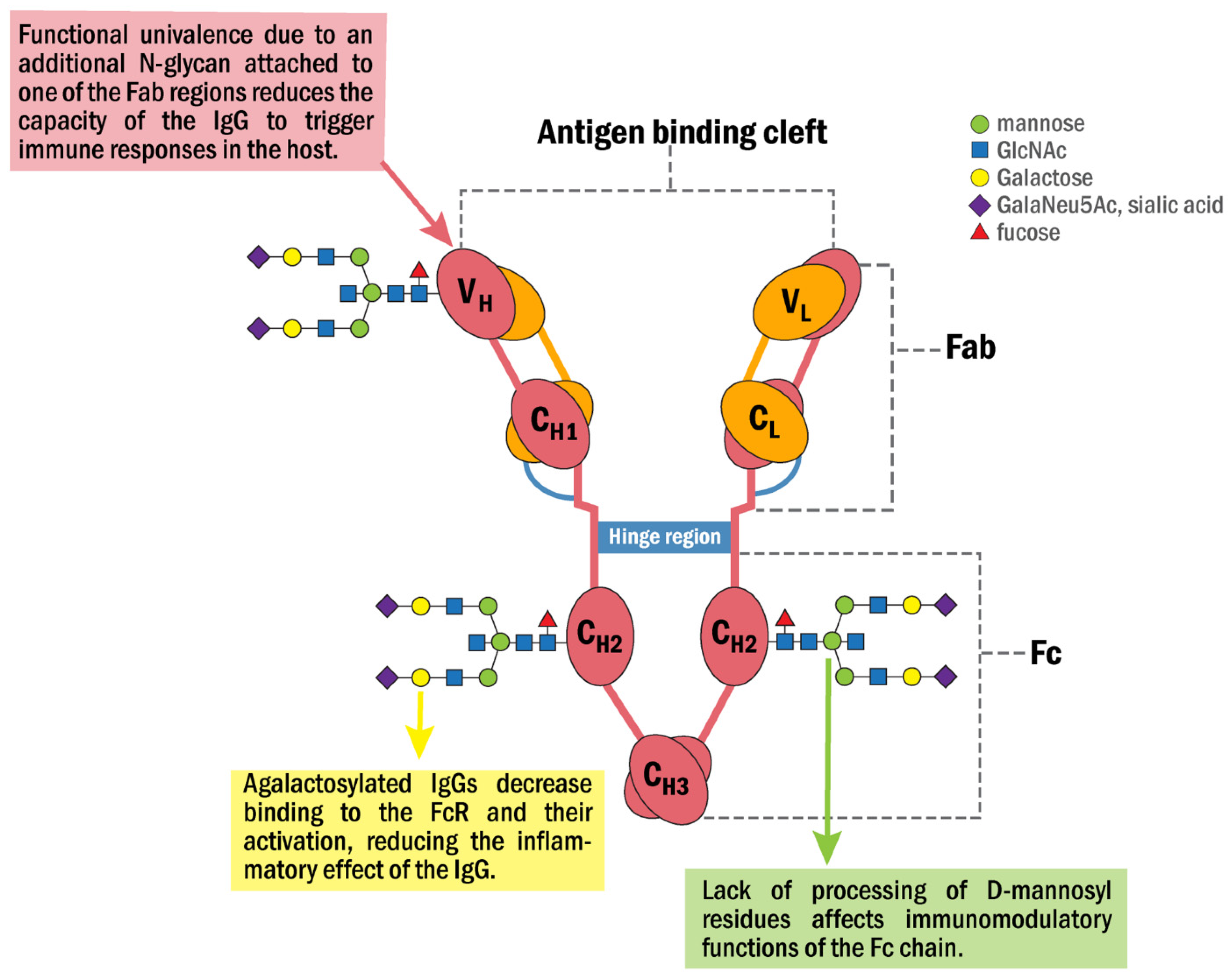
  36. Van de Boyenkamp, F.S.; Hafkenscheid, L.; Rispens, T.; Rombouts, Y. The emerging importance of IgG Fab glycosylation in immunity. J. Immunol. 2016, 196, 1435–1441.
  37. Wright, A.; Morrison, S.L. Antibody variable region glycosylation: Biochemical and clinical effects. Springer Semin. Immunopathol. 1993, 15, 259–273.
  38. Russel, A.; Adua, E.; Ugrina, I.; Laws, S.; Wang, W. Unravelling immunoglobulin G Fc N-glycosylation: A dynamic marker potentiating predictive, preventive and personalized medicine. Int. J. Mol. Sci. 2018, 19, 390.
  39. Lee, D.M.; Weinblatt, M.E. Rheumatoid arthritis. Lancet 2001, 358, 903–911.
  40. Stevens, C.R.; Williams, R.B.; Farrell, A.J.; Blake, D.R. Hypoxia and inflammatory synovitis: Observations and speculation. Ann. Rheum. Dis. 1991, 50, 124–132.
  41. Yoo, S.-A.; You, S.; Yoon, H.-J.; Kim, D.-H.; Kim, H.-S.; Lee, K.; Ahn, J.H.; Hwang, D.; Lee, A.S.; Kim, K.-J.; et al. A novel pathogenic role of ER chaperone GRP78/BiP in rheumatoid arthritis. J. Exp. Med. 2012, 209, 871–886.
  42. Masson-Bessière, C.; Sabbag, M.; Durieux, J.J.; Nogueira, L.; Vincent, C.; Girbal-Neuhausere, E.; Durroux, R.; Cantagrel, A.; Serre, G. In the rheumatoid pannus, anti-filaggrin autoantibodies are produced by local plasma cells and constitute a higher proportion of IgG than in synovial fluid and serum. Clin. Exp. Immunol. 2000, 119, 544–552.
  43. Kim, H.R. Anti-citrullinated antibodies in rheumatoid arthritis: A bridge between genetic predisposition and autoimmunity. Korean J. Intern. Med. 2013, 28, 25–28.
  44. Yamada, R. Peptidylarginine deiminase type 4, anticitrullinated peptide antibodies, and rheumatoid arthritis. Autoimmun. Rev. 2005, 4, 201–206.
  45. Suzuki, A.; Yamada, R.; Chang, X.; Tokuhiro, S.; Sawada, T.; Suzuki, M.; Nagasaki, M.; Nakayama-Hamada, M.; Kawaida, R.; Ono, M.; et al. Functional haplotypes of PADI4, encoding citrullinating enzyme peptidylarginine deiminase 4, are associated with rheumatoid arthritis. Nat. Genet. 2003, 34, 395–402.
  46. Shoda, H.; Fujio, K.; Shibuya, M.; Okamura, T.; Sumitomo, S.; Okamoto, A.; Sawada, T.; Yamamoto, K. Detection of autoantibodies to citrullinated BiP in rheumatoid arthritis patients and pro-inflammatory role of citrullinated BiP in collagen-induced arthritis. Arthritis. Res. Ther. 2011, 13, R191.
  47. Rombouts, Y.; Willemze, A.; van Beers, J.B.C.; Shi, J.; Kerkman, P.F.; van Toorn, L.; Janssen, G.M.C.; Zaldumbide, A.; Hoeben, R.C.; Pruijn, G.J.M.; et al. Extensive glycosylation of ACPA-IgG variable domains modulates binding to citrullinated antigens in rheumatoid arthritis. Ann. Rheum. Dis. 2016, 75, 578–585.
  48. Van Zeben, D.; Rook, G.A.; Hazes, J.M.; Zwinderman, A.H.; Zhang, Y.; Ghelani, S.; Rademacher, T.W.; Breedveld, F.C. Early agalactosylation of Igg is associated with a more progressive disease course in patients with rheumatoid arthritis: Results of a follow-up study. Rheumatology 1994, 33, 36–43.
  49. Rombouts, Y.; Ewing, E.; van de Stadt, L.A.; Selman, M.H.J.; Trouw, L.A.; Deelder, A.M.; Huizinga, T.W.J.; Wuhrer, M.; Van Schaardenburg, D.; Toes, R.; et al. Anti-citrullinated protein antibodies acquire a pro-inflammatory Fc glycosylation phenotype prior to the onset of rheumatoid arthritis. Ann. Rheum. Dis. 2015, 74, 234–241.
  50. Ercan, A.; Cui, J.; Chatterton, D.E.W.; Deane, K.D.; Hazen, M.M.; Brintnell, W.; O’Donnell, C.I.; Derber, L.A.; Weinblatt, M.E.; Shadick, N.A.; et al. Aberrant IgG galactosylation precedes disease onset, correlates with disease activity, and is prevalent in autoantibodies in rheumatoid arthritis. Arthritis Rheum. 2010, 62, 2239–2248.
  51. Aghamollaei, H.; Gargari, S.L.M.; Ghanei, M.; Rasaee, M.J.; Amani, J.; Bakherad, H.; Famoosh, G. Structure prediction, expression and antigenicity of c-terminal of GRP78. Biochem. Mol. Biol. Inc. 2017, 64, 117–126.
  52. Lu, M.C.; Lai, N.S.; Yu, H.C.; Huang, H.B.; Hsieh, S.C.; Yu, C.L. Anti-citrullinated protein antibodies bind surface-expressed citrullinated grp78 on monocyte-macrophages and stimulate tumor necrosis factor alpha production. Arthritis Rheum. 2010, 62, 1213–1223.
  53. Lu, M.C.; Lai, N.S.; Yin, W.Y.; Yu, H.C.; Huang, H.B.; Tung, C.H.; Huang, K.Y.; Yu, C.L. Anti-citrullinated protein antibodies activated ERK1/2 and JNK antigen-activated protein kinases via binding to surface-expressed citrullinated GRP78 on mononuclear cells. J. Clin. Immunol. 2013, 33, 558–566.
  54. Gharesi-Fard, B.; Zare, M.; Kamali-Sarvestani, E. The reaction of placerntal GRP78 protein with sera from women with multiple sclerosis. Iran. J. Immunol. 2017, 14, 306–315.
  55. Matsueda, Y.; Arinuma, Y.; Nagai, T.; Hirohata, S. Elevation of sereum anti-glucose-regulated protein 78 antibodies in neuropsychiatric systemic lupus erythematosus. Lupus Sci. Med. 2018, 5, e000281.
  56. Shimizu, F.; Ogawa, R.; Mizukami, Y.; Watanabe, K.; Hara, K.; Kadono, C.; Takahashi, T.; Misu, T.; Takeshita, Y.; Sano, Y.; et al. GRP78 antibodies are associated with blood-brain barrier breakdown in anti-myelin oligodendrocyte glycoprotein antibody-associated disorder. Neurol. Neuroimmunol. Neuroinflamm. 2022, 9, e1038.
  57. Shimizu, F.; Schaller, K.L.; Owens, G.P.; Clotleur, A.C.; Kellner, D.; Takeshita, Y.; Obermeier, B.; Kryzer, T.J.; Sano, Y.; Kanda, T.; et al. Glucose-regulated protein 78 autoantibody associates with blood-brain barrier disruption in neuromyelitis optica. Sci. Transl. Med. 2017, 9, eaai9111.
  58. Noseworthy, F.D.; Lucchinetti, C.; Rodriguez, M.; Weinshenker, B.G. Multiple sclerosis. N. Engl. J. Med. 2000, 343, 938–952.
  59. Wuhrer, M.; Selman, M.H.; McDonnell, L.A.; Kümpfel, T.; Derfuss, T.; Khademi, M.; Olsson, T.; Hohlfeld, R.; Meinl, E.; Krumbholz, M. Pro-inflammatory pattern of IgG1 Fc glycosylation in multiple sclerosis cerebrospinal fluid. J. Neuroinflamm. 2015, 12, 235.
  60. Liang, M.H.; Corzillius, M.; Bae, S.C.; Lew, R.A.; Fortin, P.R.; Gordon, C.; Isenberg, D.; Alarcón, G.S.; Straaton, K.V.; Denburg, J. The American College of Rheumatology nomenclature and case definitions for neuropsychiatric lupus syndromes. Arthritis Rheum. 1999, 42, 599–608.
  61. Vučković, F.; Krištić, J.; Gudelj, I.; Teruel, M.; Keser, T.; Pezer, M.; Pučić-Baković, M.; Štambuk, J.; Trbojević-Akmačić, I.; Barrios, C.; et al. Association of Systemic Lupus Erythematosus With Decreased Immunosuppressive Potential of the IgG Glycome. Arthritis Rheumatol. 2015, 67, 2978–2989.
  62. Titulaer, M.J.; Lang, B.; Verschuuren, J.J. Lambert-Eaton myathenic syndrome from clinical characteristics to therapeutic strategies. Lancet 2011, 10, 1098–1107.
  63. Jarius, S.; Wildemann, B. Thed history of neuromyelitis optica. J. Neuroinflamm. 2013, 10, 8.
  64. Lennon, V.A.; Kryzer, T.J.; Pittock, S.J.; Verkman, A.S.; Hinson, S.R. IgG marker of optic-spinal multiple sclerosis binds to the aquaporin-4 water channel. J. Exp. Med. 2005, 202, 473–477.
  65. Waters, P.J.; McKeon, A.; Leite, M.I.; Rajasekharan, S.; Lennon, V.A.; Villalobos, A.; Palace, J.; Mandrekar, J.N.; Vincent, A.; Bar-Or, A.; et al. Serologic diagnosis of NMO: A multicenter comparison of aquaporin-4-IgG assays. Neurology 2012, 78, 665–671.
  66. Roemer, S.F.; Parisi, J.E.; Lennon, V.A.; Benarroch, E.E.; Lassmann, H.; Bruck, W.; Mandler, R.N.; Weinshenker, B.G.; Pittock, S.J.; Wingerchuk, D.M.; et al. Pattern-specific loss of aquaporin-4 immunoreactivity distinguishes neuromyelitis optica from multiple sclerosis. Brain 2007, 10, 1194–1205.
  67. Shimizu, F.; Sano, Y.; Takahashi, T.; Haruki, H.; Saito, K.; Koga, M.; Kanda, T. Sera from neuromyelitis optica patients disrupt the blood-brain barrier. J. Neurol. Neurosurg. Psychiatry 2012, 83, 288–297.
  68. Renaudineau, Y.; Duguè, C.; Dueymes, M.; Youinou, P. Antiendothelial cell antibodies in systemic lupus erythematosus. Autoimmun. Rev. 2002, 1, 365–372.
  69. Yu, Y.J.; Watts, R.J. Developing therapeutic antibodies for neurodegenerative disease. Neurotherapeutics 2013, 10, 459–472.
More
Information
Subjects: Cell Biology
Contributors MDPI registered users' name will be linked to their SciProfiles pages. To register with us, please refer to https://encyclopedia.pub/register : ,
View Times: 386
Revisions: 2 times (View History)
Update Date: 16 Jun 2022
1000/1000