Submitted Successfully!
To reward your contribution, here is a gift for you: A free trial for our video production service.
Thank you for your contribution! You can also upload a video entry or images related to this topic.
Version Summary Created by Modification Content Size Created at Operation
1 -- 5461 2022-06-15 04:47:21

Video Upload Options

Do you have a full video?

Confirm

Are you sure to Delete?
Cite
If you have any further questions, please contact Encyclopedia Editorial Office.
Dunne, N.; Tzagiollari, A.; Mccarthy, H.; Levingstone, T. Bioadhesives for Bone Fracture Repair. Encyclopedia. Available online: https://encyclopedia.pub/entry/24042 (accessed on 28 April 2024).
Dunne N, Tzagiollari A, Mccarthy H, Levingstone T. Bioadhesives for Bone Fracture Repair. Encyclopedia. Available at: https://encyclopedia.pub/entry/24042. Accessed April 28, 2024.
Dunne, Nicholas, Antzela Tzagiollari, Helen Mccarthy, Tanya Levingstone. "Bioadhesives for Bone Fracture Repair" Encyclopedia, https://encyclopedia.pub/entry/24042 (accessed April 28, 2024).
Dunne, N., Tzagiollari, A., Mccarthy, H., & Levingstone, T. (2022, June 15). Bioadhesives for Bone Fracture Repair. In Encyclopedia. https://encyclopedia.pub/entry/24042
Dunne, Nicholas, et al. "Bioadhesives for Bone Fracture Repair." Encyclopedia. Web. 15 June, 2022.
Bioadhesives for Bone Fracture Repair
Edit

Bone defects and complex fractures present significant challenges for orthopaedic surgeons. Current surgical procedures involve the reconstruction and mechanical stabilisation of complex fractures using metal hardware (i.e., wires, plates and screws). However, these procedures often result in poor healing. An injectable, biocompatible, biodegradable bone adhesive that could glue bone fragments back together would present a highly attractive solution.

bone fractures bioadhesives bone repairing biomimetic adhesives

1. Introduction

Bone fractures are common injuries resulting from trauma or diseases such as osteoporosis and bone cancer [1]. A patient’s age, gender, lifestyle and pre-existing medical conditions are all important factors affecting the risk of a fracture occurring and the likelihood that complications will occur during the repair process [2][3]. Overall, according to a Global Burden of Disease study, an estimated 178 million individuals (53% males and 47% females) worldwide suffered bone fractures in 2019, leading to an increase of approximately 34% since 1990 [4].
During the normal bone fracture healing process, three overlapping stages occur: (1) inflammation, (2) bone production and (3) bone remodelling (Figure 1). Initial bleeding into the fracture area is followed by inflammation and clotting of blood at the fracture site. These processes involve haematopoietic and immune cells within the bone marrow and mesenchymal stem cells (MSCs) from the surrounding tissue and bone marrow [5][6]. Clotted blood is replaced with fibrous tissue and cartilage (soft callus) within 2 to 4 weeks. Callus formation around the fractured bone provides early stabilisation and protects the repair tissue from external forces [7]. Subsequently, the calcium formation that is laid down in the matrix within the next 4 to 12 months results in the callus becoming visible on radiographic images. The successful restoration of the original shape and structure of bone (i.e., bone remodelling) is the final stage in the normal healing process. In some incidences, bone healing does not occur in accordance with the normal bone repair processes. For example, micromotion at the repair site can interrupt the healing process and lead to other possible complications, such as bleeding into a joint space that causes the joint to swell (haemarthrosis) and blood clot formation that can cause blockage within a blood vessel, locally or elsewhere in the body. Non-union fractures occur when the broken bones are not able to heal due to insufficient nutrition, limited blood supply or inadequate stability (poor immobilisation). In many cases, the healing process can last from months to years [8].
Figure 1. Stages of bone healing: (1) haematoma formation from stem and macrophage cells at the fracture site (week 0–1), (2) soft callus formation at the fracture site, from chondroblast, osteoblast, fibroblast and osteoclast, replaces the hematoma (week 1–4) and (3) hard callus replaces the soft callus, using chondroblast cells and, after week 6–8, bone starts to replace the hard callus (week 4–48).

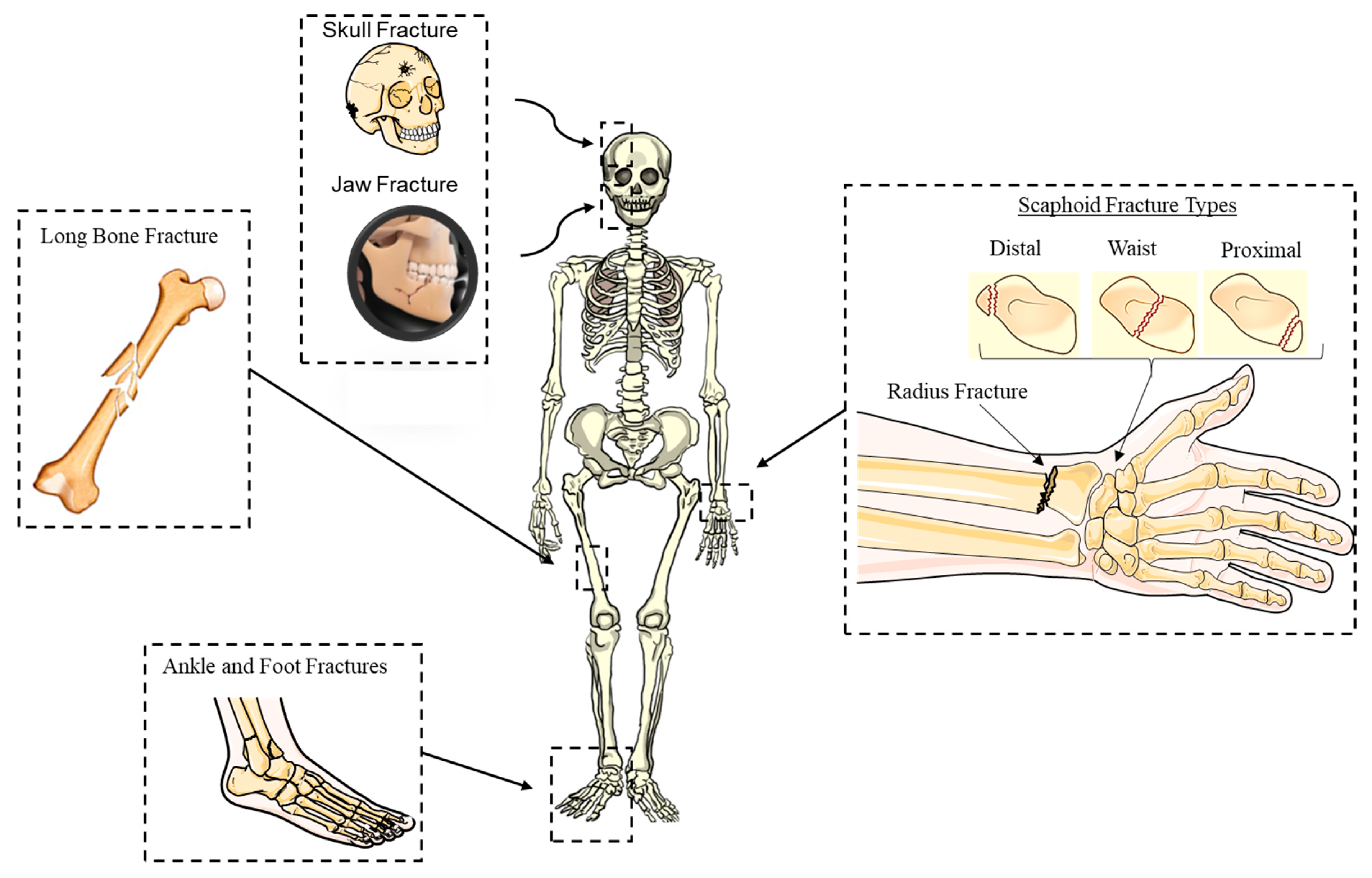
2. Complex Bone Fractures

Complex bone fractures generally consist of multiple fragments and usually require complicated surgical intervention (Figure 2). These fractures, therefore, present significant challenges for orthopaedic surgeons [9] and often lead to poor clinical outcomes. Complex fractures can vary significantly from one patient to another and may be further complicated due to joint dislocation and loss of bone fragments, leading to a painful and difficult recovery process for the patient [10]. The most common types of challenging bone fractures are distal radius fractures [3], facial bone fractures [11] and foot/ankle bone fractures [12]. Currently, 20% of distal radius fractures [13] and 71% of facial fractures require surgical intervention, with almost 20% of facial fracture requiring secondary surgical procedures [14]. The incidence of fractures that require surgical intervention is reportedly increasing among the younger patient population, with 45% of fractures in the age group under 25 years requiring surgical intervention and 37.5% of fractures in the age group 25–30 years [14].
Figure 2. Complex fractures occur most frequently in the long bones, carpal, facial and ankle–foot bones. The wrist, facial and ankle–foot bones contain several small bones close to each other, leading to complex fractures with several bone fragments after a fracture.
An analytical distribution of wrist fractures, as well as the eight carpal bones of different shapes and sizes, can be seen in Figure 3. Scaphoid fractures are the most common carpal bone fractures (70% of all carpal bone fractures) [15] that cause long-term pain and frequently require surgery. The remaining 30% of carpal bone fractures are divided across the other six bones of the wrist and can cause significant disability. Trapezium fractures can occur within the body of the trapezium or at the ridge and usually result from a direct blow or an avulsion injury [16].
Figure 3. Percentage of fracture incidences per carpal bone.
Facial bone fractures occur frequently, with an increased number of fractures being reported annually [17][18]. Facial fractures are categorised as: (1) isolated with lower energy trauma or (2) complex. In terms of the isolated fractures [19], the most common type is the fracture of the nasal bone, accounting for 40% of the cases, followed by mandible fracture at 30%. The fracture of the inferior region is the most common type of complex injury, with 14%—the highest frequency—being a tripod fracture (zygomaticomaxillary complex fracture, also known as a quadripod fracture, quadramalar fracture) [19].
It is estimated there are nine million incidents of long bone fractures worldwide per annum [20] caused by medical conditions (e.g., osteoporosis). According to Fisher et al. [21], 20% of incidents result in one or more complications such as deep infections (i.e., pain, erythema and pus discharge), fixation or implant failures (i.e., loosening of the screws and re-fracture following mobilisation), delayed union/non-union due to deep infection or failure of implant/fixation and re-fracture through the site of original injury or the screw hole. Treatment of long bone fractures at more than one anatomical site presents many clinical challenges and requirements due to the weakness of the osseous tissue [22], which ultimately leads to poor clinical outcomes [23]. Another fracture that appears complex and challenging to manage and treat due to the complexity of the bone anatomical site is the proximal humeral fracture [24][25]. Conventional surgical treatment for fracture of the proximal humeral bone normally leads to reduction in range of motion, poor restoration of anatomical congruity, pain and the likelihood of infection [24]. A common problem encountered by athletes of all levels and ages is fractures of the foot and ankle. The navicular, talus, medial malleolus, proximal fifth metatarsal and sesamoid bone fractures, due to the rate of non-union, are high-risk and require surgical fixation, with long periods of no load-bearing activity [25]. As complex fractures are very painful and difficult to recover from, the treatment plan must be carefully designed to achieve the best clinical outcomes.

3. Current Surgical Approaches for Fracture Repair

Metallic plates and wires have been used to provide compression and stabilisation between the fractured bone fragments in internal fixation procedures for +100 years. Despite the widespread use of metal hardware, they have associated limitations and frequently result in poor healing, such as mal-unions [26]. In particular, the loosening of bone plates, screws and pins often occurs over time post-surgery and, as a result, the removal of such devices is often recommended, which leads to cortical bone loss [27].
The objective of early fracture management is to control bleeding, prevent ischemic injury (i.e., bone death) and remove sources of infection such as foreign bodies and dead tissues [28]. Fracture management includes reduction of the fracture followed by maintenance of the fraction reduction using immobilisation techniques. Currently used immobilisation techniques range from the use of a cast or wrap (i.e., non-operative therapy) for simple fractures to the use of metal hardware (i.e., operative therapy). Surgical treatment approaches are aimed at establishing stability to the broken bones above and below the fracture site with internal or external support. Another purpose of surgical intervention is to supply the fracture site and surrounding soft tissue with blood and to remove the dead bone and any poorly vascularised or scarred tissue from the fracture site to encourage healing. Sometimes, healthy soft tissue along with its underlying blood vessels may be removed from another part of the body and transplanted at the fracture site to promote healing. Furthermore, bone grafts can be used to stimulate the healing response by providing bone-forming cells and supportive cells to stimulate bone healing (stem cell therapy). More complicated fractures require surgical intervention, such as open reduction and internal fixation (ORIF) or external fixation.

4. Bioadhesives

To date, a range of synthetic, naturally-derived and biomimetic-based adhesives have been developed for use in a range of clinical applications, including bone repair. They include calcium phosphate cements [29], cyanoacrylates [30], polyester cements [31], poly(methyl methacrylate) (PMMA) bone cements [32] and fibrin [33].

4.1. Synthetic Bioadhesives

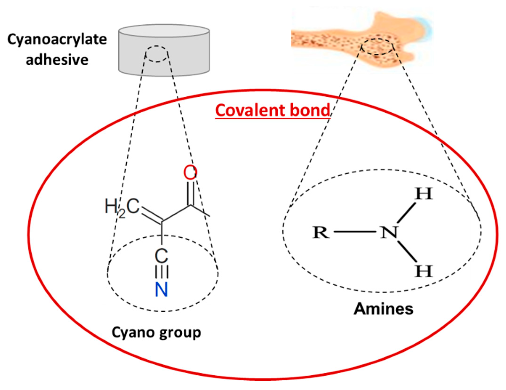
4.1.1. Cyanoacrylates

Cyanoacrylates were one of the first synthetic adhesives used as bone adhesives, demonstrating a high potential for bone bonding, together with methacrylates. Cyanoacrylate adhesives are very promising due to their ability to polymerise under wet conditions (e.g., existence of blood) and to achieve strong wet adhesion and, at the same time, via covalent bonds (Figure 4), they are able to adhere themselves with the amines on the surface of the tissue, achieving rapid curing at low cost [34]. However, the rapid polymerisation leads to an exothermal reaction that has been shown to result in the formation of a hard and brittle film on the bone, leading to cell death and tissue damage [35]. The adhesive strength provided by cyanoacrylate-based adhesives is generally reported to be lower than the bonding and fixation strength achieved using screws [36]. However, a study by Kandalam et al. explored the use of a N-butyl cyanoacrylate for the replacement of screws and plates in pig cortical bone samples and reported a higher range of shear strength (1–2 MPa) compared to that achieved using a plate and screw system (0.49 MPa) [37].
Figure 4. Covalent bond between cyano groups of the adhesive system (cyanoacrylate-based) with amines present in bone collagen matrix.
Despite the enhanced mechanical properties and the ability for adhesion in wet environments, the clinical use of cyanoacrylate-based adhesives is limited due to the toxic nature of the degradation products, which result in a chronic inflammatory response, tissue necrosis and dermatitis in vivo and cytotoxicity for cells in direct contact in vitro [38]. Lee et al. [39] compared the biocompatibility of prepolymerised allyl 2-CA (PACA)-based tissue adhesive with commercial available cyanoacrylate–based adhesive (e.g., Dermabond, Johnson & Johnson, New Brunswick, NJ, USA) and demonstrated that both adhesives were cytotoxic. However, a lower cytotoxicity and reduced tissue inflammation was observed using the PACA-based adhesive compared to the cyanoacrylate-based adhesive. In addition, despite achieving good fixation without displacement or detachment, high cytotoxicity was observed for both the unpolymerised and polymerised cyanoacrylate-based adhesives in vivo in a rabbit subcutaneous model by Pascual et al. [40]. The high cytotoxicity obtained from cyanoacrylate-based adhesives is due to the short alkyl chain length. Even though both n-butyl-2-cyanoacrylate (NBCA) and octyl-2-cyanoacrylate (OCA) are considered harmless and non-carcinogenic, there is no FDA (Food and Drug Administration) approved bone adhesive based on cyanoacrylates. In order to enhance the clinical and mechanical properties of synthetic polymers, various types of biodegradable ceramics and glasses have been added. For instance, bioactive glasses, due to their excellent osteoconductivity [41], have been encapsulated and combined with octyl cyanoacrylate, aiming to increase the migration of bone-derived mesenchymal stromal cells into the adhesive layer and promote their differentiation into osteocytes [42][43]. While instant bonding with high mechanical properties and high efficiency of bone regeneration was achieved, the toxicity of the octyl cyanoacrylate limited further improvement. Furthermore, a hydroxyethyl methacrylate (HEMA) adhesive reinforced with bioactive glass nano particles was developed, demonstrating double tensile strength and significantly enhanced biomineralization and biodegradation compared to the pure HEMA adhesives [44]. Excellent mechanical properties and osteoconductivity can also be achieved with the addition of different calcium phosphates, such as nano-hydroxyapatite [45]. This research combined a biodegradable polymer and an acrylic polymer augmented with bioactive nano-hydroxyapatite; histological results provided high biocompatibility and osteointegration with improved bioactivity [44].

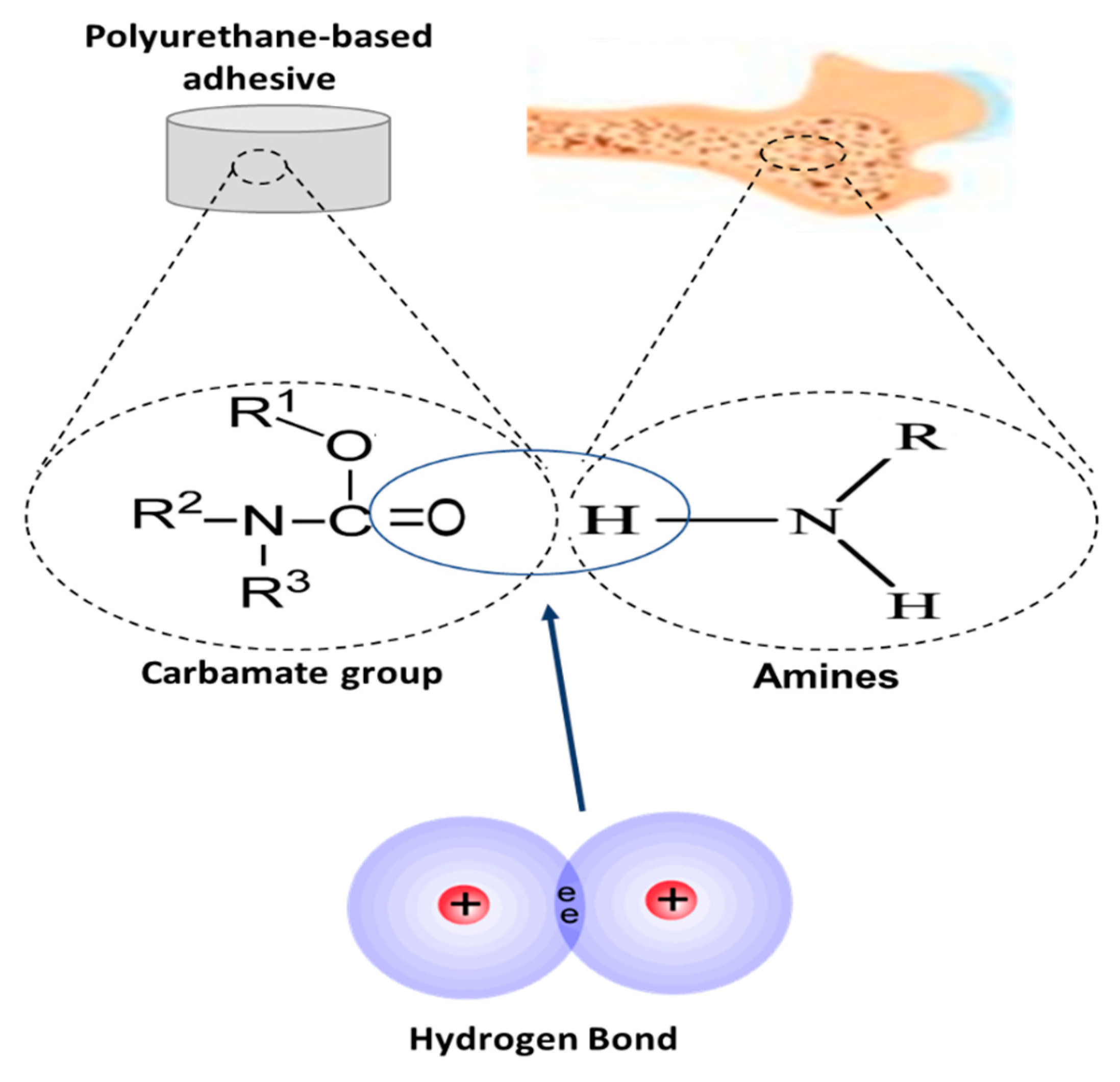
4.1.2. Polyurethanes

Polyurethanes are produced by combining polyisocyanates and polyols in the presence of a catalyst or ultra-violet light. Polyurethane-based adhesives have shown promise for orthopaedic applications as they are biocompatible and demonstrate a high adhesion strength, which is achieved through chemical and/or physical bonding between bone and the adhesive (Figure 5). For example, a polyurethane-based adhesive led to a successful adhesion of bone with a high tensile and adhesion strength on unprimed and primed bone, however, it demonstrated limited biodegradability [46][47]. Changing important factors such as molecular composition, degree of crosslinking, active chemical groups and molecular stiffness can lead to a significant change in the bonding within these polymers and, as a result, can improve biodegradation. To date, a minimal degree of biodegradability has been achieved, which has largely been reported to occur via either a hydrolysis or enzymatic process [48]. The successful closure of bone fractures using a polyurethane-based adhesive without any reaction has been reported in vivo—however, mechanical and functional performance under in vivo conditions was not investigated. Despite advances, currently, the main drawbacks of polyurethane-based adhesives (e.g., premature failure, interfacial bond failure between bone and adhesive, wound infection and tissue necrosis) outweigh the benefits (e.g., high adhesive and/or cohesive strength, osteogenic, non-toxic, high workability and the ability to be delivered by minimally invasive means). As a result, their use in biomedical applications was discontinued in 1990 when a formulation of a novel non-elastomeric polyurethane-based adhesive with calcium and phosphate was developed [49]. Furthermore, in 2012, an FDA approved castor oil-derived polyurethane-based cement, KryptoniteTM (Doctors Research Group Inc., Southbury, CT, USA), was recalled by the FDA because it failed to meet the necessary clinical standards in terms of product safety, as well as its exceptionally long hardening time [50][51].
Figure 5. Chemical and/or physical bonding of polyurethane-based adhesives with bone. Hydrogen bonding occurs between the carbamate group of adhesive system and the amines present in bone collagen matrix.

4.1.3. Polyesters

In bone tissue engineering applications, the resorbable aliphatic polyester poly(L-lactide) (PLLA) has been used as a scaffold in bone regeneration [52]. Copolymers of PLLA with superior mechanical properties have been developed as bone tissue engineered scaffolds, but the influence of copolymerisation, the osteogenic potential is unclear. For instance, biodegradable polymers that can be shaped in situ and adhere to living tissues were developed from the copolymerisation of D,L-lactide polymerisation or D,L-lactide-epsilon-caprolactone (50:50). These polyester copolymers demonstrated faster degradation under wet conditions compared to polyurethane copolymers [53]. In spite of the improved degradation properties compared to standard polyether copolymers, inflammation at the application site remains a limitation. Agarwal et al. [54] reported high adhesion strength for polyester-based adhesives. These adhesives demonstrated low yield strength and significant cytotoxicity during in vitro studies. Therefore, despite the enhanced functional properties of these adhesives, the limitations preclude use as an adhesive for bone tissue engineering applications.
These types of adhesives continue to attract much attention, with recent studies focusing on the investigation and development of polyester-based adhesives leading to enhanced combined properties. Polyethylene glycol (PEG)-based adhesives comprised of PEG ester and glutaryl-succinimidyl ester have been tested for repair of cranial and spinal injuries. The PEG-based adhesive offered high bonding strength due to covalent bonding (i.e., between thiol group and carbonyl group of succinimidyl ester), as well as normal wound healing rates with no post-operative complications. As a result, PEG-based adhesives such as DuraSeal™ (Covidien, Mansfield, MA, USA), which is composed of tetra-PEG-succinimidyl ester and trilysine amine, have been FDA approved and used for cranial surgery [55]. Since the synthesis of the first poly(glycerol sebacate) (PGS) as a tough biodegradable polyester in 2002, a number of modifications have been implemented to enable its clinical application [56]. Pure PGS modified and/or combined with other materials has achieved novel properties [57]. For example, with the addition of a thermoplastic polymer, poly(ε-caprolactone) (PCL), the PCL-modified PGS demonstrated good biocompatibility and cytocompatibility, higher mechanical properties, degradation rate and hydrophilicity [58], while the addition of PEGylated-CH nanoparticles to the PCL-modified PGS resulted in improved antibacterial properties, effective drug release and accelerated wound healing [59][60]. Moreover, good biocompatibility, decreased water contact angle, improved surface hydrophilicity and enhanced cell adhesion was achieved by incorporating poly (vinyl alcohol (PVA)) to PGS, resulting in a promising biodegradable PVA–PGS bioadhesive [61]. In addition to PVA–PGS, similar improved performance was achieved by blending PGS with different types of nanoparticles [62] such as PGS urethane (PGSU)/renewable cellulose nanocrystals (CNCs) [63] and hybrid elastomers PGS–silica glass. Specifically, PGS–silica glass modified adhesive demonstrated controlled production of matrix mineralisation with increased alkaline phosphatase (ALP) activity and osteoinductive capability, tunable elastic properties and biodegradation and enhanced osteoblast proliferation [64][65]. The incorporation of nanoparticles in the PGS offers a new choice for bone tissue repair and regeneration. For instance, the blending of PGS with β-TCP nanoparticles for guided bone regeneration resulted in a bioadhesive with improved mechanical properties and a controlled degradation rate [66]. PEGS/β-TCP promoted cell attachment/viability and superior bone tissue regeneration. Facilitation of the osteogenic differentiation was also observed due to the enhanced mineralisation and the ALP activity resulting from the presence of β-TCP.

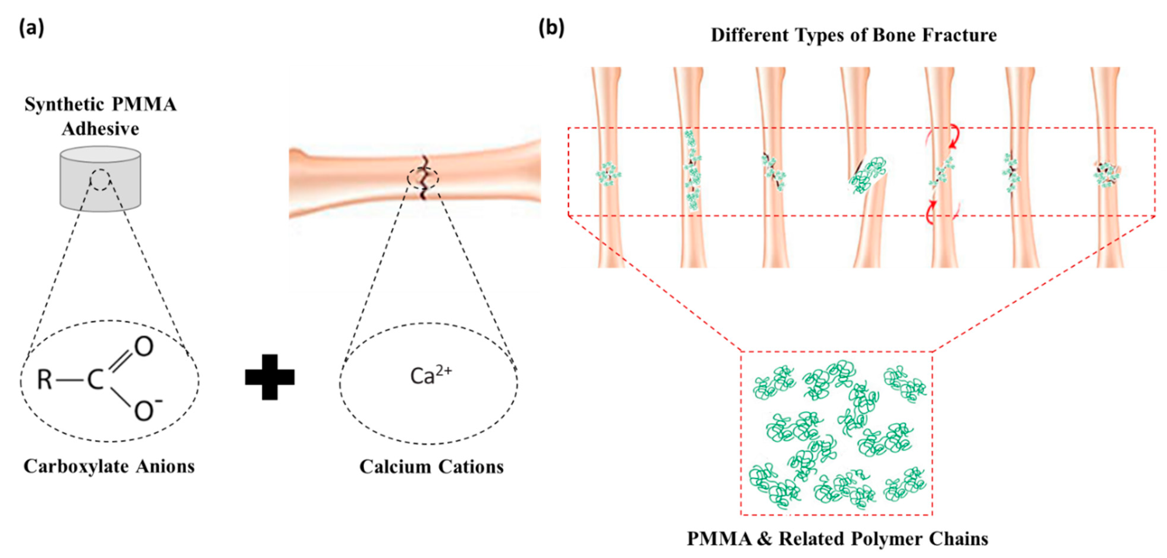
4.1.4. Poly-methyl Methacrylates (PMMA)

PMMA-based adhesives are the most commonly used adhesives in dentistry (since the 1930s) and orthopaedics (since 1958) for total joint replacement applications [67]. PMMA-based adhesives are used to support the prosthetic implant within the bone cavity, where they act as a grouting agent between the bone and implant, in addition to providing fixation [68][69]. Synthetic PMMA adhesives can create chemical and/or physical bonding through ionic interactions (Figure 6a), while PMMA-based adhesives can create a mechanical interlock between bones through the pressurised infiltration of the polymer into surface irregularities (Figure 6b). Even though PMMA-based adhesives are widely used, they exhibit low adhesive strength due to hydrophobic properties. Another drawback of these adhesives is that, in the absence of bone pretreatment or polymer chemical modification, the exothermal reaction that occurs during the polymerisation reaction can lead to considerable thermal necrosis of bone tissue [35]. The potential for carcinogenesis has not been associated with PMMA-based adhesives, although mutagenesis has been reported in bacteria [70]. Many attempts to overcome these challenges have been reported, such as the chemical modification of the PMMA combined with the enrichment of the cement with hydroxyapatite particles to enhance the functional properties [71]. The hydroxyapatite-modified PMMA cement showed higher adhesion than unmodified PMMA bone cement, being used as adhesives in dentistry, replacing the conventional PMMA adhesives. Despite clinical use, the lack of biodegradability of PMMA-based adhesives remains a significant limitation.
Figure 6. Mechanisms of action of PMMA-based adhesive materials. (a) Chemical and/or physical bonding through ionic interaction between carboxylate anions of adhesive system with Ca2+ present on the surface of bone and (b) mechanical interlocking through infiltration of the polymer chains into surface irregularities.
Approaches to overcome these challenges have involved the synthesis of different copolymers with combination properties [72]. Initial attempts focused on the combination of methyl methacrylate reactivity with the biocompatibility and biodegradability of polylactides, since the mechanism of degradation is well established. The adhesive qualities of PMMA to bone have been improved through the use of liquid acrylic resin, phosphoric acid etching or tributyl borane [73]. Despite the synthesis of copolymers with PMMA, different polymerisation techniques have also been used to achieve favourable biocompatibility, biodegradability and improved adhesion [74]. These PMMA–based adhesives demonstrated acceptable biocompatibility and adhesion, while the degradation did not interfere with physiological fracture healing. While good short-term results have been reported with respect to the use of these adhesives in mandibular fractures, spine fractures and isolated long bone fractures, issues relating to late displacement and non-union have prevented clinical use as an adhesive for the treatment of bone fractures [75][76].
The different application sites as well as properties and drawbacks of the synthetic-based bone-adhesive materials described in this section are summarised in Table 1.
Table 1. Comparison of the different properties of all the synthetic-based adhesives.

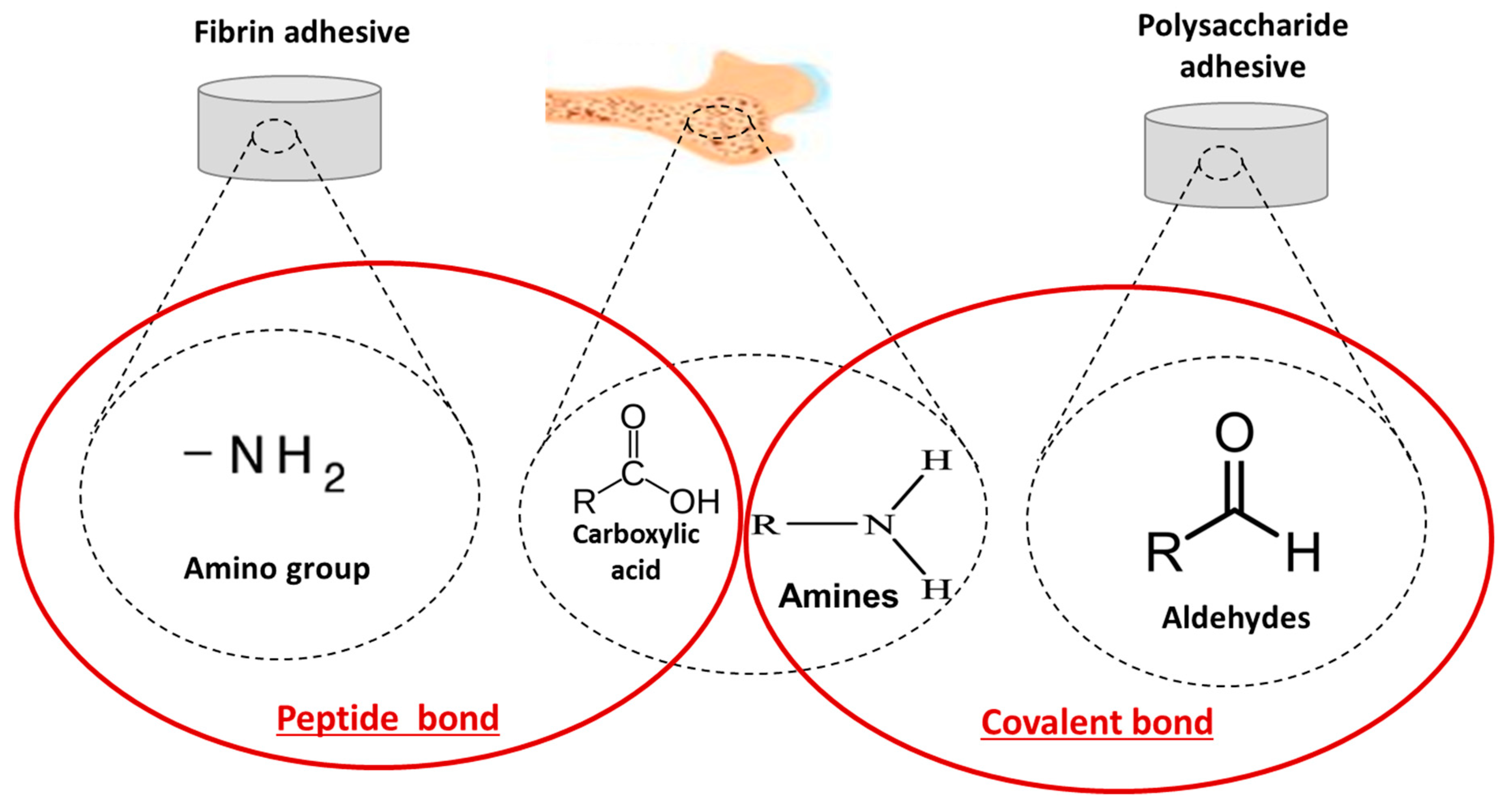
4.2. Naturally-Derived Bioadhesives

The first reported biological bone adhesive, which combined fibrous protein and collagen, was developed in 1931 [77]. The largest group of biologically derived adhesives and sealants is fibrin sealants. Other biological polymer-based adhesives include gelatin–resorcin–aldehyde adhesives, protein–aldehyde adhesives, collagen-based adhesives and polysaccharide-based adhesives. Naturally-derived bioadhesives create bonds with the bone through chemical and/or physical bonding due to amines and carboxylic acid groups present in the bone collagen matrix, respectively (Figure 7). In particular, a peptide bond (chemical bond) is formed when the carboxyl group of one molecule reacts with the amino group of the other molecule, releasing a molecule of water for fibrin adhesives while a covalent bond results in the creation of amines and aldehydes in polysaccharide-based adhesives.
Figure 7. Covalent bond between amino groups of fibrin/fibronectin and/or aldehydes of polysaccharide-based adhesive system, with carboxylic acid groups and amines present in bone collagen matrix.

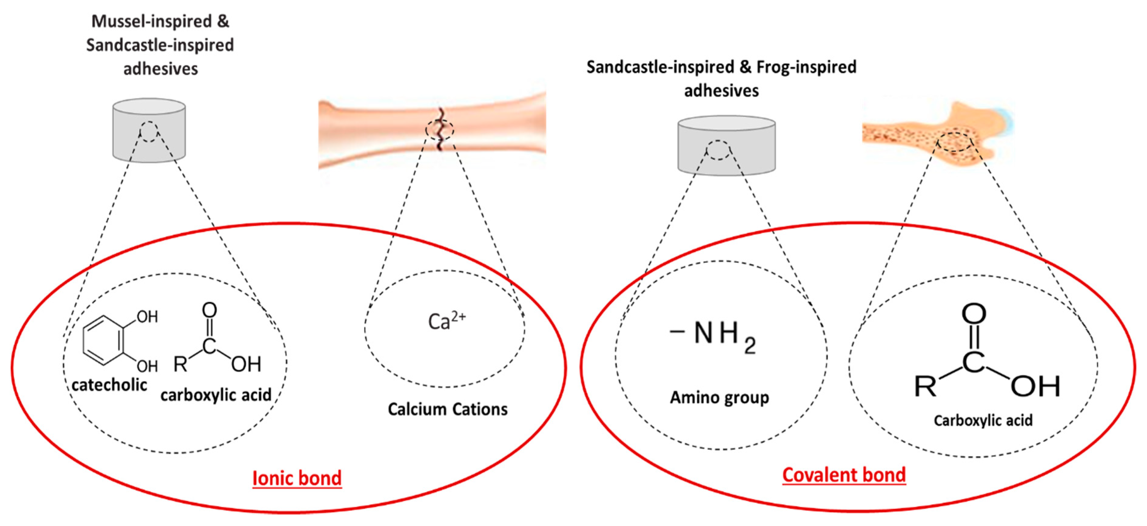
4.3. Biomimetic-Based Adhesives

Some terrestrial organisms as well as marine plants and animals use combinations of proteins and polysaccharides for the formulation of bioadhesives to meet specific requirements to function in the natural environment (e.g., settlement, hunting and defence) [47]. In many cases, these bioadhesives demonstrate higher mechanical properties compared to the currently developed synthetic or natural polymer-based adhesives and adhesion within a wet environment. Specifically, these types of adhesives are able to create ionic and/or covalent bonds with the bone surface or bone collagen (Figure 8). The ability to cure at physiological temperatures and to achieve a high bonding strength to biological materials including bone materials has prompted research into its use as a bioadhesive for bone tissue engineering applications. To date, a number of bioadhesives that mimic these animals and plants have been investigated and/or developed, but the bioadhesives produced have not yet been translated for clinical use for bone tissue engineering applications. The different types of biomimetic adhesives discussed and their properties are summarised in Table 2.
Figure 8. Ionic bond between catecholic hydroxyl and carboxylic acid groups of adhesive systems with Ca2+ present on the surface of bone as a mechanism of adhesion of mussel- and sandcastle-inspired adhesives, and covalent bond between carboxylic acid of adhesive system with amines present in bone collagen matrix for frog- and sandcastle-inspired adhesives.
Table 2. Comparison of the different natural-based adhesives.

Biomimetic Adhesives


Description

Application

Advantages

Disadvantages

Notaden bennetti frog bioadhesives

[68][78]

Protein-based elastic glue

Bone adhesion and fragments fixation (cartilage bone repair)

Binding to biological tissues as well as other surfaces

Better biocompatibility and biodegradation than fibrin glues

Function in moist environments

Lower adhesion strength than cyanoacrylates

Caddisfly silk bioadhesives [79][80][81]

Phosphate-functionalised and amino acid-based polyester copolymers

Bovine bone adhesion (orthopaedic)

Scaffold materials for spinal cord injury

Mesh grafts to treat hernias, ulcers and burns

Adhesion strength of 1.17 MPa

Biodegradable in vitro and in vivo

Higher interface compliance

Cohesive failure

Low curing kinetics and adhesive properties on translationally relevant substrates

Balanus hameri barnacle bioadhesives [82][83][84]

Polyacrylamide-based copolymer with hydroxyl and hexyl groups

Repeatable and robust underwater adhesion to various substrates

Material transfer, temporary fixation (orthopaedics) and material separation

Bovine bone adhesion

Tensile shear strength of 2 MPa

Enhanced toughness and cohesion strength

Good elastic properties

Rapid and reversible adhesion in water

Poor adhesion to bovine bone approx. 363 kPa

Low mechanical strength

Mytilus edulis blue mussel bioadhesives [85][86][87][88]

Adhesives based on complex interaction between different proteins

Strong attachment to inorganic/organic surfaces at dry/wet environment

Reliable crosslinking using oxidation agents, such as iron

Suitable for joining titanium implants to a bone and/or bonding sternal bones

Non-immunogenicity and low cytotoxicity

Greater adhesion on various substrates with adhesion strength of up to 10 MPa

Good biodegradability

Low exothermic reaction for the bonding of sternal bones

Difficulties relating to protein extraction resulting in high production costs, hampering the practical use

Further research needed to determine the suitability of this adhesive as bone adhesive

Calfornica sandcastle worm bioadhesives [81][89][90][91]

Polyphenolic protein and phosphoserine-based adhesive

Strong attachment in a wet environment

Reconstruction of craniofacial fractures

Bonding of wet bone fragments

Bond tissues to metallic and polymeric biomaterials

Maximum adhesion strength and hardness in <30 s

Osteointegration, bone ingrowth and resorbability

Small amount of adhesive needed to achieve the optimal properties

Biodegradable and osteoconductive

Further in vitro and in vivo studies need to be conducted to verify the suitability to natural bone adhesion

5. Bioadhesives for Bone Fracture Repair

A number of the synthetic, naturally-derived and biomimetic-based adhesives that have been previously discussed have been explored and adapted for use in bone repair applications, including fracture fixation, bone defect repair and prosthetic implant bonding to soft/hard tissue. These bioadhesives have the potential to overcome the disadvantages of conventional invasive surgical techniques and meet clinical requirements. Early investigations into the use of bioadhesives in bone repair applications involved the development of epoxy resin-based bioadhesives, such as phenol–formaldehyde resins. While these materials offered a high mechanical strength, they have been reported to lack biocompatibility [92]. Cyanoacrylate- (e.g., cyacrin) and polyurethane-based synthetic polymers have also been proposed as bone bioadhesives due to their high bonding strength and ability to achieve adhesion in a wet environment [93]. However, these cyanoacrylate- and polyurethane-based bioadhesives have demonstrated high tensile and adhesion properties—high infection rates, non-union (e.g., fracture displacement), low biodegradation and severe local reactions have been reported [40][92]. The poor outcomes from these initial materials resulted in research into alternative bioadhesives with more suitable functional properties and improved clinical outcomes.
One such study investigated the application of a non-elastomeric crosslinked polyurethane-based bioadhesive for the stabilisation and repair of bone fragments from the tibia [49]. This bioadhesive was developed via the reaction of a polyisocyanate and polyol in conjunction with a catalyst. The bioadhesive was improved by incorporating calcium and phosphate compounds. In vivo results demonstrated that stabilisation and bonding of the bone fragments as well as a de novo bone growth were achieved, with no evidence of inflammation/infection at the fracture site, as well as some biodegradation and good biocompatibility [49]. A similar polyurethane-based bioadhesive was developed by Schreader et al. for bone-to-bone fixation. This material consisted of a foam-like bioadhesive containing 4,4-methylene diphenyl diisocyanate (MDI) and caprolactone-based diol (polyol) reinforced with hydroxyapatite nanoparticles [46]. The crosslinking occurred via moisture-curing polyurethane chemistry, which can influence the physical properties. However, the final physicochemical and functional properties were dependent on the chemistry and structure of polyol. This bioadhesive demonstrated strong bone-to-bone bonding with an adhesion strength of 4.47 MPa after 20 h, which is four-fold greater than conventional PMMA-based bone cement.
Several studies have focused on the development of PMMA-based bioadhesives for bone repair applications. These bioadhesives have been predominantly used in dentistry and orthodontics due to the weak adhesion to bone, especially in a wet environment. Another issue is the exothermal reaction that occurs during the polymerisation that can lead to cellular death and bone tissue necrosis. Enhancement of the adhesive strength of PMMA-based bioadhesives has been reported by enriching the adhesive with hydroxyapatite particles. However, despite the increase in adhesion strength, the lack of biodegradability has limited the clinical application as a bioadhesive for bone repair applications [94]. A bioadhesive that shows improvements in adhesive properties, particularly in an environment with high humidity, as well as improved biodegradation, have been achieved by Wistlich et al. [95]. They developed a bioadhesive for bone repair applications using a photocurable poly(ethylene glycol) dimethacrylate (PEGDMA) matrix, adding an isocyanate functional (six-armed) star-shaped prepolymer with ethylene oxide and propylene oxide copolymerised (NCO-sP(EO-stat-PO)) in a ratio of 4:1. The NCO-sP(EO-stat-PO enhanced the biodegradation properties and demonstrated a low level of cytotoxicity. Furthermore, the improved adhesive properties were achieved by modifying the matrix PEGDMA with biodegradable ceramic adjuvants (e.g., struvite (MgNH4PO4·6H2O), newberyite (MgHPO4·3H2O) or gypsum (CFaSO4·2H2O). In addition to improving the adhesive properties of the bioadhesive, these ceramic-based adjuvants also increased the porosity of the adhesive, leading to ingrowth of new bone via ion release. This bioadhesive has also been shown to be cytocompatible, easy to apply and demonstrate appropriate bone-to-bone adhesion in a wet environment, as well as supporting bone formation during fracture healing.
Fibrin-based natural polymers have also been applied clinically as bone adhesives, providing biocompatibility, biodegradability and cost effectiveness. These bioadhesives have been extensively used in bone tissue engineering applications, mainly for the acceleration, union and revascularisation of the osteochondral fragments [96][97]. An in vivo study demonstrated the formation of a dense network of osteoid tissue around tricalcium phosphate particles. Le Nihouannen et al. developed a bioadhesive by incorporating macro- and micro-porous biphasic calcium phosphate (MBCP) ceramic granules within a fibrin-based sealant (i.e., Tissucol®) [98]. In particular, 60% hydroxyapatite and 40% beta-tricalcium phosphate (β-TCP) were incorporated into the fibrin-based sealant and the osteoinductive properties evaluated. The formation of a well mineralised ectopic bone was observed between the MBCP particles, proving the ability of the MBCP-fibrin-based sealant to promote osteogenesis. Cassaro et al. developed a bioadhesive that included a fibrin-based biopolymer, which demonstrated haemostatic, sealant, adhesive, scaffolding and drug-delivery properties, and biphasic calcium phosphate (BCP) particles and mesenchymal stem cells (MSCs) [99]. Cassaro et al. demonstrated the bioadhesive to be cost-effective to manufacture, offering good biocompatibility as well as effective repair of the fractured bone and the formation of new bone.
Polysaccharide-based bioadhesives have also been developed for bone repair applications. For instance, Kumbar et al. [100] investigated bioadhesives from cellulose derivatives such as cellulose acetate and ethyl cellulose, which are linear polysaccharides of D-glucose units linked by β(1→4) glycosidic bonds. The hydrogen-bonded structure resulting from the β(1→4) glycosidic bonds led to good biocompatibility and high mechanical properties. This study reported that the polysaccharide-based bioadhesive can form adhesive bonds between cellulose and bone through the carboxylic acid groups, as well as demonstrate a compressive strength (27–33 MPa) close to human trabecular bone. Two component bioadhesives derived from polysaccharides were developed by combining biocompatible chitosan or dextran with degradable starch [101] Initially, the polysaccharides were oxidised with periodic acid (L-3,4-dihydroxy-l-phenylalanine (DOPA)) to generate aldehyde groups, which is the main component found in mussels to help them adhere to the surface of a rock. In this bioadhesive, a covalent bond that is developed enabled a strong adhesion bond at the bone–bone interface as well as a high cohesion strength within the bioadhesive. This bioadhesive demonstrated excellent biocompatibility, with higher mechanical properties than fibrin glues.
L-DOPA, a hydroxylated form of tyrosine, has also been incorporated with the functional binder (mussel-derived adhesive protein (MAP)) to effectively retain deproteinised bovine bone mineral (DBBM) within the bone defect for bone tissue engineering applications [102]. Assessment of the biomechanical properties demonstrated the formation of an aggregate by the binding of the DBBM particles. An improvement in osteoconductivity and acquisition of osteoinductivity was observed, which resulted in an acceleration in bone remodelling and regeneration, with the density of new bone being similar to the normal bone.
Sandcastle worm-based adhesives have shown particular promise in bone repair applications due to the ability to achieve rapid high strength adhesion in a wet environment. One such example is a water-borne adhesive modelled on the proteins from the sandcastle worm-based adhesive which was developed via the incorporation of phosphate, primary amine and catechol sidechains [91]. In particular, polymerised monoacryloxyethyl phosphate (MAEP), dopamine methacrylamide (DMA), acrylamide (Aam) and fluoroscein isothiocyanate (FITC)-methacrylamide were mixed together and applied to bond and stabilise bone fragments. The resultant bioadhesive demonstrated an adhesive strength 40% higher than cyanoacrylate-based bioadhesives. In vitro data demonstrated the ability of the sandcastle worm-based bioadhesive to bond bone fragments back together in a wet environment, while also exhibiting good biocompatibility and osteoconductivity.
Gall et al. [103] developed a sandcastle worm-derived bioadhesive comprised of O-phospho-L-serine, a component of many proteins that exist in natural secretions, resulting in the development of a biodegradable bioadhesive that demonstrates almost instantaneous adhesion (≤10 s). O-phospho-L-serine is a phosphor-related amino acid component of osteopontin (OPN), which has a similar sequence to peptides of adhesion proteins and, when combined with calcium phosphates, leads to the development of a bioadhesive with high biodegradability and mechanical strength (i.e., adhesive and cohesive strength) within a short period [104]. An alternative approach by Kirillova et al., consisting of O-phosphoserine and tetracalcium phosphate, led to the development of another bioadhesive which exhibited a setting time of less than 10 min and the ability to achieve high bone-to-bone adhesive strength [81]. This bioadhesive demonstrated a shear strength ten-fold higher than calcium phosphate cements and PMMA bone cements. In addition to the high adhesive strength achieved, both sandcastle worm-derived bioadhesives also demonstrated osteointegration, bone ingrowth and biodegradability.

References

  1. Augat, P.; Simon, U.; Liedert, A.; Claes, L. Mechanics and mechano-biology of fracture healing in normal and osteoporotic bone. Osteoporos. Int. 2005, 16 (Suppl. 2), 36–43.
  2. Gupta, H.S.; Zioupos, P. Fracture of bone tissue: The ‘hows’ and the ‘whys’. Med. Eng. Phys. 2008, 30, 1209–1226.
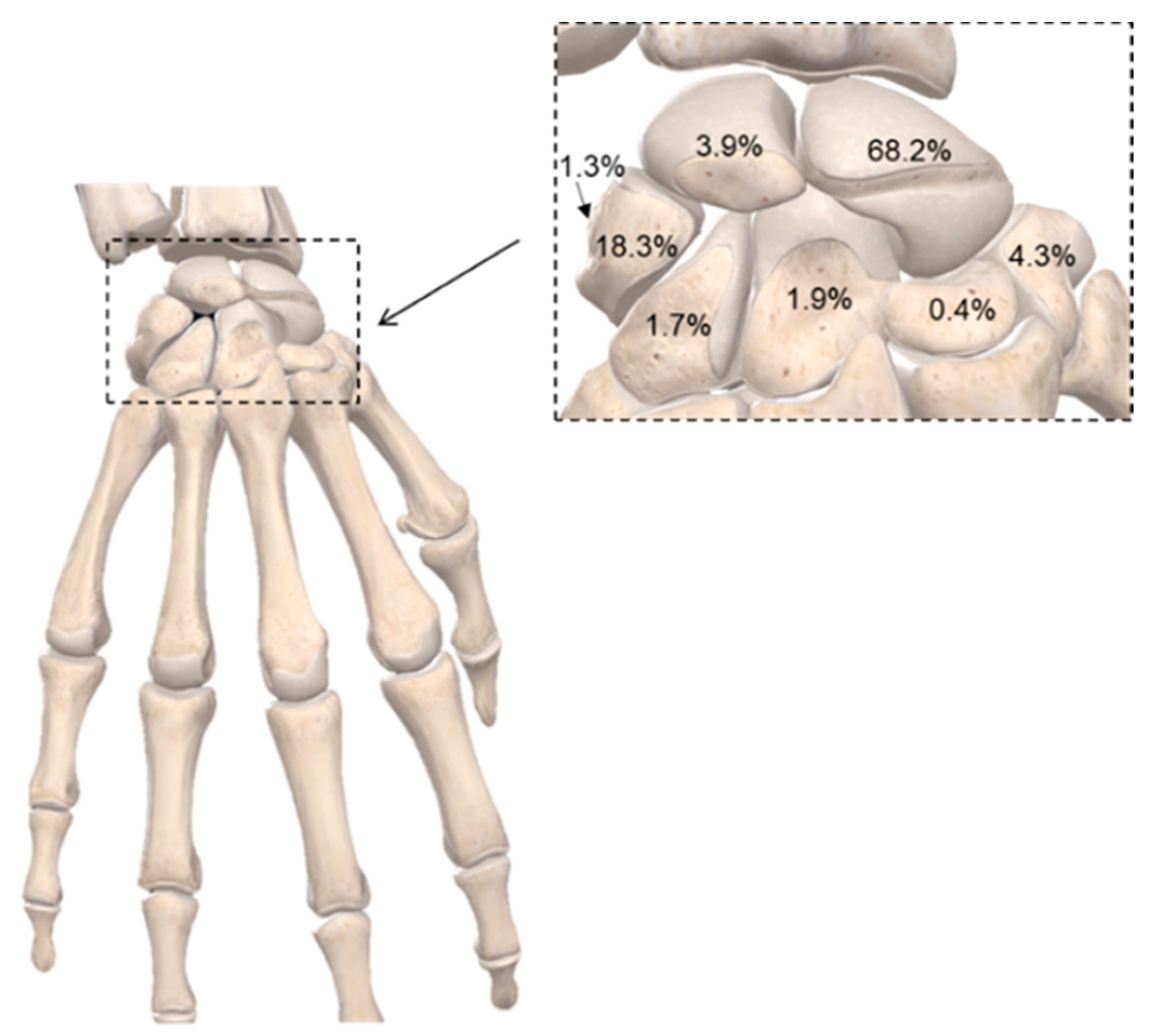
  3. Nellans, K.W.; Kowalski, E.; Chung, K.C. The Epidemiology of Distal Radius Fractures. Hand Clin. 2012, 28, 113–125.
  4. Wu, A.-M.; Bisignano, C.; James, S.L.; Abady, G.G.; Abedi, A.; Abu-Gharbieh, E.; Alhassan, R.K.; Alipour, V.; Arabloo, J.; Asaad, M.; et al. Global, regional, and national burden of bone fractures in 204 countries and territories, 1990–2019: A systematic analysis from the Global Burden of Disease Study 2019. Lancet Health Longev. 2021, 2, e580–e592.
  5. Zhu, G.; Zhang, T.; Chen, M.; Yao, K.; Huang, X.; Zhang, B.; Li, Y.; Liu, J.; Wang, Y.; Zhao, Z. Bone physiological microenvironment and healing mechanism: Basis for future bone-tissue engineering scaffolds. Bioact. Mater. 2021, 6, 4110–4140.
  6. Kovach, T.K.; Dighe, A.S.; Lobo, P.I.; Cui, Q. Interactions between MSCs and immune cells: Implications for bone healing. J. Immunol. Res. 2015, 248.
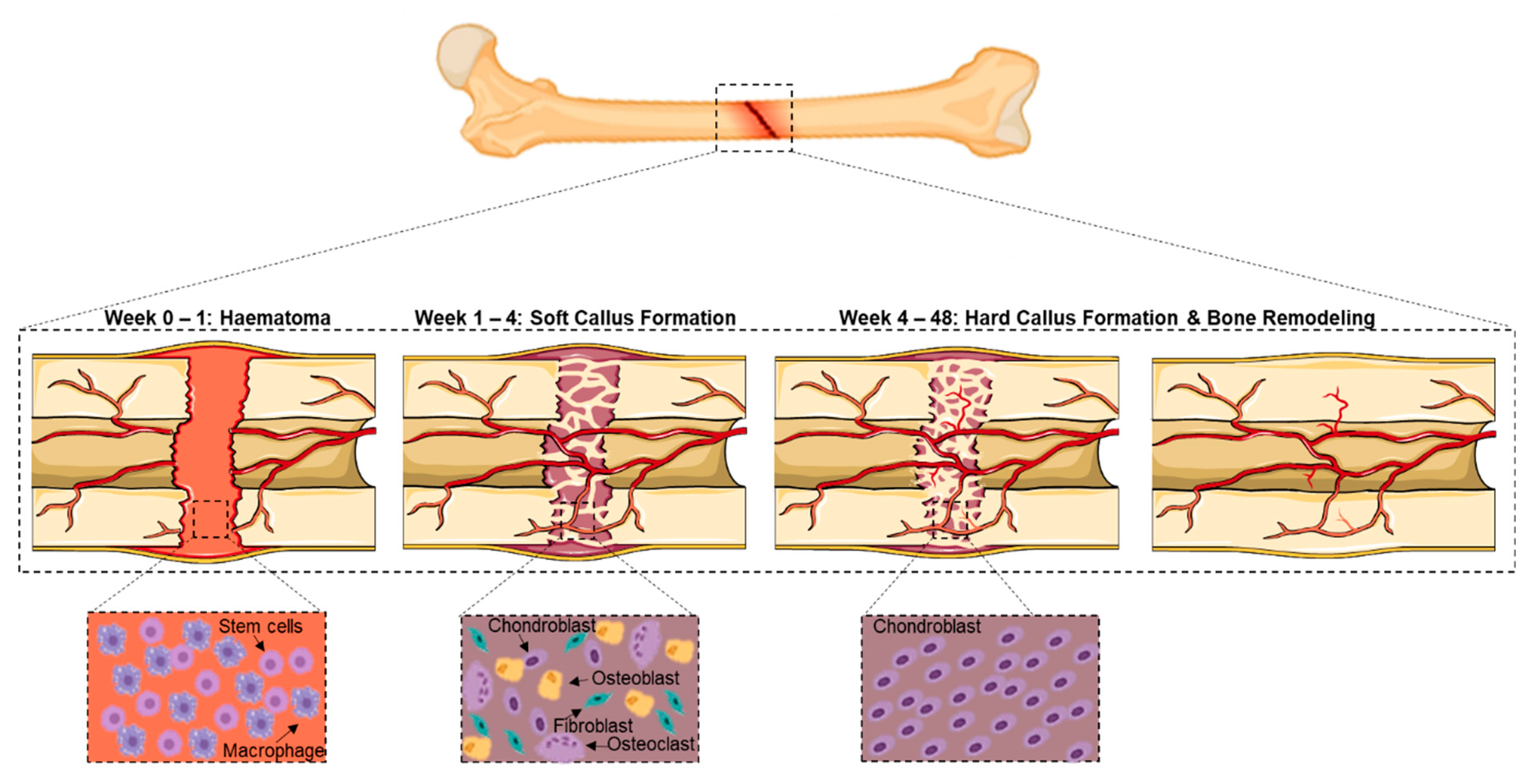
  7. Bahney, C.S.; Zondervan, R.L.; Allison, P.; Theologis, A.; Ashley, J.W.; Ahn, J.; Miclau, T.; Marcucio, R.S.; Hankenson, K.D. Cellular biology of fracture healing. J. Orthop. Res. 2019, 37, 35–50.
  8. Elliott, D.S.; Newman, K.J.H.; Forward, D.P.; Hahn, D.M.; Ollivere, B.; Kojima, K.; Handley, R.; Rossiter, N.D.; Wixted, J.J.; Smith, R.M.; et al. A unified theory of bone healing and nonunion. Bone Jt. J. 2016, 98B, 884–891.
  9. Jalil, M.A.A.; Shuid, A.N.; Muhammad, N. Role of medicinal plants and natural products on osteoporotic fracture healing. Evid.-Based Complement. Altern. Med. 2012, 2012, 714512.
  10. Vriens, J.; Moos, K. Morbidity of the infraorbital nerve following orbitozygomatic complex fractures. J. Cranio-Maxillofac. Surg. 1995, 23, 363–368.
  11. Muraoka, M.; Nakai, Y. Twenty years of statistics and observation of facial bone fracture. Acta Oto-Laryngol. Suppl. 1998, 118, 261–265.
  12. Kasimova, G.; Yunusov, D.; Sakhibova, M.; Yaxudaev, E.; Batirova, B. Analysis of fracture of the foot bones in children according to the andijan region. Ann. Rom. Soc. Cell Biol. 2021, 25, 6186–6192.
  13. Meena, S.; Sharma, P.; Sambharia, A.K.; Dawar, A. Fractures of distal radius: An overview. J. Fam. Med. Prim. Care 2014, 3, 325–332.
  14. Souyris, F.; Klersy, F.; Jammet, P.; Payrot, C. Malar bone fractures and their sequelae. J. Cranio-Maxillofac. Surg. 1989, 17, 64–68.
  15. Welling, R.D.; Jacobson, J.A.; Jamadar, D.A.; Chong, S.; Caoili, E.M.; Jebson, P.J.L. MDCT and radiography of wrist fractures: Radiographic sensitivity and fracture patterns. Am. J. Roentgenol. 2008, 190, 10–16.
  16. Jones, W.A.; Ghorbal, M.S. Fractures of the trapezium a report on three cases. J. Hand Surg. Am. 1985, 10, 227–230.
  17. Erol, B.; Tanrikulu, R.; Görgün, B. Maxillofacial fractures. Analysis of demographic distribution and treatment in 2901 patients (25-year experience). J. Cranio-Maxillofac. Surg. 2004, 32, 308–313.
  18. Hogg, N.J.V.; Stewart, T.C.; Armstrong, J.E.A.; Girotti, M.J. Epidemiology of maxillofacial injuries at trauma hospitals in Ontario, Canada, between 1992 and 1997. J. Trauma Inj. Infect. Crit. Care 2000, 49, 425–432.
  19. Hwang, K.; You, S.H. Analysis of facial bone fractures: An 11-year study of 2,094 patients. Indian J. Plast. Surg. 2010, 43, 42–48.
  20. Johnell, O.; Kanis, J.A. An estimate of the worldwide prevalence and disability associated with osteoporotic fractures. Osteoporos. Int. 2006, 17(12), 1726–1733.
  21. Fisher, W.D.; Hamblen, D.L. Problems and pitfalls of compression fixation of long bone fractures: A review of results and complications. Injury 1979, 10, 99–107.
  22. Larsson, S. Treatment of osteoporotic fractures. Scandinavian J. Surg. 2002, 91, 140–146.
  23. Xue, X.H.; Yan, S.G.; Cai, X.Z.; Shi, M.M.; Lin, T. Intramedullary nailing versus plating for extra-articular distal tibial metaphyseal fracture: A systematic review and meta-analysis. Injury 2014, 45, 667–676.
  24. Misra, A.; Kapur, R.; Maffulli, N. Complex proximal humeral fractures in adults- A systematic review of management. Injury 2001, 32, 363–372.
  25. Gross, C.E.; Nunley, J.A. Navicular Stress Fractures. Foot Ankle Int. 2015, 36, 1117–1122.
  26. Jackson, L.C.; Pacchiana, P.D. Common complications of fracture repair. Clin. Tech. Small Anim. Pract. 2004, 19, 168–179.
  27. Uhthoff, H.K.; Poitras, P.; Backman, D.S. Internal plate fixation of fractures: Short history and recent developments. J. Orthop. Sci. 2006, 11, 118–126.
  28. Mucha, P.E.; Farnell, M.B. Analysis of Pelvic Fracture Management. J. Trauma Inj. Infect. Crit. 1984, 24, 379–386.
  29. Xu, H.H.; Wang, P.; Wang, L.; Bao, C.; Chen, Q.; Weir, M.D.; Chow, L.C.; Zhao, L.; Zhou, X.; Reynolds, M.A. Calcium phosphate cements for bone engineering and their biological properties. Bone Res. 2017, 5, 1–19.
  30. Petrie, E.M. Cyanoacrylate adhesives in surgical applications: A critical review. Rev. Adhes. Adhes. 2014, 2, 253–310.
  31. Chen, C.F.; Shen, H.H.; Lin, T.Y.; Yu, Y.J.; Chen, W.C.; Chu, I.M. Studies on the preparation and characterization of mPEG-polyester biodegradable bioglue for bone defect repair. J. Med. Biol. Eng. 2011, 31, 13–17.
  32. Arora, M.; Chan, E.K.S.; Gupta, S.; Diwan, A.D. Polymethylmethacrylate bone cements and additives: A review of the literature. World J. Orthop. 2013, 4, 67–74.
  33. Schlag, G.; Redl, H. Fibrin Sealant in Orthopedic Surgery. Clin. Orthop. Relat. Res. 1988, 227, 269–285.
  34. Sánchez-Fernández, M.J.; Hammoudeh, H.; Lanao, R.P.F.; van Erk, M.; van Hest, J.C.M.; Leeuwenburgh, S.C.G. Bone-Adhesive Materials: Clinical Requirements, Mechanisms of Action, and Future Perspective. Adv. Mater. Interfaces 2019, 6, 1802021.
  35. Dunne, N.J.; Orr, J.F. Thermal characteristics of curing acrylic bone cement. ITBM-RBM 2001, 22, 88–97.
  36. Gosain, A.K. The current status of tissue glues: I. For bone fixation. Plast. Reconstr. Surg. 2002, 109, 2581–2583.
  37. Kandalam, U.; Bouvier, A.; Casas, S.; Smith, R.; Gallego, A.; Rothrock, J.; Thompson, J.; Huang, C.-Y.; Stelnicki, E. Novel bone adhesives: A comparison of bond strengths in vitro. Int. J. Oral Maxillofac. Surg. 2013, 42, 1054–1059.
  38. Kukleta, J.F.; Freytag, C.; Weber, M. Efficiency and safety of mesh fixation in laparoscopic inguinal hernia repair using n-butyl cyanoacrylate: Long-term biocompatibility in over 1, 300 mesh fixations. Hernia 2012, 16, 153–162.
  39. Lee, Y.J.; Jung, G.B.; Choi, S.; Lee, G.; Kim, J.H.; Son, H.S.; Bae, H.; Park, H.-K. Biocompatibility of a novel cyanoacrylate based tissue adhesive: Cytotoxicity and biochemical property evaluation. PLoS ONE 2013, 8, e79761.
  40. Pascual, G.; Sotomayor, S.; Rodríguez, M.; Pérez-Köhler, B.; Kühnhardt, A.; Fernández-Gutiérrez, M.; Román, J.S.; Bellón, J.M. Cytotoxicity of cyanoacrylate-based tissue adhesives and short-term preclinical in vivo biocompatibility in abdominal hernia repair. PLoS ONE 2016, 11, e0157920.
  41. Baino, F.; Hamzehlou, S.; Kargozar, S. Bioactive glasses: Where are we and where are we going? J. Funct. Biomater. 2018, 9, 25.
  42. Zhang, M.; Liu, J.; Zhu, T.; Le, H.; Wang, X.; Guo, J.; Liu, G.; Ding, J. Functional Macromolecular Adhesives for Bone Fracture Healing. ACS Appl. Mater. Interfaces 2021, 14, 1–19.
  43. Xu, L.; Gao, S.; Zhou, R.; Zhou, F.; Qiao, Y.; Qiu, D. Bioactive Pore-Forming Bone Adhesives Facilitating Cell Ingrowth for Fracture Healing. Adv. Mater. 2020, 32, 1–7.
  44. Shahbazi, S.; Moztarzadeh, F.; Sadeghi, G.M.M.; Jafari, Y. In vitro study of a new biodegradable nanocomposite based on poly propylene fumarate as bone glue. Mater. Sci. Eng. C 2016, 69, 1201–1209.
  45. Brandt, J.; Henning, S.; Michler, G.; Hein, W.; Bernstein, A.; Schulz, M. Nanocrystalline hydroxyapatite for bone repair: An animal study. J. Mater. Sci. Mater. Med. 2009, 21, 283–294.
  46. Schreader, K.J.; Bayer, I.S.; Milner, D.J.; Loth, E.; Jasiuk, I. A polyurethane-based nanocomposite biocompatible bone adhesive. J. Appl. Polym. Sci. 2012, 127, 4974–4982.
  47. Bhagat, V.; Becker, M.L. Degradable Adhesives for Surgery and Tissue Engineering. Biomacromolecules 2017, 18, 3009–3039.
  48. Meereboer, K.W.; Misra, M.; Mohanty, A.K. Review of recent advances in the biodegradability of polyhydroxyalkanoate (PHA) bioplastics and their composites. Green Chem. 2020, 22, 5519–5558.
  49. Brandeis, E.; Katz, D.; Silbermann, M.; Zinman, C. A new bioadhesive for in vivo bone adhesion. J. Mater. Sci. Mater. Med. 1993, 4, 543–546.
  50. Böker, K.O.; Richter, K.; Jäckle, K.; Taheri, S.; Grunwald, I.; Borcherding, K.; von Byern, J.; Hartwig, A.; Wildemann, B.; Schilling, A.F.; et al. Current state of bone adhesives-Necessities and hurdles. Materials 2019, 12, 3975.
  51. Jain, R.; Wairkar, S. Recent developments and clinical applications of surgical glues: An overview. Int. J. Biol. Macromol. 2019, 137, 95–106.
  52. Idris, S.B.; Arvidson, K.; Plikk, P.; Ibrahim, S.; Finne-Wistrand, A.; Albertsson, A.-C.; Bolstad, A.I.; Mustafa, K. Polyester copolymer scaffolds enhance expression of bone markers in osteoblast-like cells. J. Biomed. Mater. Res. Part A 2010, 9999A, 631–639.
  53. Kobayashi, H.; Hyon, S.-H.; Ikada, Y. Water-curable and biodegradable prepolymers. J. Biomed. Mater. Res. 1991, 25, 1481–1494.
  54. Shi, Y.; Zhou, P.; Jérôme, V.; Freitag, R.; Agarwal, S. Enzymatically Degradable Polyester-Based Adhesives. ACS Biomater. Sci. Eng. 2015, 1, 971–977.
  55. Annabi, N.; Yue, K.; Tamayol, A.; Khademhosseini, A. Elastic sealants for surgical applications. Eur. J. Pharm. Biopharm. 2015, 95, 27–39.
  56. Wang, Y.; Ameer, G.A.; Sheppard, B.J.; Langer, R. A tough biodegradable elastomer. Nat. Biotechnol. 2002, 20, 602–606.
  57. Vogt, L.; Ruther, F.; Salehi, S.; Boccaccini, A.R. Poly(Glycerol Sebacate) in Biomedical Applications—A Review of the Recent Literature. Adv. Healthc. Mater. 2021, 10, 2002026.
  58. Ferrari, P.F.; Aliakbarian, B.; Lagazzo, A.; Tamayol, A.; Palombo, D.; Perego, P. Tailored electrospun small-diameter graft for vascular prosthesis. Int. J. Polym. Mater. Polym. Biomater. 2017, 66, 635–643.
  59. Kalakonda, P.; Aldhahri, M.A.; Abdel-Wahab, M.S.; Tamayol, A.; Moghaddam, K.M.; Ben Rached, F.; Pain, A.; Khademhosseini, A.; Memic, A.; Chaieb, S. Microfibrous silver-coated polymeric scaffolds with tunable mechanical properties. RSC Adv. 2017, 7, 34331–34338.
  60. Memic, A.; Aldhahri, M.; Tamayol, A.; Mostafalu, P.; Abdel-Wahab, M.S.; Samandari, M.; Moghaddam, K.M.; Annabi, N.; Bencherif, S.A.; Khademhosseini, A. Nanofibrous silver-coated polymeric scaffolds with tunable electrical properties. Nanomaterials 2017, 7, 63.
  61. Saudi, A.; Rafienia, M.; Kharazi, A.Z.; Salehi, H.; Zarrabi, A.; Karevan, M. Design and fabrication of poly (glycerol sebacate)-based fibers for neural tissue engineering: Synthesis, electrospinning, and characterization. Polym. Adv. Technol. 2019, 30, 1427–1440.
  62. Sha, D.; Wu, Z.; Zhang, J.; Ma, Y.; Yang, Z.; Yuan, Y. Development of modified and multifunctional poly(glycerol sebacate) (PGS)-based biomaterials for biomedical applications. Eur. Polym. J. 2021, 161, 110830.
  63. Wu, T.; Frydrych, M.; Kelly, K.O.; Chen, B. Poly(glycerol sebacate urethane)—Cellulose Nanocomposites with Water-Active Shape-Memory Effects. Biomacromolecules 2014, 15, 2663–2671.
  64. Zhao, X.; Wu, Y.; Du, Y.; Chen, X.; Lei, B.; Xue, Y.; Ma, P.X. A highly bioactive and biodegradable poly(glycerol sebacate)-silica glass hybrid elastomer with tailored mechanical properties for bone tissue regeneration. J. Mater. Chem. B 2015, 3, 3222–3233.
  65. Kerativitayanan, P.; Tatullo, M.; Khariton, M.; Joshi, P.; Perniconi, B.; Gaharwar, A.K. Gaharwar, Nanoengineered Osteoinductive and Elastomeric Scaffolds for Bone Tissue Engineering. ACS Biomater. Sci. Eng. 2017, 3, 590–600.
  66. Yu, S.; Shi, J.; Liu, Y.; Si, J.; Yuan, Y.; Liu, C. A mechanically robust and flexible PEGylated poly(glycerol sebacate)/β-TCP nanoparticle composite membrane for guided bone regeneration. J. Mater. Chem. B 2019, 7, 3279–3290.
  67. Hasandoost, L.; Rodriguez, O.; Alhalawani, A.; Zalzal, P.; Schemitsch, E.H.; Waldman, S.D.; Papini, M.; Towler, M.R. The role of poly(methyl methacrylate) in management of bone loss and infection in revision total knee arthroplasty: A review. J. Funct. Biomater. 2020, 11, 25.
  68. Farrar, D.F. Bone adhesives for trauma surgery: A review of challenges and developments. Int. J. Adhes. Adhes. 2012, 33, 89–97.
  69. Lu, J. Orthopedic bone cements. Biomechanics and Biomaterials in Orthopedics, 2nd ed.; Springer: London, UK, 2016; pp. 123–138.
  70. Mikkelsen, D.B.; Pedersen, C.; Højbjerg, T.; Schønheyder, H.C. Culture of multiple peroperative biopsies and diagnosis of infected knee arthroplasties. APMIS 2006, 114, 449–452.
  71. Sa, Y.; Wang, M.; Deng, H.; Wang, Y.; Jiang, T. Beneficial effects of biomimetic nano-sized hydroxyapatite/antibiotic gentamicin enriched chitosan-glycerophosphate hydrogel on the performance of injectable polymethylmethacrylate. RSC Adv. 2015, 5, 91082–91092.
  72. Grossterlinden, L.; Janssen, A.; Schmitz, N.; Priemel, M.; Pogoda, P.; Amling, M.; Rueger, J.M.; Linhart, W. Deleterious tissue reaction to an alkylene bis(dilactoyl)-methacrylate bone adhesive in long-term follow up after screw augmentation in an ovine model. Biomaterials 2006, 27, 3379–3386.
  73. Meyer, G.; Muster, D.; Schmitt, D.; Jung, P.; Jaeger, J.H. Bone bonding through bioadhesives: Present status. Biomater. Med. Devices. Artif. Organs 1979, 7, 55–71.
  74. Heiss, C.; Kraus, R.; Peters, F.; Henn, W.; Schnabelrauch, M.; Berg, A.; Pautzsch, T.; Weisser, J.; Schnettler, R. Development of a bioresorbable self-hardening bone adhesive based on a composite consisting of polylactide methacrylates and β-tricalcium phosphate. J. Biomed. Mater. Res. Part B Appl. Biomater. 2009, 90B, 55–66.
  75. Jensen, M.E.; Evans, A.J.; Mathis, J.M.; Kallmes, D.F.; Cloft, H.J.; Dion, J.E. Percutaneous polymethylmethacrylate vertebroplasty in the treatment of osteoporotic vertebral body compression fractures: Technical aspects. Am. J. Neuroradiol. 1997, 18, 1897–1904.
  76. Ali, U.; Karim, K.J.B.A.; Buang, N.A. A Review of the Properties and Applications of Poly (Methyl Methacrylate) (PMMA). Polym. Rev. 2015, 55, 678–705.
  77. Shah, N.V.; Meislin, R. Current state and use of biological adhesives in orthopedic surgery. Orthopedics 2013, 36, 945–956.
  78. Millar, N.L.; Bradley, T.A.; Walsh, N.A.; Appleyard, R.C.; Tyler, M.J.; Murrell, G.A.C. Frog glue enhances rotator cuff repair in a laboratory cadaveric model. J. Shoulder Elb. Surg. 2009, 18, 639–645.
  79. Bhagat, V.; O’Brien, E.; Zhou, J.; Becker, M.L. Caddisfly Inspired Phosphorylated Poly(ester urea)-Based Degradable Bone Adhesives. Biomacromolecules 2016, 17, 3016–3024.
  80. Duarte, A.P.; Coelho, J.F.; Bordado, J.C.; Cidade, M.T.; Gil, M.H. Surgical adhesives: Systematic review of the main types and development forecast. Prog. Polym. Sci. 2011, 37, 1031–1050.
  81. Kirillova, A.; Kelly, C.; von Windheim, N.; Gall, K. Bioinspired Mineral-Organic Bioresorbable Bone Adhesive. Adv. Healthc. Mater. 2018, 7, e1800467.
  82. Yamamoto, H.; Nagai, A.; Okada, T.; Nishida, A. Synthesis and adhesive studies of barnacle model proteins. Mar. Chem. 1989, 26, 331–338.
  83. Nishida, J.; Higaki, Y.; Takahara, A. Synthesis and characterization of barnacle adhesive mimetic towards underwater adhesion. Chem. Lett. 2015, 44, 1047–1049.
  84. Fan, H.; Wang, J.; Gong, J.P. Barnacle Cement Proteins-Inspired Tough Hydrogels with Robust, Long-Lasting, and Repeatable Underwater Adhesion. Adv. Funct. Mater. 2020, 31, 2009334.
  85. Silverman, H.G.; Roberto, F.F. Understanding marine mussel adhesion. Mar. Biotechnol. 2007, 9, 661–681.
  86. Flammang, P.; Lambert, A.; Bailly, P.; Hennebert, E. Polyphosphoprotein-containing marine adhesives. J. Adhes. 2009, 85, 447–464.
  87. Yamamoto, H.; Kuno, S.; Nagai, A.; Nishida, A.; Yamauchi, S.; Ikeda, K. Insolubilizing and adhesive studies of water-soluble synthetic model proteins. Int. J. Biol. Macromol. 1990, 12, 305–310.
  88. Meredith, H.J.; Jenkins, C.L.; Wilker, J.J. Enhancing the adhesion of a biomimetic polymer yields performance rivaling commercial glues. Adv. Funct. Mater. 2014, 24, 3259–3267.
  89. Li, A.; Jia, M.; Mu, Y.; Jiang, W.; Wan, X. Humid bonding with a water-soluble adhesive inspired by mussels and sandcastle worms. Macromol. Chem. Phys. 2014, 216, 450–459.
  90. Bhagat, V.; O’Brien, E.; Zhou, J.; Becker, M.L. A novel class of injectable bioceramics that glue tissues and biomaterials. Materials 2018, 11, 2492.
  91. Shao, H.; Bachus, K.N.; Stewart, R.J. A water-borne adhesive modeled after the sandcastle glue of P. californica. Macromol. Biosci. 2009, 9, 464–471.
  92. Gardziella, A.; Mueller, R. Phenolic Resins. Kunstst. Ger. Plast. 1990, 80, 66–68.
  93. Donkerwolcke, M.; Burny, F.; Muster, D. Tissues and bone adhesives historical aspects. Biomaterials 1998, 19, 1461–1466.
  94. Ishihara, K.; Nakabayashi, N. Adhesive bone cement both to bone and metals: 4-META in MMA initiated with tri-n-butyl borane. J. Biomed. Mater. Res. 1989, 23, 1475–1482.
  95. Wistlich, L.; Rücker, A.; Schamel, M.; Kübler, A.C.; Gbureck, U.; Groll, J. A Bone Glue with Sustained Adhesion under Wet Conditions. Adv. Healthc. Mater. 2017, 6, 1600902.
  96. Liu, X.; Pujari-Palmer, M.; Wenner, D.; Procter, P.; Insley, G.; Engqvist, H. Adhesive cements that bond soft tissue ex vivo. Materials 2019, 12, 2473.
  97. Hulsart-Billström, G.; Stelzl, C.; Procter, P.; Pujari-Palmer, M.; Insley, G.; Engqvist, H.; Larsson, S. In vivo safety assessment of a bio-inspired bone adhesive. J. Mater. Sci. Mater. Med. 2020, 31, 24.
  98. Le Nihouannen, D.; Saffarzadeh, A.; Gauthier, O.; Moreau, F.; Pilet, P.; Spaethe, R.; Layrolle, P.; Daculsi, G. Bone tissue formation in sheep muscles induced by a biphasic calcium phosphate ceramic and fibrin glue composite. J. Mater. Sci. Mater. Med. 2008, 19, 667–675.
  99. Cassaro, C.V.; Justulin, L.A., Jr.; De Lima, P.R.; Golim, M.D.A.; Biscola, N.P.; de Castro, M.V.; De Oliveira, A.L.R.; Doiche, D.P.; Pereira, E.J.; Ferreira, R.S., Jr.; et al. Fibrin biopolymer as scaffold candidate to treat bone defects in rats. J. Venom. Anim. Toxins Incl. Trop. Dis. 2019, 25, e20190027.
  100. Kumbar, S.G.; Toti, U.S.; Deng, M.; James, R.; Laurencin, C.T.; Aravamudhan, A.; Harmon, M.; Ramos, D.M. Novel mechanically competent polysaccharide scaffolds for bone tissue engineering. Biomed. Mater. 2011, 6, 065005.
  101. Hoffmann, B.; Volkmer, E.; Kokott, A.; Augat, P.; Ohnmacht, M.; Sedlmayr, N.; Schieker, M.; Claes, L.; Mutschler, W.; Ziegler, G. Characterisation of a new bioadhesive system based on polysaccharides with the potential to be used as bone glue. J. Mater. Sci. Mater. Med. 2009, 20, 2001–2009.
  102. Choi, B.-H.; Cheong, H.; Ahn, J.-S.; Zhou, C.; Kwon, J.J.; Cha, H.J.; Jun, S.H. Engineered mussel bioglue as a functional osteoinductive binder for grafting of bone substitute particles to accelerate in vivo bone regeneration. J. Mater. Chem. B 2015, 3, 546–555.
  103. Brown, M.; Kay, G.W.; Cochran, D.; Fiorellini, J.; Hess, B. From bench-to-bedside: Licensing and development of a mineral-organic bone adhesive for bone repair. In Society for Biomaterials Annual Meeting and Exposition 42nd Annual Meeting; Society for Biomaterials: Mount Laurel, NJ, USA, 2019; Volume 40, 169p.
  104. Bystrom, J.L.; Pujari-Palmer, M. Phosphoserine functionalized cements preserve metastable phases, and reprecipitate octacalcium phosphate, hydroxyapatite, dicalcium phosphate, and amorphous calcium phosphate, during degradation, in vitro. J. Funct. Biomater. 2019, 10, 54.
More
Information
Contributors MDPI registered users' name will be linked to their SciProfiles pages. To register with us, please refer to https://encyclopedia.pub/register : , , ,
View Times: 1.6K
Revision: 1 time (View History)
Update Date: 15 Jun 2022
1000/1000