Submitted Successfully!
To reward your contribution, here is a gift for you: A free trial for our video production service.
Thank you for your contribution! You can also upload a video entry or images related to this topic.
Version Summary Created by Modification Content Size Created at Operation
1 -- 2883 2022-06-13 10:38:46 |
2 format correction Meta information modification 2883 2022-06-13 11:17:04 |

Video Upload Options

Do you have a full video?

Confirm

Are you sure to Delete?
Cite
If you have any further questions, please contact Encyclopedia Editorial Office.
De Francesco, F.; Zavan, B.; , .; Riccio, M. Regenerative Treatment for Osteoarthritis Disease. Encyclopedia. Available online: https://encyclopedia.pub/entry/23963 (accessed on 24 April 2024).
De Francesco F, Zavan B,  , Riccio M. Regenerative Treatment for Osteoarthritis Disease. Encyclopedia. Available at: https://encyclopedia.pub/entry/23963. Accessed April 24, 2024.
De Francesco, Francesco, Barbara Zavan,  , Michele Riccio. "Regenerative Treatment for Osteoarthritis Disease" Encyclopedia, https://encyclopedia.pub/entry/23963 (accessed April 24, 2024).
De Francesco, F., Zavan, B., , ., & Riccio, M. (2022, June 13). Regenerative Treatment for Osteoarthritis Disease. In Encyclopedia. https://encyclopedia.pub/entry/23963
De Francesco, Francesco, et al. "Regenerative Treatment for Osteoarthritis Disease." Encyclopedia. Web. 13 June, 2022.
Regenerative Treatment for Osteoarthritis Disease
Edit

Osteoarthritis (OA) is a chronic debilitating disorder causing pain and gradual degeneration of weight-bearing joints with detrimental effects on cartilage volume as well as cartilage damage, generating inflammation in the joint structure. The etiology of OA is multifactorial. Currently, therapies are mainly addressing the physical and occupational aspects of osteoarthritis using pharmacologic pain treatment and/or surgery to manage the symptomatology of the disease with no specific regard to disease progression or prevention.

osteoarthritis stromal vascular fraction mechanical disaggregation regenerative surgery adipo micrografts

1. Introduction

OA is an idiopathic disorder, and the management reflects the lack of understanding of the disease. The non-surgical approach involves the usage of treatment such as physiotherapy, kinesitherapy, weight control, drugs. The main drugs used are hormones (parathyroid hormones, calcitonin, leptin), which act as regulating molecular pathways of cartilage metabolism; bisphosphonates (zoledronic acid, alendronate) which act as improving bone metabolism, reducing bone reabsorption; monoclonal antibodies (bevacizumab, adalimumab,) which act on articular cartilage promoting collagen production; statins (atorvastatin, which reduces cartilage degradation); supplements (glucosamine, chondroitin sulfate, vitamin C, vitamin D, Selenium, Zinc, Magnesium), which act as a cartilage nourishment [1][2].

2. Regenerative Treatment for Osteoarthritis Disease

2.1. Platelet-Rich Plasma (PRP)

Platelet-Rich Plasma (PRP) may be described as plasma volume platelet concentration derived from centrifuged whole blood [3]. Numerous platelets or thrombocyte functions involve damage to tissue, and platelet activity leads to the release of proteins and molecules that are related to vasoconstriction, inflammation, immune reaction, angiogenesis and the repair of tissue. PRP is rich in growth factors such as platelet-derived growth factor (PDGF), vascular endothelial growth factor (VEGF), transforming growth factor beta (TGF-b), epidermal growth factor (EGF), fibroblast growth factor (FGF) and insulin-like growth factor (IGF) [4]. Various preparations have been obtained [5], with or without leukocytes [6][7]. Some authors have highlighted that, in the case of intra-articular infiltration, the presence of leukocytes is of vital importance as they produce metalloproteinases and cytokines that are able to reduce inflammation and pain [8][9] as well as release mediators that trigger a cartilage repair process [10]. In contrast with the evidence reported by “in vitro” studies, where a cellular pro-inflammatory response appears to be induced by the presence of leukocytes, other authors suggest that the presence of leukocyte-rich PRP does not induce a relevant in vivo upregulation of pro-inflammatory mediators [11]. PRP is efficient in mediating fundamental elements such as chemokines, cytokines, growth factors, adhesive proteins, proteases and other small molecules (ADP, Serotonin, Calcium, Histamine and Epinephrine). In addition, safety of PRP has been observed in repeated administration of intra-articular PRP to manage moderate pain, swelling and effusion [12]. Furthermore, investigations report beneficial outcomes of PRP to reduce joint pain in the knee affected by OA in a period lasting from 6 to 12 months [13]. Novel trends are considering the application of PRP intraosseously [14]. Besides, an observational study has reported improved results at 6 and 12 months on intraosseous and intra-articular application of PRP compared to the intra-articular administration alone [15]. However, evidence of overall benefits is still low and is most likely due to scarce standardization of platelet-rich-plasma therapeutics.

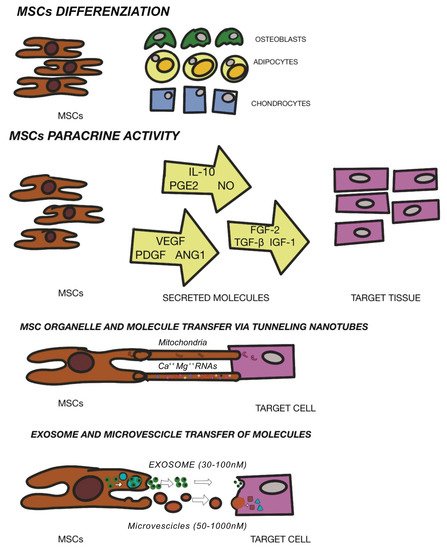
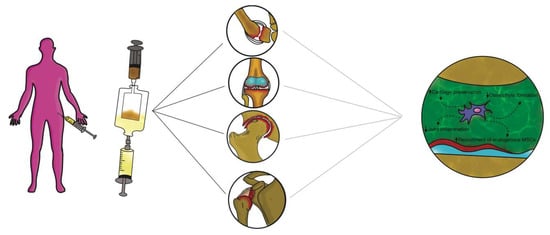
2.2. Mesenchymal Stem Cells Therapy

Different approaches have been performed as potential regenerative solution for osteochondral replacement: osteochondral autografts and allografts or autologous chondrocyte implantation [16]. Moreover, the clinical use of these techniques is limited by tissue availability, donor site morbidity and unsuccessful integration. In response to limitation with the use of cells in the osteochondral grafts, mesenchymal stem cells (MSCs) have been identified in the field of regenerative surgery. Mesenchymal stem cells (MSCs) are located in numerous tissues and may be defined as specialized precursor cells. MSCs are able to self-generate and, via relevant signals, may differentiate into different tissue-specific adult cells. In that way, MSCs will substitute aged or impaired cells [17]. MSCs form the tissues of the mesodermal line such as cartilage, bone and adipose tissue, as well as tissue such as the intervertebral disc, ligaments and muscles [18]. The International Society of Cellular Therapy [19] established a set of defining characteristics for MSCs which include the ability to adhere to plastic, expression of surface markers CD73, CD90, CD105 and a lack of hematopoietic markers CD34, Cd45, CD14, CD19. Moreover, characteristics would include tripotent differentiation into chondrogenic, osteogenic, and adipogenic phenotypes. In addition to their differentiating capacities, MSCs also represent noteworthy potential in regenerative medicine due to their anti-inflammatory and immunomodulatory potential [20]. MSCs are considered fundamental in tissue engineering since they are able to differentiate into terminal specialized cells, although currently, MSCs are exploited to “convince” the tissue or organ to self-regenerate. Regenerative medicine intends restoration principally via cell provision and specific stem cells that further enhance regeneration. It is therefore valid to define regenerative medicine as the restoration of human cells, tissue or organs to maintain regular functionality [21]. As aforementioned, the beneficial effects of MSCs are due to enhancement of both viability and proliferation of native cells, mitigation of cell death, delay cell senescence and anti-inflammatory and immunomodulatory effects. These reparative actions are obtained through MSC-secreting paracrine growth factors and cytokines, dynamic and direct cellular inter-communication along with extracellular vesicle release (defined exosomes)-containing peptides, mRNA and microRNAs [22][23][24]. The regulation of stem cell renewal and differentiation occur in the “niche” [25]. Multiple niches may be observed in different tissues [26] and stem cells contained here within have been utilized to repair cartilage. MSCs have been found in bone marrow [27], adipose tissue [28], dental pulp [29], umbilical cord tissue [30], but also in resident joint tissue such as the articular cartilage, synovium, periosteum, infrapatellar fat pad and trabecular bone [31]. The efficacy of stem cell treatment for OA has not yet been defined, but the secretion of anti-scarring (KGF, SDF1, MIP1a, MIP1b), anti-apoptotic (STC-1, SFRP2, TGFbeta1, HGF), angiogenic (VEGF), and mitogenic (TGF-a, TGF-b, HGF, IGF-1, FGF-2, EGF) factors may explain the natural repair mechanisms [32]. Other investigations have demonstrated a possible interaction between immune cells and MSCs as well as the potential to restrain propagation of inflammatory T cells and development of monocytes and the ability to impede B cell activity, which interfere with the underlying pathological or inflammatory process [33]. AI Caplan has proposed that the pericyte is released from its position in the vascular network in the case of a focal injury and have an immunomodulatory function. This immune modulation turns off T-cell surveillance of the injured tissue and thus provides a blockage of immune responses, while its trophic activity ensures that the field of damage is limited, that scarring does not occur and that tissue-intrinsic progenitors replace the expired cells. Angiogenesis occurs via MSC secretion of bioactive factors, such as VEGF, and via stabilization of newly forming vessels [34]. The new era of cell-mediated therapy (Figure 1) in the clinical trial database is promising. Multiple ongoing trials involving MSCs are evidence of the growing interest and viability of these cells. Further investigations are required to assess safety and subsequent efficacy with an urgency of broad diffusion of publications within the scientific communities to better understand therapeutic options.
Figure 1. Mechanism of MSC mediated repair. Modified from Fellows CR et al. Front Genet. 2016.

2.3. Intra-articular Application of Autologous Microfragmented Adipose Tissue with Stromal Vascular Fraction

Adipose stem cells (ASCs) are able to renew themselves and create multiple lineages [35]; besides, they can readily and rapidly expand in vitro, will not age easily and provoke fewer morbidities in patients [36]. ASCs have also displayed significant potential to propagate and differentiate into mesoderm-like tissue in relation to bone marrow derived- MSCs or other sources [37]. Importantly, ASCs are easily isolated and particularly accessible from subcutaneous adipose tissue [38][39][40]. ASCs have also shown efficient chondrogenic differentiation during in vitro expansion under adequate conditions [41]. In any case, physical and mechanical factors are required to perform an adequate formation of the cartilage tissue in vivo such as mechanical stimuli or a particular texture of the scaffolds [42][43]. The clinical use of ASCs is strictly regulated, because these products are considered “drugs” and therefore particularly restricted in clinical practice in Europe and the USA [44]. Such restrictions have led to novel studies regarding ASC alternative therapeutics considering “minimal manipulation” [45]. In particular, if ASCs are not expanded in vitro but extracted from the adipose tissue within the operating room without substantial manipulation and without use of collagenase, then the United States Food and Drug Administration (US FDA)/European Medicine Agency (EMA) allow such treatments [46]. Enzymatic tissue digestions are considered by the FDA (and EMA) as “substantial manipulations” and have accordingly imposed important restrictions. The issue of ASC “minimal manipulation” is considered during the isolation of several cells’ populations using mechanical processes to adhere to the regulations set by the FDA and EMA worldwide [47]. Additionally, alteration of biological, physiological, or structural features of cells or tissues is considered as important manipulation. The bone marrow aspirate concentration is an invasive procedure provoking donor morbidity, while the liposuction for obtaining SVF is a minimally invasive procedure [48]. Albeit efficient, the enzymatic digestion necessitates xenogenic substances that may cause immune reactions and is discordant with the European Good Manufacturing Practice (eGMP) Guidelines (Regulation (EC) No. 1394/2007 of the European Parliament and the European Council). To elude this problem, single devices have been adopted to separate and isolate SVF from adipose tissue [49][50]. Non-enzymatic methods to isolate SVF use mechanical or physical forces to manipulate the structural integrity of adipose tissue. These procedures are less specific and are sufficiently able to displace SVF cells from their own niche, and some authors have consequently introduced the concept of a stromal vascular niche [51][52]. The end product acquired via non-enzymatic digestion is not strictly cellular stromal vascular material, as would be generally acquired via enzymatic digestion, but a combination of cellular debris, blood cells, and components of ECM [53]. Moreover, the mechanical devices can preserve cells in clusters, or rather, in their native environment, which will aid in retaining cell function, including exosome discharge and secretion. The stromal vascular niche, therefore, protects the activated ASCs, enhancing their potency in the recipient environment, but also actuates a cascade of biological events that mimic the natural healing process. Non-enzymatic procedures have been proposed including mechanical dissociation of adipose tissue using closing devices and operator-dependent tools (Figure 2).
Figure 2. Non-enzymatic procedures have been proposed including mechanical dissociation of adipose tissue using automated closing devices and non-operator-dependent tools.
These devices differ from each other in the isolation protocol, in time and in the category of tissue dissociation, but also vary in the final SVF product. Non-enzymatic isolation methods are based on centrifugation force, pressure, filtration and washing. Mechanical systems commonly used to harvest and purify adipose tissue to obtain SVF are: Puregraft (Bimini Technologies LLC, Plano, Texas, USA), LipiVage (Genesis-Byosystems-Inc, Lewisville, Texas, USA), Lipogems (Lipogems Int Spa, Milan, Italy), Rigenera (HBW srl, Turin, Italy), Lipo-Kit GT (Medikan-International Inc, Seul, Korea), Hy-Tissue Nanofat (Fidia Farmaceutici, Abano Terme, Italy), Hy-Tissue SVF (Fidia Farmaceutici, Abano Terme, Italy), StromaCell (Micro-Aire-Surgical Instruments, Charlottesville, Virginia, USA), MyStem (MyStem LLC, Wilmington, NC, USA), Revolve (Life Cell Corporation, Branchburg, New Jersey, USA), Wal Body-Jet and Q-Graft system (Human Med AG, Schwerin, Germany), IntelliCell (Biosciences Inc, New York, NY, USA). Many of the devices reported have received evaluations in pre-clinical and clinical trials. Older systems are LipiVage and PureGraft, which were among the first products to be commercialized [54][55]. The LipiVage collection, washing and transfer technology is a device that allows collection of adipose grafts in controlled conditions with low vacuum, avoiding centrifugation or decantation. The lipoaspirate fat, inside the cannula, is separated from oils and fluids by an integrated filter in an extremely short time (15 min). In addition, fragmented adipose tissue from LipiVage showed no differences by normal adipose tissue, yielding large-sized grafts. However, an analysis of particles has not yet been conducted. The PureGraft technology is based on the filtration of adipose tissue through a particular membrane, an equally rapid procedure (15 min). In addition, grafts from PureGraft displayed larger particles (>1000 μm) and were able to operate a “dialysis” of the adipose tissue without resorting to other, more destructive methods such as centrifugation [56]. The main use of these technologies is in the field of fat grafting for breast volumes [57]. The most studied and commonly used system in clinical practice is the LIPOGEMS device. This technology is a closed device that allows collection of uniform products containing pericytes/ASCs with a slight mechanical force The end product is adipose tissue reduced into small fragments (600 / 400 μm), which progressively reduce in size and are without oil or blood residues, rich ASCs [58][59]. This device has been widely used especially in orthopedics for the treatment of tendinopathies and osteoarthritis [60][61]. Moreover, some authors have devised a “pure” system of mechanical disintegration [62] of tissues that is easy to use, less expensive and faster. This technology, called Rigenera micrografting technology, can disaggregate autologous tissue, with a calibrated size of 80 mm, collecting autologous micrografts enriched in progenitor cells, growth factors, and particles of ECM, by in vitro studies [45][63]. Some authors have performed comparative analyses between different mechanical and enzymatic systems. Raposio et al. [64][65] compared two procedures for isolation of ASCs, based on enzymatic + mechanical (centrifugation/vibrating plus collagenase) and mechanical (centrifugation or vibrating) methods. The authors showed that the enzymatic + mechanical procedure endorsed a major number of ASCs compared to the mechanical method alone. Indeed, Domenis et al. [66] showed that ASCs obtained from a mechanical device (Fastem kit) was less efficient in relation to the enzymatic tools (Lipo-kit and Celution). All three procedures, nevertheless, were able to maintain the amount of adipose tissue and thickness in the reconstructed breast. Additionally, Senesi et al. [67] showed good cell viability, CD markers expression, and differentiation potency of ASCs obtained from mechanical devices (Rigenera and Lipogems) compared to enzymatic digestion. Furthermore, the authors asserted that the mechanical methods acted differently on the release of the ASCs from the SVF perivascular niches. Only enzymatic digestion was able to acquire a “pure” cell population and ASCs could rapidly differentiate into all mesodermal lines. Of the two mechanical systems analyzed, only the micro-grafts obtained by Rigenera (compared to Lipogems) were able to differentiate into all mesodermal lines, albeit more slowly than by enzymatic digestion. Some authors have studied a new promising device (Hy-Tissue SVF) that allow the stromal vascular fraction to isolate in the form of free cells and micro-fragments (30 / 70 μm) of connective tissue containing stromal cells and extracellular matrix [68]. This system is able to disaggregate autologous adipose tissue using a double bag with an inner filter bag of 120 μm mesh by using a small plastic rod. The main structural and morphological unit, the adipose niche, is maintained after disintegration and protects the activated ASCs, strengthening their effectiveness in the receiving environment. This is the main difference between this system and the others, because the preservation of the adipose structural niches increases the effectiveness of the ASCs. In addition, the elimination of enzymatic action will reduce tissue trauma while maintaining cellular integrity. The reduction in the size of adipose clusters favors engraftment because of a more convenient, more effective, and rapid revascularization of the micrograft owing to the interaction with the receiving vascular microenvironment.

2.4. Exosome and Extracellular Vescicles (EVs)

Studies have revealed that MSCs are able to modulate the gene expression of the surrounding cells through miRNA secretion and provide relevant exosome involvement in the benefits of MSC-based therapy [69]. Exosomes are extracellular vesicles with a diameter measurement ranging from 30 to 150 nm. In the course of multi-vesicular body development, inward budding of endosomal membranes is observed which contributes to the fundamental inter-cell communication. The multi-vesicular body endosomes fuse with the cell membrane leading to secretion of exosomes [70][71]. Most of the MSC paracrine factors are crucial to tissue regeneration and lined to the discharge of EVs. MSC exosomes (Figure 3) originate in adipose tissue, the bone marrow and other tissues and bear a rich and complex load of nucleic acid (mRNA and miRNA), proteins and lipids [72].
Figure 3. Different modes of action of exosomes.
In OA, the in vitro studies revealed chondroprotective and anti-inflammatory functions of exosomes, as observed in chondrocyte models [73]. Moreover, in various studies, the significance of exosomes has been demonstrated as regards the benefits of MSC-based therapies in treating cartilage lesions and OA. Outcomes in pre-clinical trials have shown efficacy of MSC exosomes in cartilage repair and renewal, enhancing chondrocytes to amalgamate type II collagen and reduce production and expression of ADAMTS-5. Such a development will ensure ECM [74] as well as boost restoration of cartilage via paracrine signaling mechanisms along with secretion of soluble trophic factors [75]; reduction in inflammatory markers (iNOS) [76] and downregulation of inflammatory signals by secretion of IL-1, IL-6, IL8, MMP-1 and MMP-13 [77]. EVs that are isolated from human ASCs use various chondroprotective mechanisms to reduce inflammatory mediators (TNF-alpha, IL-6, PGE2, NO) and minimize MMP activity therefore enhancing generation of the anti-inflammatory cytokine IL-10 [78]. Many authors demonstrated, also, that exosomes from ASCs could inhibit and defer cartilage deterioration in OA models through the suppression of catabolic molecules [79] and through the immunoregulatory stimuli of hyaluronan [80]. These pioneering findings have consolidated the positive outcomes of ASC-derived EVs as a new therapeutic alternative for OA. Adequate investigations are scarce, especially considering these therapeutic options as authentic products of mechanical digestion activated by the various available medical devices. In any case, a detailed analysis to investigate the functions and mechanisms of exosomes in clinical practice is urgently required taking into account the positive outcomes of preclinical studies. In clinical trials, therapy based on exosomes derived from ASCs should aim to optimize criteria involving exosome concentration and dosage, with injection times and intervals. Additionally, the immune response in individuals is to be assessed following exosome administration.

References

  1. Apostu, D.; Lucaciu, O.; Mester, A.; Oltean-Dan, D.; Baciut, M.; Baciut, G.; Bran, S.; Onisor, F.; Piciu, A.; Pasca, R.D.; et al. Systemic drugs with impact on osteoarthritis. Drug Metab. Rev. 2019, 51, 498–523.
  2. Apostu, D.; Lucaciu, O.; Mester, A.; Oltean-Dan, D.; Gheban, D.; Benea, H.R.C. Tibolone, alendronate, and simvastatin enhance implant osseointegration in a preclinical in vivo model. Clin. Oral Implant. Res. 2020, 31, 655–668.
  3. Everts, P.; Onishi, K.; Jayaram, P.; Lana, J.F.; Mautner, K. Platelet-rich plasma: New performance understandings and therapeutic considerations in 2020. IJMS 2020, 21, 7794.
  4. Foster, T.E.; Puskas, B.L.; Mandelbaum, B.R.; Gerhardt, M.B.; Rodeo, S.A. Platelet-rich plasma: From basic science to clinical applications. Am. J. Sports Med. 2009, 37, 2259–2272.
  5. Russell, R.P.; Apostolakos, J.; Hirose, T.; Cote, M.P.; Mazzocca, A.D. Variability of platelet-rich plasma preparations. Sports Med. Arthrosc. Rev. 2013, 21, 186–190.
  6. Xu, Z.; Yin, W.; Zhang, Y. Comparative evaluation of leukocyte and platelet-rich plasma and pure platelet-rich plasma for cartilage regeneration. Sci. Rep. 2017, 7, 43301.
  7. Jiang, G.; Wu, Y.; Meng, J.; Wu, F.; Li, S.; Lin, M.; Gao, X.; Hong, J.; Chen, W.; Yan, S.; et al. Comparison of leukocyte-rich platelet -rich plasma and leukocyte-poor platelet-rich plasma on Achilles Tendinopathy at an early stage in a rabbit model. Am. J. Sports Med. 2020, 48, 1189–1199.
  8. Kobayashi, Y.; Saita, Y.; Nishio, H.; Ikeda, H.; Takazawa, Y.; Nagao, M.; Takaku, T.; Komatsu, N.; Kaneko, K. Leukocyte concentration and composition in platelet-rich plasma (PRP) influences the growth factor and protease concentrations. J. Orthop. Sci. 2016, 21, 683–689.
  9. Kenmochi, M. Clinical outcomes following injections of leukocyte-rich platelet-rich plasma in osteoarthritis patients. J. Orthop. 2020, 18, 143–149.
  10. Marmotti, A.; Rossi, R.; Castoldi, F.; Roveda, E.; Michielon, G.; Peretti, G.M. PRP and articular cartilage: A clinical update. Biomed. Res. Int. 2015, 2015, 542502.
  11. Mariani, E.; Canella, V.; Cattini, L.; Kon, E.; Marcacci, M.; Di Matteo, B.; Pulsatelli, L.; Filardo, G. Leukocyte-rich platelet-rich plasma injections do not up-modulate intra-articular pro-inflammatory cytokines in the osteoarthritic knee. PLoS ONE 2016, 11, e015613753V.
  12. Shen, L.; Yuan, T.; Chen, S.; Xie, X.; Zhang, C. The temporal effect of platelet-rich plasma on pain and physical function in the treatment of knee osteoarthritis: Systematic review and meta-analysis of randomized controlled trials. J. Orthop. Surg. Res. 2017, 12, 16.
  13. Paterson, K.L.; Hunter, D.J.; Metcalf, B.R.; Eyles, J.; Duong, V.; Kazsa, J.; Wang, Y.; Buchbinder, R.; Cicuttini, F.; Forbes, A.; et al. Efficacy of intra-articular injections of platelet-rich plasma as a symptom- and disease-modifying treatment for knee osteoarthritis–the RESTORE trial protocol. BMC Musculoskelet. Disord. 2018, 19, 272.
  14. Delgado, D.; Garate, A.; Vincent, H.; Bilbao, A.M.; Patel, R.; Fiz, N.; Sampson, S.; Sanchez, M. Current concepts in intraosseous platelet-rich plasma injections for knee osteoarthritis. J. Clin. Orthop. Trauma. 2019, 10, 36–41.
  15. Sanchez, M.; Delgado, D.; Pompei, O.; Perez, J.C.; Sanchez, P.; Garate, A.; Bilbao, A.M.; Fiz, N.; Padilla, S. Treating severe knee osteoarthritis with combination of intra-osseous and intra-articular infiltrations of platelet-rich plasma: An observational study. Cartilage 2019, 10, 245–253.
  16. Vyas, C.; Mishbak, H.; Cooper, G.; Peach, C.; Pereira, R.F.; Bartolo, P. Biological perspectives and current biofabrication strategies in osteochondral tissue engineering. Biomanuf. Rev. 2020, 5, 2.
  17. Bianco, P. “Mesenchymal” Stem Cells. Annu. Rev. Cell Dev. Biol. 2014, 30, 677–704.
  18. Pittenger, M.F.; Mackay, A.M.; Beck, S.C.; Jaiswal, R.K.; Douglas, R.; Mosca, J.D.; Moorman, M.A.; Simonetti, D.W.; Craig, S.; Marshak, D.R. Multilineage potential of adult human mesenchymal stem cells. Science 1999, 284, 143–147.
  19. Dominici, M.; Le Blanc, K.; Mueller, I.; Slaper-Cortenbach, I.; Marini, F.C.; Krause, D.S.; Deans, R.J.; Keating, A.; Prockop, D.J.; Horwitz, E.M. Minimal criteria for defining multipotent mesenchymal stromal cells. The international Society for Cellular Therapy position statement. Cytotherapy 2006, 8, 315–317.
  20. Pittenger, M.F.; Discher, D.E.; Peault, B.M.; Phinney, D.G.; Hare, J.M.; Caplan, A.I. Mesenchymal stem cell perspective: Cell biology to clinical progress. NPJ Regen. Med. 2019, 4, 22.
  21. Mason, C.; Dunnill, P. A brief definition of regenerative medicine. Regen. Med. 2008, 3, 1–5.
  22. Miceli, V.; Bulati, M.; Iannolo, G.; Zito, G.; Gallo, A.; Conaldi, P.G. Therapeutic properties of mesenchymal stromal/Stem Cells: The need of cell priming for cell-free therapies in regenerative medicine. Int. J. Mol. Sci. 2021, 22, 763.
  23. Nikfarjam, S.; Rezaie, J.; Zolbanin, N.M.; Jafari, R. Mesenchymal stem cell derived-exosomes: A modern approach in translational medicine. J. Transl. Med. 2020, 18, 449.
  24. Matula, Z.; Nemeth, A.; Lorincz, P.; Szepesi, A.; Brozik, A.; Buzas, E.I.; Low, P.; Nemet, K.; Uher, F.; Urban, V.S. The role of extracellular vesicle and tunneling nanotubes-mediated intercellular cross-talk between mesenchymal stem cells and human peripheral T cell. Stem Cell Dev. 2016, 25, 1818–1832.
  25. Matta, C.; Khademhosseini, A.; Mobasheri, A. Mesenchymal stem cells and their potential for microengineering the chondrocyte niche. EBioMedicine 2015, 2, 1560–1561.
  26. Jones, D.L.; Wagers, A.J. No place like home: Anatomy and function of the stem cell niche. Nat. Rev. Mol. Cell Biol. 2008, 9, 11–21.
  27. Charbord, P. Bone marrow mesenchymal stem cells: Historical overview and concepts. Hum. Gene Ther. 2010, 21, 1045–1056.
  28. De Francesco, F.; Ricci, G.; D’Andrea, F.; Nicoletti, G.F.; Ferraro, G.A. Human adipose stem cells: From bench to bedside. Tissue Eng. Part B Rev. 2015, 21, 572–584.
  29. La Noce, M.; Paino, F.; Spina, A.; Naddeo, P.; Montella, R.; Desiderio, V.; De Rosa, A.; Papaccio, G.; Tirino, V.; Laino, L. Dental pulp stem cells: State of the art and suggestions for a true translation of research into therapy. J. Dent. 2014, 42, 761–768.
  30. Mebarki, M.; Abadie, C.; Larghero, J.; Cras, A. Human umbilical cord-derived mesenchymal stem/stromal cells: A promising candidate for the development of advanced therapy medicinal products. Stem Cell Res. Ther. 2021, 12, 152.
  31. Fellows, C.R.; Matta, C.; Zakany, R.; Khan, I.M.; Mobasheri, A. Adipose, bone marrow and synovial joint-derived mesenchymal stem cells for cartilage repair. Front. Genet. 2016, 7, 213.
  32. Harrel, C.R.; Markovic, B.S.; Fellabaum, C.; Arsenijevic, A.; Volarevic, V. Mesenchymal stem cell-based therapy of osteoarthritis: Current knowledge and future perspectives. Biomed. Pharmacother. 2019, 109, 2318–2326.
  33. Glenn, J.D.; Whartenby, K.A. Mesenchymal stem cells: Emerging mechanisms of immunomodulation and therapy. World J. Stem Cells 2014, 6, 526–539.
  34. De Francesco, F.; Tirino, V.; Desiderio, V.; Ferraro, G.; D’Andrea, F.; Giuliano, M.; Libondi, G.; Pirozzi, G.; De Rosa, A.; Papaccio, G. Human CD34/CD90 ASCs are capable of growing as sphere clusters, producing high levels of VEGF and forming capillaries. PLoS ONE 2009, 4, e6537.
  35. Zuk, P.A.; Zhu, M.; Mizuno, H.; Huang, J.; Futrell, J.W.; Katz, A.J.; Benhaim, P.; Lorenz, H.P.; Hedrick, M.H. Multilineage cells from human adipose tissue: Implications for cell-based therapies. Tissue Eng. 2001, 7, 211–228.
  36. Palumbo, P.; Lombardi, F.; Siragusa, G.; Cifone, M.G.; Cinque, B.; Giuliani, M. Methods of isolation, characterization and expansion of human adipose-derived stem cells (ASCs): An overview. Int. J. Mol. Sci. 2018, 19, 1897.
  37. Strioga, M.; Viswanathan, S.; Darinskas, A.; Slaby, O.; Michalek, J. Same or not the same? Comparison of adipose tissue-derived versus bone marrow-derived m esenchymal stem and stromal cells. Stem Cells Dev. 2012, 21, 2724–2752.
  38. Ferraro, G.A.; De Francesco, F.; Nicoletti, G.; Paino, F.; Desiderio, V.; Tirino, V.; D’Andrea, F. Human adipose CD34+CD90+ stem cells and collagen scaffold constructs grafted in vivo fabricate loose connective and adipose tissue. J. Cell Biochem. 2013, 114, 1039–1049.
  39. D’Andrea, F.; De Francesco, F.; Ferraro, G.A.; Desiderio, V.; Tirino, V.; De Rosa, A.; Papaccio, G. Large-scale production of human adipose tissue from stem cells: A new tool for regenerative medicine and tissue banking. Tissue Eng. Part C Methods 2008, 14, 233–242.
  40. Nicoletti, G.F.; De Francesco, F.; D’Andrea, F.; Ferraro, G.A. Methods and procedures in adipose stem cells: State of the art and perspective for translation medicine. J. Cell Physiol. 2015, 230, 489–495.
  41. Pagani, S.; Veronesi, F.; Giavaresi, G.; Filardo, G.; Papio, T.; Romandini, I.; Fini, M. Autologous protein soluction effect on chondrogenic differentiation of mesenchymal stem cells from adipose tissue and bone marrow in an osteoarthritic environment. Cartilage 2021, 15, 1947603521993217.
  42. Gaut, C.; Sugaya, K. Critical review on the physical and mechanical factors involved in tissue engineering of cartilage. Regen. Med. 2015, 10, 665–679.
  43. Trumbull, A.; Subramanian, G.; Yildirim-Ayan, E. Mechanoresponsive musculoskeletal tissue differentiation of adipose-derived stem cells. Biomed. Eng. Online 2016, 15, 43.
  44. De Girolamo, L.; Lucarelli, E.; Alessandri, G.; Avanzini, M.A.; Bernardo, M.A.; Biagi, E.; Brini, A.T.; D’Amico, G.; Fagioli, F.; Ferrero, I.; et al. Mesenchymal Stem/Stromal Cells: A new “cells as drugs” paradigm. Effic. Crit. Asp. Cell Ther. Curr. Pharm. Design 2013, 19, 13.
  45. De Francesco, F.; Mannucci, S.; Conti, G.; Dai Prè, E.; Sbarbati, A.; Riccio, M. A Non-enzymatic method to obtain a fat tissue derivatite highly enriched in adipose stem cells (ASCs) from human lipoaspirates: Preliminary results. Int. J. Mol. Sci. 2018, 19, 2061.
  46. Yano, K.; Speidel, A.T.; Yamato, M. Four Food and Drug Administration draft guidance documents and the REGROW Act: A litmus test for future changes in human cell- and tissue-based products regulatory policy in the United States? J. Tissue Eng. Regen. Med. 2018, 12, 1579–1593.
  47. Raposio, E.; Ciliberti, R.G. Clinical use of adipose-derived stem cells: European legislative issues. Ann. Med. Surg. 2017, 24, 61–64.
  48. Gentile, P.; Calabrese, C.; De Angelis, B.; Pizzicannella, J.; Kothari, A.; Garcovich, S. Impact of the different preparation methods to obtain human adipose-derived stromal vascular fraction cells (AD-SVFs) and human adipose-derived mesenchymal stem cells (AD-MSCs): Enzymatic digestion versus mechanical centrifugation. Int. J. Mol. Sci. 2019, 20, 5471.
  49. Oberbauer, E.; Steffenhagen, C.; Wurzer, C.; Gabriel, C.; Redl, H.; Wolbank, S. Enzymatic and non-enzymatic isolation systems for adipose tissue-derived cells: Current state of the art. Cell Regen. 2015, 4, 7.
  50. Aronowitz, J.A.; Lockhart, R.A.; Hakakian, C.S. Mechanical versus enzymatic isolation of stromal vascular fraction cells from adipose tissue. Springerplus 2015, 4, 713.
  51. Tremolada, C.; Colombo, C.; Ventura, C. Adipose tissue and mesenchymal stem cells: State of the art and lipogems technology development. Curr. Stem. Cell. Rep. 2016, 2, 304–312.
  52. Carelli, S.; Messaggio, F.; Canazza, A.; Hebda, D.M.; Caremoli, F.; Latorre, E.; Grimoldi, M.G.; Colli, M.; Bulfamante, G.; Tremolada, C.; et al. Characteristics and properties of mesenchymal stem cells derived from microfragmented adipose tissue. Cell Transplant. 2015, 24, 1233–1252.
  53. Condè-Green, A.; Kotamarti, V.S.; Sherman, L.S.; Keith, J.D.; Lee, E.S.; Granick, M.S.; Rameshwar, P. Shift toward mechanical isolation of adipose-derived stromal vascular fraction: Review of upcoming techniques. Plast. Reconstr. Surg. Global Open 2016, 4, e1017.
  54. Ferguson, R.E.H.; Cui, X.; Fink, B.F.; Vasconez, H.C.; Pu, L.L.Q. The viability of autologous fat grafts harvested with the LipiVage system: A comparative study. Ann. Plast. Surg. 2008, 60, 594–597.
  55. Zhu, M.; Cohen, S.R.; Hicok, K.C.; Shanahan, R.K.; Strem, B.M.; Yu, J.C.; Arm, D.M.; Fraser, J.K. Comparison of three different fat graft preparation methods: Gravity separation, centrifugation, and simultaneous washing with filtration in a closed system. Plast. Reconstr. Surg. 2013, 131, 873–880.
  56. Fang, C.; Patel, P.; Li, H.; Huang, L.T.; Wan, H.; Collins, S.; Connell, T.L.; Xu, H. Physical, biochemical, and biologic properties of fat graft processed via different methods. Plast. Reconstr. Surg. Global Open 2020, 8, e3010.
  57. De Fazio, D.; Cingozoglu, C.A.C. Combined mastopexy and augmentation with autologous fat grafting: First results with lipopexy. Plast. Reconstr. Surg. Global Open 2020, 8, e1957.
  58. Bianchi, F.; Maioli, M.; Leonardi, E.; Olivi, E.; Pasquinelli, G.; Valente, S.; Mendez, A.J.; Ricordi, C.; Raddaini, M.; Tremolada, C.; et al. A new nonenzymatic method and device to obtain a fat tissue derivative highly enriched in pericyte-like elements by mild mechanical forces from human lipoaspirates. Cell Transplant. 2013, 22, 2063–2077.
  59. Vezzani, B.; Shaw, I.; Lesme, H.; Yong, L.; Khan, N.; Tremolada, C.; Peault, B. Higher Perycite content and secretory activity of microfragmented human adipose tissue compared to enzymatically derived stromal vascular fraction. Stem Cells Transl. Med. 2018, 7, 876–886.
  60. Randelli, P.; Menon, A.; Ragone, V.; Creo, P.; Bergante, S.; Randelli, F.; De Girolamo, L.; Montrasio, U.A.; Banfi, G.; Cabitza, P.; et al. Lipogems product treatment increases the proliferation rate of human tendon stem cells without affecting their stemness and differentiation capability. Stem Cells Int. 2016, 2016, 4373410.
  61. Jones, I.A.; Wilson, M.; Togashi, R.; Han, B.; Mircheff, A.K.; Thomas Vangsness, C., Jr. A randomized, controlled study to evaluate the efficacy of intra-articular, autologous adipose tissue injections for the treatment of mild-to-moderate knee osteoarthritis compared to hyaluronic acid: A study protocol. BMC Musculoskelet. Disord. 2018, 19, 383.
  62. Trovato, L.; Monti, M.; Del Fante, C.; Cervio, M.; Lampinen, M.; Ambrosio, L.; Redi, C.A.; Perotti, C.; Kankuri, E.; Ambrosio, G.; et al. A New medical device rigeneracons allows to obtain viable micrografts from mechanical disaggregation of human tissues. J. Cell Physiol. 2015, 230, 2299–2303.
  63. Dai Prè, E.; Busato, A.; Mannucci, S.; Vurro, F.; De Francesco, F.; Riccio, V.; Solito, S.; Biswas, R.; Bernardi, P.; Riccio, M.; et al. In Vitro characterization of adipose stem cells non-enzymatically extracted from the thigh and abdomen. Int. J. Mol. Sci. 2020, 21, 3081.
  64. Raposio, E.; Caruana, G.; Petrella, M.; Bonomini, M.P.; Grieco, A. A standardized method of isolating adipose-derived stem cells for clinical application. Ann. Plast. Surg. 2016, 76, 124–126.
  65. Raposio, E.; Simonacci, F.; Perrotta, R.E. Adipose-derived stem cells: Comparison between two methods of isolation for clinical applications. Ann. Med. Surg. 2017, 20, 87–91.
  66. Domenis, R.; Lazzaro, L.; Calabrese, S.; Mangoni, D.; Gallelli, A.; Bourkoula, E.; Manini, I.; Bergamin, N.; Toffoletto, B.; Beltrami, C.A.; et al. Adipose tissue derived stem cells: In vitro and in vivo analysis of a standard and three commercially available cell-assisted lipotransfer techniques. Stem Cell Res. Ther. 2015, 6, 2.
  67. Senesi, L.; De Francesco, F.; Farinelli, L.; Manzotti, S.; Gagliardi, G.; Papalia, G.F.; Riccio, M.; Gigante, A. Mechanical and enzymatic procedures to isolate the stromal vascular fraction from adipose tissue: Preliminary results. Front. Cell Dev. Biol. 2019, 7, 88.
  68. Busato, A.; De Francesco, F.; Biswas, R.; Mannucci, S.; Conti, G.; Fracasso, G.; Conti, A.; Riccio, V.; Riccio, M.; Sbarbati, A. Simple and Rapid Non-enzymatic procedure allows the isolation of structurally preserved connective tissue micro-fragments enriched with SVF. Cells 2020, 10, 36.
  69. Yin, K.; Wang, S.; Zhao, R.C. Exosomes from mesenchymal stem/stromal cells: A new therapeutic paradigm. Biomark. Res. 2019, 7, 8.
  70. Isola, A.; Chen, S. Exosomes: The messengers of health and disease. Curr. Neuropharmacol. 2016, 15, 157–165.
  71. Colombo, M.; Raposo, G.; Thery, C. Biogenesis, secretion, and intercellular interactions of exosomes and other extracellular vesicles. Annu. Rev. Cell Dev. Biol. 2014, 30, 255–289.
  72. Kao, C.Y.; Papoutsakis, E.T. Extracellular vesicles: Exosomes, microparticles, their parts, and their targets to enable their biomanufacturing and clinical applications. Curr. Opin. Biotechnol. 2019, 60, 89–98.
  73. Cosenza, S.; Ruiz, M.; Toupet, K.; Jorgensen, C.; Noel, D. Mesenchymal stem cells derived exosomes and microparticles protect cartilage and bone from degradation in osteoarthritis. Sci. Rep. 2017, 7, 16214.
  74. Zhang, S.; Chu, W.C.; Lai, R.C.; Lim, S.K.; Hui, J.H.; Toh, W.S. Exosomes derived from human embryonic mesenchymal stem cells promote osteochondral regeneration. Osteoarthr. Cartil. 2016, 24, 2135–2140.
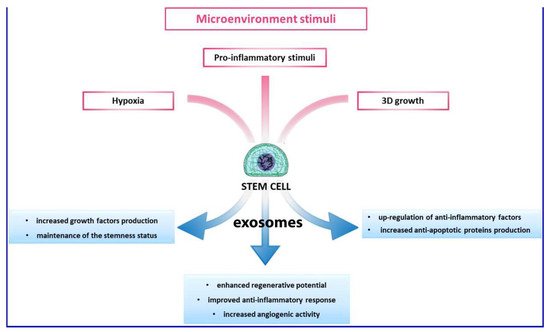
  75. Kusuma, G.D.; Carthew, J.; Lim, R.; Frith, J.E. Effect of the microenvironment on mesenchymal stem cell paracrine signaling: Opportunities to engineer the therapeutic effect. Stem Cells Dev. 2017, 9, 617–631.
  76. Mianehsaz, E.; Mirzaei, H.R.; Mahjoubin-Tehran, M.; Rezaee, A.; Sahebnasagn, R.; Pourhanifeh, M.H.; Mirzaei, H.; Hamblin, M.R. Mesenchymal stem cell-derived exosomes: A new therapeutic approach to osteoarthritis? Stem Cell Res. Ther. 2019, 10, 340.
  77. Ruiz, M.; Cosenza, S.; Maumus, M.; Jorgensen, C.; Noel, D. Therapeutic application of mesenchymal stem cells in osteoarthritis. Expert Opin. Biol. Ther. 2016, 16, 33–42.
  78. Tofino-Vian, M.; Guillen, M.I.; Perez Del Caz, M.D.; Castejon, M.A.; Alcaraz, M.J. Extracellular Vesicles from Adipose-Derived Mesenchymal Stem Cells Downregulate Senescence Features in Osteoarthritic Osteoblasts. Oxid. Med. Cell. Longev. 2017, 2017, 7197598.
  79. Woo, C.H.; Kim, H.K.; Jung, G.Y.; Jung, Y.J.; Lee, K.S.; Yun, Y.E.; Han, J.; Lee, J.; Kim, W.S.; Choi, J.S.; et al. Small extracellular vesicles from human adipose-derived stem cells attenuate cartilage degeneration. J. Extracell. Vesicles 2020, 9, 1735249.
  80. Ragni, E.; Perucca Orfei, C.; De Luca, P.; Lugano, G.; Viganò, M.; Colombini, A.; Valli, F.; Zacchetti, D.; Bollati, V.; De Girolamo, L. Interaction with hyaluronan matrix and miRNA cargo as contributors for in vitro potential of mesenchymal stem cell-derived extracellular vesicles in a model of human osteoarthritic synoviocytes. Stem Cell Res. Ther. 2019, 10, 109.
More
Information
Subjects: Surgery
Contributors MDPI registered users' name will be linked to their SciProfiles pages. To register with us, please refer to https://encyclopedia.pub/register : , , ,
View Times: 600
Revisions: 2 times (View History)
Update Date: 14 Jun 2022
1000/1000