Submitted Successfully!
To reward your contribution, here is a gift for you: A free trial for our video production service.
Thank you for your contribution! You can also upload a video entry or images related to this topic.
Version Summary Created by Modification Content Size Created at Operation
1 -- 4453 2022-05-26 11:55:50 |
2 language changed to English and deleted figures of copyright issues. -108 word(s) 4345 2022-05-27 07:57:31 | |
3 figure 11 deleted. -1 word(s) 4344 2022-06-02 07:56:41 | |
4 figures need copyright deleted. -104 word(s) 4240 2022-06-02 08:23:59 | |
5 typos corrected. + 2 word(s) 4242 2022-06-02 08:25:22 |

Video Upload Options

Do you have a full video?

Confirm

Are you sure to Delete?
Cite
If you have any further questions, please contact Encyclopedia Editorial Office.
Dongxu, Z.; Zhang, S.; , .; Zhang, L.; Wu, S. Receptor Targeted Molecular Imaging Probes for HCC Theranostics. Encyclopedia. Available online: https://encyclopedia.pub/entry/23418 (accessed on 25 April 2024).
Dongxu Z, Zhang S,  , Zhang L, Wu S. Receptor Targeted Molecular Imaging Probes for HCC Theranostics. Encyclopedia. Available at: https://encyclopedia.pub/entry/23418. Accessed April 25, 2024.
Dongxu, Zhao, Shaohua Zhang,  , Lei Zhang, Song Wu. "Receptor Targeted Molecular Imaging Probes for HCC Theranostics" Encyclopedia, https://encyclopedia.pub/entry/23418 (accessed April 25, 2024).
Dongxu, Z., Zhang, S., , ., Zhang, L., & Wu, S. (2022, May 26). Receptor Targeted Molecular Imaging Probes for HCC Theranostics. In Encyclopedia. https://encyclopedia.pub/entry/23418
Dongxu, Zhao, et al. "Receptor Targeted Molecular Imaging Probes for HCC Theranostics." Encyclopedia. Web. 26 May, 2022.
Receptor Targeted Molecular Imaging Probes for HCC Theranostics
Edit

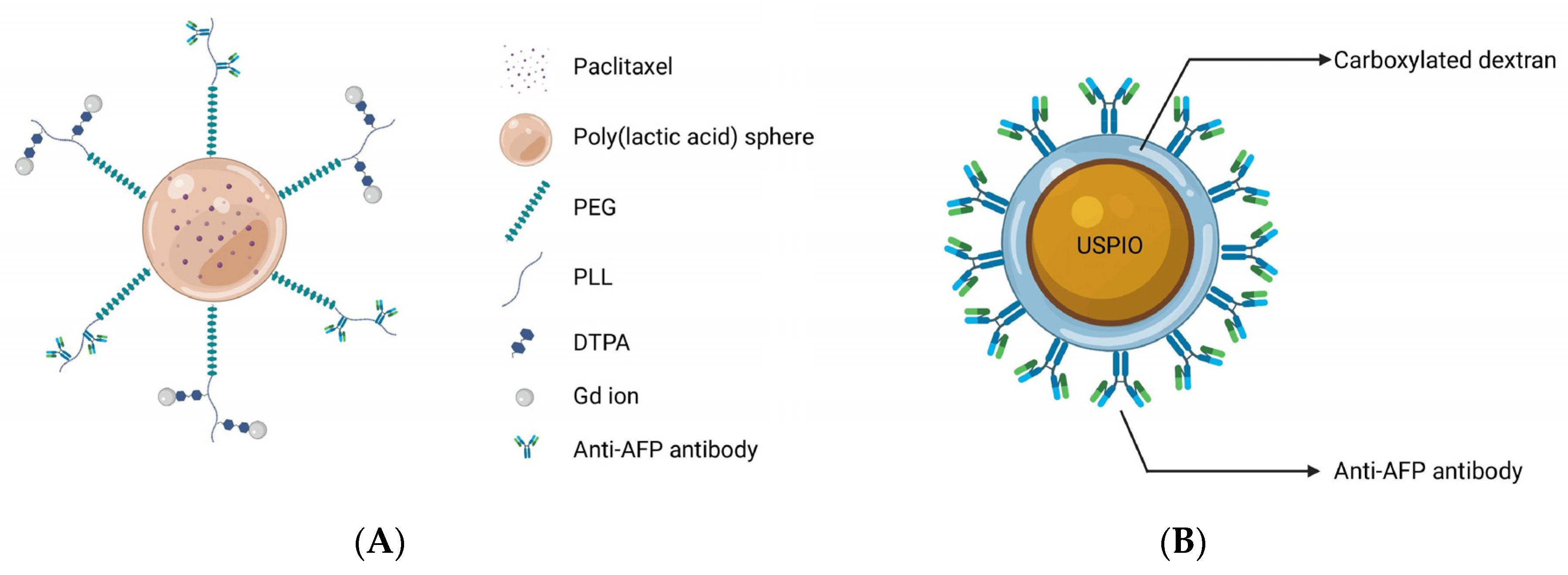
Hepatocellular carcinoma (HCC) is the sixth most commonly malignant tumor and the third leading cause of cancer-related death in the world, and the early diagnosis and treatment of patients with HCC is core in improving its prognosis. The early diagnosis of HCC depends largely on magnetic resonance imaging (MRI). MRI has good soft-tissue resolution, which is the international standard method for the diagnosis of HCC. However, MRI is still insufficient in the diagnosis of some early small HCCs and malignant nodules, resulting in false negative results. With the deepening of research on HCC, researchers have found many specific molecular biomarkers on the surface of HCC cells, which may assist in diagnosis and treatment. On the other hand, molecular imaging has progressed rapidly in recent years, especially in the field of cancer theranostics. Hence, the preparation of molecular imaging probes that can specifically target the biomarkers of HCC, combined with MRI testing in vivo, may achieve the theranostic purpose of HCC in the early stage.

molecular imaging probes magnetic resonance imaging nanomaterials hepatocellular carcinoma

1. Alpha-Fetoprotein

As one of the earliest tumor markers to be found, alpha-fetoprotein (AFP) has been routinely employed in a variety of Hepatocellular carcinoma (HCC) surveillance and detection procedures for decades [1]. Since 1964, Tatarinov et al. found a strong correlation between a high concentration of AFP and the diagnosis of HCC and further investigations demonstrated that AFP is a sensitive marker for HCC diagnosis, its efficacy evaluation, and prognosis [2][3]. AFPs can be used as molecular targets for improving diagnostic and therapeutic efficacy owing to its significant overexpression in HCC tumor cells compared to normal tissues. Liu and co-workers demonstrated the theranostic effects of polymeric micelles with the surface-linked biotinylated AFP antibodies with a biotin–avidin reaction in tumor-bearing Kunming mice after intravenous administration, and discovered that polymeric micelles demonstrated significantly higher signal intensity and a longer imaging duration in the tumor, owing to the specificity of AFP targeting effects and Gd-ion chelation with the micelles (Figure 1A) [4]. Additionally, tumors in the treated animals were greatly suppressed following therapy. Because AFP antibodies enhance the cellular uptake of micelles, chemotherapeutic agents encapsulated in the core of micelles are released to kill tumor cells. Li et al. [5] made a probe using anti-AFP antibodies, which was conjugated with carboxylated dextran-coated USPIONs. Each of the USPIOs can bind twelve antibodies, and this nanomaterial was highly efficient in HCC detection (Figure 1B). In addition, in their study of multimodal imaging, Chen and co-workers proved the feasibility of MRI/fluorescence dual-mode imaging by using AFP-modified fluorescent magnetic probes [6].
Figure 1. (A) The Gd ions used for MR imaging and the antibodies targeting AFP were connected to the outer surface of spherical micelles, and the paclitaxel were encapsulated in the core of micelles for treatment to achieve the purpose of theranostics; (B) Carboxylated dextran was used to modify the surface of USPIO directly. A single particle surface has a high coupling efficiency and can couple 12 anti-AFP ant.
Since AFP is widely expressed in HCC cells, gene imaging and treatments based on the AFP promoter are also reported in the literature. The ferritin gene, which is a reporter gene, was ligated with AFP promoter to construct the plasmid, which was delivered to the local area by a nanodrug delivery system. Hence, transferrin receptors were highly expressed in transfected HCC cells, resulting in intracellular iron deposition, which was conducive to the early detection of MRI [7][8][9]. Zhang and co-workers demonstrated that the ferritin gene was ligated with the AFP promoter for intracellular transfection by non-viral vectors for the first time. With these endogenous contrast agents, carried by a vector polyethyleneimine-β-cyclodextrin with high safety and stability, gene imaging was successfully achieved [7]. For MR images, the increased intracellular iron accumulation resulted in signal alterations in the lesion region. However, this trial was limited to the cell experiment, and the plasmid carrying the AFP promoter and ferritin gene was introduced into the cells only by transfection to achieve gene imaging. The actual imaging level in living animals remains to be further studied. In another study conducted by Lu, the endogenous imaging of the ferritin reporter gene was successfully verified in the orthotopic HCC model of living animals [8]. Researchers have constructed a nanostructured lipid vector, whose surface is modified by the A54 peptide, and the peptide can be specifically recognized and absorbed by the surface receptors of Bel-7402 cells. The lipid vector contained SPIONs and plasmids containing AFP promoter and ferritin reporter genes. Under the dual synergistic effects, the T2 value of the tumor was successfully reduced, and the sensitivity and specificity of HCC diagnosis were greatly improved.
In summary, AFP has been acknowledged in the diagnosis of HCC as the first and most extensively utilized tumor marker of HCC. Molecular imaging based on AFP also has significant promise, and when combined with several new carriers, it demonstrates excellent targeting for HCC lesions. Additionally, due to its widespread expression in HCC tissues, gene-level imaging and therapy based on the AFP promoter have also shown promising outcomes. However, while recent scientific advances are noteworthy and of interest, the sensitivity of targeting AFP still requires improvement [7]. As a result, additional research should be conducted to fill this gap.

2. Glypican-3

Glypican-3 (GPC-3) is a type of biomarker for HCC that has drawn attention because of its unique biochemistry and targeted properties [10][11][12][13][14]. GPC-3 is a membrane proteoglycan that is connected to the cell surface by a glycosylphosphatidylinositol anchor and belongs to the heparan sulphate proteoglycan family [15][16]. In recent years, GPC-3 has become a popular research topic due to its characteristics. GPC-3-based molecular imaging and treatment may be an efficient and potentially valuable method for treating HCC.
The exploration of GPC-3 for theranostic applications of HCC began in 1997. Hsu and co-workers described, for the first time, that GPC-3 mRNA was overexpressed in 74.8% of HCC tissues and proved its potential imaging and therapeutic value [17]. In 2001, Zhu et al. published similar findings, stating that the expression of GPC-3 mRNA was elevated in 83% of HCC tissues compared to nodules, liver cirrhosis tissues, and normal liver parenchyma [18]. Since then, researchers have directed their efforts to the prospective uses of GPC-3. GPC-3 monoclonal antibody, as a solution for targeted imaging, has been used in recent studies [19][20], and it can target the GPC-3 receptor expressed on HCC cell surfaces. James et al. reported the targeting ability of GPC-3 monoclonal antibody on GPC-3-expressing cells. Firstly, HepG2 cells with a high expression of GPC-3 were incubated with the biotin-conjugated GPC-3 antibodies. Then, streptavidin and NIR fluorophore were ligated on the surface-modified iron oxide nanoparticles, and streptavidin was successfully combined with the biotin on the GPC-3 antibody. Finally, the synthesized probes were incubated with cells, and dual modal imaging of near-infrared fluorescence and MR imaging were successfully achieved [19]. However, despite its strong affinity for GPC-3, the antibody’s large size may have adverse consequences, including inadequate imaging pharmacokinetics, poor tumor penetration, and higher immunogenicity [21][22]. Peptides may be another option for addressing these issues. Highly sensitive and specific peptides targeting GPC-3 can be screened using the phage-display peptide library [23]. Minimal molecular weight, simplicity of customization, and low scale-up costs are all benefits of peptide-based probes [24]. For example, by combining the strong paramagnetic properties of gadolinium ions with the excellent near infrared absorption properties of WS2, Song et al. synthesized an MRI and photoacoustic-imaging bimodal nanoprobe to provide an effective targeting specificity of tumor cells [25]. The generated nanoprobes were shown to be compatible with the physiological environment and to have no detectable toxicity both in vitro and in vivo, and outstanding imaging effects were observed in MRI and photoacoustic imaging. Furthermore, dual-modal imaging determined the macroscopic outline of the tumor and improved the effect of NIR-induced tumor ablation. Similarly, ligands screened using phage-display peptide library technology also showed excellent targeting and affinity [26]. Tian et al. modified the GPC-3 binding peptide (GBP) identified by phage display technology on the surface of a traditional Fe3O4 Core/Au shell nanocomplex (FANP), in which the photothermal effect was mediated by the Au shell and the MR imaging was mediated by Fe3O4 nanoparticles [26]. The experimental results showed that after intravenous administration, the GBP-FANP increased gradually in tumors. After 24 h, imaging showed that the local aggregation of GBP-FANP reached the peak and photothermal therapy was performed by laser. Compared with GPC-3-negative tumors, GPC-3-positive mice tumors were significantly inhibited, and no systemic toxicity was found, which proved the feasibility of this imaging method and treatment strategy.
According to previous research, GPC-3 has a greater sensitivity and specificity, as well as a higher expression level than AFP [27][28][29], with approximately 70% of HCCs detected with the high expression of GPC-3 [14]. Although AFP is widely used in clinical practice, there is still a high false-negative rate. Compared with AFP, the expression rate of GPC-3 in early HCC is significantly higher, especially in patients with tumor diameters of less than 3 cm [17]. Even in AFP-negative HCC patients, GPC-3 still can be detected with high expression [30]. Hence, due to its excellent specificity and sensitivity at early stage HCC, GPC-3 can be used to differentiate between benign and malignant hepatic nodules and in the early diagnosis of small lesions. He and co-workers proved for the first time the feasibility of the diagnosis and treatment of micro-HCC [31]. Their group created a unique organic–inorganic composite nanoprobe that was capable of dual-modal imaging (MRI/NIR-II) and non-invasive photothermal treatment. Deng et al. successfully synthesized a targeted probe FeSe2-PEG-Peptide coupled to GPC-3 for contrast-enhanced MRI and photoacoustic imaging. Nanoprobes can accurately judge a nodule whether it is HCC or liver cirrhosis [32]. In addition, since this material has excellent photothermal effects, photothermal treatment can be performed after targeted imaging.
GPC-3 can also be employed for high-efficiency-medication delivery due to its high expression rate. For instance, SPIONs and sorafenib were encapsulated in polymer micelles, which could trigger drug release through an intracellular reduction reaction and a change in the pH value. This dual-trigger mechanism ensured the precise release of drugs in situ [33]. To be more precise, endocytosis mediated by the GPC-3 receptor stimulates the entry of micelles into cells, and subsequent intracellular glutathione reduction reactions and changes in the pH value enhance the release of SPIONs and sorafenib from micelles. Furthermore, the release of SPIONs assists in noninvasive tumor identification and in the monitoring of in vivo drug administration by MRI.
Compared with simply using GPC-3 or AFP targets separately, dual-targeted imaging may enhance the early detection rate of HCC. Ma et.al successfully detected early stage malignant nodules with dual-targeted imaging [34]. In contrast to targeting AFP alone on the HCC cell surface, Ma et.al proposed that an AFP/GPC-3 double antibody-labeled probe which can target AFP and GPC-3 simultaneously could potentially increase the detection rate of HCC, and improve the efficacy in detecting heterogeneous micro malignant HCC tumors. In their study, the properties of targeting and the sensitivity of the dual-labeled probe were higher than single-labeled probes.
In summary, GPC-3 has attracted great interest in recent years due to its unique biochemical and targeted properties. The high expression of GPC-3 in tumor tissues makes it an ideal target for the imaging and treatment of HCC. Especially, in the diagnosis of tiny malignant nodules, GPC-3 showed higher sensitivity and specificity than AFP, and the experiments explored in this chapter also achieved ideal results. In terms of the corresponding ligands of GPC-3, researchers have designed a variety of ligands for selection such as antibodies, peptides and aptamers. Therefore, GPC-3 may be a molecular target with great potential in the future.

3. Folate Receptors

Folic acid is a vitamin that is required for cell growth. Folate receptors are primarily responsible for transporting folic acid within mammalian cells and tissues. Under physiologic conditions, the expression level of receptors in normal cells is relatively conservative [35]. Nevertheless, in malignant tumor tissues, rapid cell division increases the demand for folic acid, and the corresponding expression of receptors is significantly increased [36]. Thus, the surface modification of nanoparticles with folic acid may indicate significant potential for the development of a novel strategy with which to improve the efficiency of cancer diagnosis and treatment [37][38].
Compared with other ligands such as antibody-based targeting moieties, folic acid has a number of potential advantages as a targeting moiety, including ease of synthesis, low molecular weight, strong receptor affinity in tumor tissue, and outstanding stability and biocompatibility [39]. Hence, Folic acid has been covalently conjugated to anticancer drugs, dendrimers, polymers, and metallic compounds with modified surfaces for the detection and therapy of HCC [40]. For example, a folic-acid-functionalized gadolinium-loaded nanodroplet was synthesized as a dual-modal MRI/ultrasound contrast agent to target HCC cells [41]. The nanodroplets showed increased cellular absorption and selective accumulation in the tumor location, intensifying the MRI signal for tumor areas with a high r1 relaxation, which is even greater than the relaxation of the currently available clinical contrast agent (Gadovist). In another study, a folate-modified ultra-small magneto-gold nanoparticle was synthesized. This nanoparticle not only increased the imaging mode (quad-modality imaging), but also combined photothermal treatments for in situ tumors [42]. In vivo therapeutic experiments showed that folate-modified ultra-small magneto-gold nanoparticles exhibited high photothermal antitumor efficacy and reduced the tumor size compared with the control group. There are many similar experimental studies available about the targeting of folate receptors. The studies introduce the application of molecular imaging probes based on folic-acid surface modification in the diagnosis and treatment of HCC using the following four aspects: Targeted delivery of siRNA, pH-sensitive release, folate acid-modified polymer-based nanoparticles and folate acid-modified metal particles.
The targeting delivery of therapeutic drugs mediated by pH-sensitive release has also been reported in folic acid-based nanoparticles. For example, tumor pH-sensitive nanoformulated triptolide coated with folic acid for targeted drug delivery [43]. The developed nanomedicine’s physicochemical properties indicated that it was suitable for drug delivery applications due to its pH-dependent release. Additionally, in orthotopic mouse models, targeted drug delivery significantly reduced the tumor burden and improved survival without toxicity. However, combined imaging may be a better method to achieve theranostic effects than targeting drug release alone. Chi and colleagues effectively loaded the precursor medication, arsenic trioxide, into the pores of porous mesoporous silica nanoparticles, which include magnetic iron oxide particles for MR imaging in their core and generated the targeted nanoparticles. The surface modification of the as-obtained nanoparticle was accomplished by linking folic acid in order to achieve the dual function of imaging and targeted drug administration simultaneously [44]. The releasing curves demonstrate arsenite’s sensitive discharge in an acidic environment. Experiments in vivo with tumor-bearing mice revealed enhanced anticancer activity and exceptional imaging capability.

4. VEGF/VEGFR

VEGF is strongly expressed in tumor tissues and its expression has been found to be proportional to the degree of malignancy for tumors [45][46]. Additionally, the corresponding receptor is VEGFR, which is highly expressed in the majority of tumor cells as well as endothelial cells involved in tumor neovascularization [47]. The binding of receptors and ligands to activate downstream signals plays a crucial function in tumor angiogenesis, tumor tissue development, and invasion [48]. Hence, when building targeted probes, there are two options for targeting VEGF or VEGFR. One of the methods is targeting VEGF. For example, studies on probes targeting VEGF have been reported [49][50]. Liu et al. used the anti-VEGF antibody to modify polymeric particles containing gadolinium and Huang et al. synthesized the MRI-visible and VEGF targeted drug delivery system, both of which have shown the feasibility of targeting VEGF. All of them exhibited the ability to efficiently target HCC cells for early detection.
Similarly, as previously reported, VEGFR overexpresses in tumors and neovascularization [51]. Therefore, targeting VEGFR may be another promising strategy [52]. A specific magnetic imaging probe, based on PLL, and a connected paramagnetic substance, gadolinium with a DTPA chemical bond, when targeting VEGFR, successfully diagnosed HCC in the early stage [53]. A biotin–avidin reaction was used to bind VEGFR-antibodies to PLL. No obvious cytotoxicity was found in vitro, and the nanoparticles significantly increased the internalization rate of VEGFR-positive HepG2 cells. The tumor signal intensity and time of duration significantly increased in H22 mice subcutaneous tumor models. Similarly, Liu et al. synthesized a multifunctional pH-sensitive nanoparticle for the diagnosis and treatment of HCC [54]. Compared with the above synthesis process, Liu introduced pH-sensitive materials, of which gadolinium ions were connected to the external DTPA residue, and sorafenib was wrapped in nanoparticles for treatment. Drug release was achieved under the acidic condition of the tumor microenvironment. At a pH of 5.0, in vitro testing revealed that medication release might approach 99%, and for in vivo antitumor studies, compared with oral or intravenous sorafenib, these pH-sensitive materials have more obvious antitumor effects in mice bearing H22 tumors. It exhibited an improved resolution and a longer imaging period (more than 90 min) when used as a contrast agent in the diagnosis of tumor-bearing mice.

5. Integrin

Integrin is comprised of the following two subunits: the alpha and beta subunits. These heterodimeric cell surface receptors are strongly associated with malignant biological characteristics such as tumor angiogenesis, invasion, and metastasis through the mediation of cell adhesion and signal transmission [55][56][57]. Integrins are extensively expressed on neovascular endothelial cells and HCC tumor cells, whereas they are rarely found on the surface of normal hepatocytes [58][59]. As a result, integrin may be an appropriate target for the early stage detection and therapy of HCC. Integrin is a significant component of the cell-adhesion molecule family, consisting of several subtypes, and at present, the main integrin molecules used in the study of magnetic molecular imaging probes are integrin α6 and integrin αvβ3 [60][61][62][63][64][65][66][67][68].
Integrin α6 subunit can form integrin α6β1 subtype and α6β4 subtype with integrin β1 subunit or β4 subunit heterodimer. Most of them bind to extracellular matrix laminin and mediate adhesion between cells and between cells and the extracellular matrix [69][70][71]. Integrin α6 expression was shown to be considerably greater in early stage HCC tissues than in surrounding normal tissues [72][73], and was related to a worse prognosis and malignancy in previous investigations [74][75]. Hence, the overexpression of integrin α6 in early HCC has an extraordinarily high positive rate, making it a possible diagnostic biomarker for early HCC detection [60][68]. For example, Lin et al. obtained a peptide with a high affinity to integrin α6 by alanine scanning and linked it to Gd ions. An optimised MR probe, specific for integrin α6, was produced, which can detect small nodules (approximately 1 mm) in mice [60], indicating the possibility of using this integrin α6-targeted MR probe to identify HCC, particularly tiny malignant nodules.
Integrin αvβ3 is another commonly used imaging target and is composed of αv subunit and β3 subunit. The extracellular region of the α chain from integrin αvβ3 can specifically recognize arginine-glycine-aspartic acid (RGD) polypeptides. Moreover, integrin αvβ3 is highly expressed in various tumors, including HCC cells and the neovascular endothelial cells of tumors [76]. Hence, molecular imaging probes can include the RGD polypeptide, which has the potential for use as a target-imaging agent for HCC due to the high affinity of RGD polypeptides for integrin receptors [64]. Active-target T1 imaging of HCC tumors as small as 2.0 mm was carried out for the first time by Jia et al. [62]. A 2.0 mm tumor can be detected using RGD-modified Fe3O4 with T1 contrast enhancement. Before this trial, silica-coated superparamagnetic iron oxide core–shell nanoparticles connected with paramagnetic gadolinium complex and RGD peptide as ligands were successfully synthesized, and T1 and T2 weighted dual-modal imaging was achieved [66]. Chen et al. developed a novel dual-mode probe based on MR and NIRF imaging and demonstrated its viability in a nude mouse HCC model. The results indicated that it can likely improve the accuracy of liver-tumor identification and guiding during resection [67]. In the targeting delivery of siRNA or drugs, promising results were also reported. The use of RGD-modified polyethylene glycol-grafted polyethylenimine functionalized with SPIONs as a carrier for survivin siRNA administration was investigated. It has the potential to modify gene expression in the treatment of HCC and to identify the tumor in vivo as an effective MRI probe [64]. Shen and co-workers designed a novel dual-targeted nanoprobe loaded with doxorubicin. The as-obtained multi-functional nanoparticles with excellent biocompatibility showed tumor-specific accumulation behaviors and significant antitumor activity [65].
In summary, with developments in research, integrins have demonstrated a broad range of potential applications in tumor imaging and medication administration. The molecular targeting of integrins provides new ideas for the detection and treatment of HCCs. As this research is still in its early stage, efforts should be directed towards (1) the improvement of the selection of integrin-based imaging agents and drug carriers, (2) verifying the safety of human application, (3) identifying the optimization of imaging effects, and (4) exploring the pharmacokinetics of clinical drugs.

6. Endoglin (CD105)

Endoglin, also known as CD105, is an endothelial cell membrane glycoprotein that is highly expressed in the neovascularization of cancer cells including HCC [77][78], which was first discovered in 1990s and initially named as the 44G4 antigen [79][80]. Endoglin is a component of the transforming growth factor beta (TGF-β) receptor complex that plays a critical role in angiogenesis and vascular remodeling [81][82]. It is worth mentioning that growing tumor endothelial cells have higher endoglin expression than resting endothelial cells [78][83][84]. As a result, it might be a good target for imaging and treatment [80][81]. In recent studies, specific aptamers with a high affinity to CD105 were successfully screened by conducting an exponential enrichment analysis [81][85][86].
Based on endothelial glycoprotein targets, Zhou and his colleagues successfully developed a specific MRI/fluorescent imaging aptamer nanoprobe. The dual-mode nanoprobe exhibited MR and fluorescence-imaging capabilities, and was able to perform both MR imaging and fluorescence labelling simultaneously. Unfortunately, this experiment was only limited to the synthesis of probes, the optimization of experimental parameters and characterization, and was not further verified at the cellular and animal levels [85]. In the same year, Yan et al. screened a novel single-stranded DNA oligonucleotide-based aptamer by conducting an exponential enrichment analysis, and conjugated the aptamers to create an MR/optimal dual-targeted nanoprobe that successfully visualized orthotopic HCC tumors that were as small as 1–4 mm in diameter [86]. Moreover, the dual-modal probe showed excellent accuracy and potential for the edge delineation of invasive HCC and guiding tumor excision. Zhong et al. identified an aptamer that can bind to mouse endoglin molecular (m-END) specifically, by conducting an exponential enrichment analysis. On that basis, they used m-END as the targeted molecular, Fe3O4 as the magnetic material and prepared the imaging nanoprobes based on carboxymethyl chitosan nanoparticles (mEND-Fe3O4@CMCS) [81]. In vitro, this probe exhibited excellent biocompatibility and targeting ability, and during an in vivo experiment, local enhancement of the tumor lasted for more than 6 h compared with that before injection in HCC-bearing BALB/c mice.

7. Asialoglycoprotein Receptor

ASGP-R, a membrane-bound lectin [87], was suggested in a previous study to be implicated in the progression of cancer metastases [88]. ASGP-R expression is abnormally upregulated in HCC, and the receptor can bind to galactose selectively, initiating receptor-mediated endocytosis and facilitating galactose endocytosis into tumor cells [89][90][91]. ASGP-R is therefore considered as a desirable molecular target for theranostic development. By using galactosylated molecules as drug delivery vehicles or imaging media, it is possible to enhance the diagnostic and therapeutic effects of HCC.
Liang and colleagues conjugated NIRF to the surface of Fe3O4 and then modified it with galactose-containing lipids to create dual-mode imaging nanoparticles for HCC cells. By specifically targeting HCC cells overexpressing ASGP-R, the nanoparticles enabled precise imaging. The produced imaging probe exhibits outstanding biocompatibility and MRI/fluorescence performance, indicating that it has significant clinical application potential [92]. In another study, dual-modality imaging nanoparticles were prepared using gold nanoparticles for the inner core and loaded this with indocyanine green by coating polydopamine on the surface, and the shell was composed of modified lipids containing gadolinium acid and lactobionic acid that self-assemble on the outer surface. The nanoparticles successfully achieved MRI/CT dual modal imaging and targeted photothermal cytotoxicity. Particles internalized into cells were clearly observed in cell experiments, and the nanoparticles exhibited excellent NIR absorption in the region between 700 and 850 nm, and thus induced significant photothermal cytotoxicity [93]. In gene therapy, Cai et al. delivered microRNA-99a into HCC cells by targeting VEGF and ASGP-R targets simultaneously, which successfully inhibited HCC progression [94].

8. CD44

Cluster Determinant 44 (CD44) is a receptor that mediates endocytosis on the surface of liver-cell membranes [95], but the expression of CD44 receptors on HCC cell membranes can be increased significantly [96][97]. As a primary ligand with a high affinity for CD44, hyaluronic acid is particularly interesting for applications such as the tumor-targeted administration of imaging agents for the detection and treatment of HCC due to its biodegradability, biocompatibility and non-immunogenicity [98][99][100][101][102]. For instance, Yang et al. successfully developed theranostic glutathione-responsive micelles that encapsulated doxorubicin and SPIONs [100]. The in vitro drug-release data indicated that the micelles held the potential to release doxorubicin in response to reductant, which was validated by 100% doxorubicin release in the presence of 10 mM glutathione. Through the receptor-mediated mechanism between hyaluronic acid and the CD44 receptor, the micelles loaded with doxorubicin and SPIONs were internalized in HCC cells and exhibited a pronounced antitumor ability and excellent tumor-imaging potential. Wang and coworkers developed multifunctional nanoparticles modified with hyaluronic acid for dual-mode MR/CT imaging of HCC cells overexpressing CD44 receptors[98]. The as-obtained nanoparticles exhibited excellent water dispersibility, cytocompatibility, and stability. In both in vitro and in vivo cell experiments, orthotopically transplanted HCC tumors models exhibited satisfactory imaging characteristics.
An invasive but innovative approach to intraoperative transarterial infusion of imaging agents was also reported by Lee et al. [101]. The intraarterial infusion of Nd3+ doped nanoparticles combined with anti-CD44 monoclonal antibody can achieve MR and the real-time upconversion luminescence imaging of HCC in situ rat models, which is helpful for the intraoperative determination of surgical margins and the detection of small lesions.

References

  1. European Association for the Study of the Liver. EASL Clinical Practice Guidelines: Management of hepatocellular carcinoma. J. Hepatol. 2018, 69, 182–236.
  2. Tatarinov, Y.S.; Afanaseva, A.V.; Parfenova, L.F. Formation of serum proteins in human ontogeny. Fed. Proc. Transl. Suppl. 1964, 23, 437–440.
  3. Peterson, M.L.; Ma, C.; Spear, B.T. Zhx2 and Zbtb20: Novel regulators of postnatal alpha-fetoprotein repression and their potential role in gene reactivation during liver cancer. Semin. Cancer Biol. 2011, 21, 21–27.
  4. Liu, Y.; Li, J.; Liu, F.; Feng, L.; Yu, D.; Zhang, N. Theranostic Polymeric Micelles for the Diagnosis and Treatment of Hepatocellular Carcinoma. J. Biomed. Nanotechnol. 2015, 11, 613–622.
  5. Li, Y.W.; Chen, Z.G.; Zhao, Z.S.; Li, H.L.; Wang, J.C.; Zhang, Z.M. Preparation of magnetic resonance probes using one-pot method for detection of hepatocellular carcinoma. World J. Gastroenterol. 2015, 21, 4275–4283.
  6. Chen, Y.; Lu, J.; Yang, J.; Hao, K.; Li, M. Investigation of Alpha-Fetoprotein Antibody Modified Fluorescent Magnetic Probe on HepG2 Cell and Cancer Model Mouse. J. Nanosci. Nanotechnol. 2020, 20, 5147–5150.
  7. Zhang, Q.; Lu, Y.; Xu, X.; Li, S.; Du, Y.; Yu, R. MR molecular imaging of HCC employing a regulated ferritin gene carried by a modified polycation vector. Int. J. Nanomed. 2019, 14, 3189–3201.
  8. Lu, C.Y.; Ji, J.S.; Zhu, X.L.; Tang, P.F.; Zhang, Q.; Zhang, N.N.; Wang, Z.H.; Wang, X.J.; Chen, W.Q.; Hu, J.B.; et al. T2-Weighted Magnetic Resonance Imaging of Hepatic Tumor Guided by SPIO-Loaded Nanostructured Lipid Carriers and Ferritin Reporter Genes. ACS Appl. Mater. Interfaces 2017, 9, 35548–35561.
  9. Yuan, C.; An, Y.; Zhang, J.; Li, H.; Zhang, H.; Wang, L.; Zhang, D. Magnetic nanoparticles for targeted therapeutic gene delivery and magnetic-inducing heating on hepatoma. Nanotechnology 2014, 25, 345101.
  10. Capurro, M.; Wanless, I.R.; Sherman, M.; Deboer, G.; Shi, W.; Miyoshi, E.; Filmus, J. Glypican-3: A novel serum and histochemical marker for hepatocellular carcinoma. Gastroenterology 2003, 125, 89–97.
  11. Miao, H.L.; Pan, Z.J.; Lei, C.J.; Wen, J.Y.; Li, M.Y.; Liu, Z.K.; Qiu, Z.D.; Lin, M.Z.; Chen, N.P.; Chen, M. Knockdown of GPC3 inhibits the proliferation of Huh7 hepatocellular carcinoma cells through down-regulation of YAP. J. Cell. Biochem. 2013, 114, 625–631.
  12. Zittermann, S.I.; Capurro, M.I.; Shi, W.; Filmus, J. Soluble glypican 3 inhibits the growth of hepatocellular carcinoma in vitro and in vivo. Int. J. Cancer 2010, 126, 1291–1301.
  13. Sun, B.; Huang, Z.; Wang, B.; Yu, Y.; Lin, S.; Luo, L.; Wang, Y.; Huang, Z. Significance of Glypican-3 (GPC3) Expression in Hepatocellular Cancer Diagnosis. Med. Sci. Monit. 2017, 23, 850–855.
  14. Zhou, F.; Shang, W.; Yu, X.; Tian, J. Glypican-3: A promising biomarker for hepatocellular carcinoma diagnosis and treatment. Med. Res. Rev. 2018, 38, 741–767.
  15. Iglesias, B.V.; Centeno, G.; Pascuccelli, H.; Ward, F.; Peters, M.G.; Filmus, J.; Puricelli, L.; de Kier Joffé, E.B. Expression pattern of glypican-3 (GPC3) during human embryonic and fetal development. Histol. Histopathol. 2008, 23, 1333–1340.
  16. Filmus, J.; Capurro, M.; Rast, J. Glypicans. Genome Biol. 2008, 9, 224.
  17. Hsu, H.C.; Cheng, W.; Lai, P.L. Cloning and expression of a developmentally regulated transcript MXR7 in hepatocellular carcinoma: Biological significance and temporospatial distribution. Cancer Res. 1997, 57, 5179–5184.
  18. Zhu, Z.W.; Friess, H.; Wang, L.; Abou-Shady, M.; Zimmermann, A.; Lander, A.D.; Korc, M.; Kleeff, J.; Büchler, M.W. Enhanced glypican-3 expression differentiates the majority of hepatocellular carcinomas from benign hepatic disorders. Gut 2001, 48, 558–564.
  19. Park, J.O.; Stephen, Z.; Sun, C.; Veiseh, O.; Kievit, F.M.; Fang, C.; Leung, M.; Mok, H.; Zhang, M. Glypican-3 targeting of liver cancer cells using multifunctional nanoparticles. Mol. Imaging 2011, 10, 69–77.
  20. Li, Z.; Zeng, Y.; Zhang, D.; Wu, M.; Wu, L.; Huang, A.; Yang, H.; Liu, X.; Liu, J. Glypican-3 antibody functionalized Prussian blue nanoparticles for targeted MR imaging and photothermal therapy of hepatocellular carcinoma. J. Mater. Chem. B 2014, 2, 3686–3696.
  21. Primus, F.J.; Bennett, S.J.; Kim, E.E.; DeLand, F.H.; Zahn, M.C.; Goldenberg, D.M. Circulating immune complexes in cancer patients receiving goat radiolocalizing antibodies to carcinoembryonic antigen. Cancer Res. 1980, 40, 497–501.
  22. Milenic, D.E.; Detrick, B.; Reynolds, J.C.; Colcher, D. Characterization of primate antibody responses to administered murine monoclonal immunoglobulin. Int. J. Biol. Markers 1990, 5, 177–187.
  23. Hofmeister, L.H.; Lee, S.H.; Norlander, A.E.; Montaniel, K.R.; Chen, W.; Harrison, D.G.; Sung, H.J. Phage-display-guided nanocarrier targeting to atheroprone vasculature. ACS Nano 2015, 9, 4435–4446.
  24. Ye, Y.; Zhu, L.; Ma, Y.; Niu, G.; Chen, X. Synthesis and evaluation of new iRGD peptide analogs for tumor optical imaging. Bioorg. Med. Chem. Lett. 2011, 21, 1146–1150.
  25. Song, X.; Shang, W.; Peng, L.; Jiang, H.; Wang, K.; Fang, C.; Tian, J. Novel GPC3-binding WS2-Ga3+-PEG-peptide nanosheets for in vivo bimodal imaging-guided photothermal therapy. Nanomedicine 2018, 13, 1681–1693.
  26. Tian, R.; Zhu, L.; Qin, Z.; Wang, G.; Wang, J.; Zhang, H. Glypican-3 (GPC3) targeted Fe3O4 core/Au shell nanocomplex for fluorescence/MRI/photoacoustic imaging-guided tumor photothermal therapy. Biomater. Sci. 2019, 7, 5258–5269.
  27. Niu, Z.S.; Niu, X.J.; Wang, W.H.; Zhao, J. Latest developments in precancerous lesions of hepatocellular carcinoma. World J. Gastroenterol. 2016, 22, 3305–3314.
  28. Kim, H.; Park, Y.N. Role of biopsy sampling for diagnosis of early and progressed hepatocellular carcinoma. Best Pract. Res. Clin. Gastroenterol. 2014, 28, 813–829.
  29. Schütte, K.; Schulz, C.; Link, A.; Malfertheiner, P. Current biomarkers for hepatocellular carcinoma: Surveillance, diagnosis and prediction of prognosis. World J. Hepatol. 2015, 7, 139–149.
  30. Li, B.; Liu, H.; Shang, H.W.; Li, P.; Li, N.; Ding, H.G. Diagnostic value of glypican-3 in alpha fetoprotein negative hepatocellular carcinoma patients. Afr. Health Sci. 2013, 13, 703–709.
  31. He, L.; Zhang, Y.; Chen, J.; Liu, G.; Zhu, J.; Li, X.; Li, D.; Yang, Y.; Lee, C.S.; Shi, J.; et al. A multifunctional targeted nanoprobe with high NIR-II PAI/MRI performance for precise theranostics of orthotopic early-stage hepatocellular carcinoma. J. Mater. Chem. B 2021, 9, 8779–8792.
  32. Deng, H.; Shang, W.; Lu, G.; Guo, P.; Ai, T.; Fang, C.; Tian, J. Targeted and Multifunctional Technology for Identification between Hepatocellular Carcinoma and Liver Cirrhosis. ACS Appl. Mater. Interfaces 2019, 11, 14526–14537.
  33. Cai, M.; Li, B.; Lin, L.; Huang, J.; An, Y.; Huang, W.; Zhou, Z.; Wang, Y.; Shuai, X.; Zhu, K. A reduction and pH dual-sensitive nanodrug for targeted theranostics in hepatocellular carcinoma. Biomater. Sci. 2020, 8, 3485–3499.
  34. Ma, X.H.; Wang, S.; Liu, S.Y.; Chen, K.; Wu, Z.Y.; Li, D.F.; Mi, Y.T.; Hu, L.B.; Chen, Z.W.; Zhao, X.M. Development and in vitro study of a bi-specific magnetic resonance imaging molecular probe for hepatocellular carcinoma. World J. Gastroenterol. 2019, 25, 3030–3043.
  35. Yan, Y.; Yang, C.; Dai, G.; Zhang, Y.; Tu, G.; Li, Y.; Yang, L.; Shu, J. Folic Acid-Conjugated CuFeSe2 Nanoparticles for Targeted T2-Weighted Magnetic Resonance Imaging and Computed Tomography of Tumors In Vivo. Int. J. Nanomed. 2021, 16, 6429–6440.
  36. Ross, J.F.; Chaudhuri, P.K.; Ratnam, M. Differential regulation of folate receptor isoforms in normal and malignant tissues in vivo and in established cell lines. Physiologic and clinical implications. Cancer 1994, 73, 2432–2443.
  37. Ebrahimnejad, P.; Sodagar Taleghani, A.; Asare-Addo, K.; Nokhodchi, A. An updated review of folate-functionalized nanocarriers: A promising ligand in cancer. Drug Discov. Today 2022, 27, 471–489.
  38. Zhao, X.; Li, H.; Lee, R.J. Targeted drug delivery via folate receptors. Expert Opin. Drug Deliv. 2008, 5, 309–319.
  39. Bahrami, B.; Mohammadnia-Afrouzi, M.; Bakhshaei, P.; Yazdani, Y.; Ghalamfarsa, G.; Yousefi, M.; Sadreddini, S.; Jadidi-Niaragh, F.; Hojjat-Farsangi, M. Folate-conjugated nanoparticles as a potent therapeutic approach in targeted cancer therapy. Tumour Biol. 2015, 36, 5727–5742.
  40. Narmani, A.; Rezvani, M.; Farhood, B.; Darkhor, P.; Mohammadnejad, J.; Amini, B.; Refahi, S.; Abdi Goushbolagh, N. Folic acid functionalized nanoparticles as pharmaceutical carriers in drug delivery systems. Drug Dev. Res. 2019, 80, 404–424.
  41. Maghsoudinia, F.; Tavakoli, M.B.; Samani, R.K.; Hejazi, S.H.; Sobhani, T.; Mehradnia, F.; Mehrgardi, M.A. Folic acid-functionalized gadolinium-loaded phase transition nanodroplets for dual-modal ultrasound/magnetic resonance imaging of hepatocellular carcinoma. Talanta 2021, 228, 122245.
  42. Liu, J.; Wang, D.; Wang, G. Magnetic-gold theranostic nanoagent used for targeting quad modalities T1 & T2-MRI/CT/PA imaging and photothermal therapy of tumours. RSC Adv. 2021, 11, 18440–18447.
  43. Ling, D.; Xia, H.; Park, W.; Hackett, M.J.; Song, C.; Na, K.; Hui, K.M.; Hyeon, T. pH-sensitive nanoformulated triptolide as a targeted therapeutic strategy for hepatocellular carcinoma. ACS Nano 2014, 8, 8027–8039.
  44. Chi, X.; Zhang, R.; Zhao, T.; Gong, X.; Wei, R.; Yin, Z.; Lin, H.; Li, D.; Shan, H.; Gao, J. Targeted arsenite-loaded magnetic multifunctional nanoparticles for treatment of hepatocellular carcinoma. Nanotechnology 2019, 30, 175101.
  45. Zhou, Z.; Lu, Z.R. Molecular imaging of the tumor microenvironment. Adv. Drug Deliv. Rev. 2017, 113, 24–48.
  46. Wang, S.W.; Liu, S.C.; Sun, H.L.; Huang, T.Y.; Chan, C.H.; Yang, C.Y.; Yeh, H.I.; Huang, Y.L.; Chou, W.Y.; Lin, Y.M.; et al. CCL5/CCR5 axis induces vascular endothelial growth factor-mediated tumor angiogenesis in human osteosarcoma microenvironment. Carcinogenesis 2015, 36, 104–114.
  47. Pfister, N.T.; Fomin, V.; Regunath, K.; Zhou, J.Y.; Zhou, W.; Silwal-Pandit, L.; Freed-Pastor, W.A.; Laptenko, O.; Neo, S.P.; Bargonetti, J.; et al. Mutant p53 cooperates with the SWI/SNF chromatin remodeling complex to regulate VEGFR2 in breast cancer cells. Genes Dev. 2015, 29, 1298–1315.
  48. Morse, M.A.; Sun, W.; Kim, R.; He, A.R.; Abada, P.B.; Mynderse, M.; Finn, R.S. The Role of Angiogenesis in Hepatocellular Carcinoma. Clin. Cancer Res. 2019, 25, 912–920.
  49. Liu, Y.; Chen, Z.; Liu, C.; Yu, D.; Lu, Z.; Zhang, N. Gadolinium-loaded polymeric nanoparticles modified with Anti-VEGF as multifunctional MRI contrast agents for the diagnosis of liver cancer. Biomaterials 2011, 32, 5167–5176.
  50. Huang, H.; Li, Y.; Li, C.; Wang, Y.; Sun, Y.; Wang, J. A novel anti-VEGF targeting and MRI-visible smart drug delivery system for specific diagnosis and therapy of liver cancer. Macromol. Biosci. 2013, 13, 1358–1368.
  51. Liu, D.; Liu, F.; Liu, Z.; Wang, L.; Zhang, N. Tumor specific delivery and therapy by double-targeted nanostructured lipid carriers with anti-VEGFR-2 antibody. Mol. Pharm. 2011, 8, 2291–2301.
  52. Xu, S.; Olenyuk, B.Z.; Okamoto, C.T.; Hamm-Alvarez, S.F. Targeting receptor-mediated endocytotic pathways with nanoparticles: Rationale and advances. Adv. Drug Deliv. Rev. 2013, 65, 121–138.
  53. Liu, Y.; Wu, X.; Sun, X.; Wang, D.; Zhong, Y.; Jiang, D.; Wang, T.; Yu, D.; Zhang, N. Design, synthesis, and evaluation of VEGFR-targeted macromolecular MRI contrast agent based on biotin-avidin-specific binding. Int. J. Nanomed. 2017, 12, 5039–5052.
  54. Liu, Y.; Feng, L.; Liu, T.; Zhang, L.; Yao, Y.; Yu, D.; Wang, L.; Zhang, N. Multifunctional pH-sensitive polymeric nanoparticles for theranostics evaluated experimentally in cancer. Nanoscale 2014, 6, 3231–3242.
  55. Cabodi, S.; del Pilar Camacho-Leal, M.; Di Stefano, P.; Defilippi, P. Integrin signalling adaptors: Not only figurants in the cancer story. Nat. Rev. Cancer 2010, 10, 858–870.
  56. Takada, Y.; Ye, X.; Simon, S. The integrins. Genome Biol. 2007, 8, 215.
  57. Becchetti, A.; Arcangeli, A. Integrins and ion channels in cell migration: Implications for neuronal development, wound healing and metastatic spread. Adv. Exp. Med. Biol. 2010, 674, 107–123.
  58. Li, J.; Zhou, P.; Li, L.; Zhang, Y.; Shao, Y.; Tang, L.; Tian, S. Effects of Cationic Microbubble Carrying CD/TK Double Suicide Gene and αVβ3 Integrin Antibody in Human Hepatocellular Carcinoma HepG2 Cells. PLoS ONE 2016, 11, e0158592.
  59. Kumar, C.C. Integrin alpha v beta 3 as a therapeutic target for blocking tumor-induced angiogenesis. Curr. Drug Targets 2003, 4, 123–131.
  60. Lin, B.Q.; Zhang, W.B.; Zhao, J.; Zhou, X.H.; Li, Y.J.; Deng, J.; Zhao, Q.; Fu, G.; Xie, C.M.; Xu, Y.K.; et al. An Optimized Integrin α6-Targeted Magnetic Resonance Probe for Molecular Imaging of Hepatocellular Carcinoma in Mice. J. Hepatocell. Carcinoma 2021, 8, 645–656.
  61. Xu, Y.H.; Yang, J.; Meng, J.; Wang, H. Targeted MR Imaging Adopting T1-Weighted Ultra-Small Iron Oxide Nanoparticles for Early Hepatocellular Carcinoma: An in vitro and in vivo Study. Chin. Med. Sci. J. 2020, 35, 142–150.
  62. Jia, Z.; Song, L.; Zang, F.; Song, J.; Zhang, W.; Yan, C.; Xie, J.; Ma, Z.; Ma, M.; Teng, G.; et al. Active-target T1-weighted MR Imaging of Tiny Hepatic Tumor via RGD Modified Ultra-small Fe3O4 Nanoprobes. Theranostics 2016, 6, 1780–1791.
  63. Khatik, R.; Wang, Z.; Zhi, D.; Kiran, S.; Dwivedi, P.; Liang, G.; Qiu, B.; Yang, Q. Integrin alphavbeta3 Receptor Overexpressing on Tumor-Targeted Positive MRI-Guided Chemotherapy. ACS Appl. Mater. Interfaces 2020, 12, 163–176.
  64. Wu, C.; Gong, F.; Pang, P.; Shen, M.; Zhu, K.; Cheng, D.; Liu, Z.; Shan, H. An RGD-modified MRI-visible polymeric vector for targeted siRNA delivery to hepatocellular carcinoma in nude mice. PLoS ONE 2013, 8, e66416.
  65. Shen, J.M.; Li, X.X.; Fan, L.L.; Zhou, X.; Han, J.M.; Jia, M.K.; Wu, L.F.; Zhang, X.X.; Chen, J. Heterogeneous dimer peptide-conjugated polylysine dendrimer-Fe3O4 composite as a novel nanoscale molecular probe for early diagnosis and therapy in hepatocellular carcinoma. Int. J. Nanomed. 2017, 12, 1183–1200.
  66. Yang, H.; Zhuang, Y.; Sun, Y.; Dai, A.; Shi, X.; Wu, D.; Li, F.; Hu, H.; Yang, S. Targeted dual-contrast T1- and T2-weighted magnetic resonance imaging of tumors using multifunctional gadolinium-labeled superparamagnetic iron oxide nanoparticles. Biomaterials 2011, 32, 4584–4593.
  67. Chen, Q.; Shang, W.; Zeng, C.; Wang, K.; Liang, X.; Chi, C.; Liang, X.; Yang, J.; Fang, C.; Tian, J. Theranostic imaging of liver cancer using targeted optical/MRI dual-modal probes. Oncotarget 2017, 8, 32741–32751.
  68. Zhang, Y.; Zhao, J.; Cai, J.; Ye, J.C.; Xiao, Y.T.; Mei, Y.; Zeng, M.S.; Xie, C.M.; Jiang, Y.; Feng, G.K. Integrin alpha6-Targeted Magnetic Resonance Imaging of Hepatocellular Carcinoma in Mice. Mol. Imaging Biol. 2020, 22, 864–872.
  69. Brooks, D.L.; Schwab, L.P.; Krutilina, R.; Parke, D.N.; Sethuraman, A.; Hoogewijs, D.; Schörg, A.; Gotwald, L.; Fan, M.; Wenger, R.H.; et al. ITGA6 is directly regulated by hypoxia-inducible factors and enriches for cancer stem cell activity and invasion in metastatic breast cancer models. Mol. Cancer 2016, 15, 26.
  70. Kariya, Y.; Kariya, Y.; Gu, J. Roles of Integrin α6β4 Glycosylation in Cancer. Cancers 2017, 9, 79.
  71. Krebsbach, P.H.; Villa-Diaz, L.G. The Role of Integrin α6 (CD49f) in Stem Cells: More than a Conserved Biomarker. Stem Cells Dev. 2017, 26, 1090–1099.
  72. Feng, G.K.; Ye, J.C.; Zhang, W.G.; Mei, Y.; Zhou, C.; Xiao, Y.T.; Li, X.L.; Fan, W.; Wang, F.; Zeng, M.S. Integrin α6 targeted positron emission tomography imaging of hepatocellular carcinoma in mouse models. J. Control. Release 2019, 310, 11–21.
  73. Jiang, Y.; Sun, A.; Zhao, Y.; Ying, W.; Sun, H.; Yang, X.; Xing, B.; Sun, W.; Ren, L.; Hu, B.; et al. Proteomics identifies new therapeutic targets of early-stage hepatocellular carcinoma. Nature 2019, 567, 257–261.
  74. Ke, A.W.; Shi, G.M.; Zhou, J.; Huang, X.Y.; Shi, Y.H.; Ding, Z.B.; Wang, X.Y.; Devbhandari, R.P.; Fan, J. CD151 amplifies signaling by integrin α6β1 to PI3K and induces the epithelial-mesenchymal transition in HCC cells. Gastroenterology 2011, 140, 1629–1641.e1615.
  75. Ozaki, I.; Yamamoto, K.; Mizuta, T.; Kajihara, S.; Fukushima, N.; Setoguchi, Y.; Morito, F.; Sakai, T. Differential expression of laminin receptors in human hepatocellular carcinoma. Gut 1998, 43, 837–842.
  76. Ahmed, N.; Riley, C.; Rice, G.; Quinn, M. Role of integrin receptors for fibronectin, collagen and laminin in the regulation of ovarian carcinoma functions in response to a matrix microenvironment. Clin. Exp. Metastasis 2005, 22, 391–402.
  77. Yang, L.Y.; Lu, W.Q.; Huang, G.W.; Wang, W. Correlation between CD105 expression and postoperative recurrence and metastasis of hepatocellular carcinoma. BMC Cancer 2006, 6, 110.
  78. Dallas, N.A.; Samuel, S.; Xia, L.; Fan, F.; Gray, M.J.; Lim, S.J.; Ellis, L.M. Endoglin (CD105): A marker of tumor vasculature and potential target for therapy. Clin. Cancer Res. 2008, 14, 1931–1937.
  79. Gougos, A.; Letarte, M. Identification of a human endothelial cell antigen with monoclonal antibody 44G4 produced against a pre-B leukemic cell line. J. Immunol. 1988, 141, 1925–1933.
  80. Kasprzak, A.; Adamek, A. Role of Endoglin (CD105) in the Progression of Hepatocellular Carcinoma and Anti-Angiogenic Therapy. Int. J. Mol. Sci. 2018, 19, 3887.
  81. Zhong, L.; Zou, H.; Huang, Y.; Gong, W.; He, J.; Tan, J.; Lai, Z.; Li, Y.; Zhou, C.; Zhang, G.; et al. Magnetic Endoglin Aptamer Nanoprobe for Targeted Diagnosis of Solid Tumor. J. Biomed. Nanotechnol. 2019, 15, 352–362.
  82. Rosen, L.S.; Gordon, M.S.; Robert, F.; Matei, D.E. Endoglin for targeted cancer treatment. Curr. Oncol. Rep. 2014, 16, 365.
  83. Tang, S.L.; Bai, M.Y.; Wang, J.Y.; Hong, P.D. Development and application of micro-polysaccharide drug carriers incorporating doxorubicin and superparamagnetic iron oxide for bimodality treatment of hepatocellular carcinoma. Colloids Surf. B Biointerfaces 2017, 151, 304–313.
  84. Nassiri, F.; Cusimano, M.D.; Scheithauer, B.W.; Rotondo, F.; Fazio, A.; Yousef, G.M.; Syro, L.V.; Kovacs, K.; Lloyd, R.V. Endoglin (CD105): A review of its role in angiogenesis and tumor diagnosis, progression and therapy. AntiCancer Res. 2011, 31, 2283–2290.
  85. Zhou, Z.; Wang, Z.; Zhao, L.; Li, G. Preparation and properties of magnetic-fluorescent endoglin aptamer nanoprobe. In IOP Conference Series: Materials Science and Engineering; IOP Publishing: Bristol, UK, 2018; p. 022035.
  86. Yan, H.; Gao, X.; Zhang, Y.; Chang, W.; Li, J.; Li, X.; Du, Q.; Li, C. Imaging Tiny Hepatic Tumor Xenografts via Endoglin-Targeted Paramagnetic/Optical Nanoprobe. ACS Appl. Mater. Interfaces 2018, 10, 17047–17057.
  87. Ashwell, G.; Harford, J. Carbohydrate-specific receptors of the liver. Annu. Rev. Biochem. 1982, 51, 531–554.
  88. Ueno, S.; Mojic, M.; Ohashi, Y.; Higashi, N.; Hayakawa, Y.; Irimura, T. Asialoglycoprotein receptor promotes cancer metastasis by activating the EGFR-ERK pathway. Cancer Res. 2011, 71, 6419–6427.
  89. Kato, Y.; Onishi, H.; Machida, Y. Biological characteristics of lactosaminated N-succinyl-chitosan as a liver-specific drug carrier in mice. J. Control. Release 2001, 70, 295–307.
  90. Zhang, M.; Zhou, X.; Wang, B.; Yung, B.C.; Lee, L.J.; Ghoshal, K.; Lee, R.J. Lactosylated gramicidin-based lipid nanoparticles (Lac-GLN) for targeted delivery of anti-miR-155 to hepatocellular carcinoma. J. Control. Release 2013, 168, 251–261.
  91. Liu, H.; Wang, H.; Xu, Y.; Guo, R.; Wen, S.; Huang, Y.; Liu, W.; Shen, M.; Zhao, J.; Zhang, G.; et al. Lactobionic acid-modified dendrimer-entrapped gold nanoparticles for targeted computed tomography imaging of human hepatocellular carcinoma. ACS Appl. Mater. Interfaces 2014, 6, 6944–6953.
  92. Liang, J.; Zhang, X.; Miao, Y.; Li, J.; Gan, Y. Lipid-coated iron oxide nanoparticles for dual-modal imaging of hepatocellular carcinoma. Int. J. Nanomed. 2017, 12, 2033–2044.
  93. Zeng, Y.; Zhang, D.; Wu, M.; Liu, Y.; Zhang, X.; Li, L.; Li, Z.; Han, X.; Wei, X.; Liu, X. nanohybrid for MRI/CT imaging and photothermal therapy of hepatocellular carcinoma. ACS Appl. Mater. Interfaces 2014, 6, 14266–14277.
  94. Cai, C.; Xie, Y.; Wu, L.; Chen, X.; Liu, H.; Zhou, Y.; Zou, H.; Liu, D.; Zhao, Y.; Kong, X.; et al. PLGA-based dual targeted nanoparticles enhance miRNA transfection efficiency in hepatic carcinoma. Sci. Rep. 2017, 7, 46250.
  95. Saravanakumar, G.; Deepagan, V.G.; Jayakumar, R.; Park, J.H. Hyaluronic acid-based conjugates for tumor-targeted drug delivery and imaging. J. Biomed. Nanotechnol. 2014, 10, 17–31.
  96. Spadea, A.; Rios de la Rosa, J.M.; Tirella, A.; Ashford, M.B.; Williams, K.J.; Stratford, I.J.; Tirelli, N.; Mehibel, M. Evaluating the Efficiency of Hyaluronic Acid for Tumor Targeting via CD44. Mol. Pharm. 2019, 16, 2481–2493.
  97. Dhar, D.; Antonucci, L.; Nakagawa, H.; Kim, J.Y.; Glitzner, E.; Caruso, S.; Shalapour, S.; Yang, L.; Valasek, M.A.; Lee, S.; et al. Liver Cancer Initiation Requires p53 Inhibition by CD44-Enhanced Growth Factor Signaling. Cancer Cell 2018, 33, 1061–1077.e1066.
  98. Wang, R.; Luo, Y.; Yang, S.; Lin, J.; Gao, D.; Zhao, Y.; Liu, J.; Shi, X.; Wang, X. Hyaluronic acid-modified manganese-chelated dendrimer-entrapped gold nanoparticles for the targeted CT/MR dual-mode imaging of hepatocellular carcinoma. Sci. Rep. 2016, 6, 33844.
  99. Moon, M.; Thomas, R.G.; Heo, S.U.; Park, M.S.; Bae, W.K.; Heo, S.H.; Yim, N.Y.; Jeong, Y.Y. A Hyaluronic Acid-Conjugated Gadolinium Hepatocyte-Specific T1 Contrast Agent for Liver Magnetic Resonance Imaging. Mol. Imaging Biol. 2015, 17, 497–503.
  100. Yang, H.; Wang, N.; Mo, L.; Wu, M.; Yang, R.; Xu, X.; Huang, Y.; Lin, J.; Zhang, L.M.; Jiang, X. Reduction sensitive hyaluronan-SS-poly(epsilon-caprolactone) block copolymers as theranostic nanocarriers for tumor diagnosis and treatment. Mater. Sci. Eng. C Mater. Biol. Appl. 2019, 98, 9–18.
  101. Lee, J.; Gordon, A.C.; Kim, H.; Park, W.; Cho, S.; Lee, B.; Larson, A.C.; Rozhkova, E.A.; Kim, D.H. Targeted multimodal nano-reporters for pre-procedural MRI and intra-operative image-guidance. Biomaterials 2016, 109, 69–77.
  102. Zhang, F.; Jia, Y.; Zheng, X.; Shao, D.; Zhao, Y.; Wang, Z.; Dawulieti, J.; Liu, W.; Sun, M.; Sun, W.; et al. Janus nanocarrier-based co-delivery of doxorubicin and berberine weakens chemotherapy-exacerbated hepatocellular carcinoma recurrence. Acta Biomater. 2019, 100, 352–364.
More
Information
Contributors MDPI registered users' name will be linked to their SciProfiles pages. To register with us, please refer to https://encyclopedia.pub/register : , , , ,
View Times: 504
Entry Collection: Nuclear Magnetic Resonance
Revisions: 5 times (View History)
Update Date: 02 Jun 2022
1000/1000