Submitted Successfully!
To reward your contribution, here is a gift for you: A free trial for our video production service.
Thank you for your contribution! You can also upload a video entry or images related to this topic.
Version Summary Created by Modification Content Size Created at Operation
1 -- 4114 2022-05-18 11:22:45 |
2 format corrected. + 1 word(s) 4115 2022-05-18 12:09:52 |

Video Upload Options

Do you have a full video?

Confirm

Are you sure to Delete?
Cite
If you have any further questions, please contact Encyclopedia Editorial Office.
Zoladek, T.; Kamińska, J.; Soczewka, P.; Rzepnikowska, W. Yeast as a Model for VPS13-Dependent Neurodegenerative Diseases. Encyclopedia. Available online: https://encyclopedia.pub/entry/23061 (accessed on 03 May 2024).
Zoladek T, Kamińska J, Soczewka P, Rzepnikowska W. Yeast as a Model for VPS13-Dependent Neurodegenerative Diseases. Encyclopedia. Available at: https://encyclopedia.pub/entry/23061. Accessed May 03, 2024.
Zoladek, Teresa, Joanna Kamińska, Piotr Soczewka, Weronika Rzepnikowska. "Yeast as a Model for VPS13-Dependent Neurodegenerative Diseases" Encyclopedia, https://encyclopedia.pub/entry/23061 (accessed May 03, 2024).
Zoladek, T., Kamińska, J., Soczewka, P., & Rzepnikowska, W. (2022, May 18). Yeast as a Model for VPS13-Dependent Neurodegenerative Diseases. In Encyclopedia. https://encyclopedia.pub/entry/23061
Zoladek, Teresa, et al. "Yeast as a Model for VPS13-Dependent Neurodegenerative Diseases." Encyclopedia. Web. 18 May, 2022.
Yeast as a Model for VPS13-Dependent Neurodegenerative Diseases
Edit

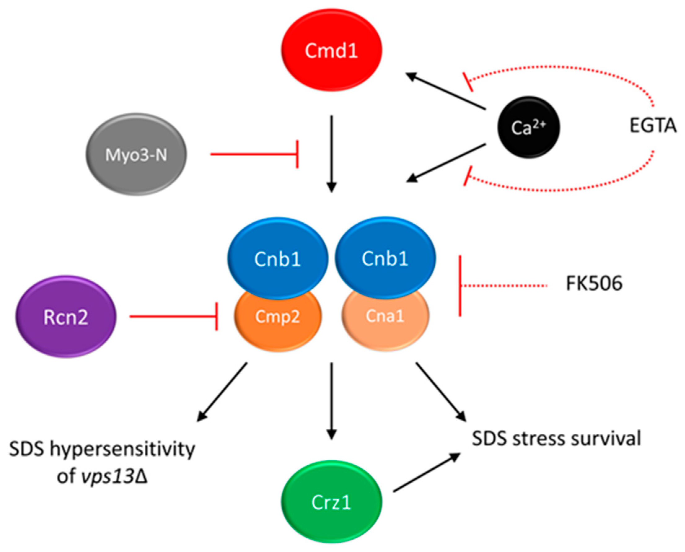
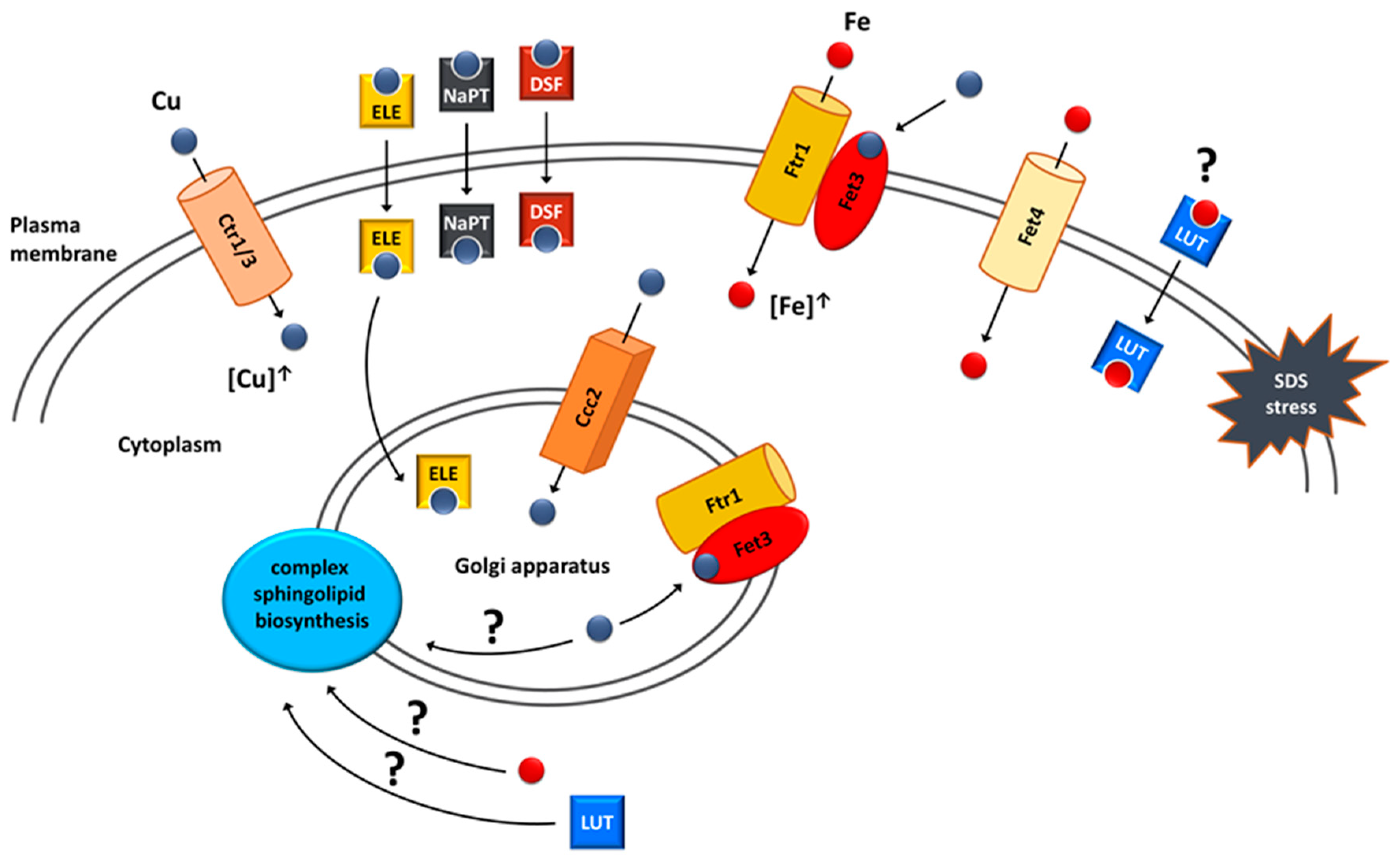
Mutations in human VPS13A-D genes result in rare neurological diseases, including chorea-acanthocytosis (ChAc). The pathogenesis of these diseases is poorly understood, and no effective treatment is available. As VPS13 genes are evolutionarily conserved, the effects of the pathogenic mutations could be studied in model organisms, including yeast, where one VPS13 gene is present. Here, the researchers summarize advancements obtained using yeast. In recent studies, vps13Δ and vps13-I2749 yeast mutants, which are models of chorea-acanthocytosis, were used to screen for multicopy and chemical suppressors. Two of the suppressors, a fragment of the MYO3 and RCN2 genes, act by downregulating calcineurin activity. In addition, vps13Δ suppression was achieved by using calcineurin inhibitors. The other group of multicopy suppressors were genes: FET4, encoding iron transporter, and CTR1, CTR3 and CCC2, encoding copper transporters. Mechanisms of their suppression rely on causing an increase in the intracellular iron content. Moreover, among the identified chemical suppressors were copper ionophores, which require a functional iron uptake system for activity, and flavonoids, which bind iron. These findings point at areas for further investigation in a higher eukaryotic model of VPS13-related diseases and to new therapeutic targets: calcium signalling and copper and iron homeostasis. Furthermore, the identified drugs are interesting candidates for drug repurposing for these diseases.

yeast chorea-acanthocytosis VPS13 VPS13A-D calcium signalling copper homeostasis iron homeostasis

1. Introduction

According to European Union law, a disease is considered rare when it affects no more than 1 person per 2000. However, with around 7000 identified rare diseases, together they affect 3.5–10% of the population, which corresponds to approximately 30 million people in Europe and 300 million around the world [1]. A multitude and variety of rare diseases pose huge diagnostic difficulties. It often takes several years from occurrence of symptoms to the correct diagnosis of a rare disease [2][3][4][5][6][7][8][9]. Despite recent advances, such as new-generation sequencing (NGS), the time and accuracy of diagnosis of rare diseases have not much improved. Another obstacle that the patients face is a great limitation of therapies. For significant majority of rare diseases current treatment is only symptomatic. There is a great need to find new drug targets and drugs for rare diseases. Drug targets can be easily identified in yeast models by genetic methods and drug librarires can be screend using yeast to find drugs for repurposing.

2. Calcium Signalling as a Potential Target for Drug Intervention in VPS13-Dependent Neurodegenerative Diseases

The knowledge about VPS13 proteins is still not sufficient to develop a specific therapy for patients. To date, gene therapy is also not available. Only symptomatic treatment is used, and new methods to alleviate the symptoms are needed. The most efforts for treatment development are reported for ChAc. The deep brain stimulation is used to relieve movement problems [10], botulinum toxin to treat dystonia and selected drugs for seizure control [11]. Based on the finding that Lyn kinase is hyperactivated in red blood cells of ChAc patients and neurons [12][13], the inhibitors of this kinase were also tested. As Lyn kinase inhibition represents a potential treatment in ChAc to restore some neuronal function [12], the trial to experimentally treat 3 patients was conducted. Although a partial restoration of the actin cytoskeleton was observed in red blood cells, the lack of improvement of neurological symptoms was noted after six-month drug administration [14]. Further, studies on mice models revealed that the drug tested, dasatinib, is not able to cross the blood–brain barrier, but another potential kinase inhibitor, nilotinib, can do that and improve not only haematological but also neurological defects in mice [15][16]. Since the variety of potential treatments is very limited, there is a great need for new drug candidates which could be effective in ChAc patients.
To find therapeutic alternatives based on the understanding of cell biology, using model organisms is necessary. Several yeast models of VPS13-dependent diseases were constructed, which show various phenotypes [17][18][19][20][21]. However, these phenotypes were not suitable for high-throughput screens. Finding novel growth phenotype of vps13, hypersensitivity to commonly used detergent sodium dodecyl sulfate (SDS), enable suppressor screens [22]. This gave better insight into the pathways which are disturbed in these cells which allowed for finding ways to overcome the observed defects.
Specifically, SDS hypersensitivity growth phenotype was used in a screen for multicopy suppressors of vps13-I2749R mutation, and several plasmids responsible for improved growth were isolated and analysed. One of them contained a fragment of the MYO3 gene (MYO3-N) encoding N-terminal part (amino acid residues (aa) 1–775) of Myo3 protein (Myo3-N) [22]. Myo3 is a type I myosin protein involved in actin cytoskeleton organisation and endocytosis [23][24]. MYO3-N overexpression was also improving growth of the vps13Δ mutant in the presence of SDS. Importantly, the suppression by MYO3-N was not limited to SDS-hypersensitivity. MYO3-N corrected two other vps13Δ defects: depolarisation of the actin cytoskeleton and hypersensitivity to canavanine, the phenotypes implying defective endocytosis. Indeed, endocytic reporters (Las17, Myo3, Myo5 and Abp1 tagged with fluorescent proteins) that localise to sites of endocytosis, were shown to be present longer on the plasma membrane in the vps13Δ cells than in the wild-type, indicating delayed endocytosis. MYO3-N overexpression shortened patch lifetimes for Las17, Myo3 and Myo5. To conclude MYO3-N overexpression could partially improve endocytosis in vps13Δ alleviating canavanine hypersensitivity [22].
In further analysis, the mechanism of MYO3-N action was elucidated. The suppressing MYO3-N fragment encodes myosin motor domain and a linker with two calmodulin binding motifs (IQ1 and IQ2). It is possible that suppression could be achieved by binding with calmodulin (Cmd1)—a conserved, calcium-binding protein mediating calcium signalling in cell [25]. To test this possibility, mutations disrupting both IQ motifs were introduced into MYO3-N and resulted myo3-iq1/2 was not able to suppress vps13Δ and the interaction between Myo3-iq1/2 and Cmd1 was abolished. These results indicated that binding of calmodulin to Myo3-N is necessary for MYO3-N-based suppression of vps13Δ [22]. The hypothesis that titrating of calmodulin by Myo3-N may result in lowering the activity of one of its downstream targets, calcineurin, was formulated. In yeast, calcineurin consists of one regulatory (Cnb1) and one of the two catalytic (Cna1 and Cmp2) subunits [26][27][28] (see Figure 1). In response to changes in calcium concentration, calcineurin regulates gene expression via its target, the Crz1 transcription factor [29][30]. In fact calcineurin activity is higher in the vps13Δ cells than in the wild-type, and MYO3-N overexpression reduces it (Figure 1) [22]. Remarkably, the Cnb1 subunit, which is essential for calcineurin activity, is required for MYO3-N-based suppression of vps13Δ. This indicates that the suppression mechanism of MYO3-N relies on downregulating calcineurin activity. However, calcineurin must not be shut off completely (Figure 1). This result resembles a study which showed that in a yeast model of PD, moderate calcineurin inhibition reduces α-synuclein toxicity, and deletion or overexpression of genes encoding calcineurin subunits exacerbate it [31].
Figure 1. The calmodulin–calcineurin pathway and its impact on yeast growth in the presence of SDS stress. Partial inhibition of calcineurin (Cnb1 + Cna1/Cmp2) activity suppresses the SDS-hypersensitivity of vps13Δ cells. Multicopy suppressors MYO3-N and RCN2 act by titrating calmodulin or blocking the activity of Cmp2 catalytic subunit, respectively. Chemical suppressors EGTA and FK-506 act by chelating the calcium ions required for calmodulin and calcineurin activities or inhibiting total calcineurin activity, respectively. As full inhibition of calcineurin activity is toxic in the presence of SDS stress, both chemical suppressors must be used in moderate concentration, indicated by dashed lines. Figure is based on results presented in Soczewka et al. 2019 and Wardaszka et al. 2021 [22][32].
The identification of the RCN2 gene as another suppressor of vps13Δ [32] is in line with the results described above. RCN2 encodes Rcn2 protein, a negative calcineurin regulator [33][34]. Interestingly, Rcn2 binds preferentially to the Cmp2 calcineurin catalytic subunit in comparison to the Cna1 catalytic subunit. Analysis indicated that the N-terminal fragment of Rcn2 is necessary to reduce calcineurin activity, maintain the Rcn2 interaction with Cmp2 and to improve growth of vps13Δ (Figure 1). Moreover, the deletion of CMP2 actually suppresses vps13Δ, contrary to CNA1 and previously shown CNB1 deletions, which negatively influence vps13Δ growth in the presence of SDS. The CNA1 and CNB1 are also required for vps13Δ suppression by RCN2 [32] as for MYO3-N, indicating that vps13Δ suppression is achieved by reducing calcineurin activity related to Cmp2 catalytic subunit (Figure 1), while the activity mediated by Cna1 is crucial for SDS stress survival. Similarly, in mammals catalytic calcineurin subunits are differently expressed and regulated [35][36][37][38]. Knowledge about the specificity of various calcineurin forms and their role in disease molecular pathologies may contribute to development of novel and specific calcineurin inhibitors. Such specific peptide inhibitors of calcineurin would allow calcineurin activity involved in a disease to be downregulated without disturbing calcineurin-related processes required for healthy organism functioning. Various peptide inhibitors of human calcineurin were already studied [39][40]. This approach could, at least partially, eliminate side effects of general calcineurin inhibitors, such as FK-506 immunosuppressant and cyclosporin A.
As partial reduction of calcineurin activity was responsible for the suppression, it is possible that vps13Δ SDS-hypersensitivity could be mitigated by pharmacological calcineurin inhibition. Indeed ethylene glycol tetraacetic acid (EGTA), which limits the calcium availability required for calmodulin and calcineurin activity, and FK-506 (Figure 1) improved the vps13Δ growth, showing that it is possible to achieve vps13Δ suppression in a pharmacological manner [22]. Moreover, FK-506 was active only in very low concentration, and higher concentration caused toxicity. This is in line with genetic experiments and shows that basal calcineurin activity is required for SDS protection.
The proper functioning of calcium signalling is crucial for cells. Especially in neurons, the concentration and storage of Ca2+ ions in specific compartments are precisely controlled. Dysregulation of Ca2+ signalling is observed in ageing neurons and neurons affected by neurodegenerative diseases such as Alzheimer's disease (AD), Parkinson's disease (PD) and Huntington disease (HD). Based on these findings, a hypothesis was formed that the dysregulation of Ca2+ signalling is the primary basis for the pathogenesis of neurodegenerative diseases. Indeed the changes in activity of calcineurin were observed in several neurological diseases [41][42], and its increased activity was reported in yeast models of PD [43]. Moreover, the store-operated calcium entry (SOCE), a mechanism of acquiring extracellular calcium triggered by Ca2+ depletion in the ER [44], was found defective in all major neurodegenerative diseases including AD, PD and HD [45]. The nature of impairment is characteristic for each of these diseases. While HD and PD are characterised by an excessive depletion of Ca2+ from ER stores in neurons, in AD neurons the ER is overloaded with Ca2+, as described in several recent reviews [45][46][47][48][49][50]. In addition, in ChAc patient-derived fibroblasts, levels of SOCE components, ORAI1 and STIM1 were reduced, and SOCE activity was downregulated [51]. Similar defects were observed in neurons generated from ChAc patient-derived induced pluripotent stem cells (iPSC), and these alteration in SOCE functioning, at least partially, contributed to neurodegeneration [52]. Despite enormous efforts, AD, PD and HD are still incurable, and only symptomatic relief drugs are available; research on effective treatment is still ongoing. One line of search for new therapeutic treatment is based on the Ca2+ signalling hypothesis of neurodegeneration. The effect of modulating the release of Ca2+ ions from ER storage sites and transport to cells was studied [53]. The relevance of Ca2+ signalling for neurodegenerative diseases further supports research focused on investigating it as a potential therapeutic target in ChAc patients.

3. Copper and Iron Homeostasis as a Potential Target for Treatment of VPS13-Dependent Neurodegenerative Diseases

Iron and copper are linked with neurodegenerative diseases with protein aggregation. AD, PD and HP are characterised by increased iron and/or copper levels in specific brain regions that are accompanied by cellular damage and oxidative stress [54][55][56][57]. Iron and copper interact with the amyloid precursor protein (APP) and its peptide derivative, amyloid beta (Aβ), both of which are involved in AD, and with α-synuclein which is involved in PD, while copper binds to huntingtin involved in HD [58][59]. It has been suggested that this interaction mediates protein aggregation and contributes to disease development [54]. Moreover, copper and iron stimulate the formation of advanced glycation end-products (AGEs), which are toxic and induce aggregation of proteins including those associated with the pathogenesis of AD [60]. However, a meta-analysis indicates a copper deficiency in the brain of AD cases [61], and most meta-analyses results suggest that overall and unbound copper are present in higher concentrations in serum samples of AD patients [62][63][64], suggesting copper dyshomeostasis. Based on these findings various metal chelators are under study for AD and PD in mouse models and in clinical trials [54][55][56].
There are not many reports on iron or copper dyshomeostasis in VPS13-dependent diseases or metal ion contribution in their pathogenesis so far [65]. Finding FET4, encoding iron transporter, and CTR3, encoding copper transporter, as suppressors of vps13Δ [66][67] provides a hint that metal disturbances could contribute to the pathogenesis of VPS13-dependent diseases and opens the possibility of discovering new drugs and drug targets aimed at the normalisation of these disturbances. For this purpose, a chemical suppressor screen of compounds from the Prestwick Chemical Library, a collection of 1280 drugs (most of which have been approved for use in humans), was performed and resulted in the identification of luteolin and tolcapone as vps13Δ chemical suppressors [66]. Luteolin is a natural compound belonging to the class of polyphenols called flavonoids which are plant secondary metabolites. They are associated with antioxidant, antiviral, antibacterial, anticancer and neuroprotective activities, and their therapeutic potential has been extensively studied [68][69][70][71]. The core of flavonoids is formed by two benzene rings connected with a heterocyclic pyranic ring. Their physico-chemical properties are determined by functional groups and their location in the flavonoid core [69][72]. Tolcapone, which is a drug used in the treatment of PD [73], has some structural similarities with luteolin, such as the location of benzene rings and hydroxyl groups on adjacent carbons. Moreover, both these drugs showed comparable activities when used in the same concentrations. These features indicate that luteolin and tolcapone could have the same mechanism of action. Tolcapone, however, exhibits serious adverse effects [74], while flavonoids are generally safe. The Prestwick Chemical Library contained one more flavonoid, ipriflavone, but it did not overcome the vps13Δ growth defect. To find out more about flavonoids as suppressors, a follow-up screen of the in-house library of approximately 50 natural compounds was performed [66]. An additional five drugs were identified as vps13Δ suppressors in this screen. Four of them—quercetin, pentaacetylquercetin, myricetin and fisetin—are flavonoids. The fifth compound, corilagin, belongs to tannins. In the collection of tested compounds, another flavonoid, kaempferol, was present but it did not improve vps13Δ growth. The only structural difference that distinguishes kaempferol from active flavonoids was that in the kaempferol structure, none of the hydroxyl groups are bound to adjacent carbon atoms. This criterion required for vps13Δ suppression by flavonoids was further confirmed in the structure–activity–relationship (SAR) analysis. The other structural criteria established during SAR analysis implicated that the heterocyclic pyranic ring must contain a double bond between C2 and C3 atoms and a carbonyl group, yet the ring itself does not necessarily have to be closed. A similar case is tolcapone, where the criteria regarding hydroxyl and carbonyl groups are met and benzyl rings are not connected by heterocyclic pyranic ring but by a carbonyl group.
The structural criteria required for vps13Δ suppression overlaps with those previously described for flavonoids responsible for the antioxidant and metal chelation properties [75][76][77]. As vps13Δ was shown to be hypersensitive to cadmium [78][79], a heavy metal that causes oxidative stress, and all of the flavonoids that suppressed vps13Δ SDS hypersensitivity were also active when tested for vps13Δ cadmium hypersensitivity, a possible mechanism of their action could rely on their antioxidant properties. Contrary to this prediction, some of the tested flavonoids, such as kaempferol, did not improve vps13Δ growth, despite having higher antioxidant potential than the suppressing flavonoids [77]. This contradiction suggests that the alleviation of oxidative stress is a rather unlikely mechanism for flavonoid suppression of vps13Δ. Thus, it was hypothesised that metal chelation properties could possibly be important for vps13Δ suppression. This was supported by the fact, that one of the identified genes in the multicopy suppressor screen was FET4 [66], which encodes a plasma membrane low-affinity iron transporter [80] (Figure 2). In addition, iron salts alone improved vps13Δ growth. These results highlight the importance of iron in the protection against SDS stress. The potential interaction between luteolin and FET4 overexpression was tested and luteolin improved vps13Δ growth regardless of FET4 overexpression, and no additive effects were observed [66]. This indicates that luteolin may involve iron in its mechanism and could act on the same process as FET4 (Figure 2). It remains to be tested directly whether luteolin acts by compensating iron deficiency in vps13Δ cells or by influencing other cellular process.
Figure 2. Suppression mechanisms of vps13Δ SDS hypersensitivity that involve copper or iron homeostasis. Suppression could be achieved by (I) overexpression of genes encoding copper transporters Ctr1, Ctr3 and Ccc2; (II) overexpression of the FET4 gene encoding the Fet4 iron transporter; (III) treatment with copper ionophores (ELE—elesclomol; NaPT—sodium pyrithione; DSF—disulfiram); (IV) treatment with luteolin (LUT). Internalised copper is targeted to the Golgi apparatus where it is incorporated into the Fet3 oxidase, a part of the complex enabling high-affinity iron uptake. This complex is targeted to the plasma membrane via vesicular trafficking and increases the intracellular iron pool used for SDS protection. Extracellular copper can also be directly incorporated into Fet3 oxidase localised in the plasma membrane. It is possible that luteolin could act as iron ionophore. A potential target of copper, iron and luteolin action could be the biosynthesis of complex sphingolipids, which are important membrane components during SDS stress. Figure from Soczewka et al. (2021) [67] was supplemented and modified.
In line with these predictions, possible links between luteolin, iron and sphingolipids (Figure 2) were tested [66]. Sphingolipids are structural components of membranes with the highest enrichment in the plasma membrane, and they are involved in signalling and regulatory processes in cells [81][82]. They are especially abundant in cells of the nervous system, and alterations in their metabolism are implicated in neurodegenerative diseases such as multiple sclerosis and Sandhoff disease [83]. Previous studies showed that the IPT1 gene, which encodes the inositol-phosphotransferase crucial for sphingolipid biosynthesis, is important for the SDS stress response in yeast [84]. Moreover, iron serves as a cofactor in enzymes of sphingolipid biosynthesis pathway [85], and luteolin was shown to increase ceramide level, one of the key sphingolipid, in cancer cell line [86]. This raised the hypothesis that both iron and luteolin could act on the sphingolipid biosynthesis pathway to suppress vps13Δ. In agreement with this, the csg2Δ strain, devoid of another enzyme from the sphingolipid biosynthesis pathway, was also hypersensitive to SDS. Moreover, deletions of both ipt1Δ and csg2Δ negatively interacted with vps13Δ, and luteolin and iron were able to suppress both of the double deletion strains [66]. The contribution of the sphingolipid biosynthesis pathway in vps13Δ phenotypes and the pathogenesis of VPS13-dependent diseases requires further study.
The primary finding that points to copper homeostasis as a potential suppression target was the identification of the CTR3 gene in the screen for multicopy suppressors of vps13-I2749R and vps13Δ [67]. CTR3 encodes the plasma membrane copper transporter [87]. Copper relevance for suppression was further confirmed by showing that overexpression of the CTR1 gene, encoding the main plasma membrane copper transporter [88][89], as well as treatment with copper salts improved the vps13Δ growth (Figure 2). Moreover, one of the identified compounds as the chemical suppressor was disulfiram [67]—a copper ionophore used for alcoholism treatment, which is gaining interest as a potential anticancer drug [90]. Two additional copper ionophores tested also suppressed vps13Δ growth defect. One was elesclomol, a candidate anticancer drug [91][92], and the other was sodium pyrithione (Figure 2), which has an anion that is used as an antimicrobial agent [93]. The fact that identified multicopy and chemical suppressors are directly related to copper strongly suggests that increasing the cellular copper concentration could be one of the mechanisms of vps13Δ suppression. Copper-based suppression was not limited only to the SDS hypersensitivity phenotype. CTR1 overexpression, as well as treatment with all three copper ionophores, mitigated vps13Δ growth defect in the presence of cadmium. When testing the suppressors for the canavanine hypersensitivity of vps13Δ, CTR1 overexpression only slightly improved vps13Δ growth, whereas elesclomol was the only active drug. This finding was quite intriguing, because despite the fact that all of the suppressors were copper-related, their mechanisms of action in the cell could be different.
When elucidating potential mechanisms of copper action, the potential copper deficiency and its compensation by improved copper uptake was tested [67]. However, copper measurements and growth tests in the presence of copper chelator did not indicate any copper deficiency in vps13Δ. Moreover, in copper-abundant conditions, the copper level in vps13Δ cells was even slightly elevated compared to the wild type. Thus, SDS hypersensitivity of vps13Δ is not caused by copper deficiency, and suppression of this vps13Δ does not rely on compensating low copper level; thus, another mechanism of action must exist.
The mechanism standing behind copper-based suppression was related to iron uptake, and its importance for SDS protection was described above [66]. Copper is required for functioning of the high-affinity iron uptake system. Upon internalisation, copper is transported to the Golgi apparatus by Ccc2 ATPase [94][95], where it is incorporated into Fet3 oxidase, which, together with Ftr1 permease, forms a complex responsible for the high-affinity iron uptake [96][97]. After binding copper, this complex is transported to the plasma membrane, where it enables iron uptake. Increasing intracellular copper pool by CTR1 overexpression would improve functioning of this system, resulting in increased iron content in the cell. Using a genetic approach, it was shown that this suppression mechanism is possible. CTR1 did not suppress vps13Δ when CCC2 or FET3 were absent, which indicates that components important for the high-affinity iron uptake system are required for CTR1-based suppression [67]. To further highlight the relevance of the high-affinity iron uptake system, it was presented that CCC2 acts as a vps13Δ multicopy suppressor; therefore, suppression could be achieved by increasing copper transport to the Golgi apparatus. Importantly, CCC2 overexpression was not effective in the absence of FET3, which further shows that the high-affinity iron uptake system is essential for copper-based suppression.
The findings from genetic experiments pointing at iron uptake as targets of copper-related suppression were further supported by measurements of iron levels in yeast cells. The results show that the iron level in vps13Δ was lower than in the wild type, and it increased upon CTR1 and, to some extent, CCC2 overexpression [67]. The iron level in vps13Δ, however, was only moderately decreased in comparison to iron levels observed in the fet3Δ mutant. It was shown that the fet3Δ strain was less sensitive to SDS than vps13Δ [66]; therefore, lower iron levels could contribute to vps13Δ SDS hypersensitivity only to a low extent. Supplementing media with copper salts greatly increased intracellular iron levels in both wild-type and vps13Δ strains, which is another indication that increasing the intracellular copper pool increases iron uptake via the Fet3–Ftr1 complex [67]. Interestingly, upon copper supplementation, the difference in iron levels between the wild-type and vps13Δ strain was higher in comparison to the measurements for yeast cultivated without copper supplementation. This indicates that in the vps13Δ mutant, copper may not be utilised by the high-affinity iron uptake system as effectively as in the wild type.
It was also tested whether copper ionophores and copper, itself, act via the high-affinity iron uptake system. Neither the tested ionophore nor copper sulphate were active in fet3Δ vps13Δ, showing that their mechanism of action relies on iron uptake by the Fet3–Ftr1 complex. Interestingly, however, copper sulphate and elesclomol but neither disulfiram nor sodium pyrithione were able to improve the growth of the ccc2Δ vps13Δ double deletion mutant when higher concentrations were applied. Copper sulphate could rescue ccc2Δ vps13Δ by the direct incorporation of copper to Fet3 on the plasma membrane [67] (Figure 2). In this mechanism, there is no need for Ccc2, which delivers copper to the Golgi apparatus where copper binding to Fet3 occurs. In agreement with this, other works showed that the effects of CCC2 deletion, but not FET3 deletion, could be overcome by copper supplementation [94][95][96][98]. In the case of elesclomol, it was proposed that the lack of CCC2 is overcome by its ability to effectively transport copper to the Golgi apparatus [67]. This is supported by previous studies in which elesclomol was able to correct defects observed in the yeast ccc2Δ mutant, and it was also effective in a Menkes disease model in which the ATP7A gene, a homologue of yeast CCC2, is mutated [99][100]. It is worth noting that elesclomol was the only ionophore that reduced vps13Δ hypersensitivity to canavanine, suggesting that its action is broader than the other tested copper ionophores.
Newly identified potential repurposable drugs which are effective in alleviating defects of yeast mutant cells, such as calcineurin inhibitors, flavonoids and copper ionophores, require intensive studies using available human cell and mouse models.

References

  1. Roessler, H.I.; Knoers, N.V.A.M.; van Haelst, M.M.; van Haaften, G. Drug Repurposing for Rare Diseases. Trends Pharmacol. Sci. 2021, 42, 255–267.
  2. Bhattacharya, K.; Balasubramaniam, S.; Choy, Y.S.; Fietz, M.; Fu, A.; Jin, D.K.; Kim, O.H.; Kosuga, M.; Kwun, Y.H.; Inwood, A.; et al. Overcoming the barriers to diagnosis of Morquio A syndrome. Orphanet J. Rare Dis. 2014, 9, 192.
  3. Blöß, S.; Klemann, C.; Rother, A.K.; Mehmecke, S.; Schumacher, U.; Mücke, U.; Mücke, M.; Stieber, C.; Klawonn, F.; Kortum, X.; et al. Diagnostic needs for rare diseases and shared prediagnostic phenomena: Results of a German-wide expert Delphi survey. PLoS ONE 2017, 12, e0172532.
  4. Bouwman, M.G.; Teunissen, Q.G.; Wijburg, F.A.; Linthorst, G.E. ‘Doctor Google’ ending the diagnostic odyssey in lysosomal storage disorders: Parents using internet search engines as an efficient diagnostic strategy in rare diseases. Arch. Dis. Child. 2010, 95, 642–644.
  5. Hallal, C.; Kieling, C.O.; Nunes, D.L.; Ferreira, C.T.; Peterson, G.; Barros, S.G.; Arruda, C.A.; Fraga, J.C.; Goldani, H.A. Diagnosis, misdiagnosis, and associated diseases of achalasia in children and adolescents: A twelve-year single center experience. Pediatr. Surg. Int. 2012, 28, 1211–1217.
  6. Kishnani, P.S.; Amartino, H.M.; Lindberg, C.; Miller, T.M.; Wilson, A.; Keutzer, J.; Advisors, P.R.B.o. Timing of diagnosis of patients with Pompe disease: Data from the Pompe registry. Am. J. Med. Genet. A 2013, 161A, 2431–2443.
  7. Prencipe, N.; Floriani, I.; Guaraldi, F.; Di Giacomo, S.V.; Cannavo, S.; Arnaldi, G.; Berton, A.; Torri, V.; Spinello, M.; Arvat, E.; et al. ACROSCORE: A new and simple tool for the diagnosis of acromegaly, a rare and underdiagnosed disease. Clin. Endocrinol. 2016, 84, 380–385.
  8. Guffon, N.; Heron, B.; Chabrol, B.; Feillet, F.; Montauban, V.; Valayannopoulos, V. Diagnosis, quality of life, and treatment of patients with Hunter syndrome in the French healthcare system: A retrospective observational study. Orphanet J. Rare Dis. 2015, 10, 43.
  9. Pierucci, P.; Lenato, G.M.; Suppressa, P.; Lastella, P.; Triggiani, V.; Valerio, R.; Comelli, M.; Salvante, D.; Stella, A.; Resta, N.; et al. A long diagnostic delay in patients with Hereditary Haemorrhagic Telangiectasia: A questionnaire-based retrospective study. Orphanet J. Rare Dis. 2012, 7, 33.
  10. Wu, Y.; Xu, Y.Y.; Gao, Y.; Li, J.M.; Liu, X.W.; Wang, M.Q.; Deng, H.; Xiao, L.L.; Ren, H.B.; Xiong, B.T.; et al. Deep brain stimulation for chorea-acanthocytosis: A systematic review. Neurosurg. Rev. 2022; online ahead of print.
  11. Dobson-Stone, C.; Rampoldi, L.; Bader, B.; Walker, R.H.; Danek, A.; Monaco, A.P. Chorea-Acanthocytosis; University of Washington: Seattle, WA, USA, 2019.
  12. Stanslowsky, N.; Reinhardt, P.; Glass, H.; Kalmbach, N.; Naujock, M.; Hensel, N.; Lübben, V.; Pal, A.; Venneri, A.; Lupo, F.; et al. Neuronal Dysfunction in iPSC-Derived Medium Spiny Neurons from Chorea-Acanthocytosis Patients Is Reversed by Src Kinase Inhibition and F-Actin Stabilization. J. Neurosci. 2016, 36, 12027–12043.
  13. De Franceschi, L.; Tomelleri, C.; Matte, A.; Brunati, A.M.; Bovee-Geurts, P.H.; Bertoldi, M.; Lasonder, E.; Tibaldi, E.; Danek, A.; Walker, R.H.; et al. Erythrocyte membrane changes of chorea-acanthocytosis are the result of altered Lyn kinase activity. Blood 2011, 118, 5652–5663.
  14. Peikert, K.; Glaß, H.; Federti, E.; Matte, A.; Pelzl, L.; Akgün, K.; Ziemssen, T.; Ordemann, R.; Lang, F.; Patients, T.N.F.T.; et al. Targeting Lyn Kinase in Chorea-Acanthocytosis: A Translational Treatment Approach in a Rare Disease. J. Pers. Med. 2021, 11, 392.
  15. Federti, E.; Matte, A.; Riccardi, V.; Peikert, K.; Alper, S.L.; Danek, A.; Walker, R.H.; Siciliano, A.; Iatcenko, I.; Hermann, A.; et al. Adaptative Up-Regulation of PRX2 and PRX5 Expression Characterizes Brain from a Mouse Model of Chorea-Acanthocytosis. Antioxidants 2021, 11, 76.
  16. Peikert, K.; Federti, E.; Matte, A.; Constantin, G.; Pietronigro, E.C.; Fabene, P.F.; Defilippi, P.; Turco, E.; Del Gallo, F.; Pucci, P.; et al. Therapeutic targeting of Lyn kinase to treat chorea-acanthocytosis. Acta Neuropathol. Commun. 2021, 9, 81.
  17. Dziurdzik, S.K.; Bean, B.D.M.; Davey, M.; Conibear, E. A VPS13D spastic ataxia mutation disrupts the conserved adaptor-binding site in yeast Vps13. Hum. Mol. Genet. 2020, 29, 635–648.
  18. Park, J.S.; Hollingsworth, N.M.; Neiman, A.M. Genetic Dissection of Vps13 Regulation in Yeast Using Disease Mutations from Human Orthologs. Int. J. Mol. Sci. 2021, 22, 6200.
  19. Rzepnikowska, W.; Flis, K.; Muñoz-Braceras, S.; Menezes, R.; Escalante, R.; Zoladek, T. Yeast and other lower eukaryotic organisms for studies of Vps13 proteins in health and disease. Traffic 2017, 18, 711–719.
  20. Rzepnikowska, W.; Flis, K.; Kaminska, J.; Grynberg, M.; Urbanek, A.; Ayscough, K.R.; Zoladek, T. Amino acid substitution equivalent to human chorea-acanthocytosis I2771R in yeast Vps13 protein affects its binding to phosphatidylinositol 3-phosphate. Hum. Mol. Genet. 2017, 26, 1497–1510.
  21. Park, J.S.; Thorsness, M.K.; Policastro, R.; McGoldrick, L.L.; Hollingsworth, N.M.; Thorsness, P.E.; Neiman, A.M. Yeast Vps13 promotes mitochondrial function and is localized at membrane contact sites. Mol. Biol. Cell 2016, 27, 2435–2449.
  22. Soczewka, P.; Kolakowski, D.; Smaczynska-de Rooij, I.; Rzepnikowska, W.; Ayscough, K.R.; Kaminska, J.; Zoladek, T. Yeast-model-based study identified myosin- and calcium-dependent calmodulin signalling as a potential target for drug intervention in chorea-acanthocytosis. Dis. Models Mech. 2019, 12, dmm036830.
  23. Goodson, H.V.; Anderson, B.L.; Warrick, H.M.; Pon, L.A.; Spudich, J.A. Synthetic lethality screen identifies a novel yeast myosin I gene (MYO5): Myosin I proteins are required for polarization of the actin cytoskeleton. J. Cell Biol. 1996, 133, 1277–1291.
  24. Geli, M.I.; Riezman, H. Role of type I myosins in receptor-mediated endocytosis in yeast. Science 1996, 272, 533–535.
  25. Cyert, M.S. Calcineurin signaling in Saccharomyces cerevisiae: How yeast go crazy in response to stress. Biochem. Biophys. Res. Commun. 2003, 311, 1143–1150.
  26. Cyert, M.S.; Kunisawa, R.; Kaim, D.; Thorner, J. Yeast has homologs (CNA1 and CNA2 gene products) of mammalian calcineurin, a calmodulin-regulated phosphoprotein phosphatase. Proc. Natl. Acad. Sci. USA 1991, 88, 7376–7380.
  27. Cyert, M.S.; Thorner, J. Regulatory subunit (CNB1 gene product) of yeast Ca2+/calmodulin-dependent phosphoprotein phosphatases is required for adaptation to pheromone. Mol. Cell Biol. 1992, 12, 3460–3469.
  28. Liu, Y.; Ishii, S.; Tokai, M.; Tsutsumi, H.; Ohki, O.; Akada, R.; Tanaka, K.; Tsuchiya, E.; Fukui, S.; Miyakawa, T. The Saccharomyces cerevisiae genes (CMP1 and CMP2) encoding calmodulin-binding proteins homologous to the catalytic subunit of mammalian protein phosphatase 2B. Mol. Gen. Genet. 1991, 227, 52–59.
  29. Stathopoulos, A.M.; Cyert, M.S. Calcineurin acts through the CRZ1/TCN1-encoded transcription factor to regulate gene expression in yeast. Genes Dev. 1997, 11, 3432–3444.
  30. Stathopoulos-Gerontides, A.; Guo, J.J.; Cyert, M.S. Yeast calcineurin regulates nuclear localization of the Crz1p transcription factor through dephosphorylation. Genes Dev. 1999, 13, 798–803.
  31. Caraveo, G.; Soste, M.; Cappelleti, V.; Fanning, S.; van Rossum, D.B.; Whitesell, L.; Huang, Y.; Chung, C.Y.; Baru, V.; Zaichick, S.; et al. FKBP12 contributes to α-synuclein toxicity by regulating the calcineurin-dependent phosphoproteome. Proc. Natl. Acad. Sci. USA 2017, 114, E11313–E11322.
  32. Wardaszka, P.; Soczewka, P.; Sienko, M.; Zoladek, T.; Kaminska, J. Partial Inhibition of Calcineurin Activity by Rcn2 as a Potential Remedy for Vps13 Deficiency. Int. J. Mol. Sci. 2021, 22, 1193.
  33. Mehta, S.; Li, H.; Hogan, P.G.; Cunningham, K.W. Domain architecture of the regulators of calcineurin (RCANs) and identification of a divergent RCAN in yeast. Mol. Cell Biol. 2009, 29, 2777–2793.
  34. Kingsbury, T.J.; Cunningham, K.W. A conserved family of calcineurin regulators. Genes Dev. 2000, 14, 1595–1604.
  35. Francis, C.E.; Bai, Y. Differential expression of cyclosporine A-Induced calcineurin isoform-specific matrix metalloproteinase 9 (MMP-9) in renal fibroblasts. Biochem. Biophys. Res. Commun. 2018, 503, 2549–2554.
  36. Usuda, N.; Arai, H.; Sasaki, H.; Hanai, T.; Nagata, T.; Muramatsu, T.; Kincaid, R.L.; Higuchi, S. Differential subcellular localization of neural isoforms of the catalytic subunit of calmodulin-dependent protein phosphatase (calcineurin) in central nervous system neurons: Immunohistochemistry on formalin-fixed paraffin sections employing antigen retrieval by microwave irradiation. J. Histochem. Cytochem. 1996, 44, 13–18.
  37. Eastwood, S.L.; Salih, T.; Harrison, P.J. Differential expression of calcineurin A subunit mRNA isoforms during rat hippocampal and cerebellar development. Eur. J. Neurosci. 2005, 22, 3017–3024.
  38. Zhang, Y.; Liu, R.B.; Cao, Q.; Fan, K.Q.; Huang, L.J.; Yu, J.S.; Gao, Z.J.; Huang, T.; Zhong, J.Y.; Mao, X.T.; et al. USP16-mediated deubiquitination of calcineurin A controls peripheral T cell maintenance. J. Clin. Investig. 2019, 129, 2856–2871.
  39. Wang, L.; Cheng, N.; Wang, P.; Li, J.; Jia, A.; Li, W.; Zhang, N.; Yin, Y.; Tong, L.; Wei, Q.; et al. A novel peptide exerts potent immunosuppression by blocking the two-site interaction of NFAT with calcineurin. J. Biol. Chem. 2020, 295, 2760–2770.
  40. Noguchi, H.; Sugimoto, K.; Miyagi-Shiohira, C.; Nakashima, Y.; Kobayashi, N.; Saitoh, I.; Watanabe, M.; Noguchi, Y. RCAN-11R peptide provides immunosuppression for fully mismatched islet allografts in mice. Sci. Rep. 2017, 7, 3043.
  41. Kipanyula, M.J.; Kimaro, W.H.; Seke Etet, P.F. The Emerging Roles of the Calcineurin-Nuclear Factor of Activated T-Lymphocytes Pathway in Nervous System Functions and Diseases. J. Aging Res. 2016, 2016, 5081021.
  42. Mizuguchi, T.; Nakashima, M.; Kato, M.; Okamoto, N.; Kurahashi, H.; Ekhilevitch, N.; Shiina, M.; Nishimura, G.; Shibata, T.; Matsuo, M.; et al. Loss-of-function and gain-of-function mutations in PPP3CA cause two distinct disorders. Hum. Mol. Genet. 2018, 27, 1421–1433.
  43. Caraveo, G.; Auluck, P.K.; Whitesell, L.; Chung, C.Y.; Baru, V.; Mosharov, E.V.; Yan, X.; Ben-Johny, M.; Soste, M.; Picotti, P.; et al. Calcineurin determines toxic versus beneficial responses to α-synuclein. Proc. Natl. Acad. Sci. USA 2014, 111, E3544–E3552.
  44. Prakriya, M.; Lewis, R.S. Store-Operated Calcium Channels. Physiol. Rev. 2015, 95, 1383–1436.
  45. Wegierski, T.; Kuznicki, J. Neuronal calcium signaling via store-operated channels in health and disease. Cell Calcium 2018, 74, 102–111.
  46. Ureshino, R.P.; Erustes, A.G.; Bassani, T.B.; Wachilewski, P.; Guarache, G.C.; Nascimento, A.C.; Costa, A.J.; Smaili, S.S.; Pereira, G.J.D.S. The Interplay between Ca2+ Signaling Pathways and Neurodegeneration. Int. J. Mol. Sci. 2019, 20, 6004.
  47. Huang, D.X.; Yu, X.; Yu, W.J.; Zhang, X.M.; Liu, C.; Liu, H.P.; Sun, Y.; Jiang, Z.P. Calcium Signaling Regulated by Cellular Membrane Systems and Calcium Homeostasis Perturbed in Alzheimer’s Disease. Front. Cell Dev. Biol. 2022, 10, 834962.
  48. Catoni, C.; Calì, T.; Brini, M. Calcium, Dopamine and Neuronal Calcium Sensor 1: Their Contribution to Parkinson’s Disease. Front. Mol. Neurosci. 2019, 12, 55.
  49. Pchitskaya, E.; Popugaeva, E.; Bezprozvanny, I. Calcium signaling and molecular mechanisms underlying neurodegenerative diseases. Cell Calcium 2018, 70, 87–94.
  50. Marambaud, P.; Dreses-Werringloer, U.; Vingtdeux, V. Calcium signaling in neurodegeneration. Mol. Neurodegener. 2009, 4, 20.
  51. Pelzl, L.; Elsir, B.; Sahu, I.; Bissinger, R.; Singh, Y.; Sukkar, B.; Honisch, S.; Schoels, L.; Jemaà, M.; Lang, E.; et al. Lithium Sensitivity of Store Operated Ca2+ Entry and Survival of Fibroblasts Isolated from Chorea-Acanthocytosis Patients. Cell Physiol. Biochem. 2017, 42, 2066–2077.
  52. Pelzl, L.; Hauser, S.; Elsir, B.; Sukkar, B.; Sahu, I.; Singh, Y.; Höflinger, P.; Bissinger, R.; Jemaà, M.; Stournaras, C.; et al. Lithium Sensitive ORAI1 Expression, Store Operated Ca. Sci. Rep. 2017, 7, 6457.
  53. Bezprozvanny, I. Calcium signaling and neurodegenerative diseases. Trends Mol. Med. 2009, 15, 89–100.
  54. Belaidi, A.A.; Bush, A.I. Iron neurochemistry in Alzheimer’s disease and Parkinson’s disease: Targets for therapeutics. J. Neurochem. 2016, 139 (Suppl. 1), 179–197.
  55. Ejaz, H.W.; Wang, W.; Lang, M. Copper Toxicity Links to Pathogenesis of Alzheimer’s Disease and Therapeutics Approaches. Int. J. Mol. Sci. 2020, 21, 7660.
  56. Gromadzka, G.; Tarnacka, B.; Flaga, A.; Adamczyk, A. Copper Dyshomeostasis in Neurodegenerative Diseases-Therapeutic Implications. Int. J. Mol. Sci. 2020, 21, 9259.
  57. Behl, T.; Madaan, P.; Sehgal, A.; Singh, S.; Anwer, M.K.; Makeen, H.A.; Albratty, M.; Mohan, S.; Bungau, S. Mechanistic Insights Expatiating the Redox-Active-Metal-Mediated Neuronal Degeneration in Parkinson’s Disease. Int. J. Mol. Sci. 2022, 23, 678.
  58. Ackerman, C.M.; Chang, C.J. Copper signaling in the brain and beyond. J. Biol. Chem. 2018, 293, 4628–4635.
  59. Fox, J.H.; Kama, J.A.; Lieberman, G.; Chopra, R.; Dorsey, K.; Chopra, V.; Volitakis, I.; Cherny, R.A.; Bush, A.I.; Hersch, S. Mechanisms of copper ion mediated Huntington’s disease progression. PLoS ONE 2007, 2, e334.
  60. Aaseth, J.; Skalny, A.V.; Roos, P.M.; Alexander, J.; Aschner, M.; Tinkov, A.A. Copper, Iron, Selenium and Lipo-Glycemic Dysmetabolism in Alzheimer’s Disease. Int. J. Mol. Sci. 2021, 22, 9461.
  61. Schrag, M.; Mueller, C.; Oyoyo, U.; Smith, M.A.; Kirsch, W.M. Iron, zinc and copper in the Alzheimer’s disease brain: A quantitative meta-analysis. Some insight on the influence of citation bias on scientific opinion. Prog. Neurobiol. 2011, 94, 296–306.
  62. Squitti, R.; Faller, P.; Hureau, C.; Granzotto, A.; White, A.R.; Kepp, K.P. Copper Imbalance in Alzheimer’s Disease and Its Link with the Amyloid Hypothesis: Towards a Combined Clinical, Chemical, and Genetic Etiology. J. Alzheimers Dis. 2021, 83, 23–41.
  63. Li, D.D.; Zhang, W.; Wang, Z.Y.; Zhao, P. Serum Copper, Zinc, and Iron Levels in Patients with Alzheimer’s Disease: A Meta-Analysis of Case-Control Studies. Front. Aging Neurosci. 2017, 9, 300.
  64. Guan, C.; Dang, R.; Cui, Y.; Liu, L.; Chen, X.; Wang, X.; Zhu, J.; Li, D.; Li, J.; Wang, D. Characterization of plasma metal profiles in Alzheimer’s disease using multivariate statistical analysis. PLoS ONE 2017, 12, e0178271.
  65. Lee, J.H.; Lee, S.M.; Baik, S.K. Demonstration of striatopallidal iron deposition in chorea-acanthocytosis by susceptibility-weighted imaging. J. Neurol. 2011, 258, 321–322.
  66. Soczewka, P.; Flis, K.; Tribouillard-Tanvier, D.; di Rago, J.P.; Santos, C.N.; Menezes, R.; Kaminska, J.; Zoladek, T. Flavonoids as Potential Drugs for VPS13-Dependent Rare Neurodegenerative Diseases. Genes 2020, 11, 828.
  67. Soczewka, P.; Tribouillard-Tanvier, D.; di Rago, J.P.; Zoladek, T.; Kaminska, J. Targeting Copper Homeostasis Improves Functioning of vps13Δ Yeast Mutant Cells, a Model of VPS13-Related Diseases. Int. J. Mol. Sci. 2021, 22, 2248.
  68. de Andrade Teles, R.B.; Diniz, T.C.; Costa Pinto, T.C.; de Oliveira Júnior, R.G.; Gama E Silva, M.; de Lavor, É.; Fernandes, A.W.C.; de Oliveira, A.P.; de Almeida Ribeiro, F.P.R.; da Silva, A.A.M.; et al. Flavonoids as Therapeutic Agents in Alzheimer’s and Parkinson’s Diseases: A Systematic Review of Preclinical Evidences. Oxid. Med. Cell. Longev. 2018, 2018, 7043213.
  69. Kumar, S.; Pandey, A.K. Chemistry and biological activities of flavonoids: An overview. Sci. World J. 2013, 2013, 162750.
  70. Rodríguez-García, C.; Sánchez-Quesada, C.; Toledo, E.; Delgado-Rodríguez, M.; Gaforio, J.J. Naturally Lignan-Rich Foods: A Dietary Tool for Health Promotion? Molecules 2019, 24, 917.
  71. Nabavi, S.F.; Braidy, N.; Habtemariam, S.; Orhan, I.E.; Daglia, M.; Manayi, A.; Gortzi, O.; Nabavi, S.M. Neuroprotective effects of chrysin: From chemistry to medicine. Neurochem. Int. 2015, 90, 224–231.
  72. Panche, A.N.; Diwan, A.D.; Chandra, S.R. Flavonoids: An overview. J. Nutr. Sci. 2016, 5, e47.
  73. Antonini, A.; Abbruzzese, G.; Barone, P.; Bonuccelli, U.; Lopiano, L.; Onofrj, M.; Zappia, M.; Quattrone, A. COMT inhibition with tolcapone in the treatment algorithm of patients with Parkinson’s disease (PD): Relevance for motor and non-motor features. Neuropsychiatr. Dis. Treat. 2008, 4, 1–9.
  74. Willman, C.; Tadi, P. StatPearls. 2022. Available online: https://www.ncbi.nlm.nih.gov/books/NBK560593/ (accessed on 29 April 2022).
  75. Bors, W.; Heller, W.; Michel, C.; Saran, M. Flavonoids as antioxidants: Determination of radical-scavenging efficiencies. Methods Enzymol. 1990, 186, 343–355.
  76. Fernandez, M.T.; Mira, M.L.; Florêncio, M.H.; Jennings, K.R. Iron and copper chelation by flavonoids: An electrospray mass spectrometry study. J. Inorg. Biochem. 2002, 92, 105–111.
  77. Rice-Evans, C.A.; Miller, N.J.; Paganga, G. Structure-antioxidant activity relationships of flavonoids and phenolic acids. Free Radic Biol. Med. 1996, 20, 933–956.
  78. Ruotolo, R.; Marchini, G.; Ottonello, S. Membrane transporters and protein traffic networks differentially affecting metal tolerance: A genomic phenotyping study in yeast. Genome Biol. 2008, 9, R67.
  79. Jiang, L.; Cao, C.; Zhang, L.; Lin, W.; Xia, J.; Xu, H.; Zhang, Y. Cadmium-induced activation of high osmolarity glycerol pathway through its Sln1 branch is dependent on the MAP kinase kinase kinase Ssk2, but not its paralog Ssk22, in budding yeast. FEMS Yeast Res. 2014, 14, 1263–1272.
  80. Dix, D.R.; Bridgham, J.T.; Broderius, M.A.; Byersdorfer, C.A.; Eide, D.J. The FET4 gene encodes the low affinity Fe(II) transport protein of Saccharomyces cerevisiae. J. Biol. Chem. 1994, 269, 26092–26099.
  81. Hechtberger, P.; Zinser, E.; Saf, R.; Hummel, K.; Paltauf, F.; Daum, G. Characterization, quantification and subcellular localization of inositol-containing sphingolipids of the yeast, Saccharomyces cerevisiae. Eur. J. Biochem. 1994, 225, 641–649.
  82. Kraft, M.L. Sphingolipid Organization in the Plasma Membrane and the Mechanisms That Influence It. Front. Cell Dev. Biol. 2016, 4, 154.
  83. Goins, L.; Spassieva, S. Sphingoid bases and their involvement in neurodegenerative diseases. Adv. Biol. Regul. 2018, 70, 65–73.
  84. López-García, B.; Gandía, M.; Muñoz, A.; Carmona, L.; Marcos, J.F. A genomic approach highlights common and diverse effects and determinants of susceptibility on the yeast Saccharomyces cerevisiae exposed to distinct antimicrobial peptides. BMC Microbiol. 2010, 10, 289.
  85. Kaniak-Golik, A.; Skoneczna, A. Mitochondria-nucleus network for genome stability. Free Radic Biol. Med. 2015, 82, 73–104.
  86. Abdel Hadi, L.; Di Vito, C.; Marfia, G.; Ferraretto, A.; Tringali, C.; Viani, P.; Riboni, L. Sphingosine Kinase 2 and Ceramide Transport as Key Targets of the Natural Flavonoid Luteolin to Induce Apoptosis in Colon Cancer Cells. PLoS ONE 2015, 10, e0143384.
  87. Pena, M.M.; Puig, S.; Thiele, D.J. Characterization of the Saccharomyces cerevisiae high affinity copper transporter Ctr3. J. Biol. Chem. 2000, 275, 33244–33251.
  88. Dancis, A.; Yuan, D.S.; Haile, D.; Askwith, C.; Eide, D.; Moehle, C.; Kaplan, J.; Klausner, R.D. Molecular characterization of a copper transport protein in S. cerevisiae: An unexpected role for copper in iron transport. Cell 1994, 76, 393–402.
  89. Dancis, A.; Haile, D.; Yuan, D.S.; Klausner, R.D. The Saccharomyces cerevisiae copper transport protein (Ctr1p). Biochemical characterization, regulation by copper, and physiologic role in copper uptake. J. Biol. Chem. 1994, 269, 25660–25667.
  90. Li, H.; Wang, J.; Wu, C.; Wang, L.; Chen, Z.S.; Cui, W. The combination of disulfiram and copper for cancer treatment. Drug Discov. Today 2020, 25, 1099–1108.
  91. Kirshner, J.R.; He, S.; Balasubramanyam, V.; Kepros, J.; Yang, C.Y.; Zhang, M.; Du, Z.; Barsoum, J.; Bertin, J. Elesclomol induces cancer cell apoptosis through oxidative stress. Mol. Cancer Ther. 2008, 7, 2319–2327.
  92. Wangpaichitr, M.; Wu, C.; You, M.; Maher, J.C.; Dinh, V.; Feun, L.G.; Savaraj, N. N′,N′-Dimethyl-N′,N′-bis(phenylcarbonothioyl) Propanedihydrazide (Elesclomol) Selectively Kills Cisplatin Resistant Lung Cancer Cells through Reactive Oxygen Species (ROS). Cancers 2009, 1, 23–38.
  93. Reeder, N.L.; Kaplan, J.; Xu, J.; Youngquist, R.S.; Wallace, J.; Hu, P.; Juhlin, K.D.; Schwartz, J.R.; Grant, R.A.; Fieno, A.; et al. Zinc pyrithione inhibits yeast growth through copper influx and inactivation of iron-sulfur proteins. Antimicrob. Agents Chemother. 2011, 55, 5753–5760.
  94. Yuan, D.S.; Dancis, A.; Klausner, R.D. Restriction of copper export in Saccharomyces cerevisiae to a late Golgi or post-Golgi compartment in the secretory pathway. J. Biol. Chem. 1997, 272, 25787–25793.
  95. Yuan, D.S.; Stearman, R.; Dancis, A.; Dunn, T.; Beeler, T.; Klausner, R.D. The Menkes/Wilson disease gene homologue in yeast provides copper to a ceruloplasmin-like oxidase required for iron uptake. Proc. Natl. Acad. Sci. USA 1995, 92, 2632–2636.
  96. Stearman, R.; Yuan, D.S.; Yamaguchi-Iwai, Y.; Klausner, R.D.; Dancis, A. A permease-oxidase complex involved in high-affinity iron uptake in yeast. Science 1996, 271, 1552–1557.
  97. Askwith, C.; Eide, D.; Van Ho, A.; Bernard, P.S.; Li, L.; Davis-Kaplan, S.; Sipe, D.M.; Kaplan, J. The FET3 gene of S. cerevisiae encodes a multicopper oxidase required for ferrous iron uptake. Cell 1994, 76, 403–410.
  98. Gaxiola, R.A.; Yuan, D.S.; Klausner, R.D.; Fink, G.R. The yeast CLC chloride channel functions in cation homeostasis. Proc. Natl. Acad. Sci. USA 1998, 95, 4046–4050.
  99. Soma, S.; Latimer, A.J.; Chun, H.; Vicary, A.C.; Timbalia, S.A.; Boulet, A.; Rahn, J.J.; Chan, S.S.L.; Leary, S.C.; Kim, B.E.; et al. Elesclomol restores mitochondrial function in genetic models of copper deficiency. Proc. Natl. Acad. Sci. USA 2018, 115, 8161–8166.
  100. Guthrie, L.M.; Soma, S.; Yuan, S.; Silva, A.; Zulkifli, M.; Snavely, T.C.; Greene, H.F.; Nunez, E.; Lynch, B.; De Ville, C.; et al. Elesclomol alleviates Menkes pathology and mortality by escorting Cu to cuproenzymes in mice. Science 2020, 368, 620–625.
More
Information
Contributors MDPI registered users' name will be linked to their SciProfiles pages. To register with us, please refer to https://encyclopedia.pub/register : , , ,
View Times: 344
Entry Collection: Neurodegeneration
Revisions: 2 times (View History)
Update Date: 18 May 2022
1000/1000