Submitted Successfully!
To reward your contribution, here is a gift for you: A free trial for our video production service.
Thank you for your contribution! You can also upload a video entry or images related to this topic.
Version Summary Created by Modification Content Size Created at Operation
1 -- 2439 2022-05-10 12:20:46 |
2 Reference format revised. -8 word(s) 2431 2022-05-11 08:46:24 |

Video Upload Options

Do you have a full video?

Confirm

Are you sure to Delete?
Cite
If you have any further questions, please contact Encyclopedia Editorial Office.
Rojas, E.A.; Gutiérrez, N.C. Genomics of Plasma Cell Leukemia. Encyclopedia. Available online: https://encyclopedia.pub/entry/22762 (accessed on 17 October 2024).
Rojas EA, Gutiérrez NC. Genomics of Plasma Cell Leukemia. Encyclopedia. Available at: https://encyclopedia.pub/entry/22762. Accessed October 17, 2024.
Rojas, Elizabeta A., Norma C. Gutiérrez. "Genomics of Plasma Cell Leukemia" Encyclopedia, https://encyclopedia.pub/entry/22762 (accessed October 17, 2024).
Rojas, E.A., & Gutiérrez, N.C. (2022, May 10). Genomics of Plasma Cell Leukemia. In Encyclopedia. https://encyclopedia.pub/entry/22762
Rojas, Elizabeta A. and Norma C. Gutiérrez. "Genomics of Plasma Cell Leukemia." Encyclopedia. Web. 10 May, 2022.
Genomics of Plasma Cell Leukemia
Edit

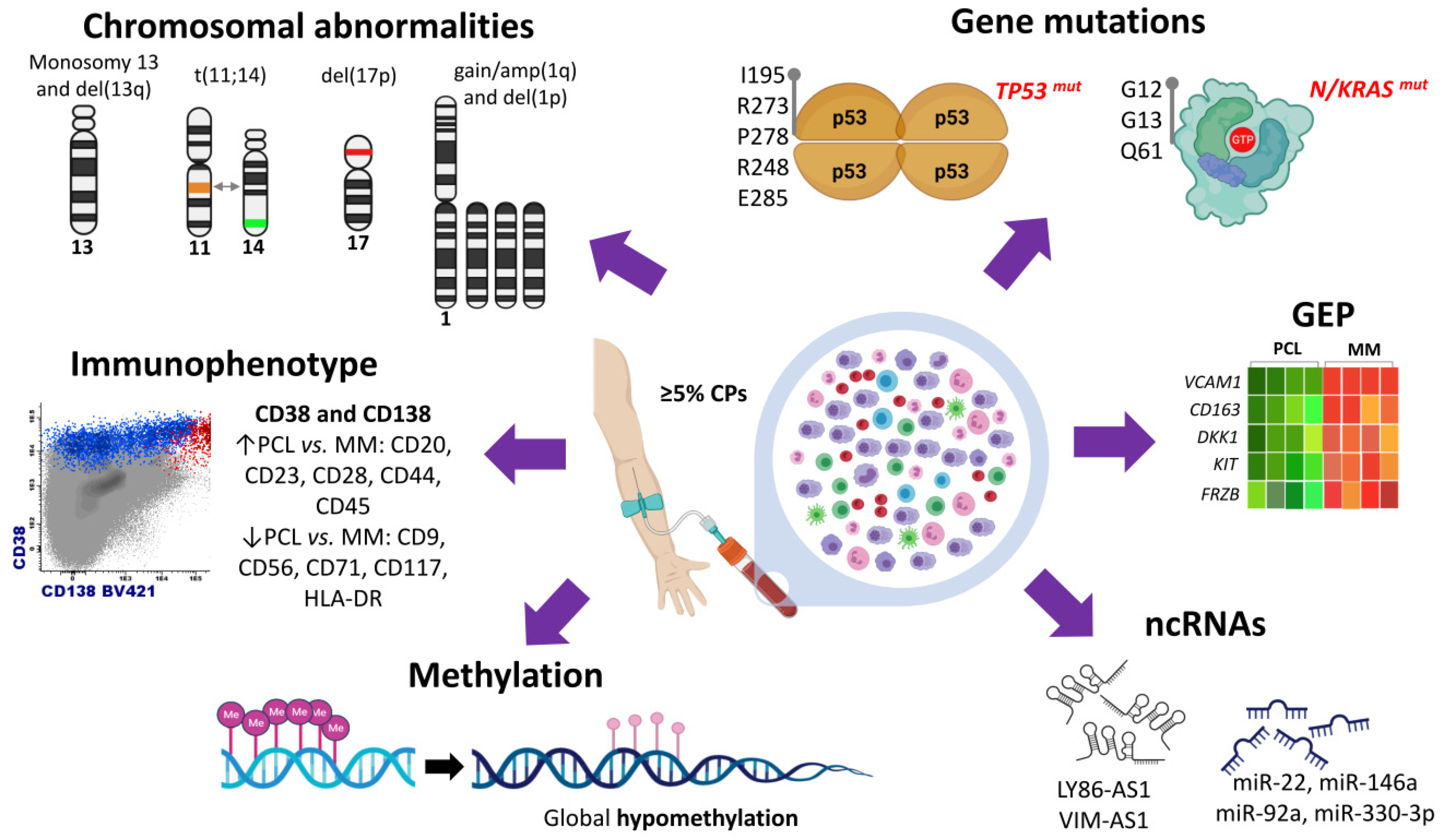
Plasma cell leukemia (PCL) is a rare and highly aggressive plasma cell dyscrasia characterized by the presence of clonal circulating plasma cells in peripheral blood. PCL accounts for approximately 2–4% of all multiple myeloma (MM) cases. PCL can be classified in primary PCL (pPCL) when it appears de novo and in secondary PCL (sPCL) when it arises from a pre-existing relapsed/refractory MM. The development of new high-throughput technologies, such as microarrays and new generation sequencing (NGS), has contributed to a better understanding of the peculiar biological and clinical features of this disease. Relevant information is now available on cytogenetic alterations, genetic variants, transcriptome, methylation patterns, and non-coding RNA profiles. Additionally, attempts have been made to integrate genomic alterations with gene expression data. 

plasma cell leukemia PCL genetics

1. Introduction

Plasma cell leukemia (PCL) is an uncommon plasma cell dyscrasia with an aggressive course and poor prognosis. PCL represents less than 3% of all plasma cells neoplasms, and its incidence has been estimated at 0.04 cases per 100,000 persons/year [1][2].
Historically, PCL has been defined by the presence of more than 20% of circulating plasma cells (PCs) and an absolute number of ≥2 × 10⁹/L of PCs in peripheral blood [3]. However, in some studies, only the presence of one of these criteria had been considered to define PCL. Moreover, recent studies have shown that much lower levels of circulating PC have the same adverse prognostic impact. Accordingly, the consensus recently published by the International Myeloma Working Group (IMWG) states that PCL is defined by the presence of 5% or more circulating plasma cells in peripheral blood [4].
The clinical presentation of PCL is more aggressive than that observed in MM, including more severe cytopenias, hypercalcemia, and renal insufficiency. Higher tumor burden and proliferation activity of PCL are manifested by greater levels of B2-microglobulin and lactate dehydrogenase (LDH). Extramedullary involvement (lymph nodes, liver, spleen, pleura, and central nervous system) at diagnosis is more common in pPCL and sPCL than in MM, but osteolytic lesions are more frequent in sPCL and MM than in pPCL [5][6][7][8][9][10].
Various studies have analyzed the immunophenotype of PCL. The two common PCs markers, CD38 and CD138 antigens, are similarly expressed in MM and PCL. However, PCL displays a more immature phenotype than MM, expressing more frequently CD20, CD23, CD28, CD44, and CD45, and less frequently CD9, CD56, CD71, CD117, and HLA-DR antigens [11][12][13].

2. Cytogenetic Abnormalities

Early cytogenetic and DNA content studies carried out in PCL revealed that there was a predominance of non-hyperdiploid cases (more than 50% of pPCL) compared to that observed in MM [5][11][14]. These results were confirmed in subsequent studies using not only conventional karyotyping but also molecular cytogenetic techniques such as comparative genomic hybridization (CGH) and single nucleotide polymorphism (SNP)-arrays, which showed that pPCL had more DNA copy number changes with a predominance of chromosomal losses in contrast to MM [15][16]. As in MM, FISH has been routinely carried out to identify cytogenetic alterations present in pPCL at the time of diagnosis. Virtually all the studies reporting data provided by FISH analysis, sometimes in combination with other cytogenetic techniques, point out that the chromosomal abnormalities observed in pPCL are mostly the same recurrently found in MM, although many of them are present with greater frequency.
Figure 1. Genomic abnormalities of primary plasma cell leukemia (pPCL). The updated consensus of the IMWG defines pPCL by the presence of 5% or more circulating plasma cells in peripheral blood. Cytogenetic studies by FISH show predominance of monosomy and deletions of chromosome 13, t(11;14), del(17p), gain/amp(1q) and del(1p). Mutation studies by conventional DNA sequencing, WES, and targeted NGS detect a high frequency of mutations in TP53 and K/NRAS genes. The amino acids most frequently mutated in TP53 are I195, R273, P278, R248, and E285. Activating mutations of K/NRAS most frequently found in pPCL patients affect codons 12, 13, and 61 (G12, G13, and Q61). Immunophenotyping of plasma cells reveals expression of CD38 and CD138 in both pPCL and MM, although higher expression of CD20, CD23, CD28, CD44, and CD45 and lower expression of CD9, CD56, CD71, CD117, and HLA-DR may be found in pPCL compared to MM. Gene expression profiling in pPCL has shown downregulation of genes associated with bone marrow microenvironment and bone diseases in MM, such as DKK1, KIT, and NCAM1 genes. A global hypomethylation profile has been found in pPCL samples. Non-coding RNAs (miRNAs and lncRNAs) are dysregulated in pPCL, and some of them are associated with survival of patients (as shown in the figure).
Monosomy and deletions of chromosome 13 (del(13q)) have been observed in approximately 85% of pPCL [5][17][18]. Abnormalities of chromosome 1 are also frequent in pPCL patients. Gain (3 copies) and amplification (≥4 copies) of chromosome arm 1q21 (gain/amp(1q)) have been reported in around 70% of pPCL cases [18][19]. Although the frequency of gain/amp(1q) does not reach such a high percentage in newly diagnosed MM patients, the incidence of this abnormality increases in relapsed/refractory MM up to 50–80% [17][20][21]. Likewise, most of the studies have shown greater frequency of deletion of 1p (del(1p)) in pPCL than in MM patients (24–33% vs. 9–18%, respectively) [17][22]. While the impact of abnormalities in chromosome 1, both gain/amp(1q) and del(1p), on the survival of patients with MM is well established [23][24], their effect on the prognosis of pPCL is still poorly substantiated. Only one study has reported that del(1p), but not gain/amp(1q), is associated with shorter survival of PCL patients, although the set of sPCL included in the study may be biasing the influence that this chromosomal alteration might have on pPCL considered as a separate entity [17]. Deletion of 17p (del(17p)), although uncommon in MM at the time of diagnosis, reaches frequencies of 50% in pPCL [5][16][17][25][26]. However, it seems to have no impact on the prognosis of pPCL, unlike in MM [5][17].
The incidence of IGH translocations is significantly higher in pPCL than in MM. Several studies show that t(11;14) leading to CCND1 dysregulation are significantly more frequent in pPCL than in MM, reaching percentages as high as 45–70% in some series [5][14][16][17][22][27][28][29][30][31]; also noteworthy is the high proportion of t(14;16) detected in pPCL compared with MM (13–25% vs. 1–5%, respectively), which is supported by five studies [14][19][22][28][29]. Conversely, in most of the studies, t(4;14) has been found to be less frequent in pPCL than in MM [19][29][32].
MYC rearrangements have also been found in PCL, although the reported incidence varies between 13% and 40% [5][31][33]. Other chromosomal abnormalities have been identified in pPCL, especially the loss of chromosome 16 (80%) [15][29][34], 7 (11%) [35] and X (25%) [11], and the trisomy of chromosome 8 (43%) [11].

3. Gene Mutations

Before the availability of next-generation sequencing (NGS) technologies, the mutational status of RAS oncogenes (NRAS and KRAS), the two most prevalent mutated genes in MM, and of the tumor suppressor TP53, had been explored in pPCL using traditional DNA sequencing methodologies. Two studies demonstrated a high incidence of NRAS and KRAS activating mutations: one of them reported these mutations at codons 12, 13, or 61 in 27% of pPCL and 15% of sPCL cases [5], and in the other study NRAS and/or KRAS mutations were found in 50% of pPCL cases and in 55% of MM [36]. Strikingly, these findings were not confirmed in a subsequent study [16]. TP53 is one of the most frequently mutated genes in pPCL in all the published series, reaching frequencies of 25% [5][16][29]. The proportion of cases with biallelic inactivation of TP53 is also greater in pPCL than in MM (17–35% vs. 3–4%) [5][16]. TP53 coding mutations involving 5–8 exons were found, predicting all of them a non-functional p53 protein [5][16] (Figure 1).
The first whole-exome sequencing (WES) analysis of pPCL revealed a highly heterogeneous mutational profile [37]. Almost 2000 coding somatic non-silent variants on 1643 genes were described, with more than 160 variants per sample, although with hardly any recurrent mutations in two or more samples. Fourteen mutated genes mainly involved in cell cycle and apoptosis (CIDEC), RNA binding and degradation (DIS3, RPL17), and cell-matrix adhesion and membrane organization (SPTB, CELA1) were considered as potential cancer driver genes in pPCL. Other studies have confirmed that the number of nonsynonymous mutations per sample is higher in pPCL than in MM [19].
As in MM, activating N/KRAS mutations have been identified in pPCL using WES methodologies, although the proportions were significantly unequal between the two of the studies. The first study reported mutations of KRAS and NRAS only in two distinct samples (<10% of the pPCL). This study highlighted that KRAS and NRAS were three-fold less frequently mutated in pPCL compared to that observed in MM [37]. On the contrary, the second study also using WES methodology found that KRAS was the most frequently mutated gene in pPCL samples (around 39%), and mutations of NRAS were present in 13% of pPCL [19]. Using targeted NGS approaches, KRAS mutations were detected in 17% of pPCL, 18% of sPCL, and 33% of MM, and NRAS mutations in 4% of pPCL, 36% of sPCL, and 27% of MM [38]. Apparently, the MEK/ERK signaling pathway was less affected by mutation events in pPCL than in sPCL and MM [38].
IRF4 mutations have recently been shown to be significantly more frequent in pPCL than in MM patients (11% vs. 4%) [22]. Other gene mutations commonly observed in MM have also been reported in pPCL but with different frequencies. Schinke et al. detected DIS3 and PRMD1 mutations in 5% and 13% of patients with pPCL, respectively, while Cifola et al. identified DIS3 mutations in 25% of cases and no variants in the PRMD1 gene. Both studies have described a similar incidence of FAM46C mutation (10–12%) in pPCL patients [19][37].

4. Transcriptome Characterization

More recently, the GEP of 41 pPCL patients has been compared to that of more than 700 newly diagnosed MM [19]. In pPCL, the analysis showed overexpression of genes previously related to MM biology or prognosis, such as PHF19 and TAGLN2, and underexpression of the adhesion molecules VCAM1 and CD163, which are highly expressed in MM and have been correlated with poor survival [39][40].
RNA-seq analysis of pPCL has also shown a specific transcriptional landscape of pPCL, as previously demonstrated by GEP using microarrays. Compared to MM, pPCL showed significantly higher expression of genes involved in G2M checkpoint and MYC target genes and lower expression of genes involved in p53 pathway, hypoxia, and TNF alpha signaling via NF-κB [22]. In this regard, significant overexpression of CDKN2A, CCND3, and CCND1 genes, using quantitative RT-PCR, has been reported in PCL compared to MM samples, indicating a marked cell cycle dysregulation in the transition from MM to PCL [41].
A comprehensive molecular analysis of pPCL integrating data from FISH, SNP-arrays, and GEP has revealed a strong correlation between chromosomal imbalances and transcriptional modulation. The gene dosage effect was particularly observed in those genes mapping 1q chromosome [16]. In addition, the analysis of upregulated and downregulated transcripts in the gained and lost chromosomal regions, respectively, found that protein transport, translation, and biosynthesis functional categories were upregulated in pPCL cases with gained chromosomal regions, whereas RNA splicing, protein catabolic process, and regulation of apoptosis were downregulated in pPCL cases with deleted regions.
Differences between the gene expression signature of pPCL and MM could be partly attributed to the dissimilar distribution of genetic abnormalities between the two diseases. This fact prompted us to compare the transcriptome of pPCL and MM patients using samples with del(17p) and a similar cytogenetic background [42]. This approach revealed that pPCL and MM were separated into two differentiated clusters despite the equivalent cytogenetic profile shared by both entities. Differentially expressed genes were mostly downregulated in pPCL, among which were genes associated with bone marrow microenvironment and bone diseases in MM, such as DKK1KITNCAM1, and FRZB (Figure 1). Interestingly, the analysis focused on isoform expression showed that dysregulation of RNA splicing machinery may be a relevant molecular mechanism underlying the biological differences between the pPCL and MM.
A similar approach has been used to ascertain the differences in the transcriptome between pPCL and MM samples harboring t(11;14) [32]. Both plasma cell dyscrasias are clearly distinguishable based on the transcriptome profile despite sharing a uniform genetic background. pPCL with t(11;14) were positively associated with genes involved in IL2-STAT5 signaling but negatively associated with the regulation of cell and cell adhesion pathways. In any case, the most relevant finding of this study was that pPCL showed a different expression pattern of the BCL2 family genes and of the B-cell-associated genes, despite the presence of t(11;14) in both PCL and MM samples. These results suggest that the efficacy of venetoclax in pPCL and MM patients with t(11;14) may be associated with different molecular programs.

5. Non-Coding RNA Profile

Non-coding RNAs (ncRNAs) are classified as short (<200 nucleotides) and long (>200 nucleotides). The miRNAs are short ncRNAs of 19–22 nucleotides that regulate gene expression at the post-transcriptional level. Since their discovery, numerous studies have attributed a wide variety of functions for ncRNAs in the pathogenic mechanisms of MM [43][44][45].
One study analyzing the expression pattern of miRNAs in pPCL [46]. The analysis of 18 pPCL identified 42 upregulated and 41 downregulated miRNAs in pPCL when compared with MM samples. Moreover, seven miRNAs were found to be differentially expressed depending on the type of IGH translocation. Three miRNAs (let-7e, miR-135a, and miR-148a) were overexpressed in PCL patients with t(4;14); three (miR-7, miR-7-1, and miR-454) underexpressed in PCL with t(14;16); and the miR-342-3p was underexpressed in PCL with t(11;14). Notably, four miRNAs, miR-22, miR-146a, miR-92a, and miR-330-3p, were found to have an impact on the survival of pPCL patients. The overexpression of miR-146a, which was associated with shorter progression-free survival (PFS) in pPCL cases, and miR-22, which was associated with longer PFS, showed a pro- and anti-survival effect, respectively, in myeloma cell lines [46]. Accordingly, one study has demonstrated that MM cells stimulate the overexpression of miR-146a in mesenchymal stromal cells, resulting in more cytokine secretion and enhancing cell viability of MM cells [47] (Figure 1).
LncRNAs expression profile has also been investigated in a large cohort of PC dyscrasias, including samples from MGUS, SMM, MM, and PCL together with NPC [48]. Differential expression of 160 lncRNAs between NPC and the four premalignant and malignant entities was detected. In particular, expression levels of 15 lncRNAs were progressively increased from NPC to PCL patients, while six lncRNAs showed a significant decrease in the transition from NPC and premalignant entities to more aggressive forms.

6. Methylation Patterns

The analysis of global methylation patterns in pPCL using high-density arrays has identified a global hypomethylation profile in pPCL samples [49] (Figure 1). The comparison of methylation levels between pPCL, MM, MGUS, and NPC samples revealed that genes highly methylated in NPC underwent a progressive decrease in the levels of methylation as the aggressiveness of the disease increased from MGUS to MM and pPCL. Curiously, pPCL patients showed distinct methylation profiles depending on the presence of DIS3 gene mutations, t(11;14), and t(14;16). On the contrary, Walker et al. [50] had previously found gene-specific hypermethylation of almost 2000 genes in the transition from MM to PCL, although the number of PCL cases was quite small.

References

  1. Gundesen, M.T.; Lund, T.; Moeller, H.E.H.; Abildgaard, N. Plasma Cell Leukemia: Definition, Presentation, and Treatment. Curr. Oncol. Rep. 2019, 21, 8.
  2. Gonsalves, W.I.; Rajkumar, S.V.; Go, R.S.; Dispenzieri, A.; Gupta, V.; Singh, P.P.; Buadi, F.K.; Lacy, M.Q.; Kapoor, P.; Dingli, D.; et al. Trends in Survival of Patients with Primary Plasma Cell Leukemia: A Population-Based Analysis. Blood 2014, 124, 907–912.
  3. Fernández de Larrea, C.; Kyle, R.A.; Durie, B.G.M.; Ludwig, H.; Usmani, S.; Vesole, D.H.; Hajek, R.; San Miguel, J.F.; Sezer, O.; Sonneveld, P.; et al. Plasma Cell Leukemia: Consensus Statement on Diagnostic Requirements, Response Criteria and Treatment Recommendations by the International Myeloma Working Group. Leukemia 2013, 27, 780–791.
  4. Fernández de Larrea, C.; Kyle, R.; Rosiñol, L.; Paiva, B.; Engelhardt, M.; Usmani, S.; Caers, J.; Gonsalves, W.; Schjesvold, F.; Merlini, G.; et al. Primary Plasma Cell Leukemia: Consensus Definition by the International Myeloma Working Group According to Peripheral Blood Plasma Cell Percentage. Blood Cancer J. 2021, 11, 192.
  5. Tiedemann, R.E.; Gonzalez-Paz, N.; Kyle, R.A.; Santana-Davila, R.; Price-Troska, T.; Van Wier, S.A.; Chng, W.J.; Ketterling, R.P.; Gertz, M.A.; Henderson, K.; et al. Genetic Aberrations and Survival in Plasma Cell Leukemia. Leukemia 2008, 22, 1044–1052.
  6. Chaulagain, C.P.; Diacovo, M.-J.; Van, A.; Martinez, F.; Fu, C.-L.; Jimenez Jimenez, A.M.; Ahmed, W.; Anwer, F. Management of Primary Plasma Cell Leukemia Remains Challenging Even in the Era of Novel Agents. Clin. Med. Insights Blood Disord. 2021, 14.
  7. Mina, R.; D’Agostino, M.; Cerrato, C.; Gay, F.; Palumbo, A. Plasma Cell Leukemia: Update on Biology and Therapy. Leuk. Lymphoma 2017, 58, 1538–1547.
  8. Jimenez-Zepeda, V.H.; Dominguez-Martinez, V.J. Plasma Cell Leukemia: A Highly Aggressive Monoclonal Gammopathy with a Very Poor Prognosis. Int. J. Hematol. 2009, 89, 259–268.
  9. Jurczyszyn, A.; Radocha, J.; Davila, J.; Fiala, M.A.; Gozzetti, A.; Grząśko, N.; Robak, P.; Hus, I.; Waszczuk-Gajda, A.; Guzicka-Kazimierczak, R.; et al. Prognostic Indicators in Primary Plasma Cell Leukaemia: A Multicentre Retrospective Study of 117 Patients. Br. J. Haematol. 2018, 180, 831–839.
  10. Ramsingh, G.; Mehan, P.; Luo, J.; Vij, R.; Morgensztern, D. Primary Plasma Cell Leukemia: A Surveillance, Epidemiology, and End Results Database Analysis between 1973 and 2004. Cancer 2009, 115, 5734–5739.
  11. García-Sanz, R.; Orfão, A.; González, M.; Tabernero, M.D.; Bladé, J.; Moro, M.J.; Fernández-Calvo, J.; Sanz, M.A.; Pérez-Simón, J.A.; Rasillo, A.; et al. Primary Plasma Cell Leukemia: Clinical, Immunophenotypic, DNA Ploidy, and Cytogenetic Characteristics. Blood 1999, 93, 1032–1037.
  12. Kraj, M.; Pogłód, R.; Kopeć-Szlezak, J.; Sokołowska, U.; Woźniak, J.; Kruk, B. C-Kit Receptor (CD117) Expression on Plasma Cells in Monoclonal Gammopathies. Leuk. Lymphoma 2004, 45, 2281–2289.
  13. Kraj, M.; Kopeć-Szlęzak, J.; Pogłód, R.; Kruk, B. Flow Cytometric Immunophenotypic Characteristics of 36 Cases of Plasma Cell Leukemia. Leuk. Res. 2011, 35, 169–176.
  14. Avet-Loiseau, H.; Daviet, A.; Brigaudeau, C.; Callet-Bauchu, E.; Terré, C.; Lafage-Pochitaloff, M.; Désangles, F.; Ramond, S.; Talmant, P.; Bataille, R. Cytogenetic, Interphase, and Multicolor Fluorescence in Situ Hybridization Analyses in Primary Plasma Cell Leukemia: A Study of 40 Patients at Diagnosis, on Behalf of the Intergroupe Francophone Du Myélome and the Groupe Français de Cytogénétique Hématologique. Blood 2001, 97, 822–825.
  15. Gutiérrez, N.C.; Hernández, J.M.; García, J.L.; Cañizo, M.C.; González, M.; Hernández, J.; González, M.B.; Garciá-Marcos, M.A.; San Miguel, J.F. Differences in Genetic Changes between Multiple Myeloma and Plasma Cell Leukemia Demonstrated by Comparative Genomic Hybridization. Leukemia 2001, 15, 840–845.
  16. Mosca, L.; Musto, P.; Todoerti, K.; Barbieri, M.; Agnelli, L.; Fabris, S.; Tuana, G.; Lionetti, M.; Bonaparte, E.; Sirchia, S.M.; et al. Genome-Wide Analysis of Primary Plasma Cell Leukemia Identifies Recurrent Imbalances Associated with Changes in Transcriptional Profiles. Am. J. Hematol. 2013, 88, 16–23.
  17. Chang, H.; Qi, X.; Yeung, J.; Reece, D.; Xu, W.; Patterson, B. Genetic Aberrations Including Chromosome 1 Abnormalities and Clinical Features of Plasma Cell Leukemia. Leuk. Res. 2009, 33, 259–262.
  18. Yu, T.; Xu, Y.; An, G.; Tai, Y.-T.; Ho, M.; Li, Z.; Deng, S.; Zou, D.; Yu, Z.; Hao, M.; et al. Primary Plasma Cell Leukemia: Real-World Retrospective Study of 46 Patients From a Single-Center Study in China. Clin. Lymphoma Myeloma Leuk. 2020, 20, e652–e659.
  19. Schinke, C.; Boyle, E.M.; Ashby, C.; Wang, Y.; Lyzogubov, V.; Wardell, C.; Qu, P.; Hoering, A.; Deshpande, S.; Ryan, K.; et al. Genomic Analysis of Primary Plasma Cell Leukemia Reveals Complex Structural Alterations and High-Risk Mutational Patterns. Blood Cancer J. 2020, 10, 70.
  20. Hanamura, I.; Stewart, J.P.; Huang, Y.; Zhan, F.; Santra, M.; Sawyer, J.R.; Hollmig, K.; Zangarri, M.; Pineda-Roman, M.; van Rhee, F.; et al. Frequent Gain of Chromosome Band 1q21 in Plasma-Cell Dyscrasias Detected by Fluorescence in Situ Hybridization: Incidence Increases from MGUS to Relapsed Myeloma and Is Related to Prognosis and Disease Progression Following Tandem Stem-Cell Transplantation. Blood 2006, 108, 1724–1732.
  21. Hanamura, I. Gain/Amplification of Chromosome Arm 1q21 in Multiple Myeloma. Cancers (Basel) 2021, 13, 256.
  22. Cazaubiel, T.; Buisson, L.; Maheo, S.; Do Souto Ferreira, L.; Lannes, R.; Perrot, A.; Hulin, C.; Avet-Loiseau, H.; Corre, J. The Genomic and Transcriptomic Landscape of Plasma Cell Leukemia. Blood 2020, 136, 48–49.
  23. Chang, H.; Qi, X.; Jiang, A.; Xu, W.; Young, T.; Reece, D. 1p21 Deletions Are Strongly Associated with 1q21 Gains and Are an Independent Adverse Prognostic Factor for the Outcome of High-Dose Chemotherapy in Patients with Multiple Myeloma. Bone Marrow Transpl. 2010, 45, 117–121.
  24. Chang, H.; Qi, C.; Jiang, A.; Xu, W.; Trieu, Y.; Reece, D.E. Loss of Chromosome 1p21 Band Is Associated with Disease Progression and Poor Survival in Multiple Myeloma Patients Undergoing Autologous Stem Cell Transplantation. Blood 2008, 112, 1702.
  25. Lionetti, M.; Barbieri, M.; Manzoni, M.; Fabris, S.; Bandini, C.; Todoerti, K.; Nozza, F.; Rossi, D.; Musto, P.; Baldini, L.; et al. Molecular Spectrum of TP53 Mutations in Plasma Cell Dyscrasias by next Generation Sequencing: An Italian Cohort Study and Overview of the Literature. Oncotarget 2016, 7, 21353–21361.
  26. Nandakumar, B.; Kumar, S.K.; Dispenzieri, A.; Buadi, F.K.; Dingli, D.; Lacy, M.Q.; Hayman, S.R.; Kapoor, P.; Leung, N.; Fonder, A.; et al. Clinical Characteristics and Outcomes of Patients With Primary Plasma Cell Leukemia in the Era of Novel Agent Therapy. Mayo Clin. Proc. 2021, 96, 677–687.
  27. Avet-Loiseau, H.; Li, J.Y.; Facon, T.; Brigaudeau, C.; Morineau, N.; Maloisel, F.; Rapp, M.J.; Talmant, P.; Trimoreau, F.; Jaccard, A.; et al. High Incidence of Translocations t(11;14)(Q13;Q32) and t(4;14)(P16;Q32) in Patients with Plasma Cell Malignancies. Cancer Res. 1998, 58, 5640–5645.
  28. Avet-Loiseau, H.; Facon, T.; Grosbois, B.; Magrangeas, F.; Rapp, M.-J.; Harousseau, J.-L.; Minvielle, S.; Bataille, R. Intergroupe Francophone du Myélome Oncogenesis of Multiple Myeloma: 14q32 and 13q Chromosomal Abnormalities Are Not Randomly Distributed, but Correlate with Natural History, Immunological Features, and Clinical Presentation. Blood 2002, 99, 2185–2191.
  29. Chiecchio, L.; Dagrada, G.P.; White, H.E.; Towsend, M.R.; Protheroe, R.K.M.; Cheung, K.L.; Stockley, D.M.; Orchard, K.H.; Cross, N.C.P.; Harrison, C.J.; et al. Frequent Upregulation of MYC in Plasma Cell Leukemia. Genes Chromosomes Cancer 2009, 48, 624–636.
  30. Pagano, L.; Valentini, C.G.; De Stefano, V.; Venditti, A.; Visani, G.; Petrucci, M.T.; Candoni, A.; Specchia, G.; Visco, C.; Pogliani, E.M.; et al. Primary Plasma Cell Leukemia: A Retrospective Multicenter Study of 73 Patients. Ann. Oncol. 2011, 22, 1628–1635.
  31. Papadhimitriou, S.I.; Terpos, E.; Liapis, K.; Pavlidis, D.; Marinakis, T.; Kastritis, E.; Dimopoulos, M.-A.; Tsitsilonis, O.E.; Kostopoulos, I.V. The Cytogenetic Profile of Primary and Secondary Plasma Cell Leukemia: Etiopathogenetic Perspectives, Prognostic Impact and Clinical Relevance to Newly Diagnosed Multiple Myeloma with Differential Circulating Clonal Plasma Cells. Biomedicines 2022, 10, 209.
  32. Todoerti, K.; Taiana, E.; Puccio, N.; Favasuli, V.; Lionetti, M.; Silvestris, I.; Gentile, M.; Musto, P.; Morabito, F.; Gianelli, U.; et al. Transcriptomic Analysis in Multiple Myeloma and Primary Plasma Cell Leukemia with t(11;14) Reveals Different Expression Patterns with Biological Implications in Venetoclax Sensitivity. Cancers (Basel) 2021, 13, 4898.
  33. Avet-Loiseau, H.; Gerson, F.; Magrangeas, F.; Minvielle, S.; Harousseau, J.L.; Bataille, R. Intergroupe Francophone du Myélome Rearrangements of the C-Myc Oncogene Are Present in 15% of Primary Human Multiple Myeloma Tumors. Blood 2001, 98, 3082–3086.
  34. Jonveaux, P.; Berger, R. Chromosome Studies in Plasma Cell Leukemia and Multiple Myeloma in Transformation. Genes Chromosomes Cancer 1992, 4, 321–325.
  35. Dimopoulos, M.A.; Palumbo, A.; Delasalle, K.B.; Alexanian, R. Primary Plasma Cell Leukaemia. Br. J. Haematol. 1994, 88, 754–759.
  36. Bezieau, S.; Devilder, M.C.; Avet-Loiseau, H.; Mellerin, M.P.; Puthier, D.; Pennarun, E.; Rapp, M.J.; Harousseau, J.L.; Moisan, J.P.; Bataille, R. High Incidence of N and K-Ras Activating Mutations in Multiple Myeloma and Primary Plasma Cell Leukemia at Diagnosis. Hum. Mutat. 2001, 18, 212–224.
  37. Cifola, I.; Lionetti, M.; Pinatel, E.; Todoerti, K.; Mangano, E.; Pietrelli, A.; Fabris, S.; Mosca, L.; Simeon, V.; Petrucci, M.T.; et al. Whole-Exome Sequencing of Primary Plasma Cell Leukemia Discloses Heterogeneous Mutational Patterns. Oncotarget 2015, 6, 17543–17558.
  38. Lionetti, M.; Barbieri, M.; Todoerti, K.; Agnelli, L.; Marzorati, S.; Fabris, S.; Ciceri, G.; Galletti, S.; Milesi, G.; Manzoni, M.; et al. Molecular Spectrum of BRAF, NRAS and KRAS Gene Mutations in Plasma Cell Dyscrasias: Implication for MEK-ERK Pathway Activation. Oncotarget 2015, 6, 24205–24217.
  39. Andersen, M.N.; Abildgaard, N.; Maniecki, M.B.; Møller, H.J.; Andersen, N.F. Monocyte/Macrophage-Derived Soluble CD163: A Novel Biomarker in Multiple Myeloma. Eur. J. Haematol. 2014, 93, 41–47.
  40. Terpos, E.; Migkou, M.; Christoulas, D.; Gavriatopoulou, M.; Eleutherakis-Papaiakovou, E.; Kanellias, N.; Iakovaki, M.; Panagiotidis, I.; Ziogas, D.C.; Fotiou, D.; et al. Increased Circulating VCAM-1 Correlates with Advanced Disease and Poor Survival in Patients with Multiple Myeloma: Reduction by Post-Bortezomib and Lenalidomide Treatment. Blood Cancer J. 2016, 6, e428.
  41. Kryukov, F.; Dementyeva, E.; Kubiczkova, L.; Jarkovsky, J.; Brozova, L.; Petrik, J.; Nemec, P.; Sevcikova, S.; Minarik, J.; Stefanikova, Z.; et al. Cell Cycle Genes Co-Expression in Multiple Myeloma and Plasma Cell Leukemia. Genomics 2013, 102, 243–249.
  42. Rojas, E.A.; Corchete, L.A.; Mateos, M.V.; García-Sanz, R.; Misiewicz-Krzeminska, I.; Gutiérrez, N.C. Transcriptome Analysis Reveals Significant Differences between Primary Plasma Cell Leukemia and Multiple Myeloma Even When Sharing a Similar Genetic Background. Blood Cancer J. 2019, 9, 90.
  43. Chen, D.; Yang, X.; Liu, M.; Zhang, Z.; Xing, E. Roles of MiRNA Dysregulation in the Pathogenesis of Multiple Myeloma. Cancer Gene Ther. 2021, 28, 1256–1268.
  44. Carrasco-Leon, A.; Ezponda, T.; Meydan, C.; Valcárcel, L.V.; Ordoñez, R.; Kulis, M.; Garate, L.; Miranda, E.; Segura, V.; Guruceaga, E.; et al. Characterization of Complete LncRNAs Transcriptome Reveals the Functional and Clinical Impact of LncRNAs in Multiple Myeloma. Leukemia 2021, 35, 1438–1450.
  45. Misiewicz-Krzeminska, I.; Krzeminski, P.; Corchete, L.A.; Quwaider, D.; Rojas, E.A.; Herrero, A.B.; Gutiérrez, N.C. Factors Regulating MicroRNA Expression and Function in Multiple Myeloma. Noncoding RNA 2019, 5, 9.
  46. Lionetti, M.; Musto, P.; Di Martino, M.T.; Fabris, S.; Agnelli, L.; Todoerti, K.; Tuana, G.; Mosca, L.; Gallo Cantafio, M.E.; Grieco, V.; et al. Biological and Clinical Relevance of MiRNA Expression Signatures in Primary Plasma Cell Leukemia. Clin. Cancer Res. 2013, 19, 3130–3142.
  47. De Veirman, K.; Wang, J.; Xu, S.; Leleu, X.; Himpe, E.; Maes, K.; De Bruyne, E.; Van Valckenborgh, E.; Vanderkerken, K.; Menu, E.; et al. Induction of MiR-146a by Multiple Myeloma Cells in Mesenchymal Stromal Cells Stimulates Their pro-Tumoral Activity. Cancer Lett. 2016, 377, 17–24.
  48. Ronchetti, D.; Agnelli, L.; Taiana, E.; Galletti, S.; Manzoni, M.; Todoerti, K.; Musto, P.; Strozzi, F.; Neri, A. Distinct LncRNA Transcriptional Fingerprints Characterize Progressive Stages of Multiple Myeloma. Oncotarget 2016, 7, 14814–14830.
  49. Todoerti, K.; Calice, G.; Trino, S.; Simeon, V.; Lionetti, M.; Manzoni, M.; Fabris, S.; Barbieri, M.; Pompa, A.; Baldini, L.; et al. Global Methylation Patterns in Primary Plasma Cell Leukemia. Leuk. Res. 2018, 73, 95–102.
  50. Walker, B.A.; Wardell, C.P.; Chiecchio, L.; Smith, E.M.; Boyd, K.D.; Neri, A.; Davies, F.E.; Ross, F.M.; Morgan, G.J. Aberrant Global Methylation Patterns Affect the Molecular Pathogenesis and Prognosis of Multiple Myeloma. Blood 2011, 117, 553–562.
More
Information
Subjects: Hematology
Contributors MDPI registered users' name will be linked to their SciProfiles pages. To register with us, please refer to https://encyclopedia.pub/register : ,
View Times: 556
Revisions: 2 times (View History)
Update Date: 11 May 2022
1000/1000
ScholarVision Creations