Submitted Successfully!
To reward your contribution, here is a gift for you: A free trial for our video production service.
Thank you for your contribution! You can also upload a video entry or images related to this topic.
Version Summary Created by Modification Content Size Created at Operation
1 -- 2974 2022-04-21 19:36:45 |
2 format correct -32 word(s) 2942 2022-04-22 04:58:23 |

Video Upload Options

We provide professional Video Production Services to translate complex research into visually appealing presentations. Would you like to try it?

Confirm

Are you sure to Delete?
Cite
If you have any further questions, please contact Encyclopedia Editorial Office.
Teixidó, C.; Castillo, P.; Vila, C.; , .; Alos, L. Molecular Markers and Targets in Melanoma. Encyclopedia. Available online: https://encyclopedia.pub/entry/22139 (accessed on 18 November 2024).
Teixidó C, Castillo P, Vila C,  , Alos L. Molecular Markers and Targets in Melanoma. Encyclopedia. Available at: https://encyclopedia.pub/entry/22139. Accessed November 18, 2024.
Teixidó, Cristina, Paola Castillo, Clara Vila,  , Llucia Alos. "Molecular Markers and Targets in Melanoma" Encyclopedia, https://encyclopedia.pub/entry/22139 (accessed November 18, 2024).
Teixidó, C., Castillo, P., Vila, C., , ., & Alos, L. (2022, April 21). Molecular Markers and Targets in Melanoma. In Encyclopedia. https://encyclopedia.pub/entry/22139
Teixidó, Cristina, et al. "Molecular Markers and Targets in Melanoma." Encyclopedia. Web. 21 April, 2022.
Molecular Markers and Targets in Melanoma
Edit

Melanoma develops as a result of several genetic alterations, with UV radiation often acting as a mutagenic risk factor. Deep knowledge of the molecular signaling pathways of different types of melanoma allows better characterization and provides tools for the development of therapies based on the intervention of signals promoted by these cascades. The latest World Health Organization classification acknowledged the specific genetic drivers leading to melanoma and classifies melanocytic lesions into nine distinct categories according to the associate cumulative sun damage (CSD), which correlates with the molecular alterations of tumors. The largest groups are melanomas associated with low-CSD or superficial spreading melanomas, characterized by frequent presentation of the BRAFV600 mutation. High-CSD melanomas include lentigo maligna type and desmoplastic melanomas, which often have a high mutation burden and can harbor NRAS, BRAFnon-V600E, or NF1 mutations. Non-CSD-associated melanomas encompass acral and mucosal melanomas that usually do not show BRAF, NRAS, or NF1 mutations (triple wild-type), but in a subset may have KIT or SF3B1 mutations. To improve survival, these driver alterations can be treated with targeted therapy achieving significant antitumor activity.

melanoma molecular pathways markers target therapy BRAF NRAS KIT NTRK

1. Introduction

1.1. Epidemiology

Melanoma is the most aggressive and deadly skin cancer. Its incidence has increased steadily in the last decades, especially in the Caucasian population, posing a heightened challenge to the global healthcare system [1][2]. Relevant geographical variations exist, depending on the clinical phenotype, the genetic background of individuals, and the extent of ultraviolet (UV) radiation exposure [3]. Currently, it is one of the most frequent cancers in fair-skinned people, especially those with blond or red hair, who have light-colored eyes. Unlike other solid tumors, melanoma mainly affects young and middle-aged people [4]. Melanoma-related mortality has increased in parallel with the increase in the incidence rate over the years, reaching a mortality rate of one in four deaths [5]. Nevertheless, the therapeutic landscape of unresectable stage III and IV melanoma has been revolutionized by immunotherapies and targeted therapies. Both strategies have shown markedly improved survival compared with the use of chemotherapy (ChT) regimens [6]. Melanoma mortality has decreased significantly since the US Food and Drug Administration (FDA) approved ipilimumab in 2011, the first immune checkpoint inhibitor (ICI) to improve survival in the advanced setting [7][8], and vemurafenib, a v-raf murine sarcoma viral oncogene homolog B1 (BRAF) tyrosine kinase inhibitor, first in class [9][10].
Melanoma develops from cutaneous melanocytes, located in the basal layer of the epidermis. UV radiation represents a major contributor to cutaneous melanomagenesis through its harmful effects on the skin and direct DNA damage [11], and it triggers the acceleration of tumorigenesis. Intense and intermittent sun exposure, as well as exposure to UV-A rays from artificial sources, has also been linked to an increased risk of melanoma development [12].
Host risk factors, such as the number of nevi, both congenital or acquired, genetic susceptibility, and a family history of melanoma, are relevant risk factors for the development of melanoma. About 25% of cutaneous melanomas arise from a nevus [13]. Polymorphisms of the melanocortin 1 receptor (MC1R) gene represent the most relevant gene for susceptibility to melanoma [14].
A family history of melanoma is present in 5–15% of patients with cutaneous melanoma, but true hereditary melanoma due to a transmitted genetic mutation is less common, such as familial atypical multiple mole-melanoma (FAMMM) syndrome and its variant, melanoma-astrocytoma syndrome. Germline mutations in cyclin-dependent kinase inhibitor 2A (CDKN2A) and, less common, mutations in cyclin-dependent kinase 4 (CDK4) are the most frequent genetic abnormalities identified in these families [15]. Other inherited conditions, such as xeroderma pigmentosum, familial retinoblastoma, Lynch syndrome type II, and Li–Fraumeni cancer syndrome, may also be related to an increased risk of melanoma development [16].

2. Molecular Pathways of Melanoma Development

Cancer results from uncontrolled cellular growth of malignant tumor cells caused by a combination of genetic alterations that lead to neoplastic transformation and escape from the inhibitory signals. Several steps in this process are known as the hallmark of cancers [17].
Several key molecular pathways have been discovered to be involved in the onset, proliferation, survival, progression, and invasion. In this section, researchers summarize the major signaling pathways that are currently known to be dysregulated and involved in melanoma disease.

2.1. MAPK Pathway

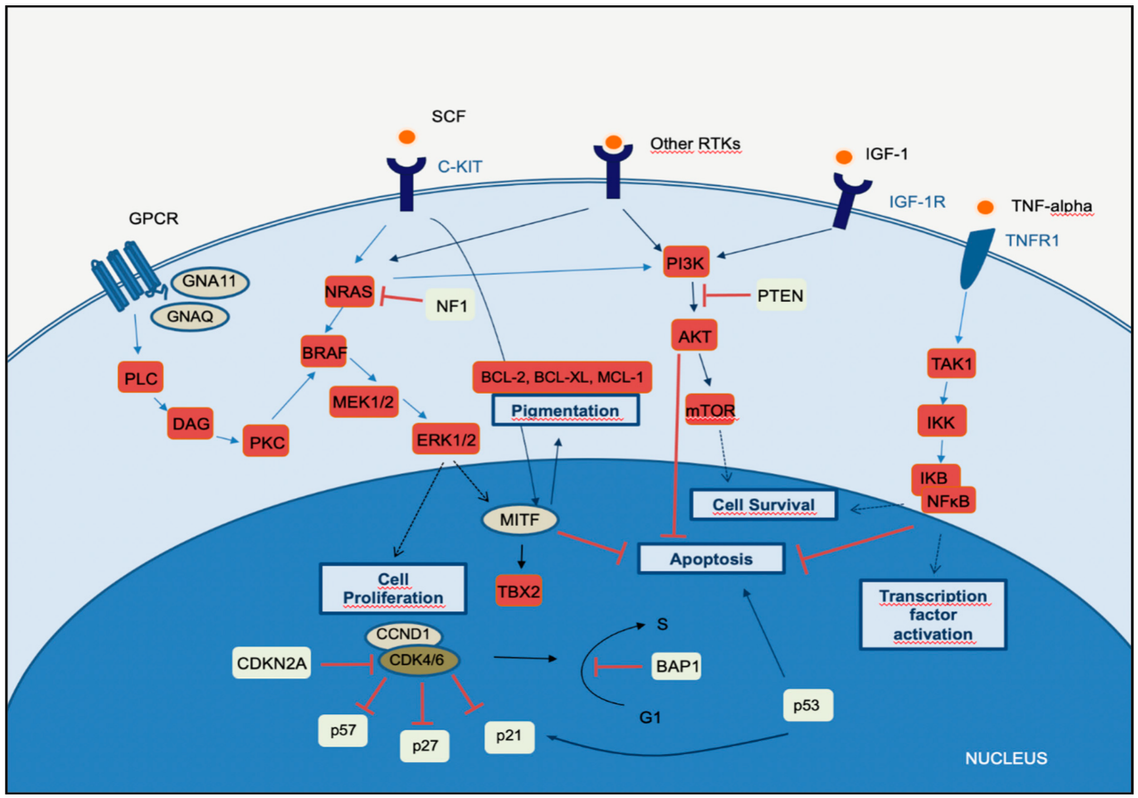
Melanomagenesis occurs after mutational events that produce signaling pathways critical for cell survival. Mitogen-activated protein kinase (MAPK) is a signal transduction pathway, involved in a variety of physiological programs, such as cell proliferation, differentiation, development, migration, apoptosis, and transformation, and is the most relevant in the development of melanoma (Figure 1) [18]. The MAPK pathway is activated by the binding of a growth factor to a receptor tyrosine kinase (RTK) on the cell surface and stimulates the guanosine triphosphatases (GTPase) activity of RAS. The signal propagates through the RAF, mitogen-activated protein kinase kinase 1 (MAP2K1), and extracellular signal-related kinase (ERK) cascade, which enters the nucleus to activate transcription factors and promote the cell cycle (Figure 1) [18].
Figure 1. Melanoma key signaling pathways.
The MAPKPI3K, and NFκB pathways intersect significantly in melanoma pathogenesis. Briefly, in the MAPK-ERK pathway, stimulation of GPCR results in activation of PLC. This promotes DAG and then activates PKC, which stimulates the MAPK pathway. Receptor tyrosin kinases (RTKs) are activated by binding of extracellular growth factor ligands and activate the tyrosine kinase activity of the cytoplasmic domain receptor, starting the cascade of signals. Activated RAS activates the protein kinase activity of RAF isoforms (RAF1, BRAF, ARAF). Each RAF isoform possesses a distinct capacity to activate MEK, with BRAF being the strongest activator. MEK phosphorylates and activates downstream proteins, such as ERK1 and ERK2ERK can translocate to the nucleus and phosphorylate different transcription factors, which leads to the control of cell cycle progression. MITF is a target of ERK and controls the production of the pigment melanin, cell cycling, and survival. The binding of the ligand to KIT (SCF) results in activation of the MAPK and PI3K pathways. In the PI3K-AKT pathway, ligand binding to the RTK leads to dimerization and autophosphorylation of the receptor and activation. Activated RTK recruits PI3K to the plasma membrane. PI3K activates AKT, whereas PTEN antagonizes this process. PI3K may also be activated by GPCRIGF-1R, and RAS. Both ERK and AKT activate the mTOR-signaling pathway, which mediates cell survival and proliferation. In the TNFR pathway (canonical NF-κB pathway), binding of the TNF-alpha cytokine to its receptor TNFR1 results in TAK1 activation. TAK1 leads to the aggregation of a downstream kinase complex, the IKK complex. Phosphorylation of IκB by the IKK complex results in the release of NFκBNFκB translocates to the nucleus and activates genes involved in cell survival and anti-apoptosis.
Fourteen MAPKs have been identified in mammals, and these kinases are typically divided into three main subfamilies: ERKs, c-Jun N-terminal kinases (JNKs), and P38 kinases. Each of these MAPKs is activated through phosphorylation by an MAPK kinase (MAP2K), which in turn is activated by an MAPKK kinase (MAP3K) [18]. The ERK pathway is the best-characterized MAPK pathway, which has a relevant role in the development and progression of melanoma. On this MAPK axis, the role of MAP3K is played by the RAF family of serine/threonine kinases, which is characterized by an RAS/GTP-binding domain. RAS proteins vHa-ras Harvey rat sarcoma viral oncogene homolog (HRAS), NRAS, and v-Ki-ras2 Kirsten rat sarcoma viral oncogene homolog (KRAS) are small GTPases located in the plasma membrane that act as activators in several pathways, apart from MAPK. Additionally, activation signals via RAS on the inner surface of the cell membrane increase ERK activity. Consequently, there is an increase in cellular proliferation, greater cell survival, and resistance to apoptosis. Activated ERK can also induce the metastatic potential of melanoma through the expression of integrins that promote tumor invasion [19].
In melanoma, dysregulated MAPK signaling and sustained ERK activation can eventually lead to cascade hyperactivity and subsequent cell proliferation, survival, invasion, metastasis, and angiogenesis. The BRAF gene is frequently mutated in several cancers, and BRAFV600 is the most common mutation of the skin. Mutated BRAFV600 leads to elevated BRAF kinase activity and sustained activation of downstream targets, in addition to unresponsive negative feedback mechanisms [20]. The mutant KRASQ61, the most frequent mutation of KRAS in melanoma, leads to an important decrease in its intrinsic hydrolytic activity and a sustained active state of KRAS. Mutations in other molecules may also lead to RAS overstimulation, such as loss-of-function mutations in neurofibromin 1 (NF1). In most melanomas with altered NF1, a loss-of-function mutation is found, in which neurofibromin loses its ability to inactivate RAS and promotes stimulation of the RAF and its downstream targets, leading to stimulation of the MAPK pathway and consequent cell proliferation and survival [21].
Telomerase reverse transcriptase (TERT) promoter mutations frequently occur in melanoma and, according to The Cancer Genome Atlas (TCGA) data, mainly in the mutated subtypes BRAF (75% of cases), RAS (72% of cases), and NF1 (83% of cases), suggesting a link between MAPK activation and TERT expression. The active MAPK pathway promotes phosphorylation and activation of the ETS1 transcription factor by ERK (the mutated TERT promoter bears ETS-binding sites) [22].

2.2. PI3K-AKT Pathway

The phosphatidylinositol-3-kinases (PI3Ks) comprise a family of lipid kinases with regulatory roles in many cellular mechanisms, including cell survival and growth, differentiation, proliferation, transcription, and translation. The pathway transduces signals from a variety of growth factors and cytokines and is the major downstream effector of RTKs and G-protein-coupled receptors (GPCRs) (Figure 1). Activated PI3K leads to the formation of phosphatidylinositol-3,4,5-trisphosphate (PIP3) through phosphorylation of phosphatidylinositol-4,5-diphosphate (PIP2) in the plasma membrane. PIP3 is essential for the recruitment of the serine-threonine protein kinase AKT to the plasma membrane. AKT is crucial in this signaling pathway, transmitting signals by phosphorylating different downstream effector targets [23]. Once AKT is phosphorylated and fully activated, it turns on a major downstream effector of the PI3K pathway, inhibiting or activating a variety of targets and regulating important cellular processes, such as apoptosis, DNA repair, cell cycle, glucose metabolism, cell growth, motility, invasion, and angiogenesis. The main target of AKT is the mammalian target of rapamycin (mTOR), which has a central role in the PI3K-AKT pathway and cancer disease. mTOR plays a crucial part in regulating cell growth and proliferation by monitoring nutrient availability, cellular energy, oxygen levels, and mitogenic signals.
PI3K-AKT signaling has negative regulators, to control any persistent and long-term activation. A major regulator of PI3K-AKT signaling is the tumor suppressor phosphatase and tensin homolog (PTEN), which antagonizes the PI3K activity through its intrinsic lipid phosphatase activity, converting PIP3 back to PIP2. Loss of PTEN results in constitutive activation of AKT and has been largely associated with tumor development in malignant melanoma. Indeed, PTEN loss has been shown to be predictive of shorter overall survival (OS) [24][25].
The PI3K signaling cascade is upregulated in different types of cancer, including melanoma. More than two-thirds of primary and metastatic melanomas show high levels of phosphorylated AKT, suggesting that this alteration is an early event in melanoma pathogenesis. Oncogenic events that activate PI3K-AKT may include mutations or copy number variations in certain components of the pathway. RAS gene mutations and mutated or amplified expression of RTK may also hyperactivate the PI3K-AKT pathway [20]. Mutations in the mTOR gene are present in approximately 10% of melanomas, and this molecular event leads to shorter survival and worse prognosis [26]PI3K-AKT signaling may also be activated in melanoma due to loss of function of the negative regulator PTEN, which occurs in 10–30% of cutaneous melanomas, leading to constitutive activation of the PI3K pathway. Interestingly, PTEN gene alterations are mutually exclusive with NRAS mutations, and approximately 20% of melanomas with loss of PTEN function also have BRAFV600E mutations [27].

2.3. CDKN2A, Cell Cycle, and Apoptosis Regulation

The CDKN2A gene encodes two proteins, p16CDKN2A and p14CDKN2A, which have a tumor suppressor function. The cyclin proteins bind and activate CDKs, which has catalytic kinase activity. Several cyclin/CDK complexes have been identified that functionally act in different cell cycle phases: in the pre-replicative stage (G1), DNA duplication (S), and promotion of progression through the S phase to mitosis (Figure 1) [28]. p16CDKN2A and p14CDKN2A proteins have an inhibitory function, interfering with the activity of the cyclin/CDK complexes. p16CDKN2A inhibits the cyclin D1 (CCND1)/CDK4 complex, which, in turn, phosphorylates pRb and allows progression through the G1–S checkpoint. p14CDKN2A is an antagonist of the mouse double minute 2 homolog (MDM2) protein. This protein degrades p53 and eliminates p53 control of cell growth. The p14CDKN2A protein inhibits the oncogenic actions of MDM2 by blocking its actions on p53 [28]. p53 is a transcription factor that functions as a major negative regulator of cell proliferation and survival. Inactivation of the TP53 gene results in intracellular accumulation of genetic damage, which promotes melanoma development and progression. TP53 can be inactivated through silencing or mutation, the latter occurring most frequently in high-cumulative solar damage-associated (CSD-associated) melanomas [29].
Somatic impairment of the CDKN2A gene in melanoma can occur by genetic deletions, inactivated mutations, or promoter hypermethylation and leads to a decrease of the function of p16CDKN2A and/or p14CDKN2A proteins, with consequent loss of cell cycle control. This situation is associated with a higher melanoma invasion potential and metastases [30].
As mentioned above, mutation of the CDKN2A gene at the germline level is the most frequent genetic alteration in patients with a strong familial history of melanoma. In addition, variants of the MC1R gene increase the melanoma risk in CDKN2A mutation carriers [31].
The CCND1 and CDK4 genes are found to be altered in a minority of melanomas, representing less than 5%, and depend on the melanoma type. CCND1 gene amplifications affect about 30% of acral melanomas, 11% of lentigo maligna melanomas, and 6% of superficial spreading melanomas. CDK4 gene amplification is frequently found in acral and mucosal melanomas [32].

2.4. MITF Pathway

The microphthalmia-associated transcription factor (MITF) acts as a master regulator of melanocyte development, function, and survival by modulating differentiation and cell cycle progression genes [33]. It is involved in the differentiation and maintenance of melanocytes and modulates melanocyte differentiation and pigmentation (Figure 1). In melanomas, MITF can behave as an oncogene, and in approximately 20% of melanomas, it amplifies and promotes the proliferation of tumor cells. Its amplification correlates with a worse prognosis and a lower OS and ChT resistance [33]MITF is activated by the MAPK and cAMP pathways and regulates the transcription of three major pigmentation enzymes (TYR, TYRP1, and DCT) [34]. In melanoma, ERK activity stimulated by BRAF is associated with MITF ubiquitin-dependent degradation. BRAF can modulate intracellular MITF protein through two opposite mechanisms. On the one hand, it can degrade the MITF protein; on the other hand, BRAF can stimulate transcription factors that increase the expression of the MITF protein. About 10–15% of melanomas harbor the BRAF mutation along with MITF amplification, suggesting that additional mechanisms are involved in ERK-dependent degradation of MITF.

3. The Integration of Histology and Molecular Diagnostics of Melanoma
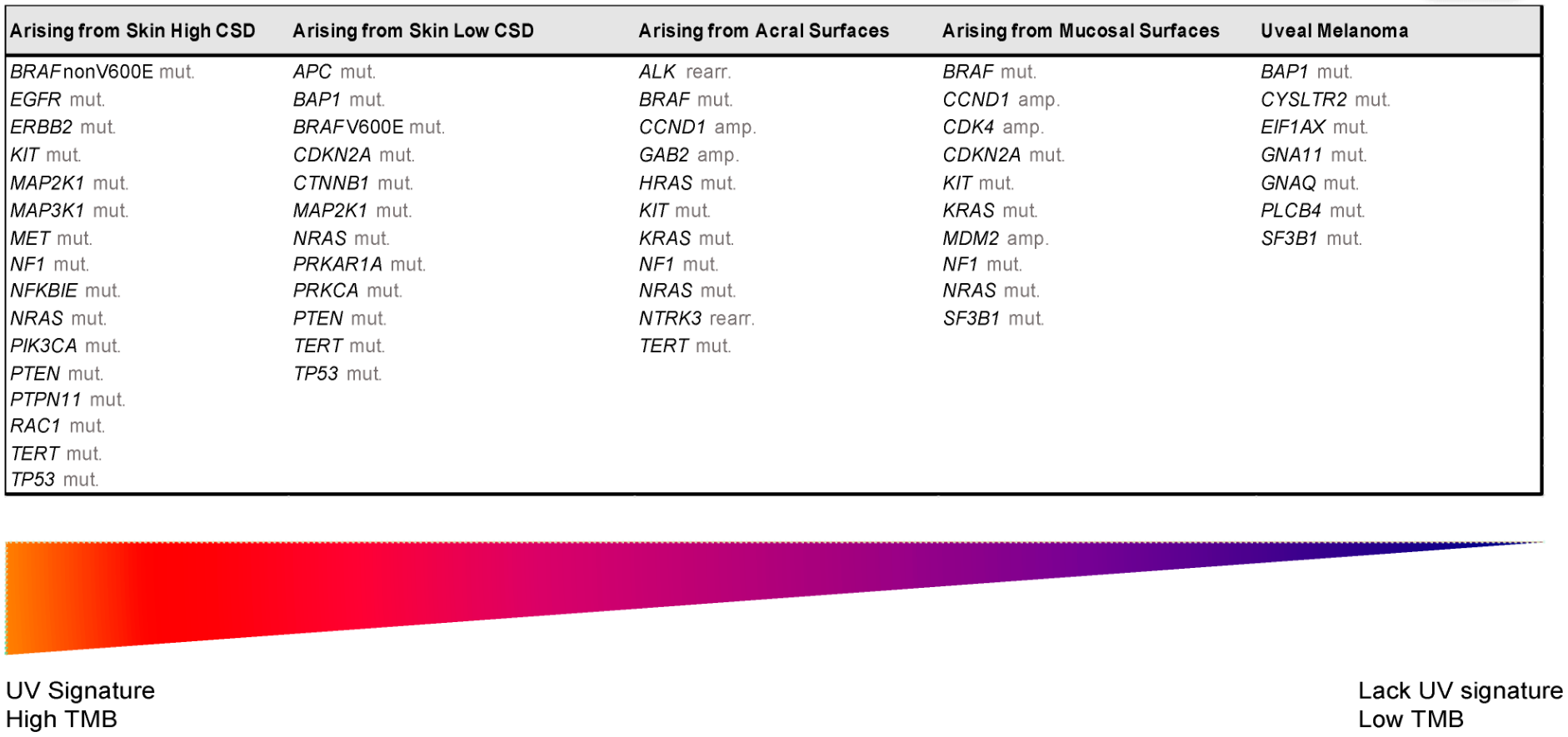
Despite recent molecular advances in melanoma characterization, paramount to diagnosis of a melanocytic skin lesion is the integration of several histopathological criteria with the clinical features. In many cases, general morphological criteria for atypia are often the subject of disagreement and inter-observer variability, especially in non-conventional lesions [35]. The World Health Organization (WHO) recognizes these challenges and incorporate the known molecular pathways in the latest WHO melanocytic tumor classification, introducing the concept of “intermediate” lesions. As stated in a recent review on the topic, this multidimensional classification showed that the view of melanocytic tumors as either benign or malignant might no longer be the proper approach [36]. Thus, WHO 2018 indicates nine categories/pathways leading to melanoma, each with specific genetic drivers (Table 1). Furthermore, melanomas can be clustered in three main subtypes, according to the degree of CSD (Table 1 and Figure 2) [37]. The largest group are melanomas associated with low-CSD or superficial spreading melanomas, which often arise on the trunk and proximal areas of the extremities. The most frequent molecular alteration in these melanomas is the BRAFV600E mutation [38]. In addition, TERT promoter mutations and CDKN2A mutations are also found in the majority of cases. PTEN and TP53 are commonly observed in advanced tumors. Lentigo maligna and desmoplastic melanomas are considered tumors associated with high-CSD. These melanomas arise on heavily sun-damaged skin, such as the face or hands, and affect older people. Molecularly, they often have a high mutation load and may harbor NRAS, BRAF non-V600E, or NF1 mutations. TERT promoter mutations and CDKN2A are also frequently found in these melanomas, and KIT mutations are found in a subset of cases. Interestingly, the number of mutations increases with the CSD grade (Figure 2), and desmoplastic melanomas harbor the highest tumor mutation burden. The category of “low to non-UV exposure/CSD” melanomas includes Spitz melanomas, acral melanomas, mucosal melanomas, melanomas developed from congenital nevi and blue nevi, and uveal melanomas. These melanomas rarely harbor BRAFNRAS, or NF1 mutations (triple wild-type) [37]. A subset of acral and mucosal melanomas may have KIT mutations, in addition to gene amplifications and structural rearrangements, most frequently of the CCND1 gene and SF3B1. Therefore, genomic studies have subsequently exemplified that acral and mucosal melanomas are biologically distinct from their cutaneous counterparts at sun-exposed sites. Spitz melanomas show a particular oncogenic signaling pathway involving tyrosine kinase or serine-threonine kinase fusions, and melanomas in blue nevus and uveal melanomas are characterized by GNA11 or GNAQ mutations [38].
Figure 2. Genomic alterations of melanoma subtypes defined by UV exposure. Abbreviations: amp, amplification; CSD, cumulative sun damage; rearr, rearrangement; TMB, tumor mutational burden; UV, ultraviolet.
Table 1. The classification of melanomas (modified from 2018 World Health Organization Classification).
UV Exposure Categories Melanoma Subtype Key Molecular Genes
Low UV/CSD I Superficial spreading melanoma BRAFV600 mut
CDKN2A mut
NRAS mut
TERT mut
PTEN mut
TP53 mut
High UV/CSD II Lentigo maligna melanoma NRAS mut
BRAFnon-V600E mut
KIT mut
TERT mut
CDKN2A mut
PTEN mut
TP53 mut
III Desmoplastic melanoma NF1 mut
NFKBIE mut
NRAS mut
PIK3CA mut
Low or no UV/CSD IV Spitz melanoma ALK rearr
NTRK1 rearr
NRTK3 rearr
CDKN2A mut
HRAS mut
V Acral melanoma KIT mut
NRAS or BRAF mut
ALK rearr
NRTK3 rearr
CDKN2A mut CCND1 amp
TERT mut
VI Mucosal melanoma KIT mut
NRAS or BRAF mut
CDKN2A mut
SF3B1 mut
CCND1 amp
CDK4 mut
MDM2 amp
VII Melanoma in congenital nevus NRAS mut BRAFV600E mut
VIII Melanoma in blue nevus GNA11 mut
GNAQ mut
CYSLTR2 mut
BAP1 mut
EIFAX mut
SF3B1 mut
IX Uveal melanoma GNA11 mut
GNAQ mut
CYSLTR2 mut
PLCB4 mut
BAP1 mut
EIFAX mut
SF3B1 mut
Abbreviations: amp, amplification; CSD, cumulative sun damage; mut, mutation; rearr, rearrangement.
Certainly, to reduce diagnostic uncertainties and maintain a diagnostic approach based on the WHO 2018 classification, histological assessment should be accompanied by basic immunohistochemistry (IHC) and molecular tests. Recent recommendations of the European Society of Pathology, the European Organization for Research and Treatment of Cancer, and the EURACAN for the diagnosis of intermediate melanocytic proliferations and melanoma variants indicate that most pathology laboratories should perform basic IHC tests, such as: HMB-45; SOX10; MITF, tyrosinase, MART-1; P16; Ki-67/MIB1; BAP1 (BRCA1-associated protein 1); β-catenin; PRAME; and at least one molecular method to detect BRAF codon 600 and NRAS mutations [36]. The most difficult cases that require complementary studies should be analyzed in specialized referral centers, where laboratories can determine a higher grade in a given lesion or the identification of molecular targets that can benefit from targeted therapy.

References

  1. Cress, R.D.; Holly, E.A. Incidence of cutaneous melanoma among non-Hispanic whites, Hispanics, Asians, and blacks: An analysis of california cancer registry data, 1988–1993. Cancer Causes Control. 1997, 8, 246–252.
  2. Ridky, T.W. Nonmelanoma skin cancer. J. Am. Acad. Dermatol. 2007, 57, 484–501.
  3. Jhappan, C.; Noonan, F.P.; Merlino, G. Ultraviolet radiation and cutaneous malignant melanoma. Oncogene 2003, 22, 3099–3112.
  4. Siegel, R.L.; Miller, K.D.; Jemal, A. Cancer statistics, 2020. CA: A Cancer J. Clin. 2020, 70, 7–30.
  5. Rastrelli, M.; Tropea, S.; Rossi, C.R.; Alaibac, M. Melanoma: Epidemiology, risk factors, pathogenesis, diagnosis and classification. In Vivo (Athens, Greece) 2014, 28, 1005–1011.
  6. Michielin, O.; van Akkooi, A.C.J.; Ascierto, P.A.; Dummer, R.; Keilholz, U. Cutaneous melanoma: ESMO Clinical Practice Guidelines for diagnosis, treatment and follow-up. Ann. Oncol. 2019, 30, 1884–1901.
  7. Hodi, F.S.; O’Day, S.J.; McDermott, D.F.; Weber, R.W.; Sosman, J.A.; Haanen, J.B.; Gonzalez, R.; Robert, C.; Schadendorf, D.; Hassel, J.C.; et al. Improved survival with ipilimumab in patients with metastatic melanoma. N. Engl. J. Med. 2010, 363, 711–723.
  8. Fellner, C. Ipilimumab (yervoy) prolongs survival in advanced melanoma: Serious side effects and a hefty price tag may limit its use. P T 2012, 37, 503–530.
  9. Chapman, P.B.; Hauschild, A.; Robert, C.; Haanen, J.B.; Ascierto, P.; Larkin, J.; Dummer, R.; Garbe, C.; Testori, A.; Maio, M.; et al. Improved survival with vemurafenib in melanoma with BRAF V600E mutation. N. Engl. J. Med. 2011, 364, 2507–2516.
  10. Sosman, J.A.; Kim, K.B.; Schuchter, L.; Gonzalez, R.; Pavlick, A.C.; Weber, J.S.; McArthur, G.A.; Hutson, T.E.; Moschos, S.J.; Flaherty, K.T.; et al. Survival in BRAF V600-mutant advanced melanoma treated with vemurafenib. N. Engl. J. Med. 2012, 366, 707–714.
  11. Sample, A.; He, Y.-Y. Mechanisms and prevention of UV-induced melanoma. Photodermatol. Photoimmunol. Photomed. 2018, 34, 13–24.
  12. D’Orazio, J.; Jarrett, S.; Amaro-Ortiz, A.; Scott, T. UV radiation and the skin. Int. J. Mol. Sci. 2013, 14, 12222–12248.
  13. Bevona, C.; Goggins, W.; Quinn, T.; Fullerton, J.; Tsao, H. Cutaneous melanomas associated with nevi. Arch. Dermatol. 2003, 139, 1620–1624; discussion 1624.
  14. Dessinioti, C.; Antoniou, C.; Katsambas, A.; Stratigos, A.J. Melanocortin 1 receptor variants: Functional role and pigmentary associations. Photochem. Photobiol. 2011, 87, 978–987.
  15. Rossi, M.; Pellegrini, C.; Cardelli, L.; Ciciarelli, V.; Di Nardo, L.; Fargnoli, M.C. Familial Melanoma: Diagnostic and Management Implications. Dermatol. Pract. Concept 2019, 9, 10–16.
  16. Goldstein, A.M.; Tucker, M.A. Genetic epidemiology of cutaneous melanoma: A global perspective. Arch. Dermatol. 2001, 137, 1493–1496.
  17. Hanahan, D.; Weinberg, R.A. Hallmarks of Cancer: The Next Generation. Cell 2011, 144, 646–674.
  18. Raman, M.; Chen, W.; Cobb, M.H. Differential regulation and properties of MAPKs. Oncogene 2007, 26, 3100–3112.
  19. Eliceiri, B.P.; Klemke, R.; Strömblad, S.; Cheresh, D.A. Integrin alphavbeta3 requirement for sustained mitogen-activated protein kinase activity during angiogenesis. J. Cell Biol. 1998, 140, 1255–1263.
  20. Davies, H.; Bignell, G.R.; Cox, C.; Stephens, P.; Edkins, S.; Clegg, S.; Teague, J.; Woffendin, H.; Garnett, M.J.; Bottomley, W.; et al. Mutations of the BRAF gene in human cancer. Nature 2002, 417, 949–954.
  21. Nissan, M.H.; Pratilas, C.A.; Jones, A.M.; Ramirez, R.; Won, H.; Liu, C.; Tiwari, S.; Kong, L.; Hanrahan, A.J.; Yao, Z.; et al. Loss of NF1 in cutaneous melanoma is associated with RAS activation and MEK dependence. Cancer Res. 2014, 74, 2340–2350.
  22. Akbani, R.; Akdemir, K.C.; Aksoy, B.A.; Albert, M.; Ally, A.; Amin, S.B.; Arachchi, H.; Arora, A.; Auman, J.T.; Ayala, B.; et al. Genomic Classification of Cutaneous Melanoma. Cell 2015, 161, 1681–1696.
  23. Scheid, M.P.; Woodgett, J.R. PKB/AKT: Functional insights from genetic models. Nat. Rev. Mol. Cell Biol. 2001, 2, 760–768.
  24. Stahl, J.M.; Cheung, M.; Sharma, A.; Trivedi, N.R.; Shanmugam, S.; Robertson, G.P. Loss of PTEN promotes tumor development in malignant melanoma. Cancer Res. 2003, 63, 2881–2890.
  25. Lito, P.; Pratilas, C.A.; Joseph, E.W.; Tadi, M.; Halilovic, E.; Zubrowski, M.; Huang, A.; Wong, W.L.; Callahan, M.K.; Merghoub, T.; et al. Relief of profound feedback inhibition of mitogenic signaling by RAF inhibitors attenuates their activity in BRAFV600E melanomas. Cancer Cell 2012, 22, 668–682.
  26. Kong, Y.; Si, L.; Li, Y.; Wu, X.; Xu, X.; Dai, J.; Tang, H.; Ma, M.; Chi, Z.; Sheng, X.; et al. Analysis of mTOR Gene Aberrations in Melanoma Patients and Evaluation of Their Sensitivity to PI3K-AKT-mTOR Pathway Inhibitors. Clin. Cancer Res. 2016, 22, 1018–1027.
  27. Goel, V.K.; Lazar, A.J.F.; Warneke, C.L.; Redston, M.S.; Haluska, F.G. Examination of Mutations in BRAF, NRAS, and PTEN in Primary Cutaneous Melanoma. J. Invest. Dermatol. 2006, 126, 154–160.
  28. Brown, V.L.; Harwood, C.A.; Crook, T.; Cronin, J.G.; Kelsell, D.P.; Proby, C.M. p16INK4a and p14ARF tumor suppressor genes are commonly inactivated in cutaneous squamous cell carcinoma. J. Invest. Dermatol 2004, 122, 1284–1292.
  29. Alos, L.; Fuster, C.; Castillo, P.; Jares, P.; Garcia-Herrera, A.; Marginet, M.; Agreda, F.; Arance, A.; Gonzalvo, E.; Garcia, M.; et al. TP53 mutation and tumoral PD-L1 expression are associated with depth of invasion in desmoplastic melanomas. Ann. Transl. Med. 2020, 8, 1218.
  30. Sini, M.C.; Manca, A.; Cossu, A.; Budroni, M.; Botti, G.; Ascierto, P.A.; Cremona, F.; Muggiano, A.; D’Atri, S.; Casula, M.; et al. Molecular alterations at chromosome 9p21 in melanocytic naevi and melanoma. Br. J. Dermatol. 2008, 158, 243–250.
  31. Fargnoli, M.C.; Gandini, S.; Peris, K.; Maisonneuve, P.; Raimondi, S. MC1R variants increase melanoma risk in families with CDKN2A mutations: A meta-analysis. Eur. J. Cancer 2010, 46, 1413–1420.
  32. Curtin, J.A.; Fridlyand, J.; Kageshita, T.; Patel, H.N.; Busam, K.J.; Kutzner, H.; Cho, K.-H.; Aiba, S.; Bröcker, E.-B.; LeBoit, P.E.; et al. Distinct Sets of Genetic Alterations in Melanoma. N. Engl. J. Med. 2005, 353, 2135–2147.
  33. Garraway, L.A.; Widlund, H.R.; Rubin, M.A.; Getz, G.; Berger, A.J.; Ramaswamy, S.; Beroukhim, R.; Milner, D.A.; Granter, S.R.; Du, J.; et al. Integrative genomic analyses identify MITF as a lineage survival oncogene amplified in malignant melanoma. Nature 2005, 436, 117–122.
  34. Carreira, S.; Goodall, J.; Aksan, I.; La Rocca, S.A.; Galibert, M.D.; Denat, L.; Larue, L.; Goding, C.R. Mitf cooperates with Rb1 and activates p21Cip1 expression to regulate cell cycle progression. Nature 2005, 433, 764–769.
  35. Piepkorn, M.W.; Longton, G.M.; Reisch, L.M.; Elder, D.E.; Pepe, M.S.; Kerr, K.F.; Tosteson, A.N.A.; Nelson, H.D.; Knezevich, S.; Radick, A.; et al. Assessment of Second-Opinion Strategies for Diagnoses of Cutaneous Melanocytic Lesions. JAMA Netw Open 2019, 2, e1912597.
  36. Ferrara, G.; Argenziano, G. The WHO 2018 Classification of Cutaneous Melanocytic Neoplasms: Suggestions From Routine Practice. Front. Oncol 2021, 11, 675296.
  37. WHO. WHO Classification of Skin Tumours, 4th ed.; Elder, D.E., Massi, D., Scolyer, R.A., Willemze, R., Eds.; International Agency for Research on Cancer: Lyon, France, 2018; p. 470.
  38. Hayward, N.K.; Wilmott, J.S.; Waddell, N.; Johansson, P.A.; Field, M.A.; Nones, K.; Patch, A.M.; Kakavand, H.; Alexandrov, L.B.; Burke, H.; et al. Whole-genome landscapes of major melanoma subtypes. Nature 2017, 545, 175–180.
More
Information
Subjects: Oncology
Contributors MDPI registered users' name will be linked to their SciProfiles pages. To register with us, please refer to https://encyclopedia.pub/register : , , , ,
View Times: 727
Revisions: 2 times (View History)
Update Date: 22 Apr 2022
1000/1000
ScholarVision Creations