Submitted Successfully!
To reward your contribution, here is a gift for you: A free trial for our video production service.
Thank you for your contribution! You can also upload a video entry or images related to this topic.
Version Summary Created by Modification Content Size Created at Operation
1 + 2197 word(s) 2197 2022-02-23 04:11:46 |
2 format correction -39 word(s) 2158 2022-03-04 04:12:52 |

Video Upload Options

Do you have a full video?

Confirm

Are you sure to Delete?
Cite
If you have any further questions, please contact Encyclopedia Editorial Office.
Dick, A. Bioisosteric Replacement in Anti-HIV Drug Design. Encyclopedia. Available online: https://encyclopedia.pub/entry/20172 (accessed on 29 March 2024).
Dick A. Bioisosteric Replacement in Anti-HIV Drug Design. Encyclopedia. Available at: https://encyclopedia.pub/entry/20172. Accessed March 29, 2024.
Dick, Alexej. "Bioisosteric Replacement in Anti-HIV Drug Design" Encyclopedia, https://encyclopedia.pub/entry/20172 (accessed March 29, 2024).
Dick, A. (2022, March 03). Bioisosteric Replacement in Anti-HIV Drug Design. In Encyclopedia. https://encyclopedia.pub/entry/20172
Dick, Alexej. "Bioisosteric Replacement in Anti-HIV Drug Design." Encyclopedia. Web. 03 March, 2022.
Bioisosteric Replacement in Anti-HIV Drug Design
Edit

Bioisosteric replacement is a powerful tool for modulating the drug-like properties, toxicity, and chemical space of experimental therapeutics. The use of bioisosteres and the introduction of structural changes to the lead compound allows the chemist to alter the compound’s size, shape, electronic distribution, polarizability, dipole, polarity, lipophilicity, and pKa, while still retaining potent target engagement.

bioisosteres HIV-1 antiviral computer-aided drug design envelope reverse transcriptase protease integrase tat Vif

1. Introduction

The design and development of a lead compound into a drug is a laborious and often costly process, with most candidates failing due to metabolism and pharmacokinetics issues rather than potency. Bioisosteric replacement is a strategy used by medicinal chemists to address these limitations while still retaining the potency/efficacy of the initial lead compound. The use of bioisosteres and the introduction of structural changes to the lead compound allows the chemist to alter the compound’s size, shape, electronic distribution, polarizability, dipole, polarity, lipophilicity, and pKa, while still retaining potent target engagement. Therefore, the bioisosteric approach can be used for the rational modification of a lead compound towards a more attractive therapeutic agent with improved potency, selectivity, altered physical, metabolic, toxicological properties with the bonus of generating novel intellectual property (IP).

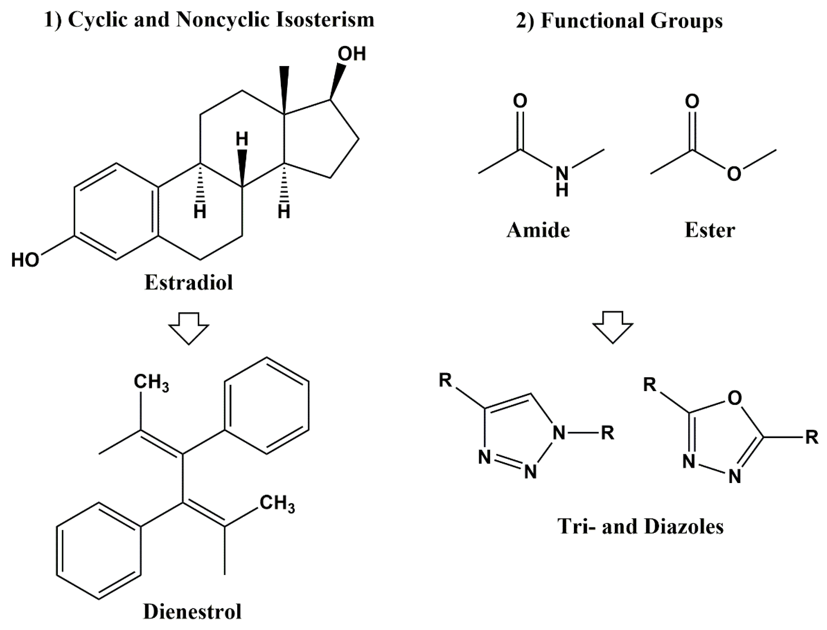
2. Principle of Bioisosterism and Historical Background

The term isosterism was first introduced by Irving Langmuir in 1919 during his studies on similarities of physicochemical properties of atoms, groups and molecules [1]. He described compounds or groups of atoms with the same number of atoms and electrons, such as N2 and CO, N2O and CO2, or N3- and NCO- as isosteres, and, based on these similarities of the arrangement of electrons, he defined 21 groups of isosteres. H. G. Grimm further developed this definition in the early 1920s. This early hypothesis of bioisosterism describes the ability of certain chemical groups to mimic other chemical groups [2][3]. Accordingly, the addition of a hydride to an atom gives to the resulting pseudoatom the properties of the atom with the next highest atomic number (Table 1) [4].
Table 1. Grimm’s Hydride Displacement Law.

C

N

O

F

Ne

Na+


CH

NH

OH

FH

-


CH2

NH2

OH2

FH2+


CH3

NH3

OH3+


CH4

NH4+

Each vertical column represents an isostere, according to Grimm. In 1932, Hans Erlenmeyer extended Grimm’s definition of isosteres as atoms, ions, and molecules in which the peripheral layers of electrons (valence electrons) are considered as identical (Table 2).
Table 2. Isosteres based on valence electron number.

Number of Valence Electrons

4

5

6

7

8

N+

P

S

Cl

ClH

P+

As

Se

Br

BrH

S+

Sb

Te

I

IH

As+


PH

SH

SH2

Sb+


PH2

PH3

Based on its application in biological systems, Harris Friedman introduced the term “bioisostere” in 1950 that included all atoms and molecules which fit the broadest definition for isosteres and have similar biological activity, either agonistic or antagonistic [5]. Today, the even more broadened definition of bioisosteres introduced by Alfred Burger in the early 1990s is in use. Accordingly, bioisosteres are “Compounds or groups that possess near-equal molecular shapes and volumes, approximately the same distribution of electrons, and similar physical properties” [6].

3. Classical and Non-Classical Bioisosteres

In the 1970s, Alfred Bruger defined bioisosteres as either classical (atom number, number of valence electrons, and degree of unsaturation) or non-classical (similar pKa, electrostatic potentials, orbital occupation/HOMOs and LUMOs) [7]. Classical bioisosteres can be further subdivided into five classes: 1) monovalent atoms or groups (D and H; F and H; C and Si; Cl, Br, SH, and OH; NH and OH; RSH and ROH, –Cl, –PH2, –SH), 2) divalent atoms or groups (–CH2, –NH, –O, –S, –Se–, -COCH2-), 3) trivalent atoms or groups (–CH=, –N=, -P=, -As=), 4) tetravalent atoms or groups (>C<, >Si< and =C=, =N+=, =P+=), and 5) ring equivalents (-CH=CH-, -S- (e.g., benzene, thiophene), -CH=, -N= (e.g., benzene, pyridine), -O-, -S-, -CH2- (e.g., tetrahydrofuran, tetrahydrothiophene, cyclopentane) [4][8]. Non-classical bioisosteres are a more sophisticated mimicry of the emulated counterparts and they do not strictly obey the steric and electronic definition of classical isosteres. As non-classical bioisosteres can significantly differ in electronic distribution, physicochemical, steric and topological properties, they have found beneficial applications in drug discovery research. Non-classical bioisosteres are subdivided into two groups: 1) cyclic vs. non-cyclic and 2) exchangeable functional groups (Figure 1) [4].
Figure 1. Non-Classical Bioisosterism.

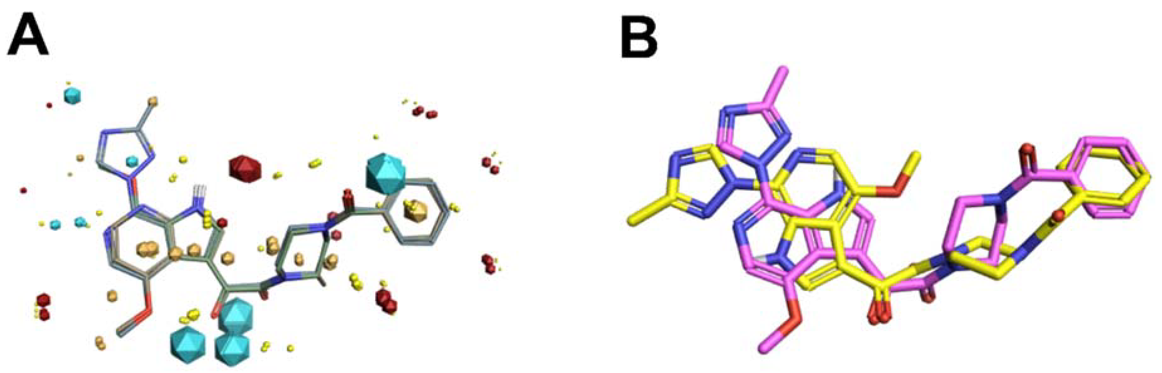
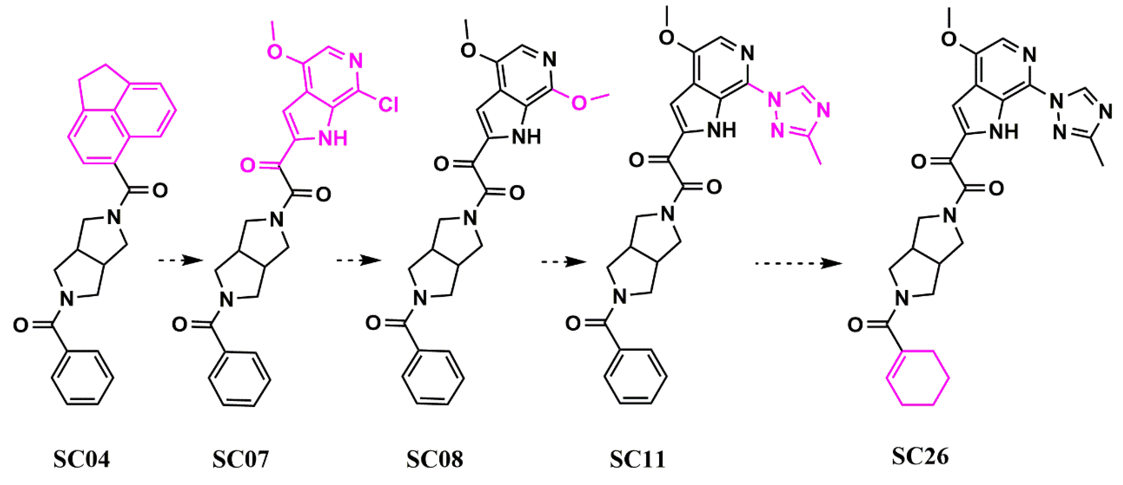
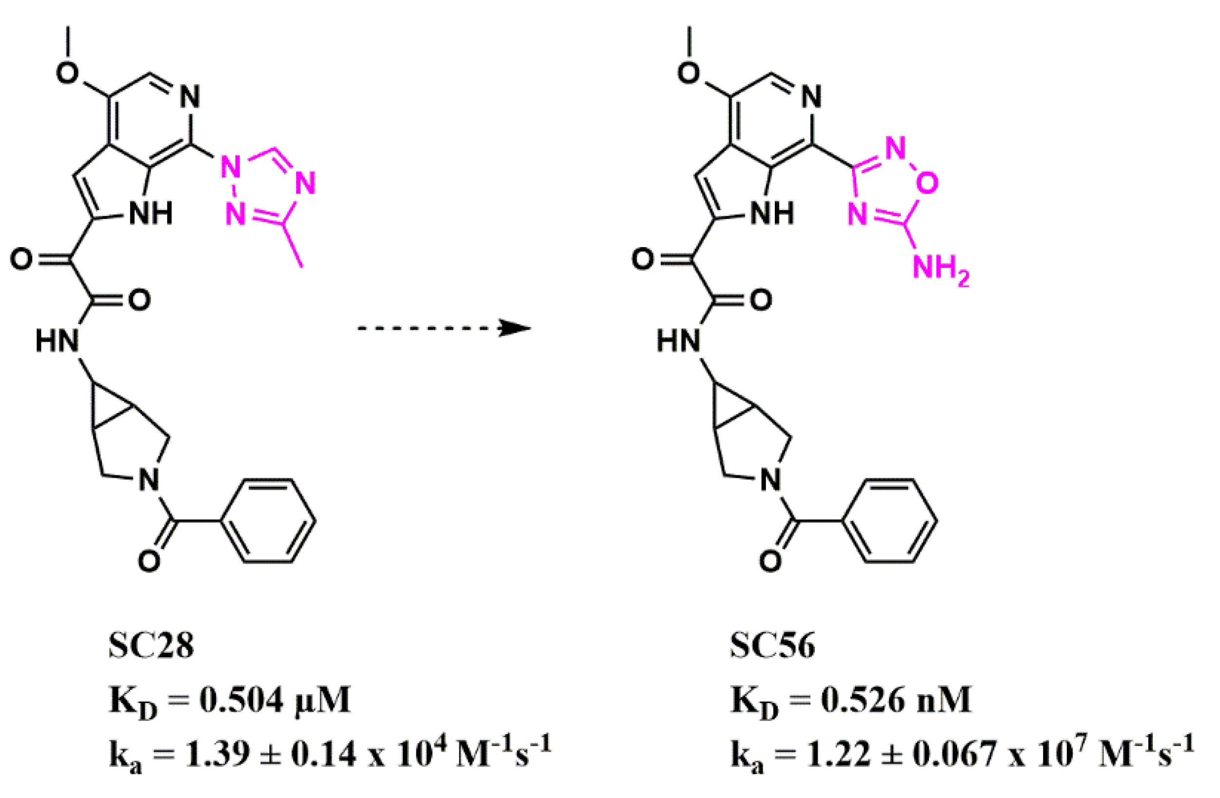
4. In Silico Molecular Field-Based Scaffold Hopping for Non-Classical Bioisostere Identification

Scaffold hopping using bioisosteric replacement has always been an integral part of the conventional drug development process but has expanded in recent years due to the availability of facile computational methods. High throughput screening (HTS), which tests thousands or millions of compounds, is a mainstay in the pharmaceutical industry. However, by applying computationally driven bioisosteric replacement to such initial hits, chemotype diversification can be readily achieved, which in turn increases the probability of successfully carrying one of these chemotypes through to the clinic.
Typical approaches that do not use molecular field points tend to view the bioisosteric fragment in a vacuum, taking only fragment comparison into account and not factoring in the effect of the replacement across the whole molecule. Using molecular fields to facilitate a comparison of the entire spatial physicochemical properties across the entire molecule, rather than the structure alone, can and has led to discoveries of non-intuitive bioisosteric replacements that have positively modulated drug-like properties while retaining target engagement [9][10][11][12][13]. However, molecular fields are continuous and vary with the conformation of the compound, and sampling at defined points in the three-dimensional grid can be computationally expensive (Figure 2A). Therefore, program collections such as Cresset (Cresset, Litlington, UK) use only extrema of these fields, the so-called field point, where intermolecular interactions most likely occur (Figure 2B).
Figure 2. Electrostatic (blue/red), van der Waals (yellow) and hydrophobic (beige) contours (A) and Field Point maxima (B) of an HIV-1 entry inhibitor.
This calculation and comparison of field points is based on the eXtended Electron Density Force Field Theory (XED) [14][15]. XED takes into account positive and negative electrostatic, Van der Waals, and hydrophobic field points. In contrast to most molecular mechanics force field methods that apply atom-centered partial charges, XED generates negative pseudo-orbitals surrounding the positive nuclei and distributes partial charges according to the orbital location. XED, therefore, mimics the electron distribution in a more accurate way by taking π-clouds and lone pairs into account, which play important roles in biological systems. In practice, Field Points of two or more compounds can be compared by sampling conformations to maximize the fields for optimal alignment, and in contrast to a structural alignment, new compounds with different structures but similar numbers and distribution of Field Points can be found. This so-called “templating” of molecules can also be used to generate a high-content pharmacophore for therapeutically interesting targets for which there is no known information regarding the small molecule binding site.

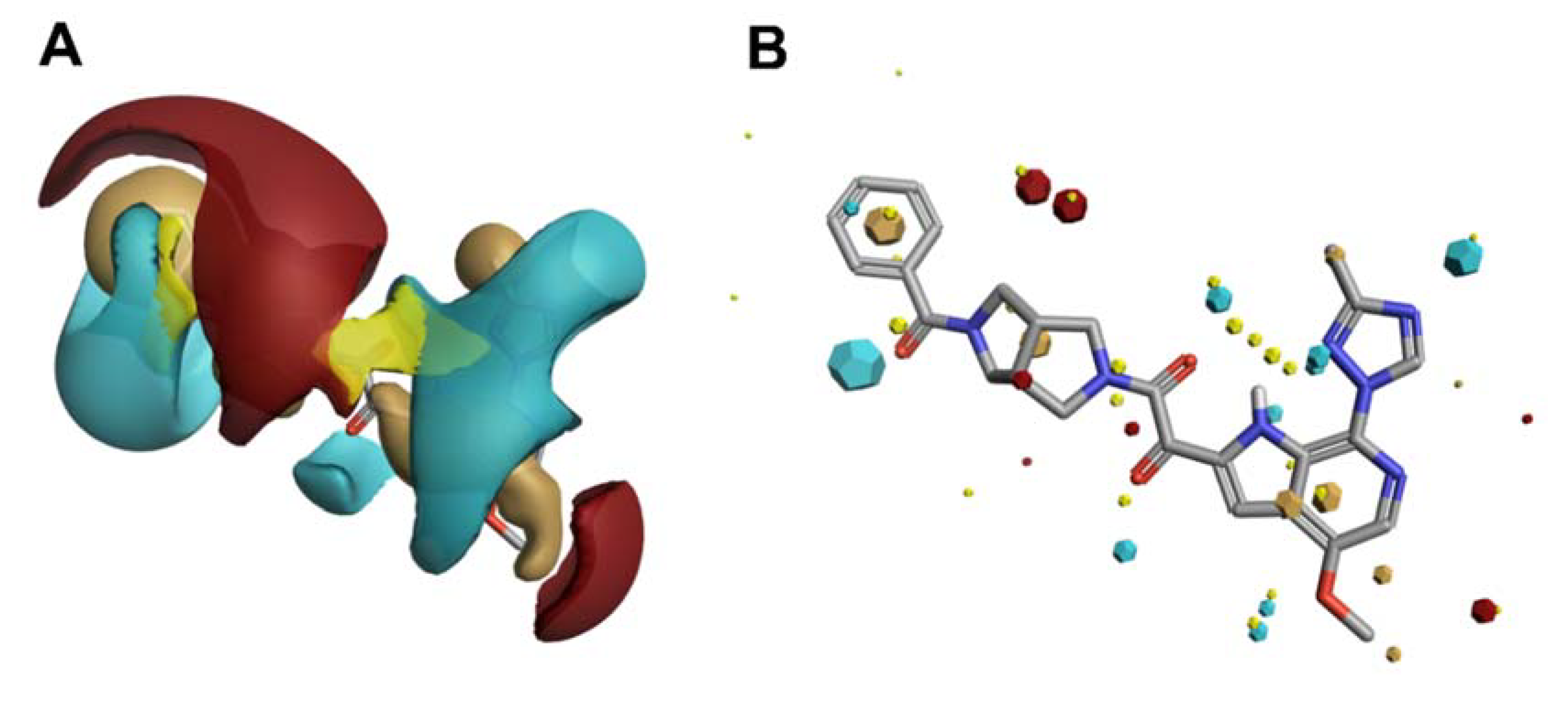
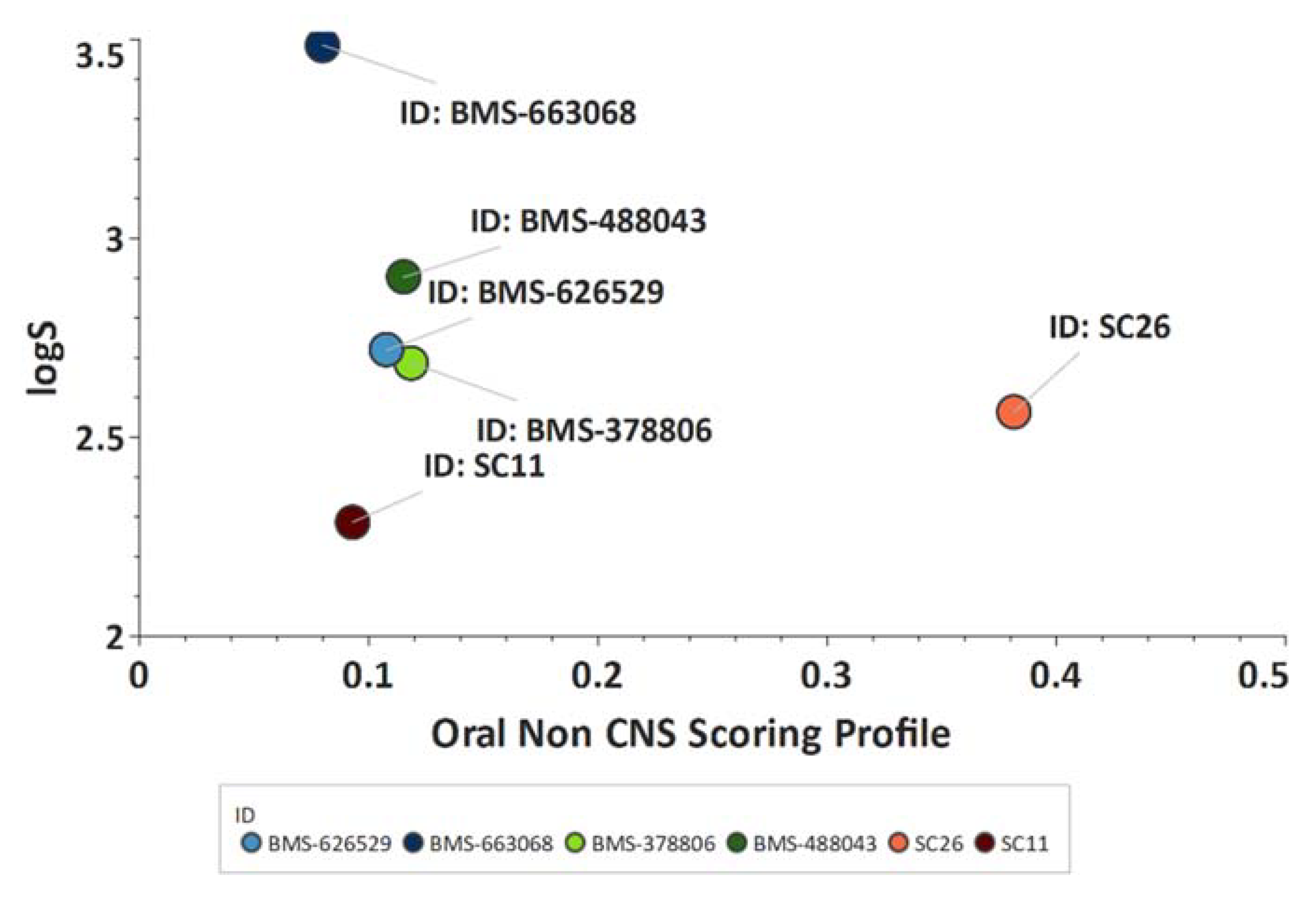
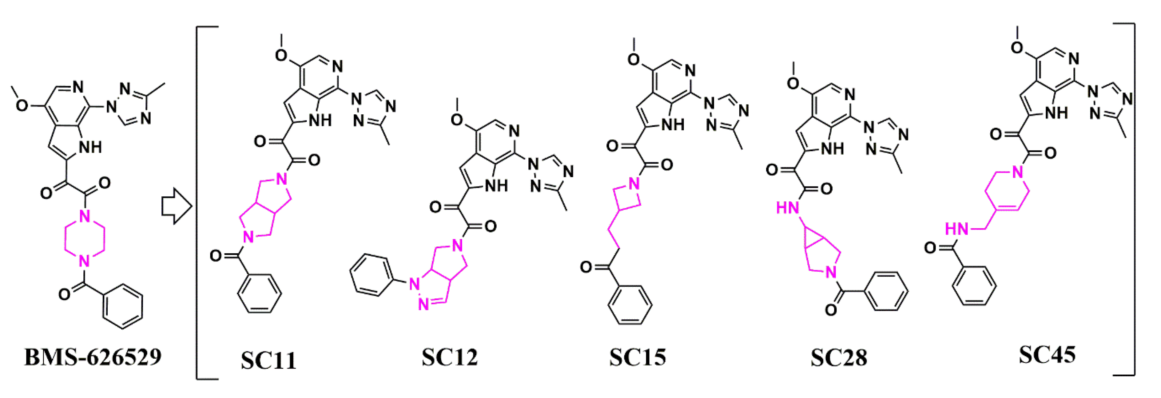
Scaffold Hopping for Potency and ADME Improvement of HIV-1 Entry Leads

The inhibition of HIV-1 entry is an attractive, yet underexploited therapeutic approach for several reasons. First, much of the entry process occurs in water-soluble compartments easily accessible to drugs. Second, both viral and host cell components involved in HIV-1 entry have been identified and can be targeted. Third, because inhibition of virus entry prevents the host cell from becoming permanently infected, such inhibitors are potentially useful in many different modalities, including pre-exposure prophylactics, prophylactics, and microbicides [16].
The most potent and broadly acting HIV-1 inhibitors to date are piperazine-based introduced by Bristol-Myers Squibb [17]. However, this class has low bioavailability, resulting from low solubility and poor dissolution. Bristol-Myers Squibb has partially overcome these limitations by adopting a prodrug approach with BMS-663068 that shows increased solubility in the gut. However, after removal of the phosphonooxymethyl-solubilizing group in the gut, the properties of the active compound, BMS-626529, dominate and, despite its potency, 1.2 g per day is required to achieve an effective plasma concentration [18]. Clearly, an entry inhibitor with more intrinsic drug-like properties would be preferable [19]. Therefore, the researchers used field-based three-dimensional similarity virtual screening experiments using Blaze (Cresset, Litlington, UK) with a high-content field-based pharmacophore template derived from BMS-626529 and two of its predecessors, BMS-488043 and BMS-378806, to identify novel scaffolds that could function as entry inhibitors [20]. Field templating was implemented, no structural information regarding the binding site nor the bioactive conformation of the BMS compounds was available. FieldTemplater (Forge) allows the individual to generate a bioactive conformation hypothesis, and a field-point pattern pharmacophore, in the absence of structural information, as long as multiple, distinct compounds are known and are supposed to interact with the same site on the target. Figure 3 shows the field-point template generated and used in the reasearhers' HIV-1 entry inhibitor studies (Figure 3A), in addition to a comparison of the template to the experimentally determined bioactive conformation of BMS-626529 (Figure 3B) [21][22].
Figure 3. (A) Field-based template containing a single conformation of compounds BMS-378806 (pink), BMS-488043 (lime green), and BMS-626529 (teal green) aligned based on their three-dimensional field point patterns. Negatively charged field points are shown in blue; positively charged field points are red; van der Waals/shape field points are displayed in yellow; centers of hydrophobicity are shown in orange. The dodecahedral size of the field points is proportional to the magnitude of the extrema. (B) Structural superimposition of BMS-62529 conformer generated with FieldTemplater (pink) and experimentally obtained crystal structure in complex with HIV-1 BG505 SOSIP.664 (yellow, PDB code: 5U7O).
From this field-point pharmacophore screen using Blaze (Cresset UK), the researchers identified hits with piperazine-based cores similar to the BMS chemotypes but with potencies in the low micromolar range, ranking from 13 to 153 µM using a HIV-1 YU-2 Env pseudotyped virus in a single-round infection assay (SRIA). Therefore, to obtain a truly novel core scaffold, the researchers chose to conduct bioisosteric replacement (scaffold hopping) with Spark (Cresset, UK). The results of this indicated that replacing the piperazine group with a dipyrrolidine moiety would be viable. The resulting compound (SC04) showed lower potency than the piperazine-based compounds (IC50 HIV-1 YU-2 = 70 ± 6 μM; IC50 HIV-1 JR-CSF = 100 ± 30 μM using a pseudotyped virus in an SRIA) but demonstrated specificity, making it perfect for further optimization in potency (Figure 4).
Figure 4. Evolution (from left to right) of HIV-1 entry inhibitors to improve potency and ADME properties. Structural changes between each compound are highlighted in pink.
Using Spark (Cresset, UK) and a fragment library generated from PubChem by fragmenting compounds with similarities to BMS-488043, the researchers researched whether the dipyrrolidine could support nanomolar potency, whilst retaining specificity. Sequentially, the researchers generated SC07 (IC50 HIV-1 JR-CSF = 0.98 ± 0.06 μM in an SRIA), SC08 (IC50 HIV-1 JR-CSF = 0.09 ± 0.01 μM in an SRIA), and SC11 (IC50 HIV-1 JR-CSF values of 0.0008 ± 0.0004 μM in an SRIA). This was the first time that the piperazine core in this class of inhibitors had been successfully changed whilst retaining high potency. Finally, the replacement of the terminal phenyl in SC11 by cyclohexene in SC26 resulted in the first dipyrolrolodine-scaffolded highly potent HIV-1 entry inhibitor (IC50 HIV-1 JR-CSF of 2.0 ± 0.1 nM; IC50 HIV-1 HxBc2 of 0.6 ± 0.01 nM in an SRIA), with significantly improved predicted ADME properties (as indicated by an increase in the Oral Non-CNS Drug-like Score in StarDrop, Optibrium, UK; Figure 5).
Figure 5. Improved ADME properties of SC26. Plot showing the StarDrop (Optibrium, Ltd., Cambridge, UK)–derived logS versus the score from a multimetric oral non-CNS profile for the BMS and SC compounds. Scores range from 0 to 1, with 0 suggesting extremely non-drug-like, and 1 suggesting the perfect drug.
Following this initial success, the researchers then used similar methods to extend the core chemotypes in the HIV-1 entry inhibitor class. This resulted in the creation of five distinct core chemotypes from the original piperazine BMS-626529 that support specificity and nanomolar potencies (Figure 6 and Table 3) [23].
Figure 6. Scaffold hopping in the core region of an HIV-1 entry inhibitor. Structural changes in the core region between each compound are highlighted in pink.
Table 3. Specificity and potency (in µM) of SC compounds against HIV-1J R-CSF, HIV-1 B41, HIV-1 HxBc2 Env, and HIV-1 AMLV pseudotyped HIV-1 using a single-round infection assay.

Compound

IC50 JR-CSF

IC50 B41

IC50 HxBc2

IC50 AMLV

SC11

0.0008 ± 0.0001

0.002 ± 0.0002

0.001 ± 0.0001

N.A.

SC12

0.008 ± 0.002

0.006 ± 0.003

0.080 ± 0.020

N.A.

SC15

0.003 ± 0.001

0.007 ± 0.001

0.009 ± 0.001

N.A.

SC28

0.096 ± 0.019

0.085 ± 0.03

0.069 ± 0.014

N.A.

SC45

0.224 ± 0.017

0.350 ± 0.030

0.380 ± 0.030

N.A.

N.A. = not active; N.D. = not determined.
Stemming from these initial successes, the researchers have since applied this workflow to identify new bioisosteres for the methyltriazole-azaindole moiety, as the main potency determinant of this entry inhibitor class and identified a compound (SC56), wherein the methyltriazole in SC28 was replaced with an amine-oxadiazole (Figure 7) [24]. This compound displayed a 1000-fold increased affinity for the B41 SOSIP Env, as judged by surface plasmon resonance (SPR).
Figure 7. Modulation of binding kinetics using bioisosteric replacement in HIV-1 entry inhibitor design. The structural change between the two compounds is highlighted in pink. Affinity and kinetic parameters were determined using a surface plasmon resonance assay.
Finally, in a recently published study, the researchers have also demonstrated that SC28, with the azabicyclo-hexane core scaffold, has twice the metabolic stability of BMS-626529 [1]. This approach highlights bioisosteric replacement as a powerful tool that can be utilized to rapidly diversify chemotypes but can improve drug–target kinetic profiles and metabolism in the drug development process.

References

  1. Langmuir, I. Isomorphism, Isosterism and Covalence. J. Am. Chem. Soc. 1919, 41, 1543–1559.
  2. Grimm, H.G. Structure and Size of the Non-metallic Hydrides. Z. Electrochem. 1925, 31, 474–480.
  3. Grimm, H.G. On the Systematic Arrangement of Chemical Compounds from the Perspective of Research on Atomic Composition; and on Some Challenges in Experimental Chemistry. Naturwissenschaften 1929, 17, 557–564.
  4. Patani, G.A.; LaVoie, E.J. Bioisosterism: A Rational Approach in Drug Design. Chem. Rev. 1996, 96, 3147–3176.
  5. Friedman, H.L. Influence of Isosteric Replacements upon Biological Activity. Nasnrs 1951, 206, 295–358.
  6. Burger, A. Isosterism and Bioisosterism in Drug Design. In Progress in Drug Research/Fortschritte der Arzneimittelforschung/Progrès des Recherches Pharmaceutiques; Birkhäuser: Basel, Switzerland, 1991.
  7. Doak, G.O.; Freedman, L.E. Medicinal Chemistry, 3rd ed.; Wiley-Interscience: New York, NY, USA, 1970.
  8. Meanwell, N.A. Synopsis of some recent tactical application of bioisosteres in drug design. J. Med. Chem. 2011, 54, 2529–2591.
  9. Cheeseright, T.; Mackey, M.; Rose, S.; Vinter, A. Molecular field extrema as descriptors of biological activity: Definition and validation. J. Chem. Inf. Modeling 2006, 46, 665–676.
  10. Cheeseright, T.; Mackey, M.; Rose, S.; Vinter, A. Molecular field technology applied to virtual screening and finding the bioactive conformation. Expert Opin. Drug Discov. 2007, 2, 131–144.
  11. Cheeseright, T.J.; Holm, M.; Lehmann, F.; Luik, S.; Gottert, M.; Melville, J.L.; Laufer, S. Novel lead structures for p38 MAP kinase via FieldScreen virtual screening. J. Med. Chem. 2009, 52, 4200–4209.
  12. Cheeseright, T.J.; Mackey, M.D.; Melville, J.L.; Vinter, J.G. FieldScreen: Virtual screening using molecular fields. Application to the DUD data set. J. Chem. Inf. Modeling 2008, 48, 2108–2117.
  13. Cheeseright, T.J.; Mackey, M.D.; Scoffin, R.A. High content pharmacophores from molecular fields: A biologically relevant method for comparing and understanding ligands. Curr. Comput. Aided Drug Des. 2011, 7, 190–205.
  14. Apaya, R.P.; Lucchese, B.; Price, S.L.; Vinter, J.G. The matching of electrostatic extrema: A useful method in drug design? A study of phosphodiesterase III inhibitors. J. Comput. Aided Mol. Des. 1995, 9, 33–43.
  15. Vinter, J.G. Extended electron distributions applied to the molecular mechanics of some intermolecular interactions. J. Comput. Aided Mol. Des. 1994, 8, 653–668.
  16. Ketas, T.J.; Schader, S.M.; Zurita, J.; Teo, E.; Polonis, V.; Lu, M.; Klasse, P.J.; Moore, J.P. Entry inhibitor-based microbicides are active in vitro against HIV-1 isolates from multiple genetic subtypes. Virology 2007, 364, 431–440.
  17. Wang, T.; Zhang, Z.; Wallace, O.B.; Deshpande, M.; Fang, H.; Yang, Z.; Zadjura, L.M.; Tweedie, D.L.; Huang, S.; Zhao, F.; et al. Discovery of 4-benzoyl-1-pyridin-3-yl)oxoacetyl]-2- (R)-methylpiperazine (BMS-378806): A novel HIV-1 attachment inhibitor that interferes with CD4-gp120 interactions. J. Med. Chem. 2003, 46, 4236–4239.
  18. Landry, I.; Zhu, L.; Tarif, M.A.; Hruska, M.; Sadler, B.M.; Pitsiu, M.; Joshi, S.; Hanna, G.J.; Lataillade, M.; Boulton, D.W.; et al. Model-Based Phase 3 Dose Selection for HIV-1 Attachment Inhibitor Prodrug BMS-663068 in HIV-1-Infected Patients: Population Pharmacokinetics/Pharmacodynamics of the Active Moiety, BMS-626529. Antimicrob. Agents Chemother. 2016, 60, 2782–2789.
  19. Nowicka-Sans, B.; Gong, Y.F.; McAuliffe, B.; Dicker, I.; Ho, H.T.; Zhou, N.; Eggers, B.; Lin, P.F.; Ray, N.; Wind-Rotolo, M.; et al. In vitro antiviral characteristics of HIV-1 attachment inhibitor BMS-626529, the active component of the prodrug BMS-663068. Antimicrob. Agents Chemother. 2012, 56, 3498–3507.
  20. Tuyishime, M.; Danish, M.; Princiotto, A.; Mankowski, M.K.; Lawrence, R.; Lombart, H.G.; Esikov, K.; Berniac, J.; Liang, K.; Ji, J.; et al. Discovery and optimization of novel small-molecule HIV-1 entry inhibitors using field-based virtual screening and bioisosteric replacement. Bioorganic Med. Chem. Lett. 2014, 24, 5439–5445.
  21. Lin, P.F.; Blair, W.; Wang, T.; Spicer, T.; Guo, Q.; Zhou, N.; Gong, Y.F.; Wang, H.G.; Rose, R.; Yamanaka, G.; et al. A small molecule HIV-1 inhibitor that targets the HIV-1 envelope and inhibits CD4 receptor binding. Proc. Natl. Acad. Sci. USA 2003, 100, 11013–11018.
  22. Yang, Z.; Zadjura, L.M.; Marino, A.M.; D’Arienzo, C.J.; Malinowski, J.; Gesenberg, C.; Lin, P.F.; Colonno, R.J.; Wang, T.; Kadow, J.F.; et al. Utilization of in vitro Caco-2 permeability and liver microsomal half-life screens in discovering BMS-488043, a novel HIV-1 attachment inhibitor with improved pharmacokinetic properties. J. Pharm. Sci. 2010, 99, 2135–2152.
  23. Tuyishime, M.; Lawrence, R.; Cocklin, S. Core chemotype diversification in the HIV-1 entry inhibitor class using field-based bioisosteric replacement. Bioorganic Med. Chem. Lett. 2016, 26, 228–234.
  24. Meuser, M.E.; Rashad, A.A.; Ozorowski, G.; Dick, A.; Ward, A.B.; Cocklin, S. Field-Based Affinity Optimization of a Novel Azabicyclohexane Scaffold HIV-1 Entry Inhibitor. Molecules 2019, 24, 1581.
More
Information
Subjects: Virology
Contributor MDPI registered users' name will be linked to their SciProfiles pages. To register with us, please refer to https://encyclopedia.pub/register :
View Times: 756
Revisions: 2 times (View History)
Update Date: 11 Mar 2022
1000/1000