Submitted Successfully!
To reward your contribution, here is a gift for you: A free trial for our video production service.
Thank you for your contribution! You can also upload a video entry or images related to this topic.
Version Summary Created by Modification Content Size Created at Operation
1 + 2527 word(s) 2527 2022-02-17 04:49:49 |
2 update references and layout + 108 word(s) 2635 2022-03-04 02:40:03 |

Video Upload Options

Do you have a full video?

Confirm

Are you sure to Delete?
Cite
If you have any further questions, please contact Encyclopedia Editorial Office.
Islam, A. FNDC5/Irisin. Encyclopedia. Available online: https://encyclopedia.pub/entry/20143 (accessed on 18 August 2024).
Islam A. FNDC5/Irisin. Encyclopedia. Available at: https://encyclopedia.pub/entry/20143. Accessed August 18, 2024.
Islam, Asimul. "FNDC5/Irisin" Encyclopedia, https://encyclopedia.pub/entry/20143 (accessed August 18, 2024).
Islam, A. (2022, March 03). FNDC5/Irisin. In Encyclopedia. https://encyclopedia.pub/entry/20143
Islam, Asimul. "FNDC5/Irisin." Encyclopedia. Web. 03 March, 2022.
FNDC5/Irisin
Edit

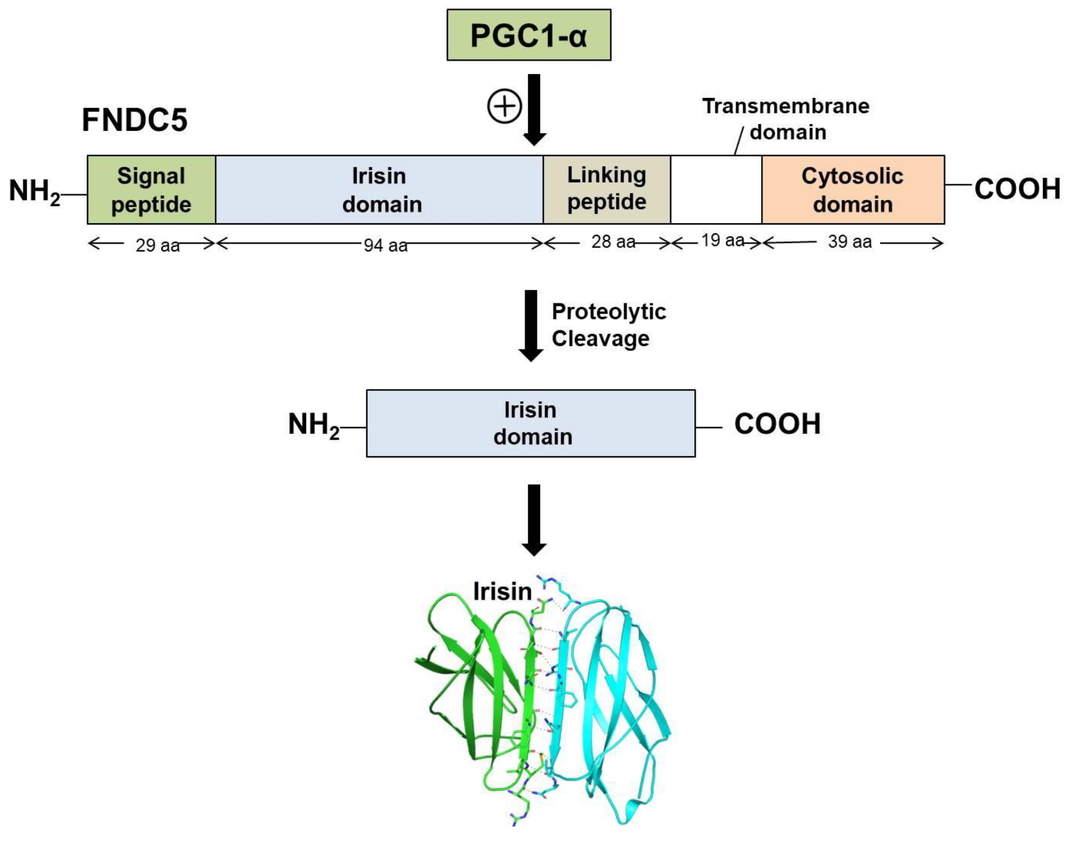
Irisin is a portion of the cell membrane protein known as FNDC5 (FNDC5 Fibronectin type III domain-containing protein 5). FNDC5 consists of a signal peptide, a fibronectin III domain, and a C-terminal domain. FNDC5 comprises 209 amino acid residues, having a signal sequence of 29 amino acids at the N-terminal end, followed by a 94-amino-acid residue fibronectin III (FNIII) 2 domains (irisin domain), a linking peptide comprising 28 amino acid residues, a 19-amino-acid residue transmembrane domain, and a cytoplasmic domain consisting of 39 amino acid residues.

irisin structural insight therapeutic potential

1. Biosynthesis and Secretion of Irisin

Irisin is mainly secreted from skeletal muscles. However, immunohistochemical studies have shown that it is also found in the pancreas, testes, liver, and stomach [1]. Irisin secretion and synthesis are induced by exercise and PGC1α [2]. PGC1-α (Peroxisome proliferator-activated receptor γ (PPAR-γ) coactivator 1-α)is a multispecific coactivator of transcription, which is competent in multiple gene regulation in response to the nutritional and the physiological signals in tissues such as brown adipose tissue, skeletal muscle, and heart and liver tissue [3]. As irisin is an exercise-induced myokine, the circulating level of irisin increases in individuals engaged in exercise-induced activities and progressively decreases in those who are sedentary and less active [4]. Prolonged exercise increases PGC1α expression mainly in the skeletal muscles and heart and improves various metabolic parameters, including AMPK(Adenosine monophosphate-activated protein kinase) activation, PGC1α phosphorylation, insulin sensitivity and signaling, and FNDC5 production, followed by the cleavage of FNDC5 to secrete irisin [4][5].
A comparative study on irisin has shown 100% identity between murine and human irisin sequences) [6]. Conversely, it has been found that in the human FNDC5 gene, there is an unusual ATA start codon [7] which was previously identified as a null mutation, and it has been suggested that in humans, this mutation would prevent irisin production and release in the blood [8]. However, in humans, FNDC5 made from the ATA-FNDC5 sequence was detected, proving that it is not a pseudogene [8]; however, it was suggested to be in the category of genes that have lost their protein-coding ability [9]. Moreover, it has already been proven that human irisin is mainly translated from its non-canonical start codon [7].
There was also a concern about the lack of specificity in anti-irisin antibodies [10]. There were contradictory remarks on the existence of irisin by experimental evidence, but many sensitive approaches—including ELISA assays and quantitative mass spectrometry— have been employed successfully to confirm irisin’s identity and to measure the circulating level of irisin in humans [7][11][12][13]. Lee et al. employed mass spectrometry for the determination of the identity of FNDC5-immunoreactive bands detectable in human serum [11]. The mass spectrometry analysis identified a unique peptide mapped to the known sequence of irisin, which validated the immunoblot identification of circulating irisin in humans [11]. Later, another study employing mass spectrometry identified and quantitated human irisin in plasma [7]. Human irisin was identified and quantitated in plasma using mass spectrometry with control peptides enriched with heavy stable isotopes as internal standards. [7]. In line with this, it was demonstrated that cold exposure increases circulating irisin levels in humans, suggesting that exercise-induced irisin could have evolved from shivering-related muscle contraction [11]. Recently, Colaianni et al. detected decreased serum irisin levels in patients with age-related bone diseases in comparison to healthy subjects [14]. Moreover, many studies have confirmed that circulating irisin levels in the body are affected by several factors, such as diet, metabolic diseases, and other pathological disorders [1][15][16]. These data support the claim that irisin does exist and is regulated by exercise.

2. Structural Features and Signaling Pathways

Irisin is a portion of the cell membrane protein known as FNDC5 [3]. FNDC5 consists of a signal peptide, a fibronectin III domain, and a C-terminal domain. FNDC5 comprises 209 amino acid residues, having a signal sequence of 29 amino acids at the N-terminal end, followed by a 94-amino-acid residue fibronectin III (FNIII) 2 domains (irisin domain), a linking peptide comprising 28 amino acid residues, a 19-amino-acid residue transmembrane domain, and a cytoplasmic domain consisting of 39 amino acid residues. The biochemical and crystallographic studies have shown that irisin exists as a homodimer, with the continuous β-sheet interactions forming the core of the dimer. The crystal structure of irisin revealed that it contains a fold which is similar to FNIII proteins. The first study which reported the crystal data of irisin shows that irisin structure is homologous to FNIII domains, as it consists of an N-terminal domain (residues 30–123) along with a C-terminal tail composed of residues 124–140 which is mostly disordered [17]. Although all of the FNIII domains have limited homology and share only 15–20% sequence identity, their structures have surprisingly similar folds, comprising a β-sandwich with four β-strands on one side and three on the other [17]. Unlike other FNIII structures, irisin constitutes a continuous inter-subunit β-sheet dimer, which has an essential implication for receptor activation and signaling. The core of the irisin dimer is formed by continuous β-sheet interactions and 10 backbone hydrogen-bonds between the two interacting four-stranded β-sheets. Hence, the irisin structure unveils the first instance of a continuous β-sheet dimer made between two FNIII domains. Irisin is a 112-amino-acid peptide that includes the 94-amino-acid residue extracellular FNIII domain, cleaved from the C-terminal end of FNDC5. Figure 1 depicts the schematic representation of the structure of FNDC5 and the formation of irisin through its proteolytic cleavage.
Figure 1. Schematic representation of FNDC5 structure and formation of irisin.
Glycosylation is a very common post-translational modification of proteins where the attachment of carbohydrates leads to greater heterogeneity in the structure of glycans. Oligosaccharides influence the protein’s physicochemical properties, which are essential to obtain accurate protein conformation and protect against proteolysis, and are also essential for their biological function in diverse metabolic processes [18]. There are two N-glycosylation sites in irisin at the Asn-7 and Asn-52 positions [19]. The molecular weight of FNDC5 proteins ranges from 20 to 32 kDa, depending on the number and structure of glycan moiety attached to the molecule of protein during the process of post-translational modification [1]. Deglycosylation lowers the molecular weight of irisin to 12–15 kDa [20]. In some studies, it is shown that post-translational modification, for example, N-glycosylation, has an important role in irisin activity. Both glycosylated and nonglycosylated forms of irisin have been used [21] and further research is required to determine the glycosylation pattern and effects of the glycosylation of irisin in various physiological conditions.
There are several intracellular signaling pathways through which FNDC5/irisin elicits its biological functions. The major pathways through which irisin exert its action in white adipocytes browning, neural differentiation, and osteoblast proliferation, are MAPK(Mitogen-activated protein kinase) signaling pathways. In addition to this, there are some other signaling cascades such as the AMPK pathway, PI3K(Phosphatidylinositol 3-kinase)/AKT, and STAT3( Signal transducer and activator of transcription 3)/Snail pathway, which mediate some other important functions of FNDC5/Irisin [22].
Major functions which the fndc5/irisin gene elicits in the body are mediated by p38 and ERK1/2 signaling. WAT (White adipose tissue) browning is induced by irisin through p38 and ERK. It was shown both in vivo and in vitro that recombinant irisin treatment increases levels of phosphorylated p38 as well as phosphorylated ERK, which in turn results in the upregulation of the UCP1(Uncoupling protein 1) expression level [19]. Irisin through p38 MAPK and ERK1/2(Extracellular signal-regulated kinase 1/2) signaling is not only responsible for the browning of WAT but also induces neural cell differentiation, osteocyte proliferation, glucose uptake by the muscles, and a reduction in insulin resistance [22]. The main physiological effects which irisin shows through MAPK signaling pathways are depicted in Figure 2. AMPK and PI3K/AKT pathways mediate the effect of irisin in proliferation, anti-inflammatory, and anti-metastatic activities. A report showed that irisin enhances the proliferation of H19-7 cells through STAT3 signaling instead of AMPK and/or ERK, so it can be inferred that irisin exerts its neuroprotective effect partly through STAT3 signaling [22]. It was demonstrated that irisin treatment activates the AMPK pathway and downregulates the mTOR(Mammalian target of rapamycin) pathway, thereby suppressing pancreatic cancer cell growth, and thus inhibits the epithelial–mesenchyme transition (EMT) of pancreatic cancer cells [23]. Irisin has also been shown to mediate its effect through the PI3/AKT pathway in lung cancers. A study showed that irisin can reduce the expression of the EMT marker and inhibits the Snail expression via PI3K/AKT pathway, thereby inhibiting the invasion, migration, and proliferation of lung cancer cells [24]. Irisin has also been found to stimulate the cAMP( Cyclic adenosine 3, 5- monophosphate)/PKA/CREB( cAMP response element binding) pathway, thereby regulating neuronal plasticity and preventing memory impairment [25]. It was demonstrated that irisin can inhibit adipogenesis through activation of the Wnt expression and following the repression of transcription factors [26]. In Figure 3, the role of irisin has been shown in different physiological conditions through pathways other than MAPK signaling.
Figure 2. Schematic representation of physiological roles of Fndc5/irisin through MAPK signaling pathways.
Figure 3. Schematic representation of physiological activities of Fndc5/irisin through pathways other than MAP Kinase signaling.

3. Irisin Receptor

At present, the receptor for irisin has not been fully identified; however, Kim et al. suggested that the αV family of integrin receptors are likely irisin receptors in thermogenic fats and osteocytes [27]. Quantitative proteomic analysis in MLO-Y4 osteocytes showed that irisin binds efficiently to the integrin β1-α1 heterodimers. The protein–protein binding assay was performed to check the binding affinity between irisin and several integrin complexes [27]. It was found that most integrin complexes, including integrin β1-α1, showed significant binding with irisin; however, αV/β5 integrins showed the highest binding affinity. HDX-MS also demonstrated that irisin binds to αV/β5 integrins which allow the mapping of binding motifs on irisin and integrin complexes. Further, it was demonstrated that a very low concentration (10 pM) of irisin treatment resulted in the activation of the classic integrin signaling pathway in MLO-Y4 osteocytes [27]. Moreover, it was revealed that when the αV integrins are chemically inhibited, the signaling and function of irisin in osteocytes and fat cells are blocked [27]. Taken together, all these data suggest that although no specific receptor of irisin has been identified yet, it exerts its action via αV/β5 integrins in bone and fat tissues. Conversely, these specific effects of irisin via interaction with αV/β5 are not completely understood in vivo, either due to the ability of αV/β5 to interact with other ligands, or the binding affinity of irisin with other integrin complexes [28]. Although αV/β5 integrins have been shown as irisin receptors in some tissues, there is also a possibility of other receptors both within and outside of the integrin family.

4. Irisin in Obesity and Diabetes

Obesity results from persistent positive energy balance, which occurs when the intake of energy is higher than the expenditure of energy. It is associated with the risk of life-threatening diseases such as type 2 diabetes, stroke, heart diseases, and so on. Fat accumulation in adipose tissue is important for energy storage and to insulate the body. However, the excessive accumulation of body fat leads to obesity. Adipose tissues are the main organ for fat storage and have a fundamental role in metabolism [29]. Based on structure and function, adipose tissues were distinguished as WAT and BAT (Brown adipose tissue). WAT consists of mainly mature white adipocytes with a nucleus that is peripherally located and a big single lipid droplet. BAT is morphologically different from WAT because BAT has a centrally located nucleus, numerous small lipid droplets, and many mitochondria. Lipids present in BAT are used primarily for heat generation and oxidative phosphorylation [1]. WAT stores energy in the form of triglyceride and releases free fatty acids when needed, whereas BAT burns fat to maintain temperature by a process called non-shivering thermogenesis [30]. The thermogenic capacity (ability to generate heat) depends upon the UCP1 which forms a pore in the inner mitochondrial membrane. Due to this, leakage of protons occurs, which dissipates the electrochemical proton gradient in the mitochondrial matrix that is required for ATP(Adenosine triphosphate) synthesis. This therefore results in blunted ATP synthesis and the release of energy as heat. Overexpression of UCP1 in WAT is suggested as the therapy for preventing obesity.
Zhang et al. demonstrated that irisin also affects WAT’s functioning, and the effects of its activity are dependent on the degree of cell differentiation [19]. In vitro studies used mature adipocytes, and undifferentiated preadipocytes, to assess the effect of irisin. Irisin induces the UCP-1 expression levels in mature fat cells, which results in reprogramming WAT to take on the phenotype of BAT by the process of fat browning [1]. Expression of the browning-associated genes and UCP1 protein is upregulated by irisin in the fresh adipose tissues, as well as in cultured primary mature adipocytes. It was observed that treating human subcutaneous WAT with irisin increases the expression of UCP1 by activating p38 MAPK and ERK signaling [6]. To confirm this, specific inhibitors were used for blocking either of these two pathways, and it was revealed that this causes the abolition of irisin-induced UCP1 upregulation [19]. Therefore, it was concluded that WAT browning is induced by irisin through p38MAPK and ERK MAPK signaling [19]. A recent study demonstrated that a lack of irisin is coupled with a poor browning response and glucose/lipid derangement [31]. It can thus be concluded that the ability of irisin to convert WAT cells into the phenotype of BAT cells can be a potential therapeutic target for obesity and other associated diseases. In Figure 4, it was shown that the secretion of irisin from FNDC5 as a mature peptide and its role in obesity through fat browning.
Figure 4. Irisin secretion and its role in fat browning.
Irisin acts as an insulin-sensitizing hormone, and it is believed that irisin improves hepatic glucose and lipid metabolism by promoting pancreatic β cell functions and helps in the amelioration of insulin resistance and type 2 diabetes [32][33]. Irisin facilitates the uptake of glucose by skeletal muscles, and also improves lipid metabolism and hepatic glucose. It shows a positive effect on hyperglycemia and hyperlipidemia caused by metabolic syndrome and obesity [34]. There is an inverse association between irisin and type 2 diabetes as shown by Choi et al. where reduced irisin concentrations were reported in diabetic patients compared to the control [35]. In line with this, another study reported significantly decreased irisin concentrations in adults with T2DM regardless of age, gender, or BMI [36]. In diabetic patients, vascular complications resulting from endothelial dysfunction are the major causes of death [37]. In type 2 diabetes, irisin has been found to alleviate endothelial dysfunction partially via reducing oxidative/nitrative stresses through inhibition of signaling pathways, implicating NF-κB/iNOS and PKC-β/NADPH oxidase [37]. These studies altogether suggest that irisin may be a potential agent for the treatment of diabetic complications.

5. Irisin in the Nervous System

Physical exercise shows beneficial effects on the functioning of the nervous system. Moderate and regular exercise enhances the differentiation and proliferation of mouse neurons, increases the survival period, and stimulates migration [38]. Exercise ameliorates negative outcomes in neurological diseases, since exercise has many positive effects on the nervous system. It was expected that exercise-induced hormone irisin would also have some beneficial influences. Irisin is found in cerebral Purkinje cells, hypothalamus, and cerebrospinal fluid, and plays some essential roles in the central nervous system [39][40]. Various evidence showed that irisin crosses the blood–brain barrier, from where it induces BDNF (Brain-derived neurotrophic factor), which is involved in regulating synaptic plasticity [28][41]. Earlier, it was reported that irisin enhances cell proliferation in H19-7 HN cells of mice [42]. Moreover, irisin plays a crucial role in activating autophagy and thus exhibits a protective role against inflammation [43]. Several reports are investigating the protective roles of irisin through activation of autophagy as an anti-inflammatory strategy [43][44].

Abbreviations

FNDC5  Fibronectin type III domain-containing protein 5

BDNF   Brain-derived neurotrophic factor

WAT     White adipose tissue

BAT      Brown adipose tissue

PGC1 α Peroxisome proliferator-activated receptor γ (PPAR-γ) coactivator 1-α

AMPK  Adenosine monophosphate-activated protein kinase

PI3K      Phosphatidylinositol 3-kinase

STAT3  Signal transducer and activator of transcription 3

MAPK  Mitogen-activated protein kinase

ERK1/2 Extracellular signal-regulated kinase 1/2

cAMP   Cyclic adenosine 3, 5- monophosphate

CREB    cAMP response element binding

ATP      Adenosine triphosphate

mTOR   Mammalian target of rapamycin

References

  1. Korta, P.; Pocheć, E.; Mazur-Biały, A. Irisin as a multifunctional protein: Implications for health and certain diseases. Medicina 2019, 55, 485.
  2. Waseem, R.; Shamsi, A.; Mohammad, T.; Alhumaydhi, F.A.; Kazim, S.N.; Hassan, M.I.; Ahmad, F.; Islam, A. Multispectroscopic and Molecular Docking Insight into Elucidating the Interaction of Irisin with Rivastigmine Tartrate: A Combinational Therapy Approach to Fight Alzheimer’s Disease. ACS Omega 2021, 6, 7910–7921.
  3. Norheim, F.; Langleite, T.M.; Hjorth, M.; Holen, T.; Kielland, A.; Stadheim, H.K.; Gulseth, H.L.; Birkeland, K.I.; Jensen, J.; Drevon, C.A. The effects of acute and chronic exercise on PGC-1α, irisin and browning of subcutaneous adipose tissue in humans. FEBS J. 2014, 281, 739–749.
  4. Clin, A. The Relationship between High-Fat Diet and Fibronectin Type-III Domain-Containing Protein 5 mRNA Expression. Anatol. Clin. J. Med. Sci. 2018, 23, 1–5.
  5. Moreno-Navarrete, J.M.; Ortega, F.; Serrano, M.; Guerra, E.; Pardo, G.; Tinahones, F.; Ricart, W.; Fernández-Real, J.M. Irisin is expressed and produced by human muscle and adipose tissue in association with obesity and insulin resistance. J. Clin. Endocrinol. Metab. 2013, 98, E769–E778.
  6. Boström, P.; Wu, J.; Jedrychowski, M.P.; Korde, A.; Ye, L.; Lo, J.C.; Rasbach, K.A.; Boström, E.A.; Choi, J.H.; Long, J.Z. A PGC1-α-dependent myokine that drives brown-fat-like development of white fat and thermogenesis. Nature 2012, 481, 463–468.
  7. Jedrychowski, M.P.; Wrann, C.D.; Paulo, J.A.; Gerber, K.K.; Szpyt, J.; Robinson, M.M.; Nair, K.S.; Gygi, S.P.; Spiegelman, B.M. Detection and quantitation of circulating human irisin by tandem mass spectrometry. Cell Metab. 2015, 22, 734–740.
  8. Raschke, S.; Elsen, M.; Gassenhuber, H.; Sommerfeld, M.; Schwahn, U.; Brockmann, B.; Jung, R.; Wisløff, U.; Tjønna, A.E.; Raastad, T. Evidence against a beneficial effect of irisin in humans. PLoS ONE 2013, 8, e73680.
  9. Vanin, E.F. Processed pseudogenes: Characteristics and evolution. Annu. Rev. Genet. 1985, 19, 253–272.
  10. Albrecht, E.; Norheim, F.; Thiede, B.; Holen, T.; Ohashi, T.; Schering, L.; Lee, S.; Brenmoehl, J.; Thomas, S.; Drevon, C.A. Irisin–a myth rather than an exercise-inducible myokine. Sci. Rep. 2015, 5, 8889.
  11. Lee, P.; Linderman, J.D.; Smith, S.; Brychta, R.J.; Wang, J.; Idelson, C.; Perron, R.M.; Werner, C.D.; Phan, G.Q.; Kammula, U.S. Irisin and FGF21 are cold-induced endocrine activators of brown fat function in humans. Cell Metab. 2014, 19, 302–309.
  12. Ruan, Q.; Zhang, L.; Ruan, J.; Zhang, X.; Chen, J.; Ma, C.; Yu, Z. Detection and quantitation of irisin in human cerebrospinal fluid by tandem mass spectrometry. Peptides 2018, 103, 60–64.
  13. Ruan, Q.; Huang, Y.; Yang, L.; Ruan, J.; Gu, W.; Zhang, X.; Zhang, Y.; Zhang, W.; Yu, Z. The effects of both age and sex on irisin levels in paired plasma and cerebrospinal fluid in healthy humans. Peptides 2019, 113, 41–51.
  14. Colaianni, G.; Errede, M.; Sanesi, L.; Notarnicola, A.; Celi, M.; Zerlotin, R.; Storlino, G.; Pignataro, P.; Oranger, A.; Pesce, V. Irisin correlates positively with BMD in a cohort of older adult patients and downregulates the senescent marker p21 in osteoblasts. J. Bone Miner. Res. 2021, 36, 305–314.
  15. Colaianni, G.; Storlino, G.; Sanesi, L.; Colucci, S.; Grano, M. Myokines and osteokines in the pathogenesis of muscle and bone diseases. Curr. Osteoporos. Rep. 2020, 18, 401–407.
  16. Young, M.F.; Valaris, S.; Wrann, C.D. A role for FNDC5/Irisin in the beneficial effects of exercise on the brain and in neurodegenerative diseases. Prog. Cardiovasc. Dis. 2019, 62, 172–178.
  17. Schumacher, M.A.; Chinnam, N.; Ohashi, T.; Shah, R.S.; Erickson, H.P. The structure of irisin reveals a novel intersubunit β-sheet fibronectin type III (FNIII) dimer: Implications for receptor activation. J. Biol. Chem. 2013, 288, 33738–33744.
  18. Korta, P.; Pocheć, E. Glycosylation of thyroid-stimulating hormone receptor. Endokrynol. Pol. 2019, 70, 86–100.
  19. Zhang, Y.; Li, R.; Meng, Y.; Li, S.; Donelan, W.; Zhao, Y.; Qi, L.; Zhang, M.; Wang, X.; Cui, T. Irisin stimulates browning of white adipocytes through mitogen-activated protein kinase p38 MAP kinase and ERK MAP kinase signaling. Diabetes 2014, 63, 514–525.
  20. Erickson, K.I.; Weinstein, A.M.; Lopez, O.L. Physical activity, brain plasticity, and Alzheimer’s disease. Arch. Med. Res. 2012, 43, 615–621.
  21. Li, X.; Duan, H.; Liu, Q.; Umar, M.; Luo, W.; Yang, X.; Zhu, J.; Li, M. Construction of a Pichia pastoris strain efficiently secreting irisin and assessment of its bioactivity in HepG2 cells. Int. J. Biol. Macromol. 2019, 124, 60–70.
  22. Rabiee, F.; Lachinani, L.; Ghaedi, S.; Nasr-Esfahani, M.H.; Megraw, T.L.; Ghaedi, K. New insights into the cellular activities of Fndc5/Irisin and its signaling pathways. Cell Biosci. 2020, 10, 1–10.
  23. Liu, J.; Song, N.; Huang, Y.; Chen, Y. Irisin inhibits pancreatic cancer cell growth via the AMPK-mTOR pathway. Sci. Rep. 2018, 8, 1–10.
  24. Shao, L.; Li, H.; Chen, J.; Song, H.; Zhang, Y.; Wu, F.; Wang, W.; Zhang, W.; Wang, F.; Li, H. Irisin suppresses the migration, proliferation, and invasion of lung cancer cells via inhibition of epithelial-to-mesenchymal transition. Biochem. Biophys. Res. Commun. 2017, 485, 598–605.
  25. Lourenco, M.V.; Frozza, R.L.; de Freitas, G.B.; Zhang, H.; Kincheski, G.C.; Ribeiro, F.C.; Gonçalves, R.A.; Clarke, J.R.; Beckman, D.; Staniszewski, A. Exercise-linked FNDC5/irisin rescues synaptic plasticity and memory defects in Alzheimer’s models. Nat. Med. 2019, 25, 165.
  26. Ma, E.B.; Sahar, N.E.; Jeong, M.; Huh, J.Y. Irisin exerts inhibitory effect on adipogenesis through regulation of Wnt signaling. Front. Physiol. 2019, 10, 1085.
  27. Kim, H.; Wrann, C.D.; Jedrychowski, M.; Vidoni, S.; Kitase, Y.; Nagano, K.; Zhou, C.; Chou, J.; Parkman, V.-J.A.; Novick, S.J. Irisin mediates effects on bone and fat via αV integrin receptors. Cell 2018, 175, 1756–1768.
  28. Pignataro, P.; Dicarlo, M.; Zerlotin, R.; Zecca, C.; Dell’Abate, M.T.; Buccoliero, C.; Logroscino, G.; Colucci, S.; Grano, M. FNDC5/Irisin System in Neuroinflammation and Neurodegenerative Diseases: Update and Novel Perspective. Int. J. Mol. Sci. 2021, 22, 1605.
  29. Rosen, E.D.; Spiegelman, B.M. Adipocytes as regulators of energy balance and glucose homeostasis. Nature 2006, 444, 847–853.
  30. Wang, W.; Seale, P. Control of brown and beige fat development. Nat. Rev. Mol. Cell Biol. 2016, 17, 691.
  31. Luo, Y.; Qiao, X.; Ma, Y.; Deng, H.; Xu, C.C.; Xu, L. Disordered metabolism in mice lacking irisin. Sci. Rep. 2020, 10, 1–10.
  32. Park, K.H.; Zaichenko, L.; Brinkoetter, M.; Thakkar, B.; Sahin-Efe, A.; Joung, K.E.; Tsoukas, M.A.; Geladari, E.V.; Huh, J.Y.; Dincer, F. Circulating irisin in relation to insulin resistance and the metabolic syndrome. J. Clin. Endocrinol. Metab. 2013, 98, 4899–4907.
  33. Liu, S.; Du, F.; Li, X.; Wang, M.; Duan, R.; Zhang, J.; Wu, Y.; Zhang, Q. Effects and underlying mechanisms of irisin on the proliferation and apoptosis of pancreatic β cells. PLoS ONE 2017, 12, e0175498.
  34. Chen, N.; Li, Q.; Liu, J.; Jia, S. Irisin, an exercise-induced myokine as a metabolic regulator: An updated narrative review. Diabetes/Metab. Res. Rev. 2016, 32, 51–59.
  35. Choi, Y.-K.; Kim, M.-K.; Bae, K.H.; Seo, H.-A.; Jeong, J.-Y.; Lee, W.-K.; Kim, J.-G.; Lee, I.-K.; Park, K.-G. Serum irisin levels in new-onset type 2 diabetes. Diabetes Res. Clin. Pract. 2013, 100, 96–101.
  36. Liu, J.-J.; Wong, M.D.; Toy, W.C.; Tan, C.S.; Liu, S.; Ng, X.W.; Tavintharan, S.; Sum, C.F.; Lim, S.C. Lower circulating irisin is associated with type 2 diabetes mellitus. J. Diabetes Its Complicat. 2013, 27, 365–369.
  37. Zhu, D.; Wang, H.; Zhang, J.; Zhang, X.; Xin, C.; Zhang, F.; Lee, Y.; Zhang, L.; Lian, K.; Yan, W. Irisin improves endothelial function in type 2 diabetes through reducing oxidative/nitrative stresses. J. Mol. Cell. Cardiol. 2015, 87, 138–147.
  38. So, J.H.; Huang, C.; Ge, M.; Cai, G.; Zhang, L.; Lu, Y.; Mu, Y. Intense exercise promotes adult hippocampal neurogenesis but not spatial discrimination. Front. Cell. Neurosci. 2017, 11, 13.
  39. Piya, M.K.; Harte, A.L.; Sivakumar, K.; Tripathi, G.; Voyias, P.D.; James, S.; Sabico, S.; Al-Daghri, N.M.; Saravanan, P.; Barber, T.M. The identification of irisin in human cerebrospinal fluid: Influence of adiposity, metabolic markers, and gestational diabetes. Am. J. Physiol. Endocrinol. Metab. 2014, 306, E512–E518.
  40. Novelle, M.G.; Contreras, C.; Romero-Picó, A.; López, M.; Diéguez, C. Irisin, two years later. Int. J. Endocrinol. 2013, 2013, 1–8.
  41. Wrann, C.D.; White, J.P.; Salogiannnis, J.; Laznik-Bogoslavski, D.; Wu, J.; Ma, D.; Lin, J.D.; Greenberg, M.E.; Spiegelman, B.M. Exercise induces hippocampal BDNF through a PGC-1α/FNDC5 pathway. Cell Metab. 2013, 18, 649–659.
  42. Moon, H.-S.; Dincer, F.; Mantzoros, C.S. Pharmacological concentrations of irisin increase cell proliferation without influencing markers of neurite outgrowth and synaptogenesis in mouse H19-7 hippocampal cell lines. Metabolism 2013, 62, 1131–1136.
  43. Pesce, M.; Ballerini, P.; Paolucci, T.; Puca, I.; Farzaei, M.H.; Patruno, A. Irisin and autophagy: First update. Int. J. Mol. Sci. 2020, 21, 7587.
  44. Xin, T.; Lu, C. Irisin activates Opa1-induced mitophagy to protect cardiomyocytes against apoptosis following myocardial infarction. Aging 2020, 12, 4474.
More
Information
Contributor MDPI registered users' name will be linked to their SciProfiles pages. To register with us, please refer to https://encyclopedia.pub/register :
View Times: 919
Revisions: 2 times (View History)
Update Date: 04 Mar 2022
1000/1000
Video Production Service