Submitted Successfully!
To reward your contribution, here is a gift for you: A free trial for our video production service.
Thank you for your contribution! You can also upload a video entry or images related to this topic.
Version Summary Created by Modification Content Size Created at Operation
1 + 1361 word(s) 1361 2022-01-30 04:44:52 |
2 format corrected. + 22 word(s) 1383 2022-02-09 02:14:52 |

Video Upload Options

Do you have a full video?

Confirm

Are you sure to Delete?
Cite
If you have any further questions, please contact Encyclopedia Editorial Office.
Makowski, T. Antibacterial Electroconductive Composite Coating of Cotton Fabric. Encyclopedia. Available online: https://encyclopedia.pub/entry/19224 (accessed on 02 July 2024).
Makowski T. Antibacterial Electroconductive Composite Coating of Cotton Fabric. Encyclopedia. Available at: https://encyclopedia.pub/entry/19224. Accessed July 02, 2024.
Makowski, Tomasz. "Antibacterial Electroconductive Composite Coating of Cotton Fabric" Encyclopedia, https://encyclopedia.pub/entry/19224 (accessed July 02, 2024).
Makowski, T. (2022, February 08). Antibacterial Electroconductive Composite Coating of Cotton Fabric. In Encyclopedia. https://encyclopedia.pub/entry/19224
Makowski, Tomasz. "Antibacterial Electroconductive Composite Coating of Cotton Fabric." Encyclopedia. Web. 08 February, 2022.
Antibacterial Electroconductive Composite Coating of Cotton Fabric
Edit

Graphene oxide (GO) was deposited on a cotton fabric and then thermally reduced to reduced graphene oxide (rGO) with the assistance of L-ascorbic acid. The GO reduction imparted electrical conductivity to the fabric and allowed for electrochemical deposition of Ag° particles using cyclic voltammetry. Only the Ag°/rGO composite coating imparted antibacterial properties to the fabric against Escherichia coli and Staphylococcus aureus. Ag°/rGO-modified fibers were free of bacterial film, and bacterial growth inhibition zones around the material specimens were found. Moreover, Ag°/rGO-modified fabric became superhydrophobic with WCA of 161°.  The obtained results on composite Ag°/rGO coating of the fabric seem to be promising for obtaining novel antibacterial materials.

electroconductive cotton graphene oxide electrochemical functionalization antibacterial activity

1. Introduction

Presently, there is a growing demand for smart and intelligent garments, including cotton cloth, that are electroconductive and able to store energy or communicate. In such applications, antimicrobial activity can eliminate microbial colonization and reduce the possibility of infection. GM particles exhibit antibacterial activity, dependent on their dispersibility, adsorption ability, number of corners and sharp edges [1], hence strongly influenced by the lateral size, shape, number of layers, surface modification, agglomeration, and dispersion. Three main mechanisms, recently postulated, include the action of sharp edges, oxidative stress, and wrapping or trapping of bacteria due to the flexible thin-film structure of GM. The action of the sharp edges, also called nanoknives, nanoblades, or cutters, is believed to be crucial. The edges, acting as cutters, can mechanically disrupt the cellular membranes and cause their integrity loss, e.g., [2][3][4], although it requires the direct contact of the bacteria with GM sheets. Recently, to obtain an antibacterial material, Han et al. [5] functionalized GO with hydrophilic polymers and used it as a vector for silver (Ag) nanoparticles and sulfadiazine. Deposition of Ag particles on carbon nanotube-coated fibrous materials, including cotton fabric, made them antibacterial [6][7][8], as such particles act against bacteria and fungi, and are also antiviral [9][10]. Metal-based nanoparticles are effective against a wide variety of microorganisms as their bonds to biomolecules are non-specific [10][11][12][13][14].

2. Insightful Analysis

The reduction of GO deposited on the fabric made the fabric electrically conductive. When the temperature of the hot stage approached 220 °C, R diminished significantly but at 220 °C it leveled off within 1 min at the value of 0.1 MΩ/sq. During cooling and further storage at RT, R increased but finally stabilized after 24 h. Such behavior was observed by us previously [15][16][17] due to partial reversibility of the reduction. Nevertheless, R of 5.4 MΩ/sq was achieved, showing the presence of conducting 3D-network of rGO on fiber surfaces.
SEM micrographs of GO and rGO-coated fibers are shown in Figure 1. Small thickness and very good adhesion to fiber surfaces make the particles hardly visible on the fibers [16]. However, the areas without typical morphology of cotton fibers are discernible, shown in the insets, which are suggestive of GO or rGO thin layers.
/media/item_content/202202/6203129c8fe2fmaterials-15-01072-g001.png
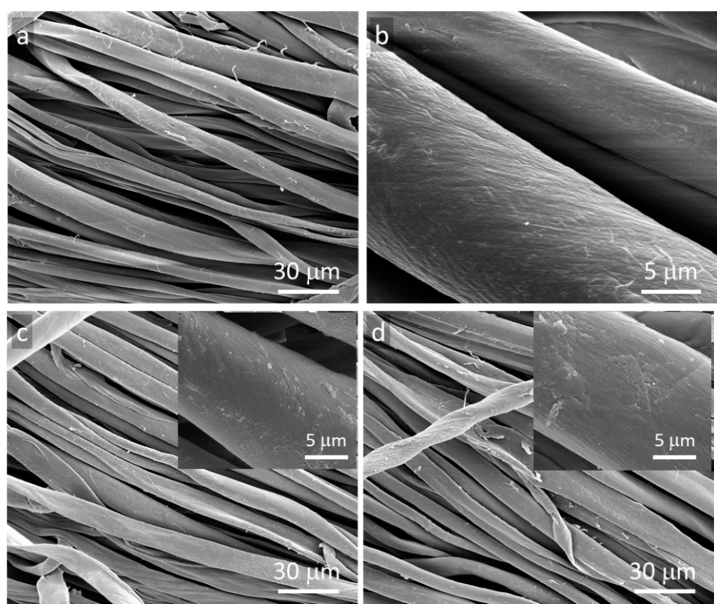
Figure 1. SEM micrographs of cotton fabric: (a,b) neat, (c) GO-coated, (d) rGO-coated.
Reduction of Ag+ ions occurred on the rGO-coated surfaces, combined with nucleation and growth of Ag° particles, which are visible in SEM micrographs in Figure 2a,b. EDS analysis confirmed the presence of Ag° particles on rGO-coated surface, as shown in Figure 2c. Reaction of silver nitrate with sodium citrate resulted in the formation of silver citrate complex stabilized by carboxyl groups of citric acid, which prevented aggregation of Ag° particles and enabled development of particles immobilized on the surface [18]. The most effective complexing occurs when [citrate]/[Ag+] >> 1 [19]. Others [20] studied GO electrochemistry and found that the reduction reactions occurred on edges of GO plates. Thus, the authors postulate that the reduction of the complex and the formation of Ag° particles, through nucleation and growth, could occur on edges or defects of rGO plates.
/media/item_content/202202/6203130ab6ae8materials-15-01072-g002.png
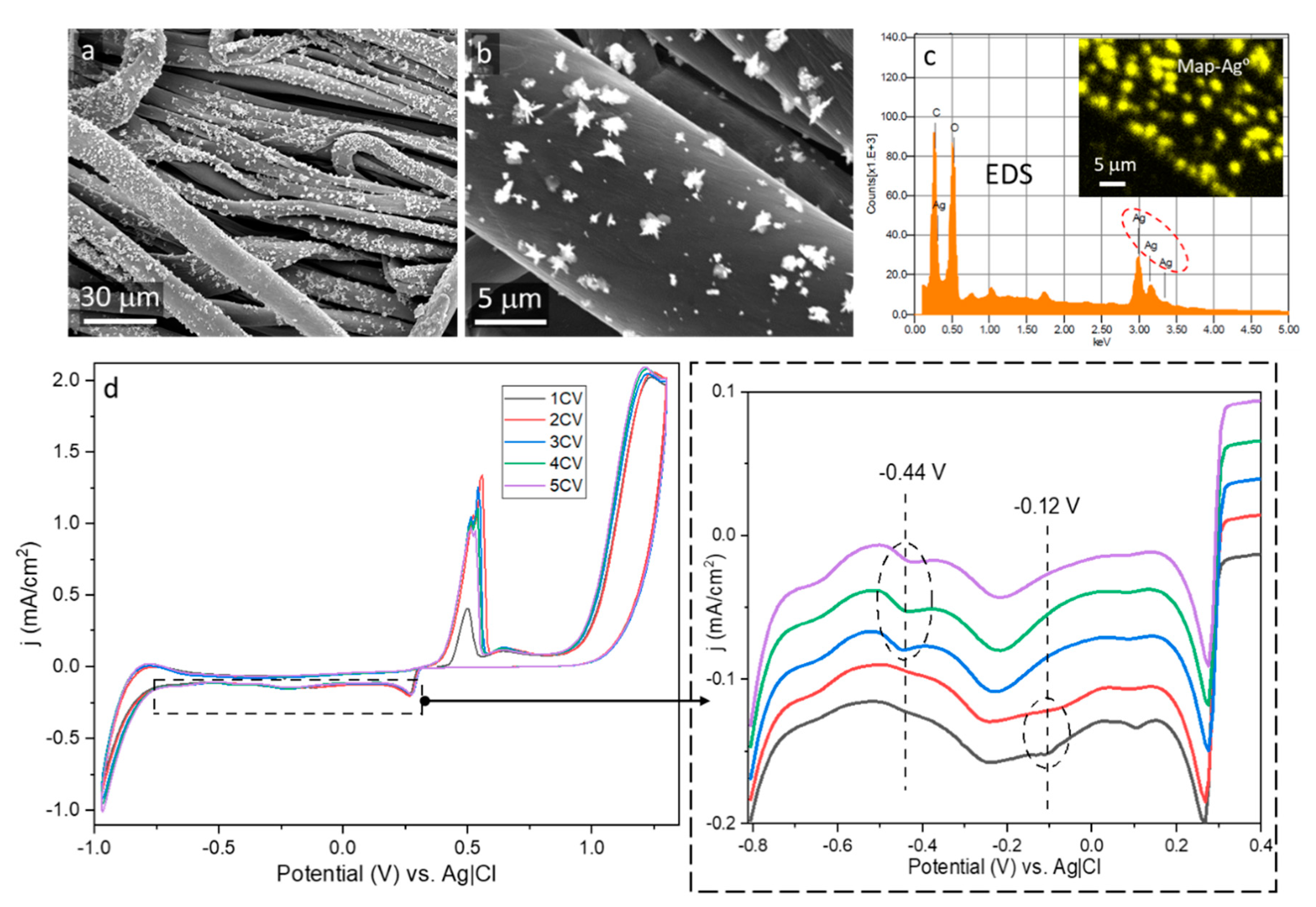
Figure 2. Ag°/rGO-coated cotton fabric: (a,b) SEM micrographs, (c) EDS Ag mapping and spectrum, (d) cyclic voltammetry profiles.
The voltammetry profile during the first and the second cycle, shown in Figure 2d, exhibits one redox weak peak at −0.12 V attributed by others to reduction of hydroxyl groups [21]. Most probably, it was related to residual hydroxyl groups of rGO, remaining due to its incomplete thermal reduction. Further reactions postulated in [21] were not observed due to a small number of other functional groups on rGO surface and competitive reactions involving Ag+ ions. During the next three cycles, a peak at −0.44 V evidenced the reduction of Ag+ ions. The proposed mechanism of the formation of the Ag° particles is shown in Scheme 1.
/media/item_content/202202/620313b43f2e6materials-15-01072-sch001.png
Scheme 1. Proposed mechanism of Ag° formation on rGO deposited on cotton fiber surfaces.
Based on the results of TGA experiments, by taking into account the residue weight at 900 °C of Ag°/rGO-modified fabric and rGO-modified fabric, the Ag content of 0.4 wt.% was determined. The successful deposition of Ag° particles confirmed the formation of the electroconductive rGO network on fiber surfaces and indicated that rGO-coated fabrics can serve as electrodes in cyclic voltammetry. R of the fabric did not change after the electrochemical modification and was equal to 5.35 MΩ/sq because Ag° was deposited on the rGO as separate particles, which did not form any continuous network. However, the Ag°/rGO-modified fabric became superhydrophobic with WCA of 161°, due to the presence of hydrophobic Ag° particles on the surface, causing the lotus effect.
As seen in Figure 3, no bacterial growth inhibition zone was found around GO-coated or rGO-coated fabric specimens. The growth inhibition zones of about 9 mm and 12 mm for Gram-positive Staphylococcus aureus and Gram-negative Escherichia coli, respectively, were only observed around the specimens of Ag°/rGO-coated fabric.
/media/item_content/202202/62031433c012fmaterials-15-01072-g003.png
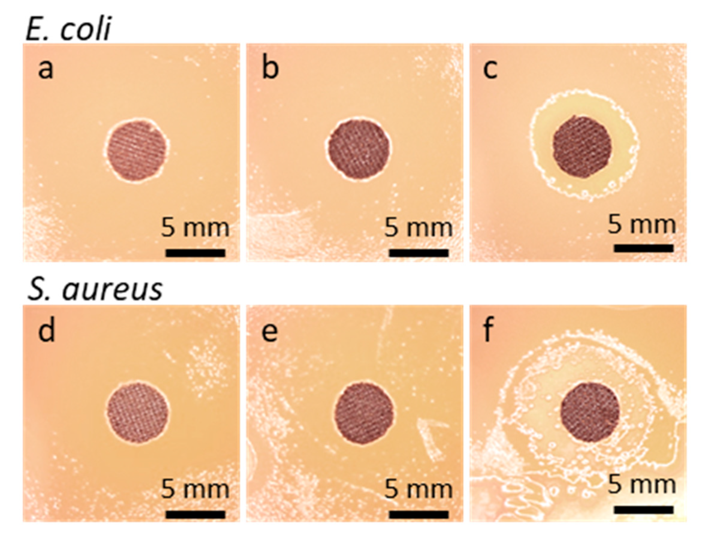
Figure 3. Disk specimens of cotton fabric on bacteria inoculated agar after 48 h incubation: E. coli (a) GO-coated, (b) rGO-coated, (c) Ag°/rGO-coated; S. aureus, (d) GO-coated, (e) rGO-coated, (f) Ag°/rGO-coated.
Ag° particles exhibit non-specific bacterial toxicity that impedes the bacteria resistance development and also broadens the spectrum of antibacterial activity [22]. The main activity mechanisms of metal nanoparticles or ions include attraction to bacterial cell walls, disruption of the cell walls increasing their permeability, and disruption of cell functions [10][11][12][22][23].
The absence of bacterial growth inhibition zones around specimens does not exclude their antibacterial activity in direct contact with microorganisms. Therefore, SEM was used to analyze the bottom surfaces of the tested fabric specimens. SEM micrographs of bottom surfaces of cotton fabric specimens in Figure 4, modified and unmodified, show that not only the neat fibers but also GO and rGO-coated fibers were inhabited by both tested bacterial strains.
/media/item_content/202202/62031539186b1materials-15-01072-g004.png
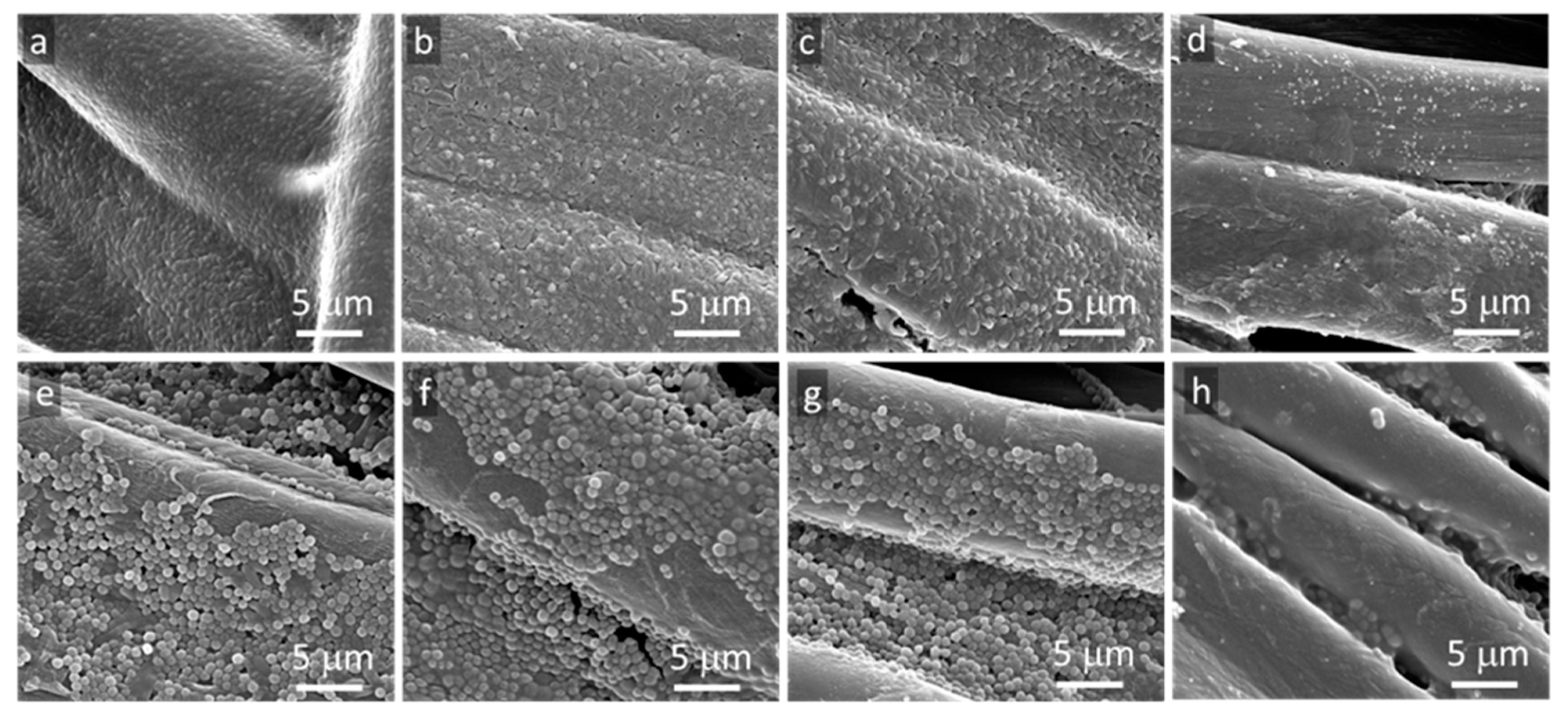
Figure 4. SEM micrographs of bottom surfaces of cotton fabric specimens removed from agar after 48 h incubation: E. coli (a) neat fabric, (b) GO-coated, (c) rGO-coated, (d) Ag°/rGO-coated, S.aureus (e) neat fabric, (f) GO-coated, (g) rGO-coated, (h) Ag°/rGO-coated.
According to the literature, there are three basic mechanisms of antibacterial activity of GO. The first one, the most commonly observed, is the effect of sharp edges, known as nanoblade effect. The nanoblades cut cell membranes causing their damage leading to the death of bacteria [2][3][4]. The second one is oxidative stress caused by reactive oxygen species, leading to bacteria DNA damage and mitochondrial dysfunction [24]. The third mechanism reported, observed rather in solutions than in coatings, is wrapping or trapping of bacteria by GO flakes, which causes their isolation from the environment [25]. The main mechanisms of antibacterial activity of rGO are related to the nanoblade effect and oxidative stress [1]. In the current study, GO or rGO flakes adhering to cotton fibers and lying flat on them did not expose sharp edges, and no wrapping or trapping of bacteria was observed by SEM. Therefore, their antibacterial activity was greatly limited. To achieve electroconductivity, a sufficient amount of GO particles had to be deposited on the fiber surfaces to form a 3D-newtork, which was then made electroconductive by GO reduction. However, the formation of 3D-network did not require full coverage of fibers with GO or rGO platelets. The uncovered areas, without coating, were prone to bacteria colonization. In turn, the activity of Ag° particles is well known and includes the release of Ag+ through oxidative dissolution, in which atmospheric O2 dissolved in water plays a role of oxidizer [23]. Due to the diffusion of thus formed ions, the antibacterial activity of Ag° modified surface does not require a close contact with microorganisms, which is reflected in the bacterial growth inhibition zones around the specimens in Figure 3.

References

  1. Cao, G.H.; Yan, J.H.; Ning, X.X.; Zhang, Q.; Wu, Q.; Bi, L.; Zhang, Y.M.; Han, Y.S.; Guo, J.B. Antibacterial and antibiofilm properties of graphene and its derivatives. Colloids Surf. B Biointerfaces 2021, 200, 111588.
  2. Nasirzadeh, N.; Azari, M.R.; Rasoulzadeh, Y.; Mohammadian, Y. An assessment of the cytotoxic effects of graphene nanoparticles on the epithelial cells of the human lung. Toxicol. Ind. Health 2019, 35, 79–87.
  3. Pulingam, T.; Thong, K.L.; Appaturi, J.N.; Nordin, N.I.; Dinshaw, I.J.; Lai, C.W.; Leo, B.F. Synergistic antibacterial actions of graphene oxide and antibiotics towards bacteria and the toxicological effects of graphene oxide on human epidermal keratinocytes. Eur. J. Pharm. Sci. 2019, 142, 105087.
  4. Zainal-Abidin, M.H.; Hayyan, M.; Ngoh, G.C.; Wong, W.F. From nanoengineering to nanomedicine: A facile route to enhance biocompatibility of graphene as a potential nano-carrier for targeted drug delivery using natural deep eutectic solvents. Chem. Eng. Sci. 2019, 195, 95–106.
  5. Han, F.; Lv, S.; Li, Z.; Jin, L.; Fan, B.; Zhang, J.; Zhang, R.; Zhang, X.; Han, L.; Li, J. Triple-synergistic 2D material-based dual-delivery antibiotic platform. NPG Asia Mater. 2020, 12, 15.
  6. Makowski, T.; Zhang, C.; Olah, A.; Piorkowska, E.; Baer, E.; Kregiel, D. Modification of dual-component fibrous materials with carbon nanotubes and methyltrichlorosilane. Mater. Des. 2018, 162, 219–228.
  7. Makowski, T.; Kowalczyk, D.; Fortuniak, W.; Brzezinski, S.; Kregiel, D. Electrochemical deposition of silver nanoparticle and polymerization of pyrrole on fabrics via conducting multiwall carbon nanotubes. Cellulose 2015, 22, 3063–3075.
  8. Farouk, A.; El-Sayed Saeed, S.; Sharaf, S.; Abd El-Hady, M.M. Photocatalytic activity and antibacterial properties of linen fabric using reduced graphene oxide/silver nanocomposite. RSC Adv. 2020, 10, 41600–41611.
  9. Galdiero, S.; Falanga, A.; Vitiello, M.; Cantisani, M.; Marra, V.; Galdiero, M. Silver Nanoparticles as Potential Antiviral Agents. Molecules 2011, 16, 8894–8918.
  10. Sánchez-López, E.; Gomes, D.; Esteruelas, G.; Bonilla, L.; Lopez-Machado, A.L.; Galindo, R.; Cano, A.; Espina, M.; Ettcheto, M.; Camins, A.; et al. Metal-Based Nanoparticles as Antimicrobial Agents: An Overview. Nanomaterials 2020, 10, 292.
  11. Anuj, S.A.; Gajera, H.; Hirpara, D.G.; Golakiya, B.A. Bacterial membrane destabilization with cationic particles of nano-silver to combat efflux-mediated antibiotic resistance in Gram-negative bacteria. Life Sci. 2019, 230, 178–187.
  12. Anuj, S.A.; Gajera, H.P.; Hirpara, D.G.; Golakiya, B.A. Interruption in membrane permeability of drug-resistant Staphylococcus aureus with cationic particles of nano-silver. Eur. J. Pharm. Sci. 2019, 127, 208–216.
  13. Qin, Z.J.; Zheng, Y.K.; Wang, Y.H.; Du, T.Y.; Li, C.M.; Wang, X.M.; Jiang, H. Versatile roles of silver in Ag-based nanoalloys for antibacterial applications. Coord. Chem. Rev. 2021, 449, 214218.
  14. Ul Hoque, M.I.; Chowdhury, A.-N.; Islam, M.T.; Firoz, S.H.; Luba, U.; Alowasheeir, A.; Rahman, M.M.; Rehman, A.U.; Ahmad, S.H.A.; Holze, R.; et al. Fabrication of highly and poorly oxidized silver oxide/silver/tin(IV) oxide nanocomposites and their comparative anti-pathogenic properties towards hazardous food pathogens. J. Hazard. Mater. 2021, 408, 124896.
  15. Makowski, T.; Svyntkivska, M.; Piorkowska, E.; Mizerska, U.; Fortuniak, W.; Kowalczyk, D.; Brzezinski, S. Conductive and superhydrophobic cotton fabric through pentaerythritol tetrakis(3-(3,5-di-tert-butyl-4-hydroxyphenyl)propionate) assisted thermal reduction of graphene oxide and modification with methyltrichlorosilane. Cellulose 2018, 25, 5377–5388.
  16. Jedrzejczyk, M.; Makowski, T.; Svyntkivska, M.; Piorkowska, E.; Mizerska, U.; Fortuniak, W.; Brzezinski, S.; Kowalczyk, D. Conductive cotton fabric through thermal reduction of graphene oxide enhanced by commercial antioxidants used in the plastics industry. Cellulose 2019, 26, 2191–2199.
  17. Mizerska, U.; Fortuniak, W.; Makowski, T.; Svyntkivska, M.; Piorkowska, E.; Kowalczyk, D.; Brzezinski, S. Electrically conductive and hydrophobic rGO-containing organosilicon coating of cotton fabric. Prog. Org. Coat. 2019, 137, 105312.
  18. Karadeniz, H.; Erdem, A.; Caliskan, A.; Pereira, C.M.; Pereira, E.M.; Ribeiro, J.A. Electrochemical sensing of silver tags labelled DNA immobilized onto disposable graphite electrodes. Electrochem. Commun. 2007, 9, 2167–2173.
  19. Patra, S.; Pandey, A.K.; Sen, D.; Ramagiri, S.V.; Bellare, J.R.; Mazumder, S.; Goswami, A. Redox Decomposition of Silver Citrate Complex in Nanoscale Confinement: An Unusual Mechanism of Formation and Growth of Silver Nanoparticles. Langmuir 2014, 30, 2460–2469.
  20. Brownson, D.A.C.; Smith, G.C.; Banks, C.E. Graphene oxide electrochemistry: The electrochemistry of graphene oxide modified electrodes reveals coverage dependent beneficial electrocatalysis. R. Soc. Open Sci. 2017, 4, 171128.
  21. Bao, Q.L.; Bao, S.J.; Li, C.M.; Qi, X.; Pan, C.X.; Zang, J.F.; Lu, Z.S.; Li, Y.B.; Tang, D.Y.; Zhang, S.; et al. Supercapacitance of Solid Carbon Nanofibers Made from Ethanol Flames. J. Phys. Chem. C 2008, 112, 3612–3618.
  22. Stensberg, M.C.; Wei, Q.S.; McLamore, E.S.; Porterfield, D.M.; Wei, A.; Sepúlveda, M.S. Toxicological studies on silver nanoparticles: Challenges and opportunities in assessment, monitoring and imaging. Nanomedicine 2011, 6, 879–898.
  23. Le Ouay, B.; Stellacci, F. Antibacterial activity of silver nanoparticles: A surface science insight. Nano Today 2015, 10, 339–354.
  24. Khan, B.; Adeleye, A.S.; Burgess, R.M.; Russo, S.M.; Ho, K.T. Effects of graphene oxide nanomaterial exposures on the marine bivalve, Crassostrea virginica. Aquat. Toxicol. 2019, 216, 105297.
  25. Akhavan, O.; Ghaderi, E.; Esfandiar, A. Wrapping Bacteria by Graphene Nanosheets for Isolation from Environment, Reactivation by Sonication, and Inactivation by Near-Infrared Irradiation. J. Phys. Chem. B 2011, 115, 6279–6288.
More
Information
Contributor MDPI registered users' name will be linked to their SciProfiles pages. To register with us, please refer to https://encyclopedia.pub/register :
View Times: 490
Entry Collection: Environmental Sciences
Revisions: 2 times (View History)
Update Date: 10 Feb 2022
1000/1000
Video Production Service