Submitted Successfully!
To reward your contribution, here is a gift for you: A free trial for our video production service.
Thank you for your contribution! You can also upload a video entry or images related to this topic.
Version Summary Created by Modification Content Size Created at Operation
1 + 2508 word(s) 2508 2022-01-11 07:35:50 |
2 format corrected. Meta information modification 2508 2022-01-18 02:53:22 |

Video Upload Options

Do you have a full video?

Confirm

Are you sure to Delete?
Cite
If you have any further questions, please contact Encyclopedia Editorial Office.
Chomova, M. Molecular Interactions in the Diabetic Brain. Encyclopedia. Available online: https://encyclopedia.pub/entry/18351 (accessed on 23 July 2024).
Chomova M. Molecular Interactions in the Diabetic Brain. Encyclopedia. Available at: https://encyclopedia.pub/entry/18351. Accessed July 23, 2024.
Chomova, Maria. "Molecular Interactions in the Diabetic Brain" Encyclopedia, https://encyclopedia.pub/entry/18351 (accessed July 23, 2024).
Chomova, M. (2022, January 17). Molecular Interactions in the Diabetic Brain. In Encyclopedia. https://encyclopedia.pub/entry/18351
Chomova, Maria. "Molecular Interactions in the Diabetic Brain." Encyclopedia. Web. 17 January, 2022.
Molecular Interactions in the Diabetic Brain
Edit

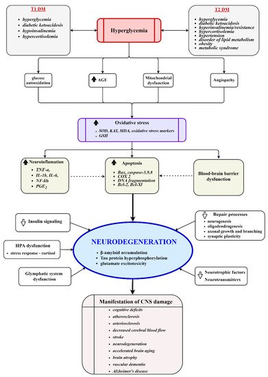
Diabetes mellitus (DM) has been associated with cognitive complications in the brain resulting from acute and chronic metabolic disturbances happening peripherally and centrally. Numerous studies have reported on the morphological, electrophysiological, biochemical, and cognitive changes in the brains of diabetic individuals. The detailed pathophysiological mechanisms implicated in the development of the diabetic cognitive phenotype remain unclear due to intricate molecular changes evolving over time and space. This study provides an insight into recent advances in understanding molecular events in the diabetic brain, focusing on cerebral glucose and insulin uptake, insulin action in the brain, and the role of the brain in the regulation of glucose homeostasis. Fully competent mitochondria are essential for energy metabolism and proper brain function; hence, the potential contribution of mitochondria to the DM-induced impairment of the brain is also discussed. 

diabetes brain mitochondria proteostasis

1. Introduction

Diabetic encephalopathies are accepted complications of DM [1]. Area-specific structural changes in the brains of diabetic patients, e.g., significantly reduced volumes of the hippocampus and prefrontal brain regions, higher rates of global cerebral atrophy, or the loss of white matter volume in the temporal lobe and inferior frontal triangle region, are the most significant changes reported by observational studies [2][3]. These changes co-occur with moderate alterations in the neurochemical profiles of N-acetyl aspartate, glutamate, myo-inositol, and choline in the white or grey matter of DM individuals and may be associated with impaired cognitive functioning [4]. The extent of metabolic (Figure 1) and cognitive alterations is determined by the interaction between disease and sensitivity of the brain to either a developmental phase (type 1—T1DM) or age (type 2—T2DM). The most evident decrement in T1DM patients is in the areas of general intelligence, psychomotor speed, mental flexibility, memory, and poor school performance. A cognitive change across the lifespan is greatest in those with an early onset of diabetes (under 6 years old) but the rate of further cognitive decline is slow—at least during the first 10 to 15 years after diagnosis [5]. Chronic hyperglycemia, microvascular complications, or recurrent episodes of hypoglycemia increase the risk of poorer cognition in older (>50 old) adults [6]. In T2DM, older adults most often show evidence of slowed information processing and poorer executive functions, and typically verbal and visual memory dysfunction, an impairment rarely associated with T1DM [7]. In elderly patients (>65 old), T2DM is associated with more severe forms of cognitive impairment. A recently published meta-analysis of 14 studies comprising over 2.3 million individuals and 102,000 cases of dementia concluded that individuals with T2DM are at a 60% greater risk of developing dementia compared with those without DM [8]. Whereas T2DM multiplies the risk of vascular dementia, the increased risk of Alzheimer’s disease is still controversial [9][10].
Figure 1. Key molecular events involved in the development of diabetic encephalopathy. Chronic hyperglycemia induces vascular injury, mitochondrial dysfunction, oxidative stress, neuroinflammation, dysfunction of the HPA axis, impairment of repair processes, reduced disposal of metabolic waste products, and activates apoptotic processes. The processes culminate in damage to the cerebral structure, neurodegeneration, and the manifestation of clinical symptoms. AGE, advanced glycation end products; SOD, superoxide dismutase; KAT, catalase; GSH, glutathione; HPA, hypothalamic–pituitary–adrenal axis.
Likewise, the results of multivariate analyses scoring the impact of prediabetes and metabolic syndrome on cognitive performance are controversial, but metabolic syndrome or impaired fasting glucose may be a risk factor for cognitive dysfunction [11]. Despite intensive research, detailed knowledge of factors and cellular mechanisms contributing to the development of the diabetic cognitive phenotype remains extremely difficult because of the multifactorial and chronic character of the disease and the biomedical and psychosocial heterogeneity of diabetic individuals.

2. Insulin–Mitochondria–ROS Interplay

Insulin action is inevitably linked to proper mitochondrial function, and, not surprisingly, aberrant mitochondria in the brain are connected to insulin resistance, metabolic syndrome, diabetic encephalopathies, neurodegenerative diseases, and aging. Alterations in electron transfer chain (ETC) function, energy metabolism [12], mitochondrial biogenesis, and fission [13], or an evident positive effect of insulin sensitizers on mitochondrial functions [14], are the most reported observations in many diabetic, obesity, or neurodegenerative studies and point to insulin–mitochondria interplay. One insulin–mitochondria interaction is “redox priming” as an intermediate phase in which oxidative modifications of sensitive cysteine residues facilitate insulin-induced receptor autophosphorylation. In neurons, insulin stimulation has been shown to generate a spike in mitochondrial H2O2 and this signal preceded activation of the IRs [15]. Although IR autophosphorylation seems to be insulin-dependent in nature, it did not occur until the H2O2 signal exceeded the threshold, even in the presence of high insulin concentrations. Moreover, the signal was ultra-sensitive to H2O2 scavenging, suggesting that a pathological increase in H2O2 scavenging antioxidant enzymes may also limit insulin signaling in the brain. Since IRs are concentrated at synapses that become enriched with mitochondria in the period of synaptic activity, a disturbance in the redox activation of IRs may represent another factor contributing to insulin resistance and cognitive impairment.
Any exogenous or endogenous stresses can perturb mitochondrial function, ROS production and ultimately impact brain energy metabolism. Neurons are especially vulnerable to mitochondrial stress since the energy stress-induced diversion of the physiological metabolic program of preferential utilization of glucose for regeneration of glutathione in the pentose phosphate cycle to the glycolytic pathway can weaken the antioxidant defense of neurons [16][17]. The hypothesis of excessive ROS production in DM is widely supported by evidence of damage to proteins, lipids, and DNA [18][19][20]. However, a significant decrease in mitochondrial membrane potential (Δψm), mitochondrial respiration, the enzymatic activities of the respiratory chain, and energy levels were reported in the cortex and hippocampus of diabetic rodent models [12][19][21], diabetic sensory neurons [22], or diabetic islets from humans and rodent models [23][24]. On the contrary, increased ROS signaling and membrane hyperpolarization have been found in peripheral diabetic cell cultures [25]. Considering a widely accepted idea of a positive correlation between mitochondrial ROS production and ETC activity/Δψm, published experimental data appear to be less conclusive. Besides numerous variations in experimental design, this inconsistency probably reflects many pitfalls of ROS measurement, especially in clearly assigning their origin to mitochondria [26].
Mitochondria are an integral part of the cellular network of adaptive and defensive responses aimed at sensing and adapting to alterations in nutrient supply or at limiting oxidative damage. In the brain, the expression and distribution of uncoupling proteins (UPCs) UCP2, UCP4, and UCP5 were induced by metabolic and oxidative challenges, suggesting the relevance of mitochondrial uncoupling to the control of neuronal, neuroendocrine, and autonomic responses [27]. Within these proteins, UCP2 is expressed abundantly in various brain areas and its key role in neuroprotection, the development of cognitive ability, resistance to anxiety, or hippocampal monoamine transmission, has been suggested [28][29]. The UCP2 uncoupler appears to be an important negative regulator of β-cell insulin secretion on the periphery, suggesting its role in the loss of glucose responsiveness in obesity-related T2DM [30]. Interestingly, the allele combination of IGF1R/IRS2/UCP2 was associated with a decreased all-cause mortality risk and with increased longevity, suggesting the combined effect of these genes on energy metabolism and the age-related metabolic remodeling capacity [31]. Observation of the downregulated UCP2 expression in rat T2DM hippocampus also indicates that its neuroprotective effect might be absent from the diabetic brain [32]. Genipin-induced inhibition of UCP2 activity not only downregulated its protein expression and enhanced ROS production in mice’s primary cortical neurons exposed to glucose fluctuation but also reduced mitochondrial biogenesis and led to the loss of neuronal synaptic integrity and cell viability. Concomitantly, inhibition of UCP2 function and the increase in metabolic and oxidative stress were compensated for with increased UCP4 expression, pointing to activated mitochondrial hormetic responses to uphold cell survival [33].
The insulin–mitochondria interplay is apparently observable in the processes of activating the mitochondrial stress response, as described in the next section.

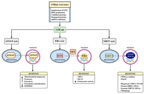
3. Mitochondrial Proteostasis in the Diabetic Brain

Metabolic disturbances in DM, such as ETC dysfunction, ROS-mediated oxidative stress, or neuroinflammation invade cellular and mitochondrial proteomes. When the proteome alters its properties, damaged and misfolded proteins and unassembled precursors can accumulate and provoke proteotoxic stress. In response to mitochondrial proteotoxic stress, the cell activates an adaptive program referred to as the mitochondrial unfolded response (UPRmt). The program, as a component of the cellular stress response pathways, represents the signaling pathway where various stress stimuli, such as misfolded and accumulated proteins, mitochondrial DNA mutations, inhibition of mitochondrial chaperones and proteases, alterations in mitochondrial dynamics, or metabolic and oxidative stress, elicit a nuclear transcriptional response to reestablish cellular and mitochondrial homeostasis [34]. Of note, several mitochondrial stress responses can be induced by other factors without any apparent connection with mitochondrial protein misfolding. A certain degree of similarity to the UPRmt can be present in these responses due to overlapping pathways, suggesting the complex nature of stress response regulation [35]. Transcription of stress-induced target genes seems to be epigenetically modulated by histone 3-specific methylation and since DNA methylation and histone posttranslational modifications differ in specific brain regions, the UPRmt may also differ in distinct types of neuronal cells [36]. Proper activation of the UPRmt supports metabolic health and increases the lifespan; however, if sustained chronically it can lead to disease, including obesity, T2D, and neurodegenerative disorders. The coordinated action of the three UPRmt axes, the canonical ATF4/5, Erα, and SIRT3 axis (Figure 2), after activation leads to increased production of chaperons, protein folding, antioxidant capacity, and protein quality control [37].
Figure 2. The mammalian UPRmt axes. Various UPRmt activators initiate gene expression via the ATF4/5, Erα, and SIRT3 branches. Triggered signaling pathways lead to a number of mitoprotective outcomes aimed at restoring mitochondrial homeostasis. ATF4/5, activating transcription factor 4/5; ERα, estrogen receptor alpha; DRP1, dynamin-related protein 1; FIS1, mitochondrial fission 1 protein; FOXO3, forkhead box protein O3; HTRA2, HtrA serine peptidase 2; MFN2, mitofusin 2; NRF1, nuclear respiratory factor 1; OPA1, mitochondrial dynamin-like GTPase; PGC1α, peroxisome proliferator-activated receptor-gamma coactivator-1 α; SIRT3, sirtuin 3; TFAM, mitochondrial transcription factor A.
The upregulation of mitochondrial chaperones and co-chaperones, mainly the group of heat shock proteins (e.g., Hsp60, Hsp70, Hsp10, HSC20, DNAJA3) and proteases (e.g., HtrA2, ClpP, Lonp1), constitutes the strongest cell response to stress. The Hsp response is predominantly cytoprotective because chaperones have the potential to attenuate pathology by the clearance of aggregated proteins, e.g., amyloid proteins. They also prevent further aggregation by inhibiting the nucleation and elongation processes of cross-seeding and facilitating cellular repair and defense mechanisms [38]. In DM, the Hsp-response is weakened and was shown to positively correlate with dysfunctional insulin signaling. In this mechanism, DM-associated increases in glucose synthase kinase-3 activity lead to abnormal phosphorylation of heat shock transcriptional factor 1 (Hsf1). Phosphorylated Hsf1 is less efficient at binding to the Hsp-transcription element and quenching the stress-induced transcriptional activity, decreasing the level of Hsp proteins [39]. The downregulation of the mitochondrial chaperone Hsp60 due to a lack of leptin signaling has been shown to be sufficient to induce hypothalamic insulin resistance in a T2DM murine model [40]. In the model of high fat diet-induced hypothalamic insulin resistance, a disrupted mitochondrial stress response led to mitochondrial dysfunction, excessive autophagy, and increased weight gain. Short-term intranasal insulin application restored expression of Atf4, Chop, Hsp60, Hsp10, ClpP, and Lonp1, suggesting that hypothalamic insulin/IGF1 signaling regulates mitochondrial stress response and ensures proper mitochondrial function [41]. Reduction in co-chaperone Hsp10, which modulates Hsp60 activity, was also sufficient to cause hypothalamic insulin resistance with acute liver insulin resistance, decreases in subunit protein levels of the ETC complexes, and mitochondrial dysfunction in T2DM mice. Interestingly, Hsp10 knockdown in murine hypothalamic cells increased saturated fatty acids (FA) and decreased monounsaturated FA content [42]. The shift from unsaturated FAs to saturated FAs in cardiolipin, the inner mitochondrial membrane phospholipid essential for the proper function of the respiratory enzymes and the assembly of the ETC into supercomplexes, was also observed in the brain cortex of streptozotocin-rats [19]. Elevated saturated FAs were also reported in patients with Hsp60 deficiency, metabolic syndrome, and in the cerebrospinal fluid of humans with obesity [43][44]. It is currently unclear how Hsp interferes with FA metabolism.
Several studies have also suggested a link between mitochondrial stress and neuroinflammation via Hsp60′s interaction with Toll-like receptors, which leads to the production of proinflammatory mediators, such as TNF-α, IL-1β, IL-6, and IL-8 [45][46]. Hyperglycemia-linked neuroinflammation in the CNS plays a key role in the development of vascular dementia in diabetic patients. Of note, Hsp60 holds many functions and occurs not only inside mitochondria but also in other intracellular locations, and it may be released from a cell too. Extracellular secretion of Hsp60 via exomes, which play an important role in cell-to-cell communication, has been documented in various inflammatory diseases including DM, suggesting that neuroinflammation could spread to neighboring neuronal cells, such as astrocytes [47][48].
Neuroinflammatory processes, a result of mitochondrial impairment, were also noticed in the model of metabolic syndrome (MS), a precondition for obesity, DM, and cardiovascular diseases. An important feature of MS is the deficiency of silent information regulator sirtuin 3 (Sirt3), the mitochondrial member of the group of NAD+-dependent lysine deacetylases. Deacetylases control a wide range of cellular processes, among them, Sirt3 controls energy metabolism processes, antioxidant defense, and mitochondrial dynamics [49]. The importance of sirtuins for cell homeostasis is highlighted by their engagement in the UPRmt stress response axis. In the brain of mice with MS, Sirt3 deficiency led to impaired mitochondrial respiration, downregulation of mitochondrial fission proteins Mfn1, Mfn2, elevated levels of brain IL-1β, and inflammasome formation [50]. Sirt3 deficiency-induced hyperacetylation of the mitochondrial proteome was also shown to spoil glucose metabolism, preferentially at the Krebs cycle, disarrange metabolic coupling between neurons and astrocytes, and decrease neurotransmitter synthesis [51]. In the mouse model of comorbid Alzheimer’s disease with amyloid pathology and MS, Sirt3 deficiency aggravated insulin resistance, glucose intolerance, amyloid plaque deposition, neuroinflammation, and microgliosis, suggesting that MS may interact with amyloid pathology during the early cellular phase of Alzheimer’s disease [52]. Thus, SIRT3 decline induced mitochondrial dysfunction and neuroinflammation in chronic metabolic diseases, such as DM, may be important participants in the cascade of molecular processes resulting in proteotoxic stress, neuronal cell damage, and late-life cognitive decline.
Healthy and fully functional mitochondria are maintained by unique equilibrium among the processes of mitochondrial biogenesis, removal of damaged mitochondria by mitophagy, and mitochondrial dynamics, which are regulated by mitofusins Mfn1, Mfn2, Drp1, and OPA1. Mitochondrial dynamics in the brain are associated with feeding, glucose homeostasis, and whole-body metabolism, and disorders of mitochondrial fission–fusion proteins are observed in obesity, DM, and neuroinflammation [53]. Mfn1 has recently emerged as a nutrient sensor in POMC neurons that influences whole-body glucose metabolism as it plays a key role in the central control of insulin release [54]. Drp1-mediated mitochondrial abnormalities have been linked to synaptic injury in the diabetic hippocampus [55]. A clinical trial in patients with T2DM-related cognitive decline observed a decrease in mitochondrial copy number, indicating that decreased mitochondrial biogenesis occurs in DM patients [56]. A decrease in the expression of PGC-1α (peroxisome proliferator-activated receptor-gamma coactivator), TFAM (mitochondrial transcription factor A), and NRFs (nuclear respiratory factors) in diabetic rat brains also corroborates dysfunctional mitochondrial biogenesis [57]. As a result, DM-linked attenuation of mitochondrial biogenesis does not restore decreased mitochondrial mass following the autophagosomal degradation of damaged mitochondria by mitophagy and leads to exacerbation of cellular damage and decline in brain functional ability.

References

  1. Sima, A.A.F. Encephalopathies: The emerging diabetic complications. Acta Diabetol. 2010, 47, 279–293.
  2. Nouwen, A.; Chambers, A.; Chechlacz, M.; Higgs, S.; Blissett, J.; Barrett, T.; Allen, H.A. Microstructural abnormalities in white and gray matter in obese adolescents with and without type 2 diabetes. NeuroImage Clin. 2017, 16, 43–51.
  3. Chen, Z.; Li, L.; Sun, J.; Ma, L. Mapping the brain in type II diabetes: Voxel-based morphometry using DARTEL. Eur. J. Radiol. 2012, 81, 1870–1876.
  4. Mangia, S.; Kumar, A.F.; Moheet, A.; Roberts, R.J.; Eberly, L.; Seaquist, E.R.; Tkac, I. Neurochemical Profile of Patients with Type 1 Diabetes Measured by 1H-MRS at 4 T. Br. J. Pharmacol. 2013, 33, 754–759.
  5. Lin, A.; Northam, E.A.; Rankins, D.; Werther, G.A.; Cameron, F.J. Neuropsychological profiles of young people with type 1 diabetes 12 yr after disease onset. Pediatr. Diabetes 2010, 11, 235–243.
  6. van Duinkerken, E.; Ryan, C.M. Diabetes mellitus in the young and the old: Effects on cognitive functioning across the life span. Neurobiol. Dis. 2020, 134, 104608.
  7. Marseglia, A.; Fratiglioni, L.; Laukka, E.J.; Santoni, G.; Pedersen, N.L.; Bäckman, L.; Xu, W. Early Cognitive Deficits in Type 2 Diabetes: A Population-Based Study. J. Alzheimer’s Dis. 2016, 53, 1069–1078.
  8. Chatterjee, S.; Peters, S.; Woodward, M.; Arango, S.M.; Batty, G.; Beckett, N.; Beiser, A.; Borenstein, A.R.; Crane, P.K.; Haan, M.N.; et al. Type 2 Diabetes as a Risk Factor for Dementia in Women Compared with Men: A Pooled Analysis of 2.3 Million People Comprising More Than 100,000 Cases of Dementia. Diabetes Care 2015, 39, 300–307.
  9. Smolina, K.; Wotton, C.; Goldacre, M.J. Risk of dementia in patients hospitalized with type 1 and type 2 diabetes in England, 1998–2011: A retrospective national record linkage cohort study. Diabetologia 2015, 58, 942–950.
  10. You, Y.; Liu, Z.; Chen, Y.; Xu, Y.; Qin, J.; Guo, S.; Huang, J.; Tao, J. The prevalence of mild cognitive impairment in type 2 diabetes mellitus patients: A systematic review and meta-analysis. Acta Diabetol. 2021, 58, 671–685.
  11. Assunção, N.; Sudo, F.K.; Drummond, C.; De Felice, F.G.; Mattos, P. Metabolic Syndrome and cognitive decline in the elderly: A systematic review. PLoS ONE 2018, 13, e0194990.
  12. Ruegsegger, G.N.; Manjunatha, S.; Summer, P.; Gopala, S.; Zabeilski, P.; Dasari, S.; Vanderboom, P.M.; Lanza, I.R.; Klaus, K.A.; Nair, K.S. Insulin deficiency and intranasal insulin alter brain mitochondrial function: A potential factor for dementia in diabetes. FASEB J. 2019, 33, 4458–4472.
  13. Edwards, J.L.; Quattrini, A.; Lentz, S.I.; Figueroa-Romero, C.; Cerri, F.; Backus, C.; Hong, Y.; Feldman, E.L. Diabetes regulates mitochondrial biogenesis and fission in mouse neurons. Diabetologia 2010, 53, 160–169.
  14. Sripetchwandee, J.; Chattipakorn, N.; Chattipakorn, S.C. Links between Obesity-Induced Brain Insulin Resistance, Brain Mitochondrial Dysfunction, and Dementia. Front. Endocrinol. 2018, 9, 496.
  15. Persiyantseva, N.A.; Storozhevykh, T.P.; Senilova, Y.E.; Gorbacheva, L.R.; Pinelis, V.G.; Pomytkin, I.A. Mitochondrial H2O2 as an enable signal for triggering autophosphorylation of insulin receptor in neurons. J. Mol. Signal. 2013, 8, 11.
  16. Bolaños, J.P.; Almeida, A.; Moncada, S. Glycolysis: A bioenergetic or a survival pathway? Trends Biochem. Sci. 2010, 35, 145–149.
  17. Chomova, M.; Zitnanova, I. Look into brain energy crisis and membrane pathophysiology in ischemia and reperfusion. Stress 2016, 19, 341–348.
  18. Kamboj, S.S.; Sandhir, R. Protective effect of N-acetylcysteine supplementation on mitochondrial oxidative stress and mitochondrial enzymes in cerebral cortex of streptozotocin-treated diabetic rats. Mitochondrion 2011, 11, 214–222.
  19. Chomova, M.; Balazova, M.; Muchova, J. Diabetes-induced abnormalities of mitochondrial function in rat brain cortex: The effect of n-3 fatty acid diet. Mol. Cell. Biochem. 2017, 435, 109–131.
  20. Poulsen, H.E.; Weimann, A.; Henriksen, T.; Kjær, L.K.; Larsen, E.L.; Carlsson, E.R.; Christensen, C.K.; Brandslund, I.; Fenger, M. Oxidatively generated modifications to nucleic acids in vivo: Measurement in urine and plasma. Free Radic. Biol. Med. 2019, 145, 336–341.
  21. Carvalho, C.; Santos, M.S.; Oliveira, C.; Moreira, P.I. Alzheimer’s disease and type 2 diabetes-related alterations in brain mitochondria, autophagy and synaptic markers. Biochim. Biophys. Acta. 2015, 1852, 1665–1675.
  22. Chowdhury, S.R.; Saleh, A.; Akude, E.; Smith, D.R.; Morrow, D.; Tessler, L.; Calcutt, N.A.; Fernyhough, P. Ciliary Neurotrophic Factor Reverses Aberrant Mitochondrial Bioenergetics Through the JAK/STAT Pathway in Cultured Sensory Neurons Derived from Streptozotocin-Induced Diabetic Rodents. Cell. Mol. Neurobiol. 2014, 34, 643–649.
  23. Lu, H.; Koshkin, V.; Allister, E.M.; Gyulkhandanyan, A.V.; Wheeler, M.B. Molecular and Metabolic Evidence for Mitochondrial Defects Associated With β-Cell Dysfunction in a Mouse Model of Type 2 Diabetes. Diabetes 2009, 59, 448–459.
  24. Gerencser, A.A. Bioenergetic Analysis of Single Pancreatic β-Cells Indicates an Impaired Metabolic Signature in Type 2 Diabetic Subjects. Endocrinology 2015, 156, 3496–3503.
  25. Siewiera, K.; Kassassir, H.; Talar, M.; Wieteska, L.; Watala, C. Higher mitochondrial potential and elevated mitochondrial respiration are associated with excessive activation of blood platelets in diabetic rats. Life Sci. 2016, 148, 293–304.
  26. Cheng, G.; Zielonka, M.; Dranka, B.; Kumar, S.N.; Myers, C.R.; Bennett, B.; Garces, A.M.; Machado, L.G.D.D.; Thiebaut, D.; Ouari, O.; et al. Detection of mitochondria-generated reactive oxygen species in cells using multiple probes and methods: Potentials, pitfalls, and the future. J. Biol. Chem. 2018, 293, 10363–10380.
  27. Marques-Aleixo, I.; Santos-Alves, E.; Balça, M.; Rizo-Roca, D.; Moreira, P.; Oliveira, P.; Magalhães, J.; Ascensao, A. Physical exercise improves brain cortex and cerebellum mitochondrial bioenergetics and alters apoptotic, dynamic and auto(mito)phagy markers. Neuroscience 2015, 301, 480–495.
  28. Wang, D.; Zhai, X.; Chen, P.; Yang, M.; Zhao, J.; Dong, J.; Liu, H. Hippocampal UCP2 is essential for cognition and resistance to anxiety but not required for the benefits of exercise. Neuroscience 2014, 277, 36–44.
  29. Bristot, V.J.D.O.; Alves, A.C.D.B.; Cardoso, L.R.; Scheffer, D.D.L.; Aguiar, A.S.J. The Role of PGC-1α/UCP2 Signaling in the Beneficial Effects of Physical Exercise on the Brain. Front. Neurosci. 2019, 13, 292.
  30. Cadenas, S. Mitochondrial uncoupling, ROS generation and cardioprotection. Biochim. Biophys. Acta Bioenerg. 2018, 1859, 940–950.
  31. Barbieri, M.; Boccardi, V.; Esposito, A.; Papa, M.; Vestini, F.; Rizzo, M.R.; Paolisso, G. A/ASP/VAL allele combination of IGF1R, IRS2, and UCP2 genes is associated with better metabolic profile, preserved energy expenditure parameters, and low mortality rate in longevity. AGE 2011, 34, 235–245.
  32. Abdul-Rahman, O.; Sasvari-Szekely, M.; Ver, A.; Rosta, K.; Szasz, B.K.; Kereszturi, E.; Keszler, G. Altered gene expression profiles in the hippocampus and prefrontal cortex of type 2 diabetic rats. BMC Genom. 2012, 13, 81.
  33. Cardoso, S.; Seiça, R.M.; Moreira, P.I. Uncoupling Protein 2 Inhibition Exacerbates Glucose Fluctuation-Mediated Neuronal Effects. Neurotox. Res. 2017, 33, 388–401.
  34. Costa-Mattioli, M.; Walter, P. The integrated stress response: From mechanism to disease. Science 2020, 368, 5314.
  35. Forsström, S.; Jackson, C.B.; Carroll, C.; Kuronen, M.; Pirinen, E.; Pradhan, S.; Marmyleva, A.; Auranen, M.; Kleine, I.-M.; Khan, N.A.; et al. Fibroblast Growth Factor 21 Drives Dynamics of Local and Systemic Stress Responses in Mitochondrial Myopathy with mtDNA Deletions. Cell Metab. 2019, 30, 1040–1054.e7.
  36. Basavarajappa, B.; Subbanna, S. Histone Methylation Regulation in Neurodegenerative Disorders. Int. J. Mol. Sci. 2021, 22, 4654.
  37. Muñoz-Carvajal, F.; Sanhueza, M. The Mitochondrial Unfolded Protein Response: A Hinge between Healthy and Pathological Aging. Front. Aging Neurosci. 2020, 12, 300.
  38. Ren, B.; Zhang, Y.; Zhang, M.; Liu, Y.; Zhang, D.; Gong, X.; Feng, Z.; Tang, J.; Chang, Y.; Zheng, J. Fundamentals of cross-seeding of amyloid proteins: An introduction. J. Mater. Chem. B 2019, 7, 7267–7282.
  39. Hooper, P.L.; Balogh, G.; Rivas, E.; Kavanagh, K.; Vigh, L. The importance of the cellular stress response in the pathogenesis and treatment of type 2 diabetes. Cell Stress Chaperones 2014, 19, 447–464.
  40. Kleinridders, A.; Lauritzen, H.P.; Ussar, S.; Christensen, J.H.; Mori, M.; Bross, P.; Kahn, C.R. Leptin regulation of Hsp60 impacts hypothalamic insulin signaling. J. Clin. Investig. 2013, 123, 4667–4680.
  41. Wardelmann, K.; Blümel, S.; Rath, M.; Alfine, E.; Chudoba, C.; Schell, M.; Cai, W.; Hauffe, R.; Warnke, K.; Flore, T.; et al. Insulin action in the brain regulates mitochondrial stress responses and reduces diet-induced weight gain. Mol. Metab. 2019, 21, 68–81.
  42. Wardelmann, K.; Rath, M.; Castro, J.; Blümel, S.; Schell, M.; Hauffe, R.; Schumacher, F.; Flore, T.; Ritter, K.; Wernitz, A.; et al. Central Acting Hsp10 Regulates Mitochondrial Function, Fatty Acid Metabolism, and Insulin Sensitivity in the Hypothalamus. Antioxidants 2021, 10, 711.
  43. Chudoba, C.; Wardelmann, K.; Kleinridders, A. Molecular effects of dietary fatty acids on brain insulin action and mitochondrial function. Biol. Chem. 2019, 400, 991–1003.
  44. Melo, H.M.; Silva, G.D.S.S.D.; Sant’Ana, M.R.; Teixeira, C.V.L.; Clarke, J.R.; Coreixas, V.S.M.; de Melo, B.C.; Fortuna, J.T.; Forny-Germano, L.; Ledo, J.H.; et al. Palmitate Is Increased in the Cerebrospinal Fluid of Humans with Obesity and Induces Memory Impairment in Mice via Pro-inflammatory TNF-α. Cell Rep. 2020, 30, 2180–2194.e8.
  45. Rosenberger, K.; Dembny, P.; Derkow, K.; Engel, O.; Krüger, C.; Wolf, S.A.; Kettenmann, H.; Schott, E.; Meisel, A.; Lehnardt, S. Intrathecal heat shock protein 60 mediates neurodegeneration and demyelination in the CNS through a TLR4- and MyD88-dependent pathway. Mol. Neurodegener. 2015, 10, 5.
  46. Juwono, J.; Martinus, R.D. Does Hsp60 Provide a Link between Mitochondrial Stress and Inflammation in Diabetes Mellitus? J. Diabetes Res. 2016, 2016, 8017571.
  47. Taha, E.A.; Ono, K.; Eguchi, T. Roles of Extracellular HSPs as Biomarkers in Immune Surveillance and Immune Evasion. Int. J. Mol. Sci. 2019, 20, 4588.
  48. Liyanagamage, D.S.N.K.; Martinus, R.D. Role of Mitochondrial Stress Protein HSP60 in Diabetes-Induced Neuroinflammation. Mediat. Inflamm. 2020, 2020, 8073516.
  49. Parodi-Rullan, R.; Chapa-Dubocq, X.R.; Javadov, S. Acetylation of Mitochondrial Proteins in the Heart: The Role of SIRT3. Front. Physiol. 2018, 9, 1094.
  50. Tyagi, A.; Nguyen, C.U.; Chong, T.; Michel, C.R.; Fritz, K.S.; Reisdorph, N.; Knaub, L.; Reusch, J.E.B.; Pugazhenthi, S. SIRT3 deficiency-induced mitochondrial dysfunction and inflammasome formation in the brain. Sci. Rep. 2018, 8, 17547.
  51. Kristian, T.; Karimi, A.J.; Fearnow, A.; Waddell, J.; McKenna, M.C. Perturbed Brain Glucose Metabolism Caused by Absent SIRT3 Activity. Cells 2021, 10, 2348.
  52. Tyagi, A.; Mirita, C.; Taher, N.; Shah, I.; Moeller, E.; Tyagi, A.; Chong, T.; Pugazhenthi, S. Metabolic syndrome exacerbates amyloid pathology in a comorbid Alzheimer’s mouse model. Biochim. Biophys. Acta Mol. Basis Dis. 2020, 1866, 165849.
  53. Haigh, J.L.; New, L.E.; Filippi, B.M. Mitochondrial Dynamics in the Brain Are Associated with Feeding, Glucose Homeostasis, and Whole-Body Metabolism. Front. Endocrinol. 2020, 11, 580879.
  54. Ramírez, S.; Gómez-Valadés, A.G.; Schneeberger, M.; Varela, L.; Haddad-Tóvolli, R.; Altirriba, J.; Noguera, E.; Drougard, A.; Flores, S.R.; Imbernón, M.; et al. Mitochondrial Dynamics Mediated by Mitofusin 1 Is Required for POMC Neuron Glucose-Sensing and Insulin Release Control. Cell Metab. 2017, 25, 1390–1399.e6.
  55. Huang, S.; Wang, Y.; Gan, X.; Fang, D.; Zhong, C.; Wu, L.; Hu, G.; Sosunov, A.A.; McKhann, G.M.; Yu, H.; et al. Drp1-Mediated Mitochondrial Abnormalities Link to Synaptic Injury in Diabetes Model. Diabetes 2014, 64, 1728–1742.
  56. Silzer, T.; Barber, R.; Sun, J.; Pathak, G.; Johnson, L.; O’Bryant, S.; Phillips, N. Circulating mitochondrial DNA: New indices of type 2 diabetes-related cognitive impairment in Mexican Americans. PLoS ONE 2019, 14, e0213527.
  57. Agrawal, R.; Zhuang, Y.; Cummings, B.P.; Stanhope, K.L.; Graham, J.; Havel, P.; Gomez-Pinilla, F. Deterioration of plasticity and metabolic homeostasis in the brain of the UCD-T2DM rat model of naturally occurring type-2 diabetes. Biochim. Biophys. Acta Mol. Basis Dis. 2014, 1842, 1313–1323.
More
Information
Contributor MDPI registered users' name will be linked to their SciProfiles pages. To register with us, please refer to https://encyclopedia.pub/register :
View Times: 353
Entry Collection: Neurodegeneration
Revisions: 2 times (View History)
Update Date: 18 Jan 2022
1000/1000
Video Production Service